capillary electrophoretic enzyme immunoassay with electrochemical detection using a noncompetitive...
TRANSCRIPT
Journal of Chromatography B, 784 (2003) 343–350www.elsevier.com/ locate/chromb
C apillary electrophoretic enzyme immunoassay with electrochemicaldetection using a noncompetitive format
1 *Zhihui He , Ning Gao, Wenrui JinSchool of Chemistry and Chemical Engineering, Shandong University, Jinan 250100,China
Received 30 May 2002; received in revised form 8 October 2002; accepted 16 October 2002
Abstract
A capillary electrophoretic enzyme immunoassay with electrochemical detection (CE-EIA-ED) using a noncompetitiveformat has been developed. In this method, antigen (Ag) reacts with an excess amount of horseradish peroxidase(HRP)-labeled antibody (Ab*). The free Ab* and the bound Ag–Ab* complex produced in the solution are separated bycapillary zone electrophoresis in a separation capillary. Then they catalyze enzyme substrate 3,39,5,59-tetramethylbenzide(TMB(Red)) and H O in a reaction capillary following the separation capillary. The reaction product, TMB(Ox), can be2 2
determined using amperometric detection on a carbon fiber microdisk bundle electrode at the outlet of the reaction capillary.Due to the amplification of the enzyme, a significant amount of TMB(Ox) can be produced for detection. Therefore, the limitof detection (LOD) of CE-EIA-ED is very low. A tumor marker (CA15-3) was used as a model, in order to test the method.
27The concentration LOD of CA15-3 is 0.024 U/ml, which corresponds to a mass detection limit of 1.3310 U. 2002 Elsevier Science B.V. All rights reserved.
Keywords: Enzyme immunoassay; Tumor marker
1 . Introduction such as high selectivity, low reagent consumptionand short incubation time. The procedure of im-
Capillary electrophoresis (CE) is a powerful tech- munoassay can be simplified by CE separation.nique for the separation of macromolecules such as Many wash steps can be eliminated. In CEIA, UVproteins and immunocomplexes [1]. With both su- detection [3,4] and laser-induced fluorescence (LIF)perior separation power and high detection sensitivi- detection [5–17] have been used. However, thety, CE can separate free antibody or antigen from major disadvantage of the UV detection is the lack ofbound antibody or antigen rapidly, and is especially sensitivity. The minimum detectable concentration
26suitable for immunoassay [2]. The method called by UV detection is around 10 mol / l. LIF detection211capillary electrophoretic immunoassay (CEIA) offers with the minimum limit of detection of 10 mol / l
several advantages over conventional immunoassays, [18] is a more general approach to improve sensitivi-ty.
Amperometric detection provides excellent sen-*Corresponding author. Fax:186-531-856-5167.
sitivity for the small dimensions associated with CE,E-mail address: [email protected](W. Jin).1 while offering a high degree of selectivity towardPresent address: Technical Center of Changde Cigarette Fac-
tory, Changde 415000, China. electroactive species. In this paper, a novel capillary
1570-0232/02/$ – see front matter 2002 Elsevier Science B.V. All rights reserved.PI I : S1570-0232( 02 )00823-1
344 Z. He et al. / J. Chromatogr. B 784 (2003) 343–350
electrophoretic enzyme immunoassay with electro- defined by two monoclonal antibodies: DF3, whichchemical detection (CE-EIA-ED) using a noncom- is found on the surface of mammary carcinoma cells,petitive format has been developed. An excess and 115D8 which is found on milk-fat globuleamount of horseradish peroxidase (HRP)-labeled membrane [19,20]. CA15-3 is significantly moreantibody (Ab*) is added to the sample to form its sensitive than carcinoembryonic antigen in thebound complex (Ag–Ab*) with the antigen (Ag) evaluation of patients with both primary and meta-present in the sample. After equilibrium is estab- static breast cancer. For healthy human, the con-lished, a small volume of incubate is injected into a centration levels of CA15-3 are lower than 30 U/mlseparation capillary, whereupon the free Ab* and the [21]. CA15-3 levels are often measured by immuno-Ag–Ab* are separated by CE, then both can catalyze radiometric assay [22–24], enzyme immunoassaythe reaction of the enzyme substrate 3,39,5,59-tetra- [25], microparticle enzyme immunoassay [25,26],methyl-benzidine (TMB(Red)) and H O . The en- chemiluminescence immunoassay [25] and immuno-2 2
zyme catalysis reaction proceeds in a catalysis fluorometric assay [27,28]. These conventional im-reaction capillary following the separation capillary. munoassays have some shortcomings such as beingThe reaction products are TMB(Ox) and H O. time-consuming, having high reagent consumption2
TMB(Ox) can be reduced at a carbon fiber microdisk and complicated operation. Therefore, CA 15-3 as abundle electrode. Thus, the activity of HRP on the model in CE-EIA-ED with a noncompetitive formatfree Ab* and the Ag–Ab* can be measured by was investigated.determining the reduction current of TMB(Ox) onthe microdisk bundle electrode at the outlet of thecatalysis reaction capillary. Since the concentration 2 . Experimentalof TMB(Ox) is much higher than those of the freeAb* and the Ag–Ab* due to the enzyme amplifica- 2 .1. Apparatustion, LOD of CE-EIA-ED should be very low. Themain advantages of CE-EIA-ED are simplicity and 2 .1.1. CE-EIA-ED systemsensitivity besides the strong points of CEIA men- The CE-EIA-ED system is illustrated in Fig. 1. Ittioned above. consisted of six main parts, a high-voltage power
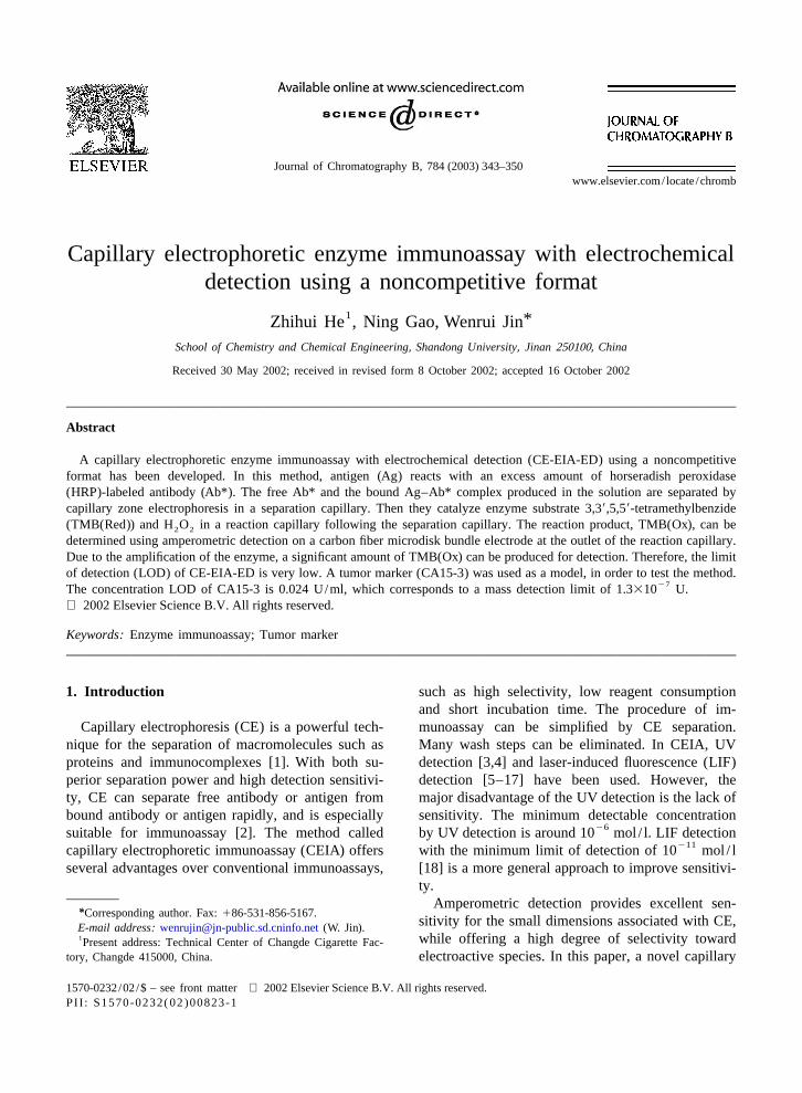
CA15-3 (Ag) is a circulating antigen which is supply (Model 9323HVPS, Beijing Institute of Newrelatively specific for human breast tissue and is Technology, Beijing, China) (1), a running buffer
Fig. 1. Overview of the CE-EIA-ED system. 1, High-voltage power supply; 2, running buffer reservoir; 3, liquid pressure buffer reservoir;4, liquid pressure substrate reservoir; 5, catalysis reactor; 6, electrochemical detector; 7, metal tubing; 8 and 89, switch; 9 and 99, rubbercover; 10, separation capillary; 11, reaction capillary; 12, hole; 13, syringe needle; 14, Pt cathode; 15, Pt anode; 16 working electrode; 17reference electrode; 18 auxiliary electrode; 19 electrochemical cell.
Z. He et al. / J. Chromatogr. B 784 (2003) 343–350 345
reservoir (2), a liquid pressure buffer reservoir (3), a electrode, and a coiled Pt wire (0.3-mm diameter, 5liquid pressure substrate reservoir (4), a Plexiglas cm in length) placed at the bottom of the cell as thecatalysis reactor (5) and an electrochemical detector auxiliary electrode. The arrangement of the electro-(6). In the system, the cylindrical running buffer chemical detection cell is illustrated in Ref. [29] inreservoir (2) (12 mm diameter and 20 mm in depth) detail. Samples were injected hydrodynamically. Thewas made from Plexiglas with a rubber cover (9). carbon fiber microdisk bundle electrodes used hereThere is a metal tubing (7) in the reservoir wall. The were described previously [30]. Before use, allmetal tubing linked up with the running buffer carbon fiber microdisk bundle electrodes werereservoir (2) and the liquid pressure buffer reservoir cleaned in alcohol and washed with double-distilled(3) through a plastic hose. On the hose, there is a water for 5 min by a ultrasonicator. During electro-switch (8) to control the flow from the liquid phoresis, the electrodes can be directly washed withpressure buffer reservoir. In this system, a metal alcohol and water in the detection cell.needle (13) (400mm I.D., 680mm O.D.) of syringewith a hole (12) in the middle passed through the 2 .1.2. Capillary treatmentcatalysis reactor (5). Both the polyacrylamide-coated The method for preparing the polyacrylamide-separation capillary (50mm I.D., 375mm O.D., 15 coated capillaries was similar to that used in Refs.cm length) (10) and the polyacrylamide-coated re- [31–34]. The polyacrylamide-coated capillaries wereaction capillary (50mm I.D., 375 mm O.D., 5 cm prepared from the uncoated fused-silica capillarieslength) (11) were inserted in the needle with a gap 35 cm long and 50mm I.D. The inner surface of the(about 10mm) between them. The separation capil- capillaries were first pretreated with 1 mol / l NaOHlary was connected to the running buffer reservoir for 30 min and then flushed with water for 30 min.through the rubber cover. The enzyme substrate The silane solution adjusted to pH 3.5 by acetic acid(TMB(Red)) solution in the liquid pressure substrate containing 0.5% (v/v)g-methacryloxypropryl-reservoir (4) was introduced into the reaction capil- trimethoxysilane (Acros Organics, NJ, USA) andlaries (11) through the gap by means of the liquid 0.5% (v/v) alcohol was sucked up into the capil-pressure. There is another switch (89) to control the laries. After the reaction proceeded for 1 h at roomflow from the liquid pressure substrate reservoir (4). temperature, the silane solution was removed. ThenA platinum wire (14) served as the grounded elec- the capillary was filled with 3.5% (w/v) deaeratedtrode in contact with the substrate solution for the acrylamide solution containing 1ml N,N,N9,N9-tetra-high potential drop across the separation capillary. methylethylenediamine and 2 mg potassium persul-Unless noted otherwise, the applied separation high- phate per ml. After 3 h, the excess (not attached)voltage was 20 kV. In the system, all the joints could polyacrylamide was sucked away and the capillariesbe fixed with epoxy adhesive. CE and ED in CE- were rinsed with water. After most of the water inEIA-ED used in this work were similar to our the capillaries was removed by aspiration, they wereprevious description [29]. Briefly, a reversible high- then dried under a N stream at 458C. Every coated2
voltage power supply (Model 9323HVPS, Beijing capillary can be used for at least 50 runs. After theInstitute of New Technology, Beijing, China) pro- experiments, the coated capillary was washed withvided a variable voltage of 0–30 kV across the water and then dried by aspiration for analysisseparation capillary with its outlet at ground po- reproducibility between days.tential. ED at a constant potential was carried outwith the electrochemical analyzer (Model CHI800, 2 .2. Reagents and solutionsCH Instruments, Austin, TX, USA). The detectioncell, the detector and the catalysis reactor were The CA15-3 EIA Kit (No. 200-10) was purchasedhoused in a Faraday cage in order to minimize the from CanAg Diagnostics AB, Gothenburg, Sweden),interference from noise of external sources. ED was which consisted of CA 15-3 standards (containing 0,carried out with a three-electrode system. It consisted 15, 50, 125 and 250 U/ml), and the anti-CA15-3of a carbon fiber microdisk bundle electrode as the antibody (50 mg/ l) labeled with horseradish per-working electrode, an SCE used as the reference oxidase (HRP). The kit was stored at 48C. The

346 Z. He et al. / J. Chromatogr. B 784 (2003) 343–350
breast cancer serum samples and the results detected and raising the vial 9 cm height for 20 s. Afterby ELISA were provided by the hematological center injection, the separation capillary was manipulatedat Qilu Hospital, Jinan, China. The serum samples down, out of the sample vial, and then immersed inwere stored at220 8C. TMB(Red) (High Pure the running buffer solution. After that, the cover (9)Grade) was obtained from Amresco Inc. (Solon, OH, of the running buffer reservoir was sealed. TheUSA). A stock standard solution of TMB(Red) switch (8) of liquid pressure buffer reservoir was(0.020 mol / l) was prepared in double-distilled water turned on, and then the liquid pressure substrateand kept in a dark bottle. The substrate solution reservoir was raised 40 cm. Finally, the separation
24 22consisted of 2.0310 mol / l TMB(Red), 1.0310 high voltage was applied across the separation23mol / l Na HPO and 5.0310 mol / l citric acid (pH capillary, the detection potential was applied at the2 4
235.0). The running buffer consisted of 2.0310 mol / working electrode and the electropherogram was24 23l H O , 2.5310 mol / l Na B O and 9.0310 recorded. During the electrophoresis, the same liquid2 2 2 4 7
mol / l H BO (pH 7.4). TMB(Red) or H O was pressure from the liquid pressure buffer reservoir and3 3 2 2
added to the buffers just before the measurement. the liquid pressure substrate reservoir was kept toThe running buffer was renewed every run. All prevent a distortion of the flat electroosmotic flowbuffers and solutions were stored at 48C until use. profile in the separation capillary. The reaction timeUnless stated otherwise, all other reagents were of could be controlled by the liquid pressure. Routinely,analytical grade or better and purchased from stan- for a 50mm I.D. reaction capillary, the applied liquiddard reagent suppliers. All solutions were prepared pressure height was 40 cm.with double-distilled water. All buffers were filtered In the electrochemical detection, the workingthrough 0.45-mm cellulose acetate membrane filters microdisk bundle electrode was cemented onto a(Shanghai Yadong Resin Co. Ltd., Shanghai, China) microscope slide, which was placed over a labora-before use. tory-made XYZ micro-manipulator and glued in
place in such a way that the microdisk end protruded2 .3. Immunoassay procedure from the edge of the slide. The position of the
microdisk bundle electrode was adjusted (under aThe immunoassay protocol was a noncompetitive microscope) against the outlet of the reaction capil-
format. A 25-ml aliquot of the CA15-3 standards or lary so that the electrode and the capillary were inserum samples, and a 5-ml aliquot of HRP-labeled contact. This arrangement allowed easy removal andanti-CA15-3 antibody were added to a microcentri- realignment of both the capillary and the electrode.fuge tube. The solution was incubated for 1 h at All potentials were measured against SCE. Allroom temperature, and then was diluted to 150ml disposable plastic wares and disposable micropipettewith the running buffer. Before injection, the levels tips used in the assay were autoclaved prior to use inof the solutions in the sample vial (not shown in Fig. order to denature any contaminants. All solutions1), the running buffer reservoir (2) and the catalysis were prepared in disposable plastic ware usingreactor (5) were kept at the same height. The liquid disposable pipette tips.pressure buffer reservoir (3) and the liquid pressuresubstrate reservoir (4) were put 40 cm over therunning buffer reservoir and the catalysis reactor, 3 . Results and discussionrespectively. The injection process was as follows:first, the liquid pressure substrate reservoir (4) was 3 .1. Optimization of CE-EIA-EDput down and keeps the solution level at the sameheight as that of the catalysis reactor (5). The switch The electropherograms of HRP-labeled anti-(89) of the liquid pressure substrate reservoir (4) was CA15-3 antibody (Ab*) in different buffers of pHturned on. The switch (8) of the liquid pressure 7.4 are shown in Fig. 2. It can be found that thebuffer reservoir (3) was turned off. Then hydro- maximum peak area,q, the minimum width at thedynamic injection was carried out by inserting the half-peak,W and the maximum number of theoret-1 / 2
23inlet of the separation capillary into the sample vial ical plates,N, were obtained in 9.0310 mol / l

Z. He et al. / J. Chromatogr. B 784 (2003) 343–350 347
23 24Fig. 2. Electropherograms of HRP-labeled anti-CA15-3 antibody, Ab*, in: (1) 9.0310 mol / l Na HPO –4.6310 mol / l citric acid; (2)2 422 23 23 24 231.0310 mol / l Tris–8.4310 mol / l HCl and (3) 9.0310 mol / l H BO –2.5310 mol / l Na B O ; 1.67 mg/ l Ab* and 2.03103 3 2 4 7
24 22 23mol / l H O in the running buffer; substrate solution, 2.0310 mol / l TMB(Red)11.0310 mol / l Na HPO –5.0310 mol / l citric acid2 2 2 4
(pH 5.0); separation capillary, 25 cm350 mm I.D., reaction capillary, 5 cm350 mm I.D.; h, 40 cm; hydrodynamic injection, 9 cm for 20 s;separation voltage, 20 kV; detection potential, 0.00 V.
24H BO –2.5310 mol / l Na B O (pH 7.4). If the The liquid heights,h, in the system shown in Fig.3 3 2 4 7
buffer concentration is higher than this value or 1 obviously exerted an influence onq. When h,40pH.7.4, q will decrease. Therefore, this buffer was cm,q increases rapidly with increasingh. Whenselected in the subsequent experiments. h.40 cm, q decreases with increasingh. q has a
The migration time,t , q, W , andN at different maximum at the liquid height of 40 cm.t decreasesm 1 / 2 m
V are listed in Table 1.t andW decrease andN slowly with increasingh. N and W are almosts m 1 / 2 1 / 2
increases very slowly with increasingV . WhenV , constants with increasingh. Therefore, 40 cm wass s
20 kV, q is a constant. WhenV .20 kV, q decreases. selected in our experiments. It was found that 2.03s
Therefore, 20 kV forV was chosen in our experi-s
ments. Fig. 3 shows the relationship betweenq andthe applied detected potential,E . WhenE is higherd d
than 0.10 V,q increases with decreasingE . WhenEd d
is lower than 0.10 V,q is almost a constant.E ofd
0.00 V was used because of largerq and lower noise.
Table 1The values ofq, t , W andN at different separation voltages,Vm 1 / 2 s
24V (kV) t (min) q (nC) W (s) 10 Ns m 1 / 2
12 16.6 1.30 10.6 4.915 16.3 1.31 10.0 5.318 16.2 1.31 9.9 5.320 16.1 1.34 9.8 5.425 15.9 1.11 9.6 5.4
Fig. 3. Relationship between the detected electric charge,q, and23 24 23Conditions: 9.0310 mol / l H BO –2.5310 mol / l the applied detected potential,E . 9.0310 mol / l H BO –2.533 3 d 3 3
24Na B O , pH 7.4; other conditions as in Fig. 2. 10 mol / l Na B O (pH 7.4). Other conditions as in Fig. 2.2 4 7 2 4 7
348 Z. He et al. / J. Chromatogr. B 784 (2003) 343–350
24 2310 mol / l TMB(Red) and 2.0310 mol / l H O2 2
were optimum in our experiments.
3 .2. CE-EIA-ED for CA15-3
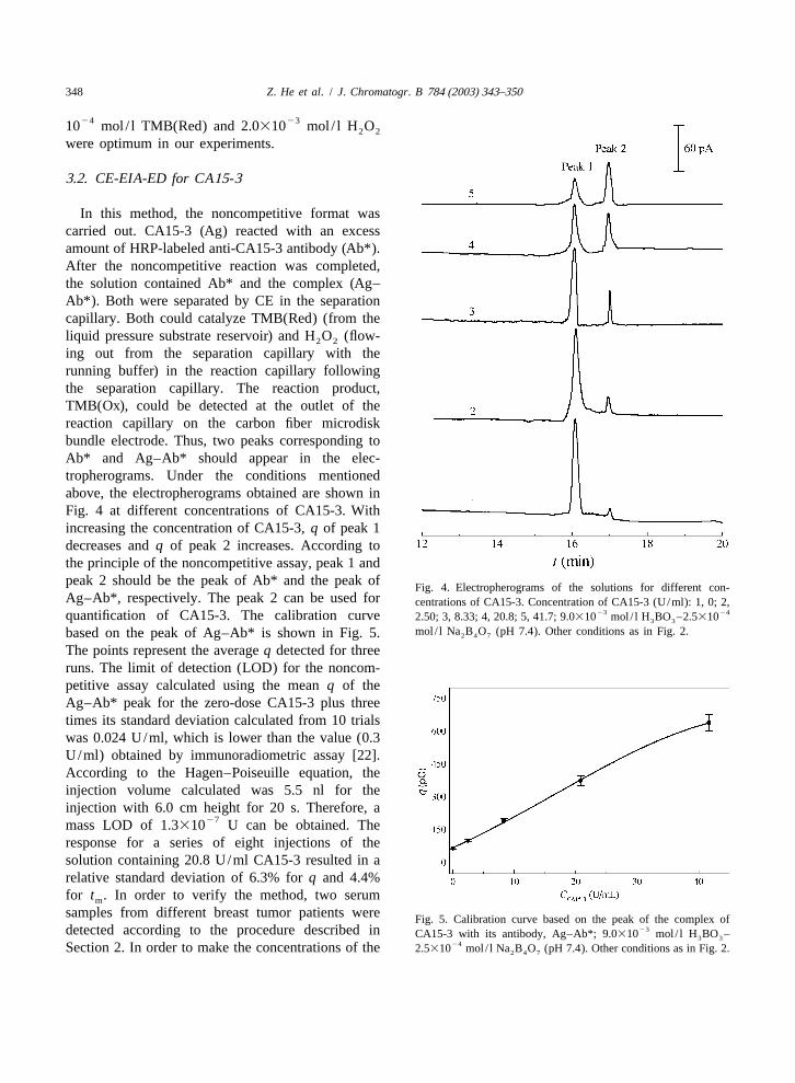
In this method, the noncompetitive format wascarried out. CA15-3 (Ag) reacted with an excessamount of HRP-labeled anti-CA15-3 antibody (Ab*).After the noncompetitive reaction was completed,the solution contained Ab* and the complex (Ag–Ab*). Both were separated by CE in the separationcapillary. Both could catalyze TMB(Red) (from theliquid pressure substrate reservoir) and H O (flow-2 2
ing out from the separation capillary with therunning buffer) in the reaction capillary followingthe separation capillary. The reaction product,TMB(Ox), could be detected at the outlet of thereaction capillary on the carbon fiber microdiskbundle electrode. Thus, two peaks corresponding toAb* and Ag–Ab* should appear in the elec-tropherograms. Under the conditions mentionedabove, the electropherograms obtained are shown inFig. 4 at different concentrations of CA15-3. Withincreasing the concentration of CA15-3,q of peak 1decreases andq of peak 2 increases. According tothe principle of the noncompetitive assay, peak 1 andpeak 2 should be the peak of Ab* and the peak of Fig. 4. Electropherograms of the solutions for different con-Ag–Ab*, respectively. The peak 2 can be used for centrations of CA15-3. Concentration of CA15-3 (U/ml): 1, 0; 2,
23 24quantification of CA15-3. The calibration curve 2.50; 3, 8.33; 4, 20.8; 5, 41.7; 9.0310 mol / l H BO –2.53103 3
mol / l Na B O (pH 7.4). Other conditions as in Fig. 2.based on the peak of Ag–Ab* is shown in Fig. 5. 2 4 7
The points represent the averageq detected for threeruns. The limit of detection (LOD) for the noncom-petitive assay calculated using the meanq of theAg–Ab* peak for the zero-dose CA15-3 plus threetimes its standard deviation calculated from 10 trialswas 0.024 U/ml, which is lower than the value (0.3U/ml) obtained by immunoradiometric assay [22].According to the Hagen–Poiseuille equation, theinjection volume calculated was 5.5 nl for theinjection with 6.0 cm height for 20 s. Therefore, a
27mass LOD of 1.3310 U can be obtained. Theresponse for a series of eight injections of thesolution containing 20.8 U/ml CA15-3 resulted in arelative standard deviation of 6.3% forq and 4.4%for t . In order to verify the method, two serumm
samples from different breast tumor patients were Fig. 5. Calibration curve based on the peak of the complex of23detected according to the procedure described in CA15-3 with its antibody, Ag–Ab*; 9.0310 mol / l H BO –3 3
24Section 2. In order to make the concentrations of the 2.5310 mol / l Na B O (pH 7.4). Other conditions as in Fig. 2.2 4 7
Z. He et al. / J. Chromatogr. B 784 (2003) 343–350 349
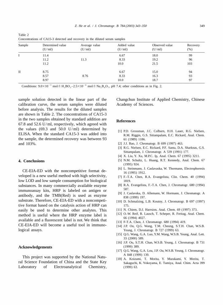
Table 2Concentrations of CA15-3 detected and recovery in the diluted serum samples
Sample Determined value Average value Added value Observed value Recovery(U/ml) (U/ml) (U/ml) (U/ml) (%)
I 11.4 6.67 18.0 9911.2 11.3 8.33 19.2 9611.2 10.0 21.5 103
II 8.75 6.67 15.0 948.57 8.76 8.33 16.3 938.97 10.0 18.7 97
23 24Conditions: 9.0310 mol / l H BO –2.5310 mol / l Na B O , pH 7.4; other conditions as in Fig. 2.3 3 2 4 7
sample solution detected in the linear part of the Changchun Institute of Applied Chemistry, Chinesecalibration curve, the serum samples were diluted Academy of Sciences.before analysis. The results for the diluted samplesare shown in Table 2. The concentrations of CA15-3in the two samples obtained by standard addition are R eferences67.8 and 52.6 U/ml, respectively, which agreed withthe values (69.3 and 50.0 U/ml) determined by
[1] P.D. Grossman, J.C. Colburn, H.H. Lauer, R.G. Nielsen,ELISA. When the standard CA15-3 was added into R.M. Riggin, G.S. Sittampalam, E.C. Rickard, Anal. Chem.the sample, the determined recovery was between 93 61 (1989) 1186.
[2] J.J. Bao, J. Chromatogr. B 699 (1997) 463.and 103%.[3] R.G. Nielsen, E.C. Rickard, P.F. Santa, D.A. Sharknas, G.S.
Sittampalam, J. Chromatogr. A 539 (1991) 177.[4] X. Liu, Y. Xu, M.P.C. Ip, Anal. Chem. 67 (1995) 3211.[5] N.M. Schultz, L. Huang, R.T. Kennedy, Anal. Chem. 674 . Conclusions
(1995) 924.[6] L. Steinmann, J. Caslavaska, W. Thormann, Electrophoresis
CE-EIA-ED with the noncompetitive format de- 16 (1995) 1912.veloped is a new useful method with high selectivity, [7] F.-T.A. Chen, R.A. Evangelista, Clin. Chem. 40 (1994)low LOD and low sample consumption for biological 1819.
[8] R.A. Evangelista, F.-T.A. Chen, J. Chromatogr. 680 (1994)substances. In many commercially available enzyme587.immunoassay kits, HRP is labeled on antigen or
[9] J. Caslavska, D. Allemann, W. Hormann, J. Chromatogr. Aantibody, and the TMB(Red) is used as enzyme 838 (1999) 197.substrate. Therefore, CE-EIA-ED with a noncompeti- [10] D. Schmalzing, L.B. Koutny, J. Chromatogr. B 697 (1997)tive format based on the catalysis action of HRP can 175.
[11] N. Chiem, D.J. Harrsion, Anal. Chem. 69 (1997) 373.easily be used to determine other analytes. This[12] O.-W. Reif, R. Lausch, T. Scheper, R. Freitag, Anal. Chem.method is useful where the HRP enzyme label is
66 (1994) 4027.available and a fluorescent label is not. We think that [13] F-T.A. Chen, J. Chromatogr. 680 (1994) 419.CE-EIA-ED will become a useful tool in immuno- [14] J.P. Ou, Q.G. Wang, T.M. Cheung, S.T.H. Chan, W.S.B.logical assays. Yeung, J. Chromatogr. B 727 (1999) 63.
[15] Q.G. Wang, G.A. Luo, Y.M. Wang, W.S.B. Yeung, Anal. Lett.33 (2000) 589.
[16] J.P. Ou, S.T.H. Chan, W.S.B. Yeung, J. Chromatogr. B 731(1999) 389.A cknowledgements
[17] Q.G. Wang, G.A. Lou, J.P. Ou, W.S.B. Yeung, J. Chromatogr.A 848 (1999) 139.
This project was supported by the National Natu- [18] A. Koizumi, T. Morita, Y. Murakami, Y. Morita, T.ral Science Foundation of China and the State Key Sakaguchi, K. Yokoyama, E. Tamiya, Anal. Chim. Acta 399Laboratory of Electroanalytical Chemistry, (1999) 63.
350 Z. He et al. / J. Chromatogr. B 784 (2003) 343–350
[19] D. Kufe, G. Inghirami, M. Abe, D. Hayes, H. Justi-Wheeler, [26] G. van Kamp, G. Bon, R. Verstraeten, D. Lynch, M. Krikau,Hybridoma 3 (3) (1984) 223. J. Fluckiger, Clin. Chem. 42 (1996) 28.
[20] J. Hilkens, F. Buijs, J. Hilgers, P. Hageman, J. Calafat, A. [27] L.F. Norum, A.M. Sauren, P.D. Rye, K. Nustada, TumourSonnenberg, M. van der Valk, Int. J. Cancer 34 (2) (1984) Biol. 22 (4) (2001) 216.197. [28] L.F. Norum, O. Nilssonb, K. Nustada, Tumour Biol. 22 (3)
[21] X. Li, X. Wu, Y. Ni, Clinical Determination of Tumor (2001) 169.Markers, People’s Healthy Press, Beijing, 1996, p. 75. [29] W. Jin, Q. Weng, J. Wu, Anal. Chim. Acta 342 (1997) 67.
[22] G.G. Bon, P. Kenemans, A.A. Verstraeten, S. Go, A. Philipi, [30] W. Jin, D. Yu, Q. Dong, X. Ye, J. Chromatogr. Sci. 38 (2000)G.J. van Kamp, H.P. van Geijn, J.M.G. van Vugt, Fetal 11.Diagn. Ther. 16 (3) (2001) 166. [31] S. Hjerten, J. Chromatogr. 347 (1985) 191.
[23] Z.Z. Chen, Z.F. Fan, J. Yang, Y. Xu, Z.Y. Tian, Chin. J. [32] N.M. Schultz, L. Huang, R.T. Kennedy, Anal. Chem. 67Oncol. 20 (2) (1998) 125. (1995) 924.
[24] R. Tobias, C. Rothwell, J. Wagner, A. Green, Y-S.V. Liu, [33] X. Liu, Y. Xu, Anal. Chem. 67 (1995) 3211.Clin. Chem. 31 (1985) 986. [34] D. Schmalzing, C.A. Piggee, F. Foret, E. Carrilho, B.L.
[25] G.G. Bon, S. von Mensdorff-Pouilly, P. Kenemans, G.J. van Karger, J. Chromatogr. A 652 (1993) 149.Kamp, R.A. Verstraeten, J. Hilgers, S. Meijer, J.B. Ver-morken, Clin. Chem. 43 (1997) 585.