cannabidiol inhibits angiogenesis by multiple mechanisms.pdf
TRANSCRIPT
RESEARCH PAPERbph_2050 1218..1231
Cannabidiol inhibitsangiogenesis by multiplemechanismsM Solinas1*, P Massi2*, AR Cantelmo3*, MG Cattaneo2*, R Cammarota3*,D Bartolini3, V Cinquina1, M Valenti1, LM Vicentini2, DM Noonan4,A Albini3** and D Parolaro1**
1Department of Biomedical, Computer and Communication Sciences, University of Insubria,
Busto Arsizio (VA), Italy, 2Department of Pharmacology, Chemotherapy and Toxicology,
University of Milan, Milan, Italy, 3Oncology Research Laboratory, Science and Technology Park,
IRCCS MultiMedica, Milan, Italy, and 4Department of Biotechnology and Life Sciences,
University of Insubria, Varese, Italy
CorrespondenceDaniela Parolaro, Department ofBiomedical, Computer andCommunication Sciences, Centreof Neuroscience, Universityof Insubria, Busto Arsizio(VA) 21052, Italy. E-mail:daniela.parolaro@uninsubria.it----------------------------------------------------------------
*These authors equallycontributed to the work.**These authors share theseniorship.----------------------------------------------------------------
Keywordscannabidiol; angiogenesis;HUVEC; migration; invasion;tube formation----------------------------------------------------------------
Received6 December 2011Revised16 May 2012Accepted21 May 2012
BACKGROUND AND PURPOSESeveral studies have demonstrated anti-proliferative and pro-apoptotic actions of cannabinoids on various tumours,together with their anti-angiogenic properties. The non-psychoactive cannabinoid cannabidiol (CBD) effectively inhibitsthe growth of different types of tumours in vitro and in vivo and down-regulates some pro-angiogenic signals producedby glioma cells. As its anti-angiogenic properties have not been thoroughly investigated to date, and given its veryfavourable pharmacological and toxicological profile, here, we evaluated the ability of CBD to modulate tumourangiogenesis.
EXPERIMENTAL APPROACHFirstly, we evaluated the effect of CBD on human umbilical vein endothelial cell (HUVEC) proliferation and viability – through[3-(4,5-dimethylthiazol-2-yl)-2,5-diphenyltetrazolium bromide] assay and FACS analysis – and in vitro motility – both in aclassical Boyden chamber test and in a wound-healing assay. We next investigated CBD effects on different angiogenesis-related proteins released by HUVECs, using an angiogenesis array kit and an ELISA directed at MMP2. Then we evaluated itseffects on in vitro angiogenesis in treated HUVECs invading a Matrigel layer and in HUVEC spheroids embedded into collagengels, and further characterized its effects in vivo using a Matrigel sponge model of angiogenesis in C57/BL6 mice.
KEY RESULTSCBD induced HUVEC cytostasis without inducing apoptosis, inhibited HUVEC migration, invasion and sprouting in vitro, andangiogenesis in vivo in Matrigel sponges. These effects were associated with the down-modulation of several angiogenesis-related molecules.
CONCLUSIONS AND IMPLICATIONSThis study reveals that CBD inhibits angiogenesis by multiple mechanisms. Its dual effect on both tumour and endothelial cellssupports the hypothesis that CBD has potential as an effective agent in cancer therapy.
AbbreviationsET-1, endothelin-1; HUVECs, human umbilical vein endothelial cells; PDGF-AA, platelet-derived growth factor-AA;TIMP1, tissue inhibitor of metalloproteinases 1; uPA, urokinase-type plasminogen activator
BJP British Journal ofPharmacology
DOI:10.1111/j.1476-5381.2012.02050.xwww.brjpharmacol.org
1218 British Journal of Pharmacology (2012) 167 1218–1231 © 2012 The AuthorsBritish Journal of Pharmacology © 2012 The British Pharmacological Society
IntroductionSeveral studies have demonstrated that cannabinoids exert aninhibitory action on the proliferation of various cancer celllines, and are able to slow down or arrest the growth ofdifferent models of tumour xenograft in experimental animals(for review see Flygare and Sander, 2008; Alexander et al.,2009; Freimuth et al., 2010; Guindon and Hohmann, 2011).These data have attracted increasing interest for clinicalexploitation of cannabinoid-based anti-cancer therapies.
Recently, in addition to their anti-proliferative and pro-apoptotic actions, it has been shown that cannabinoids canaffect other important processes in tumourigenesis, in par-ticular angiogenesis. Angiogenesis, the formation of newblood vessels from the pre-existing ones, represents an essen-tial part of tumour growth, invasion and metastasis and con-stitutes a therapeutic target for cancer therapy. Diversecomplex cellular actions are implicated in angiogenesis, suchas extracellular matrix degradation, migration and prolifera-tion of endothelial cells, morphological differentiation ofendothelial cells to form tubes. All of these processes requirea finely tuned balance between stimulating and inhibitorysignals. Stimulating signals include growth factors, such asVEGF, integrins, angiopoietins, chemokines, as well as otherfactors (Folkman, 2007; Chung et al., 2010). Molecules induc-ing inhibitory signals include thrombospondin, interferonsand other cytokines as well as other endogenous angiogenesisinhibitory factors, which may target endothelial cells eitherdirectly or indirectly (Noonan et al., 2008; 2011a; Albini et al.,2009; 2010).
Cannabinoids that bind to the CB1 and/or CB2 cannabi-noid receptors (WIN5512-2, HU210, JWH133 and THC) havebeen reported to inhibit vascular endothelial cell survivaland migration (Blázquez et al., 2003) as part of theiranti-angiogenic action. Treatment with these cannabinoidsreduces vascular density in experimental tumours (Blázquezet al., 2003; 2006; Casanova et al., 2003; Portella et al., 2003;Preet et al., 2008). Met-fluoro-anandamide, a metabolicallystable analogue of the endocannabinoid anandamide, hasbeen demonstrated to inhibit spreading of endothelial cellspheroids, reduce capillary-like tube formation in vitro andsuppress angiogenesis in an in vivo chick chorioallantoicmembrane assay (Pisanti et al., 2007). In addition, cannabi-noids are also able to suppress pro-angiogenic factor produc-tion (Casanova et al., 2003; Blázquez et al., 2004; Preet et al.,2008) as well as directly induce apoptosis of the endothelialcells.
Although cannabinoids have a favourable drug safetyprofile, their clinical use in cancer therapy is impaired bytheir psychoactivity and psychotropic side effects, mediatedlargely by their interaction with the neuronal CB1 cannabi-noid receptor, or by their immune depressant effects, medi-ated by the peripheral CB2 cannabinoid receptor subtype.More strategic approaches are aimed at the use of naturalnon-psychotropic cannabinoids that bind with very lowaffinity to cannabinoid receptors, thus excluding either psy-chotropic and/or immune/peripheral effects (Gertsch et al.,2010; Russo, 2011).
The non-psychoactive cannabinoid, cannabidiol (CBD),which has a very low affinity for both CB1 and CB2 cannabi-noid receptors, but variably interferes with transient receptor
potential (TRP) receptors (De Petrocellis et al., 2011) andPPAR receptors (O’Sullivan and Kendall, 2010), has beenreported to inhibit the growth of several tumours (Ligrestiet al., 2006; Ramer et al., 2010a,b; McAllister et al., 2011;Shrivastava et al., 2011), including glioma (Massi et al., 2004;2006; 2008; Vaccani et al., 2005; Marcu et al., 2010; Torreset al., 2011), both in vitro and in vivo. Treatment of gliomacells with CBD triggered apoptosis/autophagy, caspasecascade activation, oxidative stress as well as modulation ofthe lipoxygenase pathway and the endocannabinoid system(Massi et al., 2004; 2006; 2008; Vaccani et al., 2005; Marcuet al., 2010; Torres et al., 2011).
The anti-angiogenic properties of CBD have not beenthoroughly investigated to date. Given its very favourablepharmacological and toxicological profile, here, we investi-gated the anti-angiogenic properties of CBD on humanumbilical vein endothelial cells (HUVECs). We found thatCBD induced endothelial cell cytostasis without inducingapoptosis, inhibited endothelial cell migration, invasionand sprouting in vitro and inhibited angiogenesis in vivo.These effects were associated with a down-modulation ofseveral molecules associated with angiogenesis, includingMMP2 and MMP9, urokinase-type plasminogen activator(uPA), endothelin-1 (ET-1), platelet-derived growth factor-AA(PDGF-AA) and chemokine (c-x-c motif) ligand 16 (CXCL16).
Taken together, our results provide a wide spectrum char-acterization of the anti-angiogenic effects of CBD on HUVECsand its dual effect on both cancer and endothelial cells sup-ports the hypothesis that CBD could represent a potentialeffective agent in cancer therapy.
Methods
ReagentsStandard chemicals and cell culture reagents were purchasedfrom Sigma-Aldrich Srl (Milan, Italy).
Murine recombinant VEGFA and murine recombin-ant TNF-a were purchased from Peprotech (Offenbach,Germany); heparin was obtained from Sigma (Sigma-AldrichChemie, Taufkirchen, Germany).
CBD was a generous gift from GW Pharmaceuticals (Salis-bury, UK). It was initially dissolved in ethanol to a concen-tration of 50 mM and stored at -20°C and further diluted incomplete tissue culture medium; final ethanol concentrationnever exceeded 0.05%.
Cell culturesHUVECs were either isolated from umbilical cords by digestionwith collagenase as described by Jaffe et al. (1973) or pur-chased from Promo Cell (Heidelberg, Germany) or Lonza(Basel, Switzerland). These former cells were routinely grownin 199 medium (M199), supplemented with 20% heat-inactivated fetal bovine serum (FBS), 25 mg·mL-1 endothelialcell growth factor and 50 mg·mL-1 heparin. The latter weregrown in endothelial growth medium as indicated by theprovider. All cells were maintained at 37°C in a humidified5% CO2 atmosphere and used between the second and eighthpassage in vitro.
For in vitro studies, cells were seeded in complete medium,and after 24 h incubation, the medium was replaced by
BJPCannabidiol and angiogenesis
British Journal of Pharmacology (2012) 167 1218–1231 1219
medium with 2% FBS containing the compound to be testedat the indicated concentrations.
MTT testTo determine the effects of CBD on cell proliferation, the[3-(4,5-dimethylthiazol-2-yl)-2,5-diphenyltetrazoliumbromide] (MTT) colorimetric assay was carried out as previ-ously reported (Massi et al., 2004). Briefly, HUVECs wereseeded in complete medium in a 96-well flat bottom multi-well at a density of 1 ¥ 104 cells per well. After 24 h, themedium was replaced by medium with 2% FBS and 2 ng·mL-1
VEGF and cells were treated with CBD at the indicated con-centrations for 24 h. At the end of the incubation with thedrug, MTT (0.5 mg·mL-1 final concentration) was added toeach well and the incubation was continued for a further 4 h.The insoluble formazan crystals were solubilized by the addi-tion of 100 mL of 100% dimethyl sulfoxide. Plates were read at570 nm, using an automatic microtitre plate reader and %control was calculated as the absorbance of the treated cellsper control cells ¥100.
Cytofluorimetric analysisHUVECs (6 ¥ 105) were incubated with different concentra-tions of CBD for 24 h in 2% FBS complete medium. Cells werethen recovered, washed twice with PBS and transferred to testtubes. Cells were pelleted and resuspended in AnnexinV-binding buffer (0.01 M HEPES (pH 7.4) and 0.14 M NaCl;2.5 mM CaCl2). Fluorescein isothiocyanate Annexin V and7-amino-actinomycin D (BD Biosciences, San Jose , CA, USA)were added to each test tube and incubated for 15 min atroom temperature in the dark. Cells were then washed in PBS,supernatants discarded and resuspended in 400 mL of bindingbuffer. Samples were acquired by flow fluorocytometry usinga FACSCanto (BD Biosciences) and analysed using FACSDivaSoftware 6.1.2. The experiment was performed twice andeach condition was in duplicate.
Cell migration assay – Boyden chamber andscratch wound-healing assayHUVEC migration assays were performed in a 48-well modi-fied Boyden chamber as previously described (Cattaneo et al.,2008). Briefly, Nucleopore polyvinylpyrrolidone-free polycar-bonate filters (8 mm) coated with 10 mg·mL-1 of type IV colla-gen were placed over a bottom chamber containing M199supplemented with 10% FBS as attractant factor. The cells,suspended in M199 containing 1% fatty acid free BSA, wereincubated for 1 h with the indicated concentrations of CBD,and then added to the upper chamber at a density of 5 ¥104 cells per well. CBD was continuously present during theexperiments. After 6 h of incubation at 37°C, non-migratedcells on the upper surface of the filter were removed byscraping. The cells migrated to the lower side of the filter werestained with Diff-quick stain, and 5 unit fields per filter werecounted at 160 ¥ magnification with a microscope (Zeiss,Oberkochen, Germany). The assays were run in triplicate.
To investigate HUVEC migration, the cells were seeded ata concentration of 4 ¥ 104 cells·500 mL-1 medium per well in24-well culture plates. After 24 h, confluent monolayers were‘scratched’ with a plastic pipette tip to create a uniform,cell-free ‘wound’ area and treated with CBD at the indicated
concentrations. The gap created and the time required forcells to migrate into the area were recorded by phase contrastmicroscopy using a 10¥ objective, at 0, 16 and 24 h. At eachtime point, eight photographs of each wound area were takenand the migratory effect was quantified by counting the cellspresent in the gap using ImageJ software.
Human array kit/proteome profilerTo analyse the expression profiles of tumour-related proteins,we used the Proteome Profiler™ Human Antibody Array Kit(R&D Systems, Ltd, Abingdon, UK), according to the manu-facturer’s instructions. This kit uses an array of 55 antibodiesdirected at proteins involved in angiogenesis and invasive-ness, spotted onto a nitrocellulose membrane. Briefly,HUVECs were seeded in complete medium in a 24-well flatbottom multiwell at a density of 9.6 ¥ 104 cells per well-1. After24 h, the medium was replaced by medium with 2% FBS and2 ng·mL-1 VEGF and cells were treated with CBD at the indi-cated concentrations for 24 h. Supernatants of CBD-treatedand untreated HUVECs (1 mL) were centrifuged and mixedwith 15 mL of biotinylated detection antibodies for 1 h atroom temperature. Then the membranes were incubated withthe sample/antibody mixtures overnight at 4°C on a rockingplatform. Following a washing step to remove unbound mate-rial, streptavidin–horseradish and chemiluminescent detec-tion reagents were added sequentially. The intensity ofchemiluminescence was captured on X-ray film and the dataquantified by scanning on a transmission-mode scanner andanalysing the array image file using ImageJ analysis software.
ELISAThe release of MMP2 from CBD-treated and -untreatedHUVECs was evaluated by an ELISA according to the manu-facturer’s instructions (R&D Systems Ltd). Briefly, HUVECswere seeded in complete medium in a 24-well flat bottommultiwell at a density of 5 ¥ 104 cells per well. After 24 h, themedium was replaced by medium with 2% FBS and 2 ng·mL-1
VEGF and cells were treated with CBD at the indicated con-centrations for 24 h. At the end of the incubation period,supernatants were collected, centrifuged and protein contentwas determined according to BCA assay (Pierce, IL, USA).Samples (50 mL) were added to individual wells in a microwellplate commercially coated with a polyclonal antibody againsthuman MMP2. After 2 h at room temperature, the wells werewashed and detection antibody against MMP2 conjugated tohorseradish peroxidase was added. The wells were thenwashed and substrate solution, containing both hydrogenperoxide and tetramethylbenzidine as chromogen, was addedfor 30 min at room temperature. After the addition of thestop solution, colour intensity was measured at 450 nm in amicroplate reader (EL800, Bio-Tek, Winooski, VT, USA). Theabsorbance values of the unknown samples were within thelinearity range of the ELISA test, assessed by calibrationcurves obtained with known amounts of MMP2.
Western blottingHUVECs were plated in complete medium and after adhesionwere treated with increasing concentrations of CBD in thepresence of 2% FBS. After 24 h, cells were collected by brieftrypsinization and total lysates were prepared using Cell Lysis
BJP M Solinas et al.
1220 British Journal of Pharmacology (2012) 167 1218–1231
Buffer (Cell Signaling Technology, Beverly, MA, USA). Proteinconcentrations were evaluated by the DC Protein Assay (Bio-Rad, Hercules, CA, USA). Equal amounts of proteins for eachsample were resolved on 10% SDS-PAGE and blotted ontonitrocellulose membranes (Amersham, Biosciences, Otelfin-gen, CH). Following blocking with 5% non-fat milk powder(w v-1) in Tris-buffered saline (10 mM Tris–HCl, pH 7.5,100 mM NaCl, 0.1% Tween-20) for 1 h at room temperature,membranes were incubated with primary antibodies directedagainst the human antigens MMP2 and uPA (5B4 clone), bothkindly provided by Prof Mario Del Rosso (Department ofExperimental Pathology and Oncology, University of Flor-ence) and an anti-a-tubulin antibody used for normalizingprotein loading. The antibodies were diluted in 5% BSA,Tris-buffered saline, 0.1% Tween-20 and 5% milk powder,Tris-buffered saline, 0.1% Tween-20, respectively. The boundantibodies were visualized by horseradish-peroxidase-conjugated secondary antibodies and an enhanced chemi-luminescence detection system from Amersham Biosciences(Pittsburgh, PA, USA).
Zymographic analysisZymography was performed by electrophoresis of 10 mg ofproteins extracted from HUVECs treated with different con-centrations of CBD as for Western blotting, but withoutheating the samples, in 10% polyacrylamide containing 0.1%gelatin in the presence of SDS. After electrophoresis, the gelswere incubated for 30 min at room temperature with gentleagitation in Renaturing Buffer (Invitrogen, Eugene, OR, USA)and overnight at 37°C in the Developing Buffer (Invitrogen).The gels then were stained for 30 min with Coomassie®G-250 stain (Invitrogen), which visualizes areas where thegelatin has been removed by enzymatic activity. The resultingbands were acquired with an Epson Perfection V750 proscanner (Syngene, Cambridge, UK).
Matrigel morphogenesis assayThe effects of CBD on the ability of HUVECs to reorganize anddifferentiate into capillary-like networks were also assessed inthe in vitro Matrigel morphogenesis assay. A 24-multiwellplate, pre-chilled at -20°C, was carefully filled with 300 mL perwell of liquid Matrigel (10 mg·mL-1) at 4°C with a pre-chilledpipette, avoiding bubbles. The Matrigel was then polymerizedfor 1 h at 37°C. HUVECs (5 ¥ 104 cells per well) were suspendedin 1 mL of complete medium supplemented with 2% FBS inthe absence or presence of different concentrations of CBD atthe indicated concentrations, and carefully layered on the topof the polymerized Matrigel. Effects on the growth and mor-phogenesis of HUVECs were recorded after 6 h incubationwith an inverted microscope (Leica DM-IRB, Leitz Microsys-tems, Wetzlar, Germany) equipped with charge-coupleddevice optics and a digital analysis system.
In vitro angiogenesis assay from spheroidsHUVEC spheroids were generated as described by Korff andAugustin (1998). In brief, a specific number of HUVECs (1 ¥103 cells per well) were suspended in M199-containing 10%FBS and 0.25% (w v-1) carboxymethylcellulose, and seeded innon-adherent round bottom 96-well plates. Under these con-ditions, single suspended cells contribute to the formation ofan endothelial cell-derived spheroid.
In order to quantify in vitro angiogenesis, HUVEC sphe-roids were embedded into collagen gels. Briefly, 50–100HUVEC spheroids were suspended in 0.3 mL of 20% FBScontaining 0.9% (w v-1) carboxymethylcellulose, and mixedwith 0.3 mL of a collagen stock solution prepared by mixingat 4°C acidic rat tail collagen (5 mg·mL-1; 8 vol) with 10¥M199 and 0.1 M NaOH to adjust the pH to 7.4. The varioustest substances were added to the suspended spheroids beforeembedding them into collagen. The spheroid-containing gelwas rapidly transferred into pre-warmed 24-well plates, andincubated for 48 h at 37°C in 5% CO2. In-gel angiogenesiswas quantified by measuring the cumulative length of all ofthe capillary-like sprouts originating from the individualspheroids using the National Institute of Health Image J pro-gramme. At least 20 spheroids per experimental group weremeasured in each experiment.
AnimalsMale mice (6–7 weeks old, ~20 g body weight) were main-tained on a standard chow pellet diet and had free access towater, with a 12 h light/dark cycles. Wild-type (WT) animals(strain C57/BL6; Charles River, Italy) were used 7 days afterarrival. Groups of eight mice were used for each treatment fora total number of 40 animals. The animals were monitoreddaily for health status. All procedures were performed inadherence with the guidelines released by the Italian Ministryof Health (D.L.116/92) and the European Community direc-tives regulating animal research (86/609/EEC). The results ofall studies involving animals are reported in accordance withthe ARRIVE guidelines (Kilkenny et al., 2010; McGrath et al.,2010).
In vivo angiogenesis: Matrigel sponge assayThe ability of CBD to inhibit the formation of new bloodvessels in vivo was tested using the Matrigel sponge model asdescribed previously (Albini et al., 1994). Liquid Matrigelsolutions containing an angiogenic cocktail (100 ng·mL-1
VEGFA, 1.2 ng·mL-1 TNF-a, and 25 U·mL-1 heparin) in com-bination with different concentrations of CBD or vehiclealone were brought to a final volume of 0.6 mL and slowlyinjected s.c. into the flanks of C57/BL6 male mice (CharlesRiver) where they formed a polymerized support. Heparinwas added to avoid cytokine/growth factor trapping by pro-teoglycans in the Matrigel. The CBD concentrations werecalculated by referring to the dose that we previously injectedperitumourally in xenografts of nude mice (Massi et al.,2004). Groups of eight mice were used for each treatment.Four days after injection, the gels were recovered andweighed. For haemoglobin measurements, the recovered gelswere minced and dispersed in PBS. The haemoglobin releasedwas measured using Drabkin reagent kit (Sigma) and theconcentration calculated from a calibration curve after spec-trophotometric analysis at 540 nm.
Statistical analysisResults are presented as mean � SEM. The significance ofdifferences was evaluated by one-way ANOVA, followed bypost-hoc analysis Dunnett’s t-test, performed with the Prismsoftware package (GraphPad Software for Science, Inc., SanDiego, CA, USA).
BJPCannabidiol and angiogenesis
British Journal of Pharmacology (2012) 167 1218–1231 1221
NomenclatureThe drug/molecular target nomenclature conforms to theBJP’s Guide to Receptors and Channels (Alexander et al.,2011)
Results
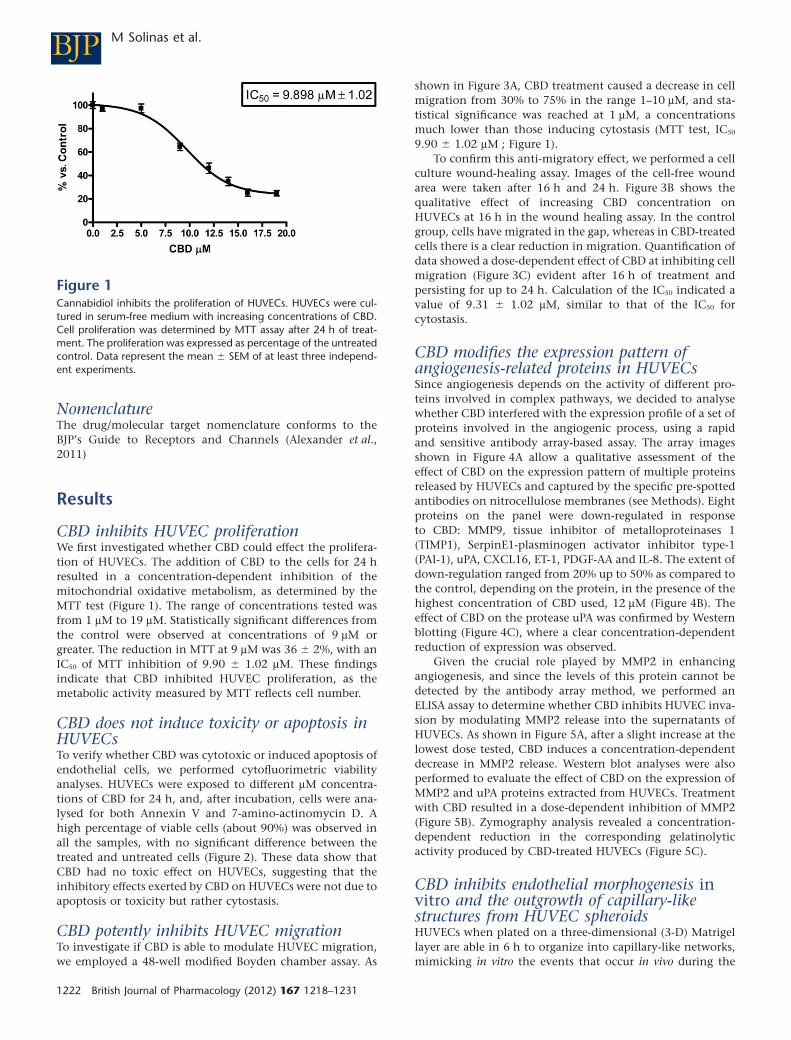
CBD inhibits HUVEC proliferationWe first investigated whether CBD could effect the prolifera-tion of HUVECs. The addition of CBD to the cells for 24 hresulted in a concentration-dependent inhibition of themitochondrial oxidative metabolism, as determined by theMTT test (Figure 1). The range of concentrations tested wasfrom 1 mM to 19 mM. Statistically significant differences fromthe control were observed at concentrations of 9 mM orgreater. The reduction in MTT at 9 mM was 36 � 2%, with anIC50 of MTT inhibition of 9.90 � 1.02 mM. These findingsindicate that CBD inhibited HUVEC proliferation, as themetabolic activity measured by MTT reflects cell number.
CBD does not induce toxicity or apoptosis inHUVECsTo verify whether CBD was cytotoxic or induced apoptosis ofendothelial cells, we performed cytofluorimetric viabilityanalyses. HUVECs were exposed to different mM concentra-tions of CBD for 24 h, and, after incubation, cells were ana-lysed for both Annexin V and 7-amino-actinomycin D. Ahigh percentage of viable cells (about 90%) was observed inall the samples, with no significant difference between thetreated and untreated cells (Figure 2). These data show thatCBD had no toxic effect on HUVECs, suggesting that theinhibitory effects exerted by CBD on HUVECs were not due toapoptosis or toxicity but rather cytostasis.
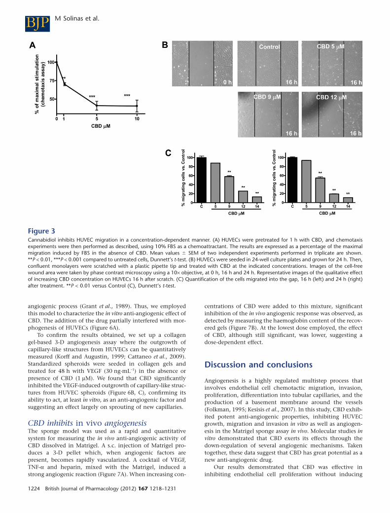
CBD potently inhibits HUVEC migrationTo investigate if CBD is able to modulate HUVEC migration,we employed a 48-well modified Boyden chamber assay. As
shown in Figure 3A, CBD treatment caused a decrease in cellmigration from 30% to 75% in the range 1–10 mM, and sta-tistical significance was reached at 1 mM, a concentrationsmuch lower than those inducing cytostasis (MTT test, IC50
9.90 � 1.02 mM ; Figure 1).To confirm this anti-migratory effect, we performed a cell
culture wound-healing assay. Images of the cell-free woundarea were taken after 16 h and 24 h. Figure 3B shows thequalitative effect of increasing CBD concentration onHUVECs at 16 h in the wound healing assay. In the controlgroup, cells have migrated in the gap, whereas in CBD-treatedcells there is a clear reduction in migration. Quantification ofdata showed a dose-dependent effect of CBD at inhibiting cellmigration (Figure 3C) evident after 16 h of treatment andpersisting for up to 24 h. Calculation of the IC50 indicated avalue of 9.31 � 1.02 mM, similar to that of the IC50 forcytostasis.
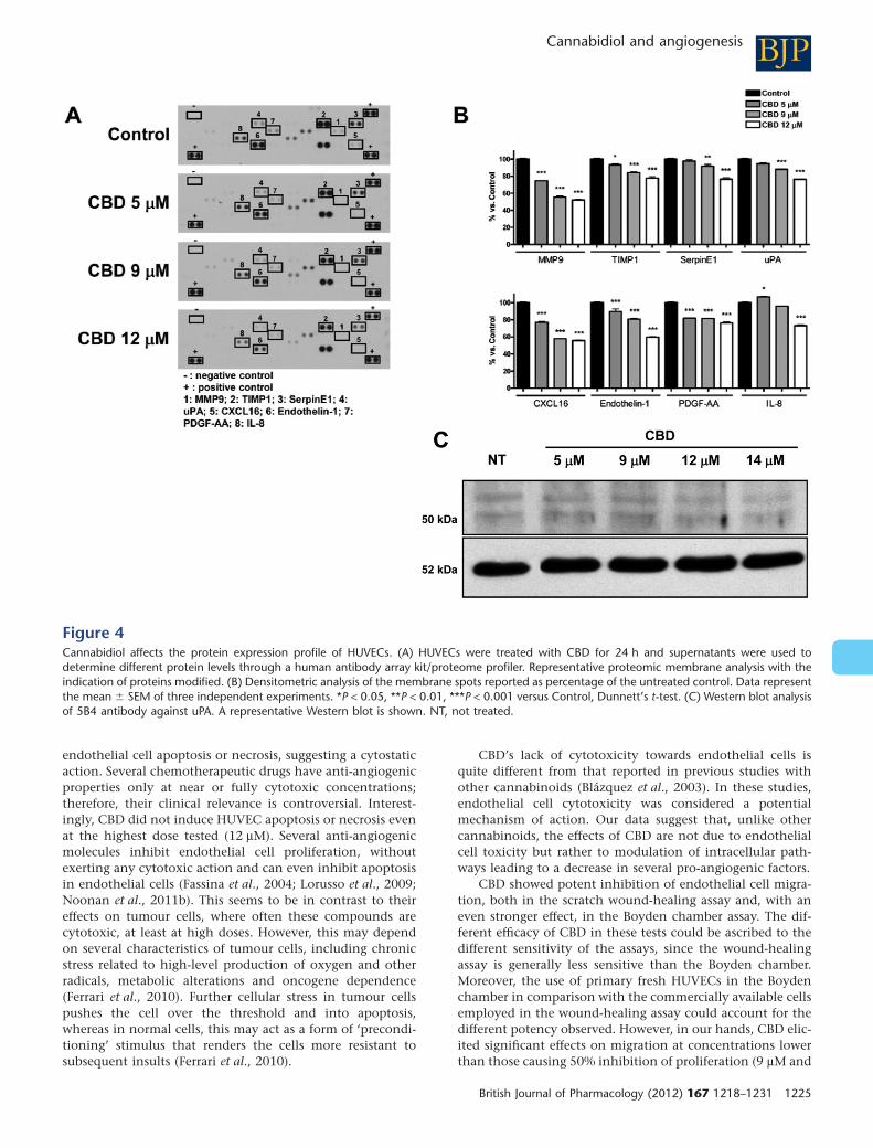
CBD modifies the expression pattern ofangiogenesis-related proteins in HUVECsSince angiogenesis depends on the activity of different pro-teins involved in complex pathways, we decided to analysewhether CBD interfered with the expression profile of a set ofproteins involved in the angiogenic process, using a rapidand sensitive antibody array-based assay. The array imagesshown in Figure 4A allow a qualitative assessment of theeffect of CBD on the expression pattern of multiple proteinsreleased by HUVECs and captured by the specific pre-spottedantibodies on nitrocellulose membranes (see Methods). Eightproteins on the panel were down-regulated in responseto CBD: MMP9, tissue inhibitor of metalloproteinases 1(TIMP1), SerpinE1-plasminogen activator inhibitor type-1(PAI-1), uPA, CXCL16, ET-1, PDGF-AA and IL-8. The extent ofdown-regulation ranged from 20% up to 50% as compared tothe control, depending on the protein, in the presence of thehighest concentration of CBD used, 12 mM (Figure 4B). Theeffect of CBD on the protease uPA was confirmed by Westernblotting (Figure 4C), where a clear concentration-dependentreduction of expression was observed.
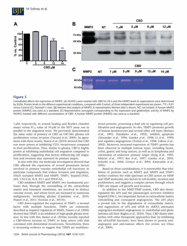
Given the crucial role played by MMP2 in enhancingangiogenesis, and since the levels of this protein cannot bedetected by the antibody array method, we performed anELISA assay to determine whether CBD inhibits HUVEC inva-sion by modulating MMP2 release into the supernatants ofHUVECs. As shown in Figure 5A, after a slight increase at thelowest dose tested, CBD induces a concentration-dependentdecrease in MMP2 release. Western blot analyses were alsoperformed to evaluate the effect of CBD on the expression ofMMP2 and uPA proteins extracted from HUVECs. Treatmentwith CBD resulted in a dose-dependent inhibition of MMP2(Figure 5B). Zymography analysis revealed a concentration-dependent reduction in the corresponding gelatinolyticactivity produced by CBD-treated HUVECs (Figure 5C).
CBD inhibits endothelial morphogenesis invitro and the outgrowth of capillary-likestructures from HUVEC spheroidsHUVECs when plated on a three-dimensional (3-D) Matrigellayer are able in 6 h to organize into capillary-like networks,mimicking in vitro the events that occur in vivo during the
Figure 1Cannabidiol inhibits the proliferation of HUVECs. HUVECs were cul-tured in serum-free medium with increasing concentrations of CBD.Cell proliferation was determined by MTT assay after 24 h of treat-ment. The proliferation was expressed as percentage of the untreatedcontrol. Data represent the mean � SEM of at least three independ-ent experiments.
BJP M Solinas et al.
1222 British Journal of Pharmacology (2012) 167 1218–1231
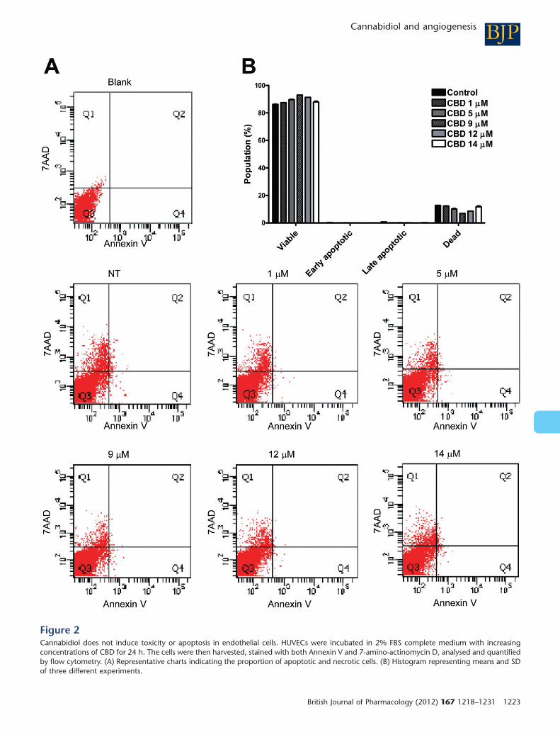
Figure 2Cannabidiol does not induce toxicity or apoptosis in endothelial cells. HUVECs were incubated in 2% FBS complete medium with increasingconcentrations of CBD for 24 h. The cells were then harvested, stained with both Annexin V and 7-amino-actinomycin D, analysed and quantifiedby flow cytometry. (A) Representative charts indicating the proportion of apoptotic and necrotic cells. (B) Histogram representing means and SDof three different experiments.
BJPCannabidiol and angiogenesis
British Journal of Pharmacology (2012) 167 1218–1231 1223
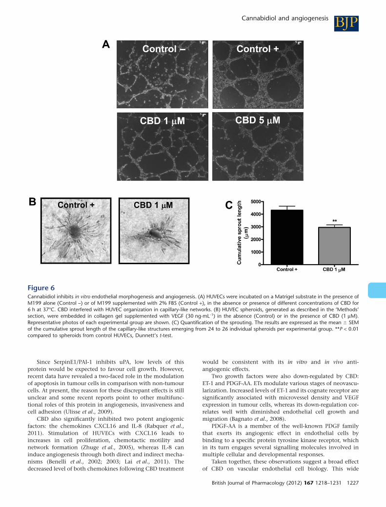
angiogenic process (Grant et al., 1989). Thus, we employedthis model to characterize the in vitro anti-angiogenic effect ofCBD. The addition of the drug partially interfered with mor-phogenesis of HUVECs (Figure 6A).
To confirm the results obtained, we set up a collagengel-based 3-D angiogenesis assay where the outgrowth ofcapillary-like structures from HUVECs can be quantitativelymeasured (Korff and Augustin, 1999; Cattaneo et al., 2009).Standardized spheroids were seeded in collagen gels andtreated for 48 h with VEGF (30 ng·mL-1) in the absence orpresence of CBD (1 mM). We found that CBD significantlyinhibited the VEGF-induced outgrowth of capillary-like struc-tures from HUVEC spheroids (Figure 6B, C), confirming itsability to act, at least in vitro, as an anti-angiogenic factor andsuggesting an effect largely on sprouting of new capillaries.
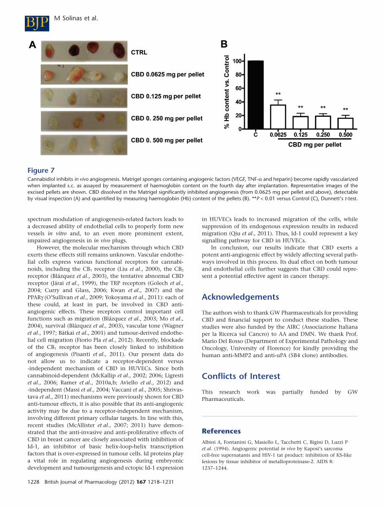
CBD inhibits in vivo angiogenesisThe sponge model was used as a rapid and quantitativesystem for measuring the in vivo anti-angiogenic activity ofCBD dissolved in Matrigel. A s.c. injection of Matrigel pro-duces a 3-D pellet which, when angiogenic factors arepresent, becomes rapidly vascularized. A cocktail of VEGF,TNF-a and heparin, mixed with the Matrigel, induced astrong angiogenic reaction (Figure 7A). When increasing con-
centrations of CBD were added to this mixture, significantinhibition of the in vivo angiogenic response was observed, asdetected by measuring the haemoglobin content of the recov-ered gels (Figure 7B). At the lowest dose employed, the effectof CBD, although still significant, was lower, suggesting adose-dependent effect.
Discussion and conclusions
Angiogenesis is a highly regulated multistep process thatinvolves endothelial cell chemotactic migration, invasion,proliferation, differentiation into tubular capillaries, and theproduction of a basement membrane around the vessels(Folkman, 1995; Kesisis et al., 2007). In this study, CBD exhib-ited potent anti-angiogenic properties, inhibiting HUVECgrowth, migration and invasion in vitro as well as angiogen-esis in the Matrigel sponge assay in vivo. Molecular studies invitro demonstrated that CBD exerts its effects through thedown-regulation of several angiogenic mechanisms. Takentogether, these data suggest that CBD has great potential as anew anti-angiogenic drug.
Our results demonstrated that CBD was effective ininhibiting endothelial cell proliferation without inducing
Figure 3Cannabidiol inhibits HUVEC migration in a concentration-dependent manner. (A) HUVECs were pretreated for 1 h with CBD, and chemotaxisexperiments were then performed as described, using 10% FBS as a chemoattractant. The results are expressed as a percentage of the maximalmigration induced by FBS in the absence of CBD. Mean values � SEM of two independent experiments performed in triplicate are shown.**P < 0.01, ***P < 0.001 compared to untreated cells, Dunnett’s t-test. (B) HUVECs were seeded in 24-well culture plates and grown for 24 h. Then,confluent monolayers were scratched with a plastic pipette tip and treated with CBD at the indicated concentrations. Images of the cell-freewound area were taken by phase contrast microscopy using a 10¥ objective, at 0 h, 16 h and 24 h. Representative images of the qualitative effectof increasing CBD concentration on HUVECs 16 h after scratch. (C) Quantification of the cells migrated into the gap, 16 h (left) and 24 h (right)after treatment. **P < 0.01 versus Control (C), Dunnett’s t-test.
BJP M Solinas et al.
1224 British Journal of Pharmacology (2012) 167 1218–1231
endothelial cell apoptosis or necrosis, suggesting a cytostaticaction. Several chemotherapeutic drugs have anti-angiogenicproperties only at near or fully cytotoxic concentrations;therefore, their clinical relevance is controversial. Interest-ingly, CBD did not induce HUVEC apoptosis or necrosis evenat the highest dose tested (12 mM). Several anti-angiogenicmolecules inhibit endothelial cell proliferation, withoutexerting any cytotoxic action and can even inhibit apoptosisin endothelial cells (Fassina et al., 2004; Lorusso et al., 2009;Noonan et al., 2011b). This seems to be in contrast to theireffects on tumour cells, where often these compounds arecytotoxic, at least at high doses. However, this may dependon several characteristics of tumour cells, including chronicstress related to high-level production of oxygen and otherradicals, metabolic alterations and oncogene dependence(Ferrari et al., 2010). Further cellular stress in tumour cellspushes the cell over the threshold and into apoptosis,whereas in normal cells, this may act as a form of ‘precondi-tioning’ stimulus that renders the cells more resistant tosubsequent insults (Ferrari et al., 2010).
CBD’s lack of cytotoxicity towards endothelial cells isquite different from that reported in previous studies withother cannabinoids (Blázquez et al., 2003). In these studies,endothelial cell cytotoxicity was considered a potentialmechanism of action. Our data suggest that, unlike othercannabinoids, the effects of CBD are not due to endothelialcell toxicity but rather to modulation of intracellular path-ways leading to a decrease in several pro-angiogenic factors.
CBD showed potent inhibition of endothelial cell migra-tion, both in the scratch wound-healing assay and, with aneven stronger effect, in the Boyden chamber assay. The dif-ferent efficacy of CBD in these tests could be ascribed to thedifferent sensitivity of the assays, since the wound-healingassay is generally less sensitive than the Boyden chamber.Moreover, the use of primary fresh HUVECs in the Boydenchamber in comparison with the commercially available cellsemployed in the wound-healing assay could account for thedifferent potency observed. However, in our hands, CBD elic-ited significant effects on migration at concentrations lowerthan those causing 50% inhibition of proliferation (9 mM and
Figure 4Cannabidiol affects the protein expression profile of HUVECs. (A) HUVECs were treated with CBD for 24 h and supernatants were used todetermine different protein levels through a human antibody array kit/proteome profiler. Representative proteomic membrane analysis with theindication of proteins modified. (B) Densitometric analysis of the membrane spots reported as percentage of the untreated control. Data representthe mean � SEM of three independent experiments. *P < 0.05, **P < 0.01, ***P < 0.001 versus Control, Dunnett’s t-test. (C) Western blot analysisof 5B4 antibody against uPA. A representative Western blot is shown. NT, not treated.
BJPCannabidiol and angiogenesis
British Journal of Pharmacology (2012) 167 1218–1231 1225
1 mM, respectively, in wound healing and Boyden chamberassays versus IC50 value of 10 mM in the MTT assay run inparallel to the migration tests). We previously demonstratedthe same order of potency of CBD on U87-MG glioma cellproliferation versus invasion (Vaccani et al., 2005). In agree-ment with these results, Marcu et al. (2010) showed that CBDwas more potent at inhibiting U251 invasiveness comparedto their proliferation. Thus, similar to glioma, CBD is highlypotent at inhibiting endothelial cell migration compared toproliferation, suggesting that factors influencing cell migra-tion and invasion may represent its primary targets.
In line with this, our molecular investigation showed thatCBD affected the expression of several prominent factorsinvolved in primary vascular endothelial cell functions; inparticular compounds that induce invasion and migration,which included MMP2 and MMP9, TIMP1, SerpinE1/PAI1,uPA, CXCL16, IL-8, ET-1 and PDGF-AA.
CBD inhibited MMP2 and MMP9, two fundamental pro-teases that, through the remodelling of the extracellularmatrix and basement membrane, are involved in distinctvascular events, and whose levels are increased in numerousmalignancies, including glioma (Cantelmo et al., 2010;Pisanti et al., 2011; Noonan et al., 2011b).
CBD down-regulated the expression of TIMP1, a stromalfactor with multiple functions. TIMPs are commonlydescribed as negative regulators of MMPs. Nasser et al. (2006)showed that TIMP1 is an inhibitor of high-grade glioma inva-sion. In line with this, Ramer et al. (2010a) recently reporteda CBD-driven increase in TIMP1 in lung cancer cells thatcorrelated with diminished invasiveness. Nevertheless, thereis increasing evidence to suggest that TIMPs are multifunc-
tional proteins, possessing a dual role in regulating cell pro-liferation and angiogenesis. In vitro, TIMP1 promotes growthof human keratinocytes and several other cell types (Bertauxet al., 1991; Hayakawa et al., 1992), inhibits apoptosis(Alexander et al., 1996; Guedez et al., 1998; Li et al., 1999)and regulates angiogenesis (Yoshiji et al., 1998; Lafleur et al.,2002). Moreover, increased expression of TIMP1 protein hasbeen observed in multiple tumour types, including breast,colon, gastric and lung cancers, as well as in lymphoma andcarcinomas of unknown primary origin (Zeng et al., 1995;Mimori et al., 1997; Ree et al., 1997; Guedez et al., 2001;Schrohl et al., 2004; Gouyer et al., 2005; Karavasilis et al.,2005).
Based on these considerations, it is noteworthy that inhi-bition of proteins such as MMP2 and MMP9 and TIMP1further confirms the wide spectrum of CBD action on MMPand TIMP molecules, key factors in cell motility, invasion andproliferation, and suggests a complex picture through whichCBD can impair cell growth and invasion.
In addition to the MMP/TIMP system, CBD also down-regulated the uPA and the plasminogen activator inhibitorSerpinE1/PAI-1, two important factors in extracellular matrixremodelling and consequent angiogenesis. The uPA playsa pivotal role in the degradation of extracellular matrix,and suppression of uPA and uPAR by shRNA attenuatesangiogenin-mediated angiogenesis in endothelial and gliob-lastoma cell lines (Raghu et al., 2010). Thus, CBD shares simi-larities with other therapeutic approaches that, by inhibitingthe uPA/uPAR functions, have been shown to possess anti-angiogenic and anti-tumour effects (for review, see Ulisseet al., 2009).
Figure 5Cannabidiol affects the expression of MMP2. (A) HUVECs were treated with CBD for 24 h and the MMP2 levels in supernatants were determinedby ELISA. Protein levels in the different experimental conditions, compared with Control, of three independent experiments are shown. **P < 0.01versus Control (C), Dunnett’s t-test. (B) Western blot analysis of MMP2. A representative Western blot is shown. NT, not treated. A human MMP2protein (MMP2) was used as a standard. (C) Representative zymogram corresponding to the expression and gelatinolytic activity of MMP2 byHUVECs treated with different concentrations of CBD. A human MMP2 protein (MMP2) was used as a standard.
BJP M Solinas et al.
1226 British Journal of Pharmacology (2012) 167 1218–1231
Since SerpinE1/PAI-1 inhibits uPA, low levels of thisprotein would be expected to favour cell growth. However,recent data have revealed a two-faced role in the modulationof apoptosis in tumour cells in comparison with non-tumourcells. At present, the reason for these discrepant effects is stillunclear and some recent reports point to other multifunc-tional roles of this protein in angiogenesis, invasiveness andcell adhesion (Ulisse et al., 2009).
CBD also significantly inhibited two potent angiogenicfactors: the chemokines CXCL16 and IL-8 (Rabquer et al.,2011). Stimulation of HUVECs with CXCL16 leads toincreases in cell proliferation, chemotactic motility andnetwork formation (Zhuge et al., 2005), whereas IL-8 caninduce angiogenesis through both direct and indirect mecha-nisms (Benelli et al., 2002; 2003; Lai et al., 2011). Thedecreased level of both chemokines following CBD treatment
would be consistent with its in vitro and in vivo anti-angiogenic effects.
Two growth factors were also down-regulated by CBD:ET-1 and PDGF-AA. ETs modulate various stages of neovascu-larization. Increased levels of ET-1 and its cognate receptor aresignificantly associated with microvessel density and VEGFexpression in tumour cells, whereas its down-regulation cor-relates well with diminished endothelial cell growth andmigration (Bagnato et al., 2008).
PDGF-AA is a member of the well-known PDGF familythat exerts its angiogenic effect in endothelial cells bybinding to a specific protein tyrosine kinase receptor, whichin its turn engages several signalling molecules involved inmultiple cellular and developmental responses.
Taken together, these observations suggest a broad effectof CBD on vascular endothelial cell biology. This wide
Figure 6Cannabidiol inhibits in vitro endothelial morphogenesis and angiogenesis. (A) HUVECs were incubated on a Matrigel substrate in the presence ofM199 alone (Control –) or of M199 supplemented with 2% FBS (Control +), in the absence or presence of different concentrations of CBD for6 h at 37°C. CBD interfered with HUVEC organization in capillary-like networks. (B) HUVEC spheroids, generated as described in the ‘Methods’section, were embedded in collagen gel supplemented with VEGF (30 ng·mL-1) in the absence (Control) or in the presence of CBD (1 mM).Representative photos of each experimental group are shown. (C) Quantification of the sprouting. The results are expressed as the mean � SEMof the cumulative sprout length of the capillary-like structures emerging from 24 to 26 individual spheroids per experimental group. **P < 0.01compared to spheroids from control HUVECs, Dunnett’s t-test.
BJPCannabidiol and angiogenesis
British Journal of Pharmacology (2012) 167 1218–1231 1227
spectrum modulation of angiogenesis-related factors leads toa decreased ability of endothelial cells to properly form newvessels in vitro and, to an even more prominent extent,impaired angiogenesis in in vivo plugs.
However, the molecular mechanism through which CBDexerts these effects still remains unknown. Vascular endothe-lial cells express various functional receptors for cannabi-noids, including the CB1 receptor (Liu et al., 2000), the CB2
receptor (Blázquez et al., 2003), the tentative abnormal CBDreceptor (Járai et al., 1999), the TRP receptors (Golech et al.,2004; Curry and Glass, 2006; Kwan et al., 2007) and thePPARg (O’Sullivan et al., 2009; Yokoyama et al., 2011): each ofthese could, at least in part, be involved in CBD anti-angiogenic effects. These receptors control important cellfunctions such as migration (Blázquez et al., 2003; Mo et al.,2004), survival (Blázquez et al., 2003), vascular tone (Wagneret al., 1997; Bátkai et al., 2001) and tumour-derived endothe-lial cell migration (Fiorio Pla et al., 2012). Recently, blockadeof the CB1 receptor has been closely linked to inhibitionof angiogenesis (Pisanti et al., 2011). Our present data donot allow us to indicate a receptor-dependent versus-independent mechanism of CBD in HUVECs. Since bothcannabinoid-dependent (McKallip et al., 2002; 2006; Ligrestiet al., 2006; Ramer et al., 2010a,b; Aviello et al., 2012) and-independent (Massi et al., 2004; Vaccani et al., 2005; Shrivas-tava et al., 2011) mechanisms were previously shown for CBDanti-tumour effects, it is also possible that its anti-angiogenicactivity may be due to a receptor-independent mechanism,involving different primary cellular targets. In line with this,recent studies (McAllister et al., 2007; 2011) have demon-strated that the anti-invasive and anti-proliferative effects ofCBD in breast cancer are closely associated with inhibition ofId-1, an inhibitor of basic helix-loop-helix transcriptionfactors that is over-expressed in tumour cells. Id proteins playa vital role in regulating angiogenesis during embryonicdevelopment and tumourigenesis and ectopic Id-1 expression
in HUVECs leads to increased migration of the cells, whilesuppression of its endogenous expression results in reducedmigration (Qiu et al., 2011). Thus, Id-1 could represent a keysignalling pathway for CBD in HUVECs.
In conclusion, our results indicate that CBD exerts apotent anti-angiogenic effect by widely affecting several path-ways involved in this process. Its dual effect on both tumourand endothelial cells further suggests that CBD could repre-sent a potential effective agent in cancer therapy.
Acknowledgements
The authors wish to thank GW Pharmaceuticals for providingCBD and financial support to conduct these studies. Thesestudies were also funded by the AIRC (Associazione Italianaper la Ricerca sul Cancro) to AA and DMN. We thank Prof.Mario Del Rosso (Department of Experimental Pathology andOncology, University of Florence) for kindly providing thehuman anti-MMP2 and anti-uPA (5B4 clone) antibodies.
Conflicts of Interest
This research work was partially funded by GWPharmaceuticals.
ReferencesAlbini A, Fontanini G, Masiello L, Tacchetti C, Bigini D, Luzzi Pet al. (1994). Angiogenic potential in vivo by Kaposi’s sarcomacell-free supernatants and HIV-1 tat product: inhibition of KS-likelesions by tissue inhibitor of metalloproteinase-2. AIDS 8:1237–1244.
Figure 7Cannabidiol inhibits in vivo angiogenesis. Matrigel sponges containing angiogenic factors (VEGF, TNF-a and heparin) become rapidly vascularizedwhen implanted s.c. as assayed by measurement of haemoglobin content on the fourth day after implantation. Representative images of theexcised pellets are shown. CBD dissolved in the Matrigel significantly inhibited angiogenesis (from 0.0625 mg per pellet and above), detectableby visual inspection (A) and quantified by measuring haemoglobin (Hb) content of the pellets (B). **P < 0.01 versus Control (C), Dunnett’s t-test.
BJP M Solinas et al.
1228 British Journal of Pharmacology (2012) 167 1218–1231
Albini A, Brigati C, Ventura A, Lorusso G, Pinter M, Morini M et al.(2009). Angiostatin anti-angiogenesis requires IL-12: the innateimmune system as a key target. J Transl Med 14: 7–5.
Albini A, Indraccolo S, Noonan DM, Pfeffer U (2010). Functionalgenomics of endothelial cells treated with anti-angiogenic orangiopreventive drugs. Clin Exp Metastasis 27: 419–439.
Alexander A, Smith PF, Rosengren RJ (2009). Cannabinoids in thetreatment of cancer. Cancer Lett 285: 6–12.
Alexander CM, Howard EW, Bissell MJ, Werb Z (1996). Rescue ofmammary epithelial apoptosis and entactin degradation by a tissueinhibitor of metalloproteinase-1 transgene. J Cell Biol 135:1669–1677.
Alexander SPH, Mathie A, Peters JA (2011). Guide to receptors andchannels (GRAC), 5th edition. Br J Pharmacol 164 (Suppl. 1):S1–S324.
Aviello G, Romano B, Borrelli F, Capasso R, Gallo L, Piscitelli F et al.(2012). Chemopreventive effect of the non-psychotropicphytocannabinoid cannabidiol on experimental colon cancer. J MolMed DOI: 10.1007/s00109-011-0856-x [Epub ahead of print].
Bagnato A, Spinella F, Rosanò L (2008). The endothelin axis incancer: the promise and the challenges of molecularly targetedtherapy. Can J Physiol Pharmacol 86: 473–484.
Bátkai S, Járai Z, Wagner JA, Goparaju SK, Varga K, Liu J et al.(2001). Endocannabinoids acting at vascular CB1 receptors mediatethe vasodilated state in advanced liver cirrhosis. Nat Med 7:827–832.
Benelli R, Morini M, Carrozzino F, Ferrari N, Minghelli S, Santi Let al. (2002). Neutrophils as a key cellular target for angiostatin:implications for regulation of angiogenesis and inflammation.FASEB J 16: 267–269.
Benelli R, Albini A, Noonan D (2003). Neutrophils andangiogenesis: potential initiators of the angiogenic cascade. In:Cassatella MA (ed.). The Neutrophil: An Emerging Regulator ofInflammatory and Immune Response. Karger: Basel, pp. 167–181.
Bertaux B, Hornebeck W, Eisen AZ, Dubertret L (1991). Growthstimulation of human keratinocytes by tissue inhibitor ofmetalloproteinases. J Invest Dermatol 97: 679–685.
Blázquez C, Casanova ML, Planas A, Gómez del Pulgar T,Villanueva C, Fernández-Aceñero MJ et al. (2003). Inhibition oftumor angiogenesis by cannabinoids. FASEB J 17: 529–531.
Blázquez C, González-Feria L, Alvarez L, Haro A, Casanova ML,Guzmán M (2004). Cannabinoids inhibit the vascular endothelialgrowth factor pathway in gliomas. Cancer Res 64: 5617–5623.
Blázquez C, Carracedo A, Barrado L, Real PJ, Fernández-Luna JL,Velasco G et al. (2006). Cannabinoid receptors as novel targets forthe treatment of melanoma. FASEB J 20: 2633–2635.
Cantelmo AR, Cammarota R, Noonan DM, Focaccetti C,Comoglio PM, Prat M et al. (2010). Cell delivery of Met docking sitepeptides inhibit angiogenesis and vascular tumor growth.Oncogene 29: 5286–5298.
Casanova ML, Blázquez C, Martínez-Palacio J, Villanueva C,Fernández-Aceñero MJ, Huffman JW et al. (2003). Inhibition of skintumor growth and angiogenesis in vivo by activation ofcannabinoid receptors. J Clin Invest 111: 43–50.
Cattaneo MG, Chini B, Vicentini LM (2008). Oxytocin stimulatesmigration and invasion in human endothelial cells. Br J Pharmacol153: 728–736.
Cattaneo MG, Lucci G, Vicentini LM (2009). Oxytocin stimulates invitro angiogenesis via a Pyk-2/Src-dependent mechanism. Exp CellRes 315: 3210–3219.
Chung AS, Lee J, Ferrara N (2010). Targeting the tumourvasculature: insights from physiological angiogenesis. Nat RevCancer 10: 505–514.
Curry FR, Glass CA (2006). TRP channels and the regulation ofvascular permeability: new insights from the lung microvasculature.Circ Res 99: 915–917.
De Petrocellis L, Ligresti A, Moriello AS, Allarà M, Bisogno T,Petrosino S et al. (2011). Effects of cannabinoids andcannabinoid-enriched Cannabis extracts on TRP channels andendocannabinoid metabolic enzymes. Br J Pharmacol 163:1479–1494.
Fassina G, Venè R, Morini M, Minghelli S, Benelli R, Noonan DMet al. (2004). Mechanisms of inhibition of tumor angiogenesis andvascular tumor growth by epigallocatechin-3-gallate. Clin CancerRes 10: 4865–4873.
Ferrari N, Tosetti F, De Flora S, Donatelli F, Noonan DM, Albini A(2010). Diet-derived phytochemicals: from cancer chemopreventionto cardio-oncological prevention. Curr Drug Targets 12: 1909–1924.
Fiorio Pla A, Avanzato D, Munaron L, Ambudkar IS (2012). Ionchannels and transporters in cancer. 6. Vascularizing the tumor:TRP channels as molecular targets. Am J Physiol Cell Physiol 302:C9–C15.
Flygare J, Sander B (2008). The endocannabinoid system incancer-potential therapeutic target? Semin Cancer Biol 18: 176–189.
Folkman J (1995). Angiogenesis in cancer, vascular, rheumatoid andother disease. Nat Med 1: 27–31.
Folkman J (2007). Angiogenesis: an organizing principle for drugdiscovery? Nat Rev Drug Discov 6: 273–286.
Freimuth N, Ramer R, Hinz B (2010). Antitumorigenic effects ofcannabinoids beyond apoptosis. J Pharmacol Exp Ther 332:336–344.
Gertsch J, Pertwee RG, Di Marzo V (2010). Phytocannabinoidsbeyond the Cannabis plant – do they exist? Br J Pharmacol 160:523–529.
Golech SA, McCarron RM, Chen Y, Bembry J, Lenz F, Mechoulam Ret al. (2004). Human brain endothelium: coexpression and functionof vanilloid and endocannabinoid receptors. Brain Res Mol BrainRes 132: 87–92.
Gouyer V, Conti M, Devos P, Zerimech F, Copin MC, Créme E et al.(2005). Tissue inhibitor of metalloproteinase-1 is an independentpredictor of prognosis in patients with non-small cell lungcarcinoma who undergo resection with curative intent. Cancer 103:1676–1684.
Grant DS, Tashiro K, Segui-Real B, Yamada Y, Martin GR,Kleinman HK (1989). Two different laminin domains mediate thedifferentiation of human endothelial cells into capillary-likestructures in vitro. Cell 58: 933–943.
Guedez L, Stetler-Stevenson WG, Wolff L, Wang J, Fukushima P,Mansoor A et al. (1998). In vitro suppression of programmed celldeath of B cells by tissue inhibitor of metalloproteinases-1. J ClinInvest 102: 2002–2010.
Guedez L, McMarlin AJ, Kingma DW, Bennett TA,Stetler-Stevenson M, Stetler-Stevenson WG (2001). Tissue inhibitorof metalloproteinase-1 alters the tumorigenicity of Burkitt’slymphoma via divergent effects of tumor growth and angiogenesis.Am J Pathol 158: 1207–1215.
BJPCannabidiol and angiogenesis
British Journal of Pharmacology (2012) 167 1218–1231 1229
Guindon J, Hohmann AG (2011). The endocannabinoid system andcancer: therapeutic implication. Br J Pharmacol 163: 1447–1463.
Hayakawa T, Yamashita K, Tanzawa K, Uchijima E, Iwata K(1992). Growth-promoting activity of tissue inhibitor ofmetalloproteinases-1 (TIMP-1) for a wide range of cells. A possiblenew growth factor in serum. FEBS Lett 298: 29–32.
Kilkenny C, Browne W, Cuthill IC, Emerson M, Altman DG (2010).NC3Rs Reporting Guidelines Working Group. Br J Pharmacol 160:1577–1579.
Jaffe EA, Nachman RL, Becker CG, Minick CR (1973). Culture ofhuman endothelial cells derived from umbilical veins.Identification by morphologic and immunological criteria. J ClinInvest 52: 2745–2756.
Járai Z, Wagner JA, Varga K, Lake KD, Compton DR, Martin BR et al.(1999). Cannabinoid-induced mesenteric vasodilation through anendothelial site distinct from CB1 or CB2 receptors. Proc Natl AcadSci U S A 96: 14136–14141.
Karavasilis V, Malamou-Mitsi V, Briasoulis E, Tsanou E, Kitsou E,Kalofonos H et al. (2005). Matrix metalloproteinases in carcinomaof unknown primary. Cancer 104: 2282–2287.
Kesisis G, Broxterman H, Giaccone G (2007). Angiogenesisinhibitors. Drug selectivity and target specificity. Curr Pharm Des13: 2795–2809.
Korff T, Augustin HG (1998). Integration of endothelial cells inmulticellular spheroids prevents apoptosis and inducesdifferentiation. J Cell Biol 143: 1341–1352.
Korff T, Augustin HG (1999). Tensional forces in fibrillarextracellular matrices control directional capillary sprouting. J CellSci 112: 3249–3258.
Kwan HY, Huang Y, Yao X (2007). TRP channels in endothelialfunction and dysfunction. Biochim Biophys Acta 1772: 907–914.
Lafleur MA, Handsley MM, Knäuper V, Murphy G, Edwards DR(2002). Endothelial tubulogenesis within fibrin gels specificallyrequires the activity of membrane-type matrix metalloproteinases(MT-MMPs). J Cell Sci 115: 3427–3428.
Lai Y, Shen Y, Liu XH, Zhang Y, Zeng Y, Liu YF (2011).Interleukin-8 induces the endothelial cell migration through theactivation of phosphoinositide 3-kinase-Rac1/RhoA pathway. Int JBiol Sci 7: 782–791.
Li G, Fridman R, Kim HR (1999). Tissue inhibitor ofmetalloproteinase-1 inhibits apoptosis of human breast epithelialcells. Cancer Res 59: 6267–6275.
Ligresti A, Moriello AS, Starowicz K, Matias I, Pisanti S,De Petrocellis L et al. (2006). Antitumor activity of plantcannabinoids with emphasis on the effect of cannabidiol onhuman breast carcinoma. J Pharmacol Exp Ther 318: 1375–1387.
Liu J, Gao B, Mirshahi F, Sanyal AJ, Khanolkar AD, Makriyannis Aet al. (2000). Functional CB1 cannabinoid receptors in humanvascular endothelial cells. Biochem J 346: 835–840.
Lorusso G, Vannini N, Sogno I, Generoso L, Garbisa S, Noonan DMet al. (2009). Mechanisms of hyperforin as an anti-angiogenicangioprevention agent. Eur J Cancer 45: 1474–1484.
Marcu JP, Christian RT, Lau D, Zielinski AJ, Horowitz MP, Lee Jet al. (2010). Cannabidiol enhances the inhibitory effects ofdelta9-tetrahydrocannabinol on human glioblastoma cellproliferation and survival. Mol Cancer Ther 9: 180–189.
Massi P, Vaccani A, Ceruti S, Colombo A, Abbracchio MP,Parolaro D (2004). Antitumor effects of cannabidiol, anonpsychoactive cannabinoid, on human glioma cell lines. JPharmacol Exp Ther 308: 838–845.
Massi P, Vaccani A, Bianchessi S, Costa B, Macchi P, Parolaro D(2006). The non-psychoactive cannabidiol triggers caspaseactivation and oxidative stress in human glioma cells. Cell Mol LifeSci 63: 2057–2066.
Massi P, Valenti M, Vaccani A, Gasperi V, Perletti G, Marras E et al.(2008). 5-Lipoxygenase and anandamide hydrolase (FAAH) mediatethe antitumor activity of cannabidiol, a non-psychoactivecannabinoid. J Neurochem 104: 1091–1100.
McAllister SD, Christian RT, Horowitz MP, Garcia A, Desprez PY(2007). Cannabidiol as a novel inhibitor of Id-1 gene expression inaggressive breast cancer cells. Mol Cancer Ther 6: 2921–2927.
McAllister SD, Murase R, Christian RT, Lau D, Zielinski AJ, Allison Jet al. (2011). Pathways mediating the effects of cannabidiol on thereduction of breast cancer cell proliferation, invasion, andmetastasis. Breast Cancer Res Treat 129: 37–47.
McKallip RJ, Lombard C, Fisher M, Martin BR, Ryu S, Grant S et al.(2002). Targeting CB2 cannabinoid receptors as a novel therapy totreat malignant lymphoblastic disease. Blood 100: 627–634.
McKallip RJ, Jia W, Schlomer J, Warren JW, Nagarkatti PS,Nagarkatti M (2006). Cannabidiol-induced apoptosis in humanleukemia cells: a novel role of cannabidiol in the regulation ofp22phox and Nox4 expression. Mol Pharmacol 70: 897–908.
McGrath J, Drummond G, McLachlan E, Kilkenny C, Wainwright C(2010). Guidelines for reporting experiments involving animals: theARRIVE guidelines. Br J Pharmacol 160: 1573–1576.
Mimori K, Mori M, Shiraishi T, Fujie T, Baba K, Haraguchi M et al.(1997). Clinical significance of tissue inhibitor of metalloproteinaseexpression in gastric carcinoma. Br J Cancer 76: 531–536.
Mo FM, Offertáler L, Kunos G (2004). Atypical cannabinoidstimulates endothelial cell migration via a Gi/Go-coupled receptordistinct from CB1, CB2 or EDG-1. Eur J Pharmacol 489: 21–27.
Nasser JA, Falavigna A, Ferraz F, Duigou G, Bruce J (2006).Transcription analysis of TIMP-1 and NM23-H1 genes in glioma cellinvasion. Arq Neuropsiquiatr 64: 774–780.
Noonan DM, De Lerma Barbaro A, Vannini N, Mortara L, Albini A(2008). Inflammation, inflammatory cells and angiogenesis:decisions and indecisions. Cancer Metastasis Rev 27: 31–40.
Noonan DM, Ventura A, Bruno A, Pagani A, Albini A (2011a). Theangiogenic switch: role of immune cells. In: Wang E, Marincola F(eds). Immunologic Signatures of Rejection. Springer: New York, pp.57–75.
Noonan DM, Sogno I, Albini A (2011b). Plants and plant-derivedproducts as cancer chemopreventive agents. In: Bagetta G,Cosentino M, Corasaniti MT, Sakurada S (eds). Herbal Medicines:Development and Validation of Plant-Derived Medicines forHuman Health. CRC Press Inc: Boca Raton, FL, pp. 285–306.
O’Sullivan SE, Kendall DA (2010). Cannabinoid activation ofperoxisome proliferator-activated receptors: potential formodulation of inflammatory disease. Immunobiology 215:611–616.
O’Sullivan SE, Sun Y, Bennett AJ, Randall MD, Kendall DA (2009).Time-dependent vascular actions of cannabidiol in the rat aorta.Eur J Pharmacol 612: 61–68.
Pisanti S, Borselli C, Oliviero O, Laezza C, Gazzerro P, Bifulco M(2007). Antiangiogenic activity of the endocannabinoidanandamide: correlation to its tumor-suppressor efficacy. J CellPhysiol 211: 495–503.
BJP M Solinas et al.
1230 British Journal of Pharmacology (2012) 167 1218–1231
Pisanti S, Picardi P, Prota L, Proto MC, Laezza C, McGuire PG et al.(2011). Genetic and pharmacologic inactivation of cannabinoidCB1 receptor inhibits angiogenesis. Blood 117: 5541–5550.
Portella G, Laezza C, Laccetti P, De Petrocellis L, Di Marzo V,Bifulco M (2003). Inhibitory effects of cannabinoid CB1 receptorstimulation on tumor growth and metastatic spreading: actions onsignals involved in angiogenesis and metastasis. FASEB J 17:1771–1773.
Preet A, Ganju RK, Groopman JE (2008).Delta9-tetrahydrocannabinol inhibits epithelial growthfactor-induced lung cancer cell migration in vitro as well as itsgrowth and metastasis in vivo. Oncogene 27: 339–346.
Qiu J, Wang G, Hu J, Peng Q, Zheng Y (2011). Id1-inducedinhibition of p53 facilitates endothelial cell migration and tubeformation by regulating the expression of beta1-integrin. Mol CellBiochem 357: 125–133.
Rabquer BJ, Tsou PS, Hou Y, Thirunavukkarasu E, Haines GK 3rd,Impens AJ et al. (2011). Dysregulated expression of MIG/CXCL9,IP-10/CXCL10 and CXCL16 and their receptors in systemicsclerosis. Arthritis Res Ther 13: R18.
Raghu H, Lakka SS, Gondi CS, Mohanam S, Dinh DH, Gujrati Met al. (2010). Suppression of uPA and uPAR attenuates angiogeninmediated angiogenesis in endothelial and glioblastoma cell lines.PLoS ONE 5: e12458.
Ramer R, Merkord J, Rohde H, Hinz B (2010a). Cannabidiol inhibitscancer cell invasion via upregulation of tissue inhibitor of matrixmetalloproteinases-1. Biochem Pharmacol 79: 955–966.
Ramer R, Rohde A, Merkord J, Rohde H, Hinz B (2010b). Decreaseof plasminogen activator inhibitor-1 may contribute to theanti-invasive action of cannabidiol on human lung cancer cells.Pharm Res 27: 2162–2174.
Ree AH, Florenes VA, Berg JP, Maelandsmo GM, Nesland JM,Fodstad O (1997). High levels of messenger RNAs for tissueinhibitors of metalloproteinases (TIMP-1 and TIMP-2) in primarybreast carcinomas are associated with development of distantmetastases. Clin Cancer Res 3: 1623–1628.
Russo EB (2011). Taming THC: potential cannabis synergy andphytocannabinoid-terpenoid entourage effects. Br J Pharmacol 163:1344–1364.
Schrohl AS, Holten-Andersen MN, Peters HA, Look MP,Meijer-van Gelder ME, Klijn JG et al. (2004). Tumor tissue levels oftissue inhibitor of metalloproteinase-1 as a prognostic marker inprimary breast cancer. Clin Cancer Res 10: 2289–2298.
Shrivastava A, Kuzontkoski PM, Groopman JE, Prasad A (2011).Cannabidiol induces programmed cell death in breast cancer cellsby coordinating the cross-talk between apoptosis and autophagy.Mol Cancer Ther 10: 1161–1172.
Torres S, Lorente M, Rodríguez-Fornés F, Hernández-Tiedra S,Salazar M, García-Taboada E et al. (2011). A combined preclinicaltherapy of cannabinoids and temozolomide against glioma. MolCancer Ther 10: 90–103.
Ulisse S, Baldini E, Sorrenti S, D’Armiento M (2009). The urokinaseplasminogen activator system: a target for anti-cancer therapy. CurrCancer Drug Targets 9: 32–71.
Vaccani A, Massi P, Colombo A, Rubino T, Parolaro D (2005).Cannabidiol inhibits human glioma cell migration through acannabinoid receptor-independent mechanism. Br J Pharmacol 144:1032–1036.
Wagner JA, Varga K, Ellis EF, Rzigalinski BA, Martin BR, Kunos G(1997). Activation of peripheral CB1 cannabinoid receptors inhaemorrhagic shock. Nature 390: 518–521.
Yokoyama Y, Xin B, Shigeto T, Mizunuma H (2011). Combinationof ciglitazone, a peroxisome proliferator-activated receptor gammaligand, and cisplatin enhances the inhibition of growth of humanovarian cancers. J Cancer Res Clin Oncol 137: 1219–1228.
Yoshiji J, Harris SR, Raso E, Gomez DE, Lindsay CK, Shibuya Met al. (1998). Mammary carcinoma cells overexpressing tissueinhibitor of metalloproteinases-1 show enhanced vascularendothelial growth factor expression. Int J Cancer 75: 81–87.
Zeng ZS, Cohen AM, Zhang ZF, Stetler-Stevenson W, Guillem JG(1995). Elevated tissue inhibitor of metalloproteinase I RNA incolorectal cancer stroma correlates with lymph node and distantmetastases. Clin Cancer Res 1: 899–906.
Zhuge X, Murayama T, Arai H, Yamauchi R, Tanaka M, Shimaoka Tet al. (2005). CXCL16 is a novel angiogenic factor for humanumbilical vein endothelial cells. Biochem Biophys Res Commun331: 1295–1300.
BJPCannabidiol and angiogenesis
British Journal of Pharmacology (2012) 167 1218–1231 1231