canine ehrlichiosis-from acute infection to chronic disease (cvbd
TRANSCRIPT
CVBD® DIGEST
ww
w.c
vbd
.org
No.7 December 2010
Cutting-edge information brought to you by the CVBD® World Forum
Canine Ehrlichiosis – from Acute Infection to Chronic Disease
Wide Distribution • Canine monocytic ehrlichiosis (CME) is caused by Ehrlichia canis.• It has been reported in all continents from tropical and subtropical regions
and is probably the most widely distributed CVBD (canine vector-borne disease).
• Distribution is driven by the global abundance of its main vector, the BrownDog tick, Rhipicephalus sanguineus.
And … Increasing • With global warming and expanding tick habitats the spread of disease to
former non-endemic areas is of great concern. • Ehrlichia vectors and infections should also be considered in non-endemic
areas due to increasing international pet travel and dog importation.
Zoonotic Potential • E. chaffeensis (monocytic ehrlichiosis) and E. ewingii (granulocytic ehrlichiosis)
also cause canine ehrlichiosis and both can affect humans.• To date, canine infections with E. chaffeensis and E. ewingii have only been
diagnosed in the United States.
Diagnostic Challenge • Multiple clinical and subclinical presentations make diagnosis challenging.• Acute and chronic phases as well as co-infection with other tick-borne
pathogens may further complicate therapy.
Silent Infections • Often, the pathogen cannot be completely eliminated, despite antibiotic
treatment and resolution of clinical signs.
Prevention • A vaccine for ehrlichiosis is not currently available.• Treatment with an ectoparasiticide product with repelling and killing
activity against ticks presents the best option for prevention.
Quick Digest
CVBD® DIGEST No.7 December 2010Canine Ehrlichiosis – from Acute Infection to Chronic Disease

Author: Juliane Straube Institute for Animal Hygiene and Veterinary Public Health,University of Leipzig, Germany
Ehrlichiosis is a globally distributed canine vector-borne disease (CVBD) transmitted by ticks. Causedby the rickettsial bacteria Ehrlichia spp., ehrlichiosisaffects dogs and humans as well as other domesticand wild animal species. With global warming, ex-panding tick habitats and increasing internationaltravel the spread of disease to former non-endemicareas is of great concern.
Ehrlichiosis can have multiple clinical and subclinicalpresentations making diagnosis challenging. Acuteand chronic phases as well as co-infection withother tick-borne pathogens may further complicatetherapy. Often, the pathogen cannot be completelyeliminated, despite antibiotic treatment and resolu-tion of clinical signs. A vaccine for ehrlichiosis is notcurrently available, so treatment with an ectopara-siticide product with repelling and killing activityagainst ticks presents the best option for preven-tion.
Pathogen/Taxonomy
Ehrlichia spp. are gram-negative obligate intracellu-lar bacteriae with tropism for hematopoietic cells.
Three different Ehrlichia species can cause canineehrlichiosis: E. canis, E. chaffeensis and E. ewingii(see Tab. 1). The term “ehrlichiosis” may still some-times be used to describe infections by organismsbelonging to the former Ehrlichiae tribe. However,with reclassification into the genera Anaplasma,Ehrlichia and Neorickettsia the term now refersspecifically to infections by species within the newlyreorganized genera (see Fig. 2 and Info Box 1).
E. canis causes canine monocytic ehrlichiosis (CME).This disease, also known as tropical canine pancy-topenia, canine rickettsiosis or canine hemorrhagicfever, was first described in Algeria in 1935 by Dona-tien and Lestoquard.1 CME has since been reportedin many parts of the world, mainly in the tropicaland subtropical regions. However, the geo graphicaldistribution of E. canis is expanding alongside thatof its main tick vector, the Brown Dog tick, Rhipi-cephalus sanguineus.
E. canis form microcolonies within a membrane-lined intracellular vacuole (so-called morula), primar-ily in monocytes and macrophages of mammalianhosts. The pathogen replicates only in the cytoplasmof monocytic cells, and the formation of morulae is a defining characteristic that can be used for diag-nosis (see Fig. 1).
Canine ehrlichiosis is also caused by the species E. chaffeensis (monocytic ehrlichiosis) and E. ewingii(granulocytic ehrlichiosis). Both species can also affect humans. Clinical signs of both related diseasesin dogs are indistinguishable from those seen withCME. Discriminating the pathogens by serologicaltesting may be difficult due to a substantial cross-reactivity, mainly between E. canis and E. chaffeen-sis, but also to a lesser degree to E. ewingii. To date, infections with E. chaffeensis and E. ewingii haveonly been diagnosed in dogs in the United States.
Cutting-edge information brought to you by the CVBD World Forum
3No.7 December 2010Canine Ehrlichiosis – from Acute Infection to Chronic Disease CVBD® DIGEST
Canine Ehrlichiosis – from Acute Infection to Chronic Disease
Fig. 1 Intracytoplasmic gram-negative E. canis in monocytes forming morulae. (With kind permission of D. Otranto, Bari, Italy)

CVBD® DIGEST No.7 December 2010Canine Ehrlichiosis – from Acute Infection to Chronic Disease
Co-infections of Ehrlichia with Anaplasma, Ricket t -sia, Babesia or Bartonella spp. occur frequently asdogs are naturally exposed to multiple tick-bornepathogens. Little is known about the clinical out-come of concurrent infections with different pa -thogens. A recently reported study looked at dogsthat were simultaneously and sequentially co-in-fected with E. canis and A. platys. Lower plateletcounts and hematocrit were seen in co-infected an-imals, along with an enhanced humoral immune re-sponse to A. platys and a slower clearance of thatpathogen.2 The awareness of co-infections is impor-tant in clinical practice, as diagnosis may be compli-cated by the presence of multiple pathogens.
Transmission/Vector
Ehrlichiae have a complex life cycle involving a tickvector and a mammalian host. Typically, tick nymphsor larvae are infected with E. canis after feeding ona persistently infected dog. Transstadial transmissionoccurs to subsequent stages of the tick vector. A newhost is infected via salivary gland secretions duringblood feeding. Transmission of the disease has alsobeen reported via blood trans fusion.7
A natural reservoir of infection is maintained in bothwild and domestic canids, including but not limitedto, dogs, wolves, coyotes, and foxes. The failure ofcanids to completely clear E. canis is one importantmechanism of this ongoing persistence and shouldbe considered when selecting canine blood donorsfrom endemic regions.
Zoonotic Potential
A few decades ago, ehrlichioses were considered toonly have veterinary relevance. The first human infection with E. chaffeensis was diagnosed in 1986raising the awareness of Ehrlichia spp. as zoonoticpathogens.8
Note: Nowadays E. canis, E. chaffeensis, andE. ewingii are all known to cause ehrlichiosisin humans.!
4
SpeciesCommonname ofdisease(s)
Common natural host(s)
Cells mostcommonly infected
Primary vector(s) Distribution
E. canisCanine mono-cytic ehrlichio-sis (CME)
Dogs and othermembers of thefamily Canidae,cats, humans
Primarily mono -nuclear cells(monocytes andlymphocytes)
Rhipicephalussanguineus,Dermacentorvariabilis
Worldwide, primarily tropi-cal, subtropical,and temperateclimates
E. chaffeensisHuman mono-cytic ehrlichio-sis (HME)
Humans, deer,horses, rodents
Monocytes, macrophages
Amblyommaamericanum,Dermacentorvariabilis
USA, Europe,Africa, Southand CentralAmerica, Korea
E. ewingii
Canine granulocyticehrlichiosis(CGE), humangranulocyticehrlichiosis (HGE)
Dogs, humansPrimarily neutrophils and eosinophils
Amblyommaamericanum,Otobius megnini
USA, Africa,Korea
E. murisNot currently associated withdisease
Rodents, humans
Mononuclearcells
Haemaphy-salis spp. Japan
E. ruminantium Heartwaterdisease Ruminants Endothelial
cellsAmblyommaspp.
Africa, Caribbean
Tab. 1 Summary of ehrlichial diseases and their related Ehrlichia pathogens.
Note: The failure of canids to completely clear E. canis is one important mechanism of this ongoing persistence.!
No.7 December 2010Canine Ehrlichiosis – from Acute Infection to Chronic Disease CVBD® DIGEST
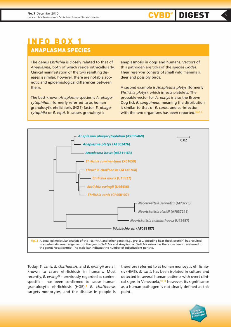
The genus Ehrlichia is closely related to that ofAnaplasma, both of which reside intracellularly.Clinical manifestation of the two resulting dis -eases is similar; however, there are notable zoo-notic and epidemiological differences betweenthem.
The best-known Anaplasma species is A. phago-cytophilum, formerly referred to as human granulocytic ehrlichiosis (HGE) factor, E. phago -cytophila or E. equi. It causes granulocytic
anaplasmosis in dogs and humans. Vectors of this pathogen are ticks of the species Ixodes. Their reservoir consists of small wild mammals,deer and possibly birds.
A second example is Anaplasma platys (formerlyEhrlichia platys), which infects platelets. The probable vector for A. platys is also the BrownDog tick R. sanguineus, meaning the distributionis similar to that of E. canis, and co-infectionwith the two organisms has been reported.3,4,5,6
I N F O B O X 1 ANAPLASMA SPECIES
Today, E. canis, E. chaffeensis, and E. ewingii are allknown to cause ehrlichiosis in humans. Most recently, E. ewingii – previously regarded as canine-specific – has been confirmed to cause human granulocytic ehrlichiosis (HGE).9 E. chaffeensistar gets monocytes, and the disease in people is
therefore referred to as human monocytic ehrlichio-sis (HME). E. canis has been isolated in culture anddetected in several human patients with overt clini-cal signs in Venezuela,10,11 however, its significanceas a human pathogen is not clearly defined at thispoint.
Anaplasma phagocytophilum (AY055469)
Anaplasma platys (AF303476)
Anaplasma bovis (AB211163)
Ehrlichia ruminantium (X61659)
Ehrlichia chaffeensis (AF416764)
Ehrlichia muris (U15527)
Ehrlichia ewingii (U96436)
Ehrlichia canis (CP000107)
Wolbachia sp. (AF088187)
Neorickettsia sennetsu (M73225)
Neorickettsia risticii (AF037211)
Neorickettsia helminthoeca (U12457)
0.02
Fig. 2 A detailed molecular analysis of the 16S rRNA and other genes (e.g., gro ESL, encoding heat shock protein) has resulted in a systematic re-arrangement of the genus Ehrlichia and Anaplasma. Ehrlichia risticii has therefore been transferred tothe genus Neorickettsia. The scale bar indicates the number of substitutions per site.
5

CVBD® DIGEST No.7 December 2010Canine Ehrlichiosis – from Acute Infection to Chronic Disease
6
To date, there is no evidence of direct transmissionof Ehrlichia spp. from dogs to humans,12,13 and dogshave not been established as a reservoir for humaninfection. Additionally, the Brown Dog tick wouldnot appear to be the main vector or reservoir involved in zoonotic transmission because it rarelybites humans.14
Distribution
E. canis organisms are found on all continentsthrough out the world but are more prevalent intropical and subtropical climates (see Fig. 3). Infec-tions with E. chaffeensis and E. ewingii in dogs areprobably restricted to the United States. With in-creasing global mobility of dogs, a diagnosis ofEhrlichia infection should not be ruled out in non-endemic areas particularly given the chronic stageof the disease.
Clinical Presentation
Clinical signs and the severity of illness seen withehrlichiosis depend on the species of Ehrlichiainvolved and the immune response of the dog. In general, all breeds of dogs are susceptible to E. canisinfection, but German shepherds seem to develop severe forms of the disease more frequently thanother breeds.15
CME is characterized by three stages, acute, subclinical and chronic. These can be difficult to definitively distinguish in practice.
Fig. 4 Pale conjunctival mucosa due to anemia caused by E. canis infection. (With permission of D. Otranto, Bari, Italy)
Fig. 5 Ecchymoses as clinical signs of canine E. canis infection.(With permission of D. Otranto, Bari, Italy)
Fig. 3 Geographical distribution of canine ehrlichiosis in different parts of the world. Countries where the endemic occur ence hasbeen reported are highlighted in red. The data were gathered by Bayer HealthCare Animal Health from recent scientificpublications to provide a comprehensive picture of the endemic situation of several CVBDs including ehrlichiosis by E. canisin Asia-Pacific (Fig. 3a), Europe (Fig. 3b) and Latin America (Fig. 3c). More specific regional information can be obtained fromwww.cvbd.org.
No occurrence
Endemic occurrence
Note: Due to international pet travel and import of dogs from endemic areas, Ehrlichiainfections have to be considered also in non-endemic areas.
!

Acute PhaseAcute disease lasts between 3 to 5 weeks with clinical findings of fever, anorexia, depression, lymph adenopathy, and splenomegaly. More variably, ocular discharge, pale mucous membranes, hemor-rhagic tendencies (dermal petechiae, ecchymoses, or epistaxis), or neurological signs are seen (see Figs.4 and 5). The most commonly observed hemato -logical abnormalities are thrombocytopenia andanemia.16
Subclinical PhaseA long-term subclinical phase usually follows thesubsidence of clinical signs and can last for severalyears.17 Dogs that are unable to eliminate the infec-
tious agent develop subclinical persistent infectionsand become asymptomatic carriers.
Chronic PhaseSome infected dogs progress to a chronic phase,which can be mild or severe. This is characterized byrecurrent clinical and hematological signs includingthrombocytopenia, anemia, and pancytopenia.
Dogs may have weight loss, depression, petechiae,pale mucous membranes, edema, and lympha d -enopathy among other signs. In severe cases, the response to antibiotic therapy is poor and dogsoften die from massive hemorrhage, severe debili-
Note: Dogs unable to eliminate the infectiousagent develop subclinical persistent in fec tionsand become asymptomatic carriers.!
No.7 December 2010Canine Ehrlichiosis – from Acute Infection to Chronic Disease CVBD® DIGEST
• A complete blood count is an important toolfor the diagnosis of CME. Moderate to severe thrombocytopenia is a characteristic finding of acute ehrlichiosis.
• Thrombocytopenia appears around day 10and peaks in the third week post-infection,with platelet counts ranging from 20,000 to52,000/µl (normal range: 200– 450,000/µl).There can also be mild anemia and leukopenia.
• In endemic regions, platelet counts on a bloodsmear are used as a screening test for CME.20
True thrombocytopenia can also be distin -guished from in vitro pseudo-thrombocytope-nia by evaluation of platelet numbers on ablood smear.19 Granular lymphocytosis canoccur occasionally during the acute phase and lead to a misdiagnosis of lymphocytic leukemia.
• Hypoalbuminemia, hyperglobulinemia, and hypergammaglobulinemia (mostly polyclonal,
rarely monoclonal) are common in CME. Alsomoderate increases in alanine aminotrans -ferase (ALT) and alkaline phosphatase (ALP)can occur due to hepatocyte damage duringthe acute phase.
• Dogs in the subclinical phase are clinically healthy, but variable degrees of thrombo -cytopenia and leukopenia may be present.Thrombocytopenia usually becomes severe in the chronic phase accompanied by markedanemia and leukopenia. Pancytopenia due tobone marrow hypoplasia is characteristic ofthe chronic severe form.21
• A hypocellular bone marrow with varying supression of the erythroid, myeloid, and megakaryocytic cells is seen on aspiration.
• E. canis can occasionally induce a protein-losing nephropathy as a result of immune-complex glomerulonephritis with consequentproteinuria and azotemia.
I N F O B O X 2 LABORATORY FINDINGS (HEMATOLOGY / BIOCHEMISTRY)
Note: The most commonly observed hemato-logical abnormalities are thrombocyto peniaand anemia.16!
7

CVBD® DIGEST No.7 December 2010Canine Ehrlichiosis – from Acute Infection to Chronic Disease
8
tation, or secondary infections. It is very likely that E. canis causes immunosup pression but currently little is known about the immunobiology of this infection. A recent study in dogs was unable todemon strate a marked immunosuppression.18
Diagnosis
Light microscopy and blood culture tend to be lesssensitive than serology and PCR. Co-infections withother tick-borne pathogens may complicate diagno-sis (see Info Box 2).
Blood Smear Microscopy Detection of typical intracellular E. canis-morulae on blood smear examination is highly specific forehrlichiosis. However, this method is time-consum-ing and not very reliable because morulae are onlyfound in low numbers in blood smears during theacute phase of infection. Microscopy has an esti-mated sensitivity of 4%.22 Detection of morulae canbe improved by evaluation of numerous buffy coatsmears.23
Cell CultureIt is possible to culture Ehrlichia species in specificmacrophage cell lines (canine macrophage cell line[DH82] or mouse macrophage cell line [J774.A1]).However, this technique is used more in research laboratories than for diagnosis in practice.
SerologyThe indirect fluorescent antibody test (IFAT) is re -commended to confirm a diagnosis of ehrlichiosis.24
Detection of specific IgG antibodies indicates previ-ous exposure to the ehrlichial pathogen, and duringthe acute disease two tests one to two weeks apartwill show rising antibody titers. However, there is extensive serologic cross-reactivity between E. canisand E. chaffeensis and E. ewingii.25 Thus, results ob-tained by IFAT need to be interpreted carefully. LowIFAT titers are of low specifity.
Enzyme-linked immunosorbent assays (ELISA) canalso be used to confirm a diagnosis of ehrlichiosisand different Dot-ELISA kits for the detection of E. canis-IgG antibodies are commercially available.Western immunoblot is a more specific test, whichcan distinguish between infections with the differ-ent organisms causing ehrlichiosis, anaplasmosis, orneorickettsiosis as well as between Ehrlichia spp., forexample E. canis and E. ewingii. Dogs will generallybecome seronegative following antibiotic treat-ment, but some dogs will show stable antibody titersfor years.26
Molecular Detection by PCRPCR techniques are now considered to be the mostreliable method to diagnose ehrlichial infection.19
PCR methods are highly sensitive and enable the detection of Ehrlichia DNA as early as 4–10 dayspost-infection prior to sero-conversion.27 Numerouscon ventional and real-time PCRs are available basedon different gene sequences.
PCR can be performed on whole blood, serum,splenic aspirates, lymph nodes, or bone marrow. Thespleen is the organ most likely to harbor E. canis parasites during the subclinical phase21 and is con-sidered to reveal higher sensitivity than testing ofbone marrow or blood samples.28,29 To evaluateelimination of Ehrlichia bacteria following treat-ment, testing of spleen samples is recommended.
Differential Diagnosis
In general, ehrlichiosis should be suspected in dogswith pancytopenia, thrombocytopenia, and aplas -tic anemia in areas endemic for the tick vector, R. sanguineus. But depending on the geographic region, similar clinical signs can occur with other relevant CVBD pathogens. Anaplasmosis, canineRocky Moun tain spotted fever (another rickettsiosis),babesiosis, bartonellosis, hepatozoonosis, and caninedistemper should all be considered as possible dif-ferential diagnoses for ehrlichiosis. Molecular char-acterization by PCR and sequencing may be requiredto finally determine the specific pathogen involved.
Note: Clinical findings with ehrlichiosis can be similar to other CVBD.!
Note: PCR techniques are suggested to be the most reliable method to diagnose ehrlichial infection.!
No.7 December 2010Canine Ehrlichiosis – from Acute Infection to Chronic Disease CVBD® DIGEST
Autoimmune-mediated thromboytopenia, systemiclupus erythematosus or neoplasia (lymphoma ormultiple myeloma) should also be considered.
Treatment
Tetracyclines are the treatment of choice for rick-ettsial diseases. For canine ehrlichiosis, tetracycline(22 mg/kg given every eight hours) or doxycycline (5 mg/kg every twelve hours) administered for fourweeks is the recognized treatment. Most dogs re-cover from the acute and subclinical phases whentreated with doxycycline or other tetracyclines at appropriate dosages for an adequate period oftime.28,29
After initiation of treatment, a rapid improvementin clinical signs is usually seen, but several weeks oftherapy are usually required to ensure a full recov-ery. Persistent infections with E. canis often remainas complete bacterial clearance is not guaranteedbut has been reported in some cases following antibiotic therapy.29–33 It has been suggested thatthe phase of CME could affect the efficacy of doxy-cycline treatment in clearing E. canis infections.33
The extent to which antibiotic treatment can prevent transmission of the pathogen from an infected dog to feeding ticks remains unclear. Experimentally infected dogs treated with doxy -cycline for 14 days were still infectious to ticks andthus reservoirs of E. canis infection.33
Supportive therapy such as blood or fluid trans-fusions and anabolic steroids may be required in severe cases. The prognosis becomes poor once dogsenter the chronic phase of disease.34 Co-in fectionswith other pathogens like Babesia or Barto nella maycontribute to the fatal outcome of chronic infec-tions.
As long-term protective immunity does not developto ehrlichiosis, dogs can be reinfected. Also recru -des cence can occur months to years after primary infection.
Prevention
There are no vaccines currently available to protectdogs from Ehrlichia spp. infections, and further re-search is needed to define the virulence factors andimmunoprotective antigens required to develop one.
The best means of preventing canine ehrlichiosis isby avoiding exposure to the tick vector. Treatmentswith ectoparasiticides that repel and kill ticks reducethe risk of disease transmission. Spot-on products areapplied topically to the dog’s skin. Recent studieshave evaluated the efficacy of a spot-on formulationcontaining imidacloprid 10% and permethrin 50%(Advantix®) to prevent tick exposure and thus E. canisinfection in dogs. Preventive efficacies of 95–100%were demonstrated in treated dogs living under natural conditions in endemic areas.35,36
Note: Due to the fact that no long-lastingprotective immunity is developed, dogs canbe reinfected with ehrlichiosis.!
CANINE EHRLICHIOSIS IN THE WEB
• Background information:www.cvbd.org/4001.0.html
• Menn B et al. Parasites & Vectors 2010, 3:34www.parasitesandvectors.com/content/3/1/34
• Gaunt SD et al. Parasites & Vectors 2010, 3:33www.parasitesandvectors.com/content/3/1/33
• U.S. Centers for Disease Control and Preven-tion: www.cdc.gov/ticks/diseases/ehrlichiosis/
• ACVIM Consensus Statement: 3 www3.interscience.wiley.com/cgi-bin/fulltext/119824370/PDFSTART
I N F O B O X 3
9

CVBD® DIGEST No.7 December 2010Canine Ehrlichiosis – from Acute Infection to Chronic Disease
References
1. Donatien, A., Lestoquard, F. (1937): State of the present knowledge concerning rickettsiosis of animals. Arch. Inst. Pasteur Alger. 5, 142–187
2. Gaunt, S., Beall, M., Stillman, B., Lorentzen, L., Diniz, P., Chandrashekar, R., Breitschwerdt, E.B. (2010):Experimental infection and co-infection of dogs with Anaplasma platys and Ehrlichia canis: hematologic, serologic and molecular findings. Parasit. Vectors 3(1), 33
3. Harrus, S., Aroch, I., Lavy, E., Bark, H. (1997): Clinical manifestations of infectious canine cyclic thrombocytopenia. Vet. Rec. 141, 247–250
4. Cardoso, L., Tuna, J., Vieira, L., Yisaschar-Mekuzas, Y., Baneth, G. (2008): Molecular detection ofAnaplasma platys and Ehrlichia canis in dogs from the North of Portugal. Vet. J. 183, 232–233
5. Gal, A., Loeb, E., Yisaschar-Mekuzas, Y., Baneth, G. (2008): Detection of Ehrlichia canis by PCR in different tissues obtained during necropsy from dogs surveyed for naturally occurring canine monocytic ehrlichiosis. Vet. J. 175, 212–217
6. Diniz, P.P.V.P., Beall, M.J., Omark, K., Chandrashekar, R., Daniluk, D.A., Cyr, K.E., Koterski, J.F., Robbins,R.G., Lalo, P.G., Hegarty, B.C., Breitschwerdt, E.B. (2010): High prevalence of tick-borne pathogens indogs from an Indian reservation in Northeastern Arizona. Vector Borne Zoonotic Dis. 10(2), 117–123
7. Ettinger, S.J., Feldman, E.C., Edward, C. (1995): In: Ettinger S.J., Feldmann E.C. (Ed.): Textbook of Veterinary Internal Medicine. 4th ed. Philadelphia, PA, W.B. Saunders Company, 422–428
8. Maeda, K., Markowitz, N., Hawley, R.C., Ristic, M., Cox, D., McDade, J.E. (1987): Human infection withEhrlichia canis, a leukocytic rickettsia. New Engl. J. Med. 316(14), 853– 856
9. Buller, R.S., Arens, M., Hmiel, S.P., Paddock, C.D., Rikhisa, Y., Unver, A., Gaudreault-Keener, M., Liddell, A.M., Schmulewitz, N., Storch, N.A. (1999): Ehrlichia ewingii, a newly recognized agent ofhuman ehrlichiosis. New Engl. J. Med. 341, 148–155
10. Perez, M., Rikihisa, Y., Wen, B. (1996): Ehrlichia canis-like agent isolated from a man in Venezuela:antigenic and genetic characterization. J. Clin. Microbiol. 34, 2133–2139
11. Perez, M., Bodor, M., Zhang, C., Xiong, Q., Rikihisa, Y. (2006): Human infection with Ehrlichia canisaccompanied by clinical signs in Venezuela. Ann. N. Y. Acad. Sci. 1078, 110–117
12. Fishbein, D.B., Sawyer, L.A., Holland, C.J. (1987): Unexplained febrile illnesses after exposure to ticks:infection with an Ehrlichia? J. A. M. A. 257, 3100–3104
13. Fishbein, D.B., Taylor, J.P., Dawson, J. (1987): Human Ehrlichiosis in the United States (Abstract no.1277). In: Program and abstracts of the Twenty-Seventh Interscience Conference on AntimicrobialAgents and Chemotherapy. Washington, DC, American Society for Microbiology, 319
14. Nelson, V.A. (1969): Human parasitism by the Brown Dog tick. J. Econ. Entomol. 62, 710–712
15. Nyindo, M., Huxsoll, D.L., Ristic, M., Kakoma, I., Brown, J.L., Carson, C.A., Stephenson, E.H. (1980): Cell-mediated and humoral immune responses of German Shepherd Dogs and Beagles to experimental infection with Ehrlichia canis. Am. J. Vet. Res. 41, 250–254
16. Harrus, S., Waner, T., Bark, H. (1997): Canine monocytic ehrlichiosis update. Compend. Contin. Educ.Pract. Vet.19, 431–444
17. Waner, T., Harrus, S., Bark, H., Bogin, E., Avidar, Y., Keysary, A. (1997): Characterization of the subclinicalphase of canine ehrlichiosis in experimentally infected Beagle dogs. Vet. Parasitol. 69, 307–317
18. Hess, P.R., English, R.V., Hegarty, B.C., Brown, G.D., Breitschwerdt, E.B. (2006): Experimental Ehrlichiacanis infection in the dog does not cause immunosuppression. Vet. Immunol. Immunopathol.109(1–2), 117–125
19. Harrus, S., Waner, T. (2010): Diagnosis of canine monocytotropic ehrlichiosis (Ehrlichia canis): an overview. Vet. J. Mar. 10, in press. [Epub ahead of print]
20. Bulla, C., Kiomi Takahira, R., Pessoa Araujo Jr., J., Aparecida Trinca, L., Souza Lopes, R., Wiedmeyer, C.E. (2004): The relationship between the degree of thrombocytopenia and infection with Ehrlichiacanis in an endemic area. Vet. Res. 35, 141–146
10

No.7 December 2010Canine Ehrlichiosis – from Acute Infection to Chronic Disease CVBD® DIGEST
21. Harrus, S., Kass, P.H., Klement, E., Waner, T. (1997): Canine monocytic ehrlichiosis: a retrospective studyof 100 cases, and an epidemiological investigation on prognostic indicators for the disease. Vet. Rec.141, 360–363
22. Woody, B.J., Hoskins, J.D. (1991): Ehrlichial diseases of dogs. Vet. Clin. North Am. Small Anim. Pract.21, 75–98
23. Mylonakis, M.E., Koutinas, A.F., Billinis, C., Leontides, L.S., Kontos, V., Papadopoulos, O., Rallis, T., Fytianou, A. (2003): Evaluation of cytology in the diagnosis of acute canine monocytic ehrlichiosis(Ehrlichia canis): a comparison between five methods. Vet. Microbiol. 91, 197–204
24. Waner, T., Harrus, S., Jongejan, F., Bark, H., Keysary, A., Cornelissen, A.W. (2001): Significance of serological testing for ehrlichial diseases in dogs with special emphasis on the diagnosis of caninemonocytic ehrlichiosis caused by Ehrlichia canis. Vet. Parasitol. 95, 1–15
25. Cardenas, A.M., Doyle, C.K., Zhang, X., Nethery, K., Corstvet, R.E., Walker, D.H., McBride, J.W. (2007):Enzyme-linked immunosorbent assay with conserved immunoreactive glycoproteins gp36 and gp19has enhanced sensitivity and provides species-specific immunodiagnosis of Ehrlichia canis infection.Clin. Vaccine Immunol. 14, 123–128
26. Breitschwerdt, E.B. (2007): Canine and Feline Anaplasmosis: Emerging Infectious Diseases. In: Proceedings of the Second Canine Vector-Borne Disease Symposium. Germany, Bayer HealthCare AG,Animal Health, 6–14
27. Iqbal, Z., Chaichanasiriwithaya, W., Rikihisa, Y. (1994): Comparison of PCR with other tests for earlydiagnosis of canine ehrlichiosis. J. Clin. Microbiol. 32, 1658 –1662
28. Harrus, S., Waner, T., Aizenberg, I., Foley, J.E., Poland, A.M., Bark, H. (1998): Amplification of ehrlichialDNA from dogs 34 months after infection with Ehrlichia canis. J. Clin. Microbiol. 36, 73–76
29. Harrus, S., Kenny, M., Miara, L., Aizenberg, I., Waner, T., Shaw, S. (2004): Comparison of simultaneoussplenic sample PCR with blood sample PCR for diagnosis and treatment of experimental Ehrlichia canisinfection. Antimicrob. Agents Chemother. 48, 4488– 4490
30. Iqbal, Z., Rikihisa, Y. (1994): Reisolation of Ehrlichia canis from blood and tissues of dogs after doxycy-cline treatment. J. Clin. Microbiol. 32, 1644 –1649
31. Harrus, S., Waner, T., Aizenberg, I., Bark, H. (1998): Therapeutic effect of doxycycline in experimentalsubclinical canine monocytic ehrlichiosis: evaluation of a 6-week course. J. Clin. Microbiol. 36,2140–2142
32. Breitschwerdt, E.B., Hegarty, B.C., Hancock, S.I. (1998): Doxycycline hyclate treatment of experimentalcanine ehrlichiosis followed by challenge inoculation with two Ehrlichia canis strains. Antimicrob.Agents. Chemother. 42, 362–368
33. Schaefer, J.J., Needham, G.R., Bremer, W.G., Rikihisis, Y., Ewing, S.A., Stich, R.W. (2007): Tick acquisitionof Ehrlichia canis from dogs treated with doxycycline hyclate. Antimicrob. Agents Chemother. 51(9),3394 –3396
34. Mylonakis, M.E., Koutinas, A.F., Breitschwerdt, E.B., Hegarty, B.C., Billinis, C.D., Leontides, L.S., Kontos,V.S. (2004): Chronic canine ehrlichiosis (Ehrlichia canis): a retrospective study of 19 natural cases. J. Am.Anim. Hosp. Assoc. 40(3),174–184
35. Otranto, D., de Caprariis, D., Lia, R.P., Tarallo, V., Lorusso, V., Testini, G., Dantas-Torres, F., Latrofa, S.,Diniz, P.P., Mencke, N., Maggi, R., Breitschwerdt, E.B., Capelli, G., Stanneck, D. (2010): Prevention of endemic canine vector-borne diseases using imidacloprid 10% and permethrin 50% in young dogs: a longitudinal field study. Vet. Parasitol. 172(3–4), 323–332
36. Otranto, D., Paradies, P., Testini, G., Latrofa, M.S., Weigl, S., Mencke, N., Capariis, D., Parisi, A., Capelli, G., Stanneck, D. (2008): Application of 10% imidacloprid/50% permethrin to preventEhrlichia canis exposure in dogs under natural conditions. Vet. Parasitol. 153, 320–328
* Members of the CVBD World Forum
11

No.7 December 2010Canine Ehrlichiosis – from Acute Infection to Chronic DiseaseCVBD® DIGEST
www.cvbd.org