cancer research visible drug delivery by supramolecular ...quently, polymeric micelles have been...
TRANSCRIPT

Integ
VisiDirePan
SachiIchio
Abst
Intro
Rectherapdrugstractalatestcific eonly slwith otypicaintersttuzumtives tfor can
AuthorMediciEngineeIntegraRadiatiAdvancof TokyRadioloUnivers
Note: SOnline
CorresEnginee7-3-1 H7138; F
doi: 10
©2010
www.a
Dow
Published OnlineFirst August 4, 2010; DOI: 10.1158/0008-5472.CAN-10-0303
Canceresearch
rated Systems and Technologies
ble Drug Delivery by Supramolecular Nanocarrierscting to Single-Platformed Diagnosis and Therapy of
R
creatic Tumor Model
ko Kaida1,3,7, Horacio Cabral1,3, Michiaki Kumagai1, Akihiro Kishimura2, Yasuko Terada4, Masaki Sekino5,
Aoki6, Nobuhiro Nishiyama1,3, Toru Tani7, and Kazunori Kataoka1,2,3ractNan
challetifuncneticchelatpancrehancehighessynchr
o developicer cells,
s' Affiliatine, Graduaring, Gradution, The Unon Researced Energy,o, Chiba, Jgical Sciencity of Medic
upplementa(http://cance
ponding Aring, Graduongo, Bunax: 81-3-58
.1158/0008-
American A
acrjourna
nloaded
oparticle therapeutics are promising platforms for cancer therapy. However, it remains a formidablenge to assess their distribution and clinical efficacy for therapeutic applications. Here, by using mul-tional polymeric micellar nanocarriers incorporating clinically approved gadolinium (Gd)–based mag-resonance imaging contrast agents and platinum (Pt) anticancer drugs through reversible metalion of Pt, simultaneous imaging and therapy of an orthotopic animal model of intractable humanatic tumor was successfully performed without any serious toxicity. The strong tumor contrast en-ment achieved by the micelles correlated with the 24 times increase of r1 of the Gd chelates, thet for the formulations using clinically approved Gd chelates reported to date. From the micro-otron radiation X-ray fluorescence spectrometry scanning of the lesions, we confirmed that bothd chelates and Pt drugs delivered by the micelles selectively colocalized in the tumor interior. Our
the Gstudy provides new insights for the design of theranostic micelles with high contrast enhancement andsite-specific clinical potential. Cancer Res; 70(18); 7031–41. ©2010 AACR.
in thetumorscaletumorIn t
tics plmericdrugsto impfects onanop(16) ananop
duction
ently, there has been explosive development of chemo-eutic agents for cancer, but the efficacies of anticancerare still insufficient particularly for the treatment of in-ble tumors, including pancreatic cancer. Although theadvances in molecular targeting agents have shown spe-fficiency, the survival time of patients is often extendedightly, even when these agents are used in combinationther anticancer drugs. Moreover, the use of such drugslly results in various characteristic side effects, such asitial pneumonia for gefitinib (1), cardiotoxicity for tras-ab (2, 3), and thrombosis for bevacizumab (4). Alterna-
ng these compounds and antibodies selectivewith the aim of modulating drug distribution
abilitycreasesuch aabilitypoundcancethe aua self-of hydwater-12). Thincorpstanceas nucby englymersspons
ons: 1Center for Disease Biology and Integrativete School of Medicine, 2Department of Materialsate School of Engineering, and 3Center for NanoBioiversity of Tokyo, Tokyo, Japan; 4Japan Synchrotronh Institute, SPring-8, Hyogo, Japan; 5Department ofGraduate School of Frontier Sciences, The Universityapan; 6Molecular Imaging Center, National Institute ofes, Chiba, Japan; and 7Department of Surgery, Shigaal Science, Shiga, Japan
ry data for this article are available at Cancer Researchrres.aacrjournals.org/).
uthor: Kazunori Kataoka, Department of Materialsate School of Engineering, The University of Tokyo,kyo-ku, Tokyo 113-8656, Japan. Phone: 81-3-5841-41-7139; E-mail: [email protected].
5472.CAN-10-0303
ssociation for Cancer Research.
ls.org
on June 4, 2020. © 2cancerres.aacrjournals.org from
body to accomplish selective drug accumulation in thesite, are thus needed, and for this purpose, nanometric-vehicles or nanocarriers directing therapeutics to thesite are a key platform.he last decade, several kinds of nanoparticle therapeu-atforms, including liposomes, nanoparticles, and poly-micelles, have been developed to selectively deliverto tumor sites (5–12). These approaches have been usedrove the therapeutic efficacy and to reduce the side ef-f drugs incorporated in delivery carriers (13, 14), andarticle therapeutics such as Doxil (15) or Abraxanere already in clinical use. The tumor targeting of thesearticle therapeutics is based on the enhanced perme-and retention (EPR) effect (in other words, the in-d accumulation of high–molecular weight compounds,s nanoparticles, in tumor tissue due to the high perme-of tumor blood vessels and the retention of these com-s because of the impaired lymphatic drainage at ther site; ref. 17). In the late 1980s, we developed one ofspicious nanoparticle therapeutics, polymeric micelles,assembly of amphiphilic block copolymers consistingrophobic segments forming the drug-loaded core andsoluble segments forming the biocompatible shell (11,e main advantages of this system are the possibility oforating a variety of drugs, including hydrophobic sub-s, metal complexes, and charged macromolecules suchleic acids, as well as controlling their release propertiesineering and modifying the micelle-forming block copo-
. Moreover, polymeric micelles can be designed to be re-ive to environmental changes and capable of target7031
010 American Association for Cancer Research.

recognmycinoxalipspectiof the(18–21micellagainsbreastceptorquentthe mof canAlth
been amatinare intheir dwouldin eactherapbutiontude o
the depeuticalizatiand tuthe trtics ofWe
baseda magtherapdiethyused Tdiamicompcore band pP(Glu)compavoidibody,under
Figurechloride
Kaida et al.
Cance7032
Dow
Published OnlineFirst August 4, 2010; DOI: 10.1158/0008-5472.CAN-10-0303
ition. Our micelle formulations incorporating Adria-, paclitaxel, SN-38, cisplatin, and DACHPt (activatedlatin; NK911, NK105, NK012, NC6004, and NC4016, re-vely) are being examined in clinical studies, and fourse formulations have advanced to phase II studies). These clinical studies have revealed that polymerices showed reduced side effects and high effectivenesst various intractable tumors, including triple-negativecancers that do not express the genes for estrogen re-, progesterone receptor, and Her2/neu (22). Conse-ly, polymeric micelles have been considered one ofost promising drug delivery systems (DDS) in the fieldcer chemotherapy.ough a crucial breakthrough in cancer treatment haschieved using several micelles, the methods for esti-g the distribution and effectiveness of the micelleseffective and inadequate. The precise monitoring ofistribution and early feedback on treatment efficacyallow clinicians to anticipate the therapeutic processh cancer patient and customize medicine for cancery. Thus, it is imperative to directly assess the biodistri-
of the micelles and their cargo as well as the magni- laxivit1. Schematic diagram of proposed self-assembly of Gd-DTPA/DACHPt–loaded m-containing medium.
r Res; 70(18) September 15, 2010
on June 4, 2020. © 2cancerres.aacrjournals.org nloaded from
velopment of micelles with both imaging and thera-functions [theranostic (23) micelles] will permit visu-on of the distribution of the micelles inside the bodymor in a real-time manner, allowing optimization ofeatment protocol according to the unique characteris-the malignancies in individual patients (24–26).developed theranostic core-shell polymeric micelleson the self-assembly of block copolymers with bothnetic resonance imaging (MRI) function and cancereutic capacity. The micelles incorporate gadolinium–lenetriaminepentaacetic acid (Gd-DTPA), a widely
1-weighted MRI (T1W) contrast agent (27), and (1,2-nocyclohexane)platinum(II) (DACHPt), the parentlex of the potent anticancer drug oxaliplatin, in theiry reversible complexation between DACHPt, Gd-DTPA,oly(ethylene glycol)-b-poly(glutamic acid) [PEG-b-; Fig. 1]. Accordingly, both the DACHPt and Gd-DTPAlexes, which can be excreted from the kidney, thusng toxicity from long-term accumulation inside theare released from the micelles in a sustained mannerphysiologic conditions. Moreover, the longitudinal re-
y (r1) of the micelles (i.e., their ability as an MRI con-f their accumulation at the cancer site. Consequently, trast agent) increased ∼24 times compared with that of free
icelles and release of Pt and Gd complexes from the micelles in
Cancer Research
010 American Association for Cancer Research.

Gd-DTlationsolid tous antrast ocanceralone,cancergestiveare exof themonitsugges
Mate
CanceMur
supplipancreAmeriwere ming 10tainin(femalfrom Cried oComm
PrepaPEG
of P(Gto theanhydthesizThen,merizCH3O-(γ-bendegreelene uof PBLonancJEOL,NaOHdeprovent: DGd-
salt byilized.nocycGmbHof Gd-24 houaddedfor 12micell
distill2,000;filtratGd-DdynamZetasGd cocoupleHewle
Fouri
Visible DDS for Diagnosis and Therapy of Solid Tumors
www.a
Dow
Published OnlineFirst August 4, 2010; DOI: 10.1158/0008-5472.CAN-10-0303
PA, enabling, jointly with the enhanced tumor accumu-provided by the EPR effect, the improved detection ofumors. We also showed that the micelles have continu-d strong anticancer effect, and enhance the MRI con-f the tumor region in an orthotopic human pancreaticxenograft model much more intensely than Gd-DTPAalthough the diagnosis and treatment of pancreatichas been considered to be the most difficult among di-cancers. Thus, the Gd-DTPA/DACHPt–loadedmicelles
pected not only to improve the effectiveness and safetyincorporated drugs but also to assist in the real-time
oring of the drug distribution and tumor accumulation, aqueoFoua FT-IresoluGd-DcomplKBr a
ArsenThe
was coGd-D(0.2 mmixedAldrictromecalibraat 660III/Gd(Sigmsolutio
ReleaGd-DTThe
micellGd-DTin a dcelles150 mbag wand GDTPArecord(V-570
KinetThe
physiolight sin thetime ping inmolecmicell
ting the great potential of visible DDSs.
rials and Methods
r cell lines and animalsine colon adenocarcinoma 26 (C-26) cells were kindlyed by the National Cancer Center. The BxPC3 humanatic adenocarcinoma cell line was obtained from thecan Type Culture Collection. C-26 and BxPC3 cellsaintained in RPMI 1640 (Sigma-Aldrich, Inc.) contain-
% fetal bovine serum in a humidified atmosphere con-g 5% CO2 at 37°C. CDF1 mice and BALB/c nude micee; 18–20 g body weight; 6 weeks old) were purchasedharles River Japan. All animal experiments were car-
ut in accordance with the policies of the Animal Ethicsittee of the University of Tokyo.
ration of Gd-DTPA/DACHPt–loaded micelles-b-P(Glu) [MwPEG = 12,000 Da; polymerization degreelu) = 20] block copolymer was synthesized accordingpreviously described method (28). Briefly, the N-carboxyride of γ-benzyl L-glutamate (Sigma Chemical) was syn-ed by the Fuchs-Farthing method using triphosgene.N-carboxy anhydride of γ-benzyl L-glutamate was poly-ed in DMF initiated by the primary amino group ofPEG-NH2 (Nippon Oil and Fats) to obtain PEG-b-polyzyl-L-glutamate) (PEG-b-PBLG). The polymerizationwas verified by comparing the proton ratios of methy-
nits in PEG (-OCH2CH2-: δ = 3.7 ppm) and phenyl groupsG (-CH2C6H5: δ = 7.3 ppm) in 1H nuclear magnetic res-e (NMR) measurement (solvent: DMSO-d6; JEOL EX270,Inc.). PEG-b-PBLGwas deprotected bymixing with 0.5 Nat room temperature to obtain PEG-b-P(Glu). Completetection was confirmed by 1H-NMR measurement (sol-2O; temperature: 25°C).DTPA (Aldrich Chemical) was converted to sodiumadjusting the pH to 7 with NaOH, and it was lyoph-A 5 mmol/L solution of bis(nitrato) (trans-l-1,2-diami-lohexane)platinum(II) [DACHPt(NO3)2; W.C. Heraeus& Co. KG] in water was mixed with the sodium saltDTPA (5 mmol/L), and the solution wasmaintained forrs at 37°C. Then, PEG-b-P(Glu) ([Glu] = 5 mmol/L) wasto this solution ([DACHPt]/[Glu] = 1.0) and reacted
0 hours at 37°C to prepare Gd-DTPA/DACHPt–loadedes. The micelles were purified by dialysis againstdistribsimult
acrjournals.org
on June 4, 2020. © 2cancerres.aacrjournals.org nloaded from
ed water [molecular weight cutoff size (MWCO):Spectra/Por-6, Spectrum Laboratories] and by ultra-ion (MWCO: 30,000). The size distribution of theTPA/DACHPt–loaded micelles was evaluated by aic light scattering (DLS) measurement at 25°C using aizer Nano ZS90 (Malvern Instruments). The Pt andntents of the micelles were determined by inductivelyd plasma–mass spectrometry (ICP-MS; 4500 ICP-MS,tt Packard).
er transform IR spectra of Gd-DTPA/DACHPtus complexrier transform IR (FT-IR) spectra were obtained usingR spectrophotometer (FT/IR 615, JASCO Corp.) with ation of 4 cm−1. To characterize the interaction betweenTPA and DACHPt, freeze-dried Gd-DTPA/DACHPtex at 1:1, 1:5, and 1:10 mixing ratios was milled withnd then pressed into a disc for analysis.
azo III colorimetric assayabsence of Gd3+ in the Gd-DTPA/DACHPt mixturenfirmed by using the arsenazo III method (29). Briefly,TPA and DACHPt were mixed at 1:1 molar ratiomol/L) in water. Then, 0.5 mL of this solution waswith 0.5 mL of arsenazo III (0.2 mmol/L; Sigma-
h). The absorbance spectra were measured with a spec-ter (V-570 UV/VIS/NIR Spectrophotometer, JASCO). Ation curve was obtained by measuring the absorbancenm of a series of standard solutions of the arsenazo3+ complex prepared by mixing solutions of GdCl3a-Aldrich) and arsenazo III in water. The pH of thens was maintained at 6.5.
se rate of DACHPt and Gd-DTPA from thePA/DACHPt–loaded micellesrelease of DACHPt and Gd-DTPA complexes from thees was studied by the dialysis method. One milliliter ofPA/DACHPt–loaded micelles solution was introducedialysis bag (MWCO: 6,000) and incubated in 99 mL mi-in physiologic conditions (i.e., 10 mmol/L PBS plusmol/L NaCl at 37°C). The solution outside the dialysisas sampled at defined periods. The concentration of Ptd was measured by ICP-MS. The UV-Vis spectra of Gd-, GdCl3, and the solution outside the dialysis bag wereed from 270 nm to 280 nm with a UV-Vis spectrometerUV/VIS/NIR Spectrophotometer).
ic stability of Gd-DTPA/DACHPt–loaded micellesstability of the Gd-DTPA/DACHPt–loaded micelles inlogic conditions was determined by DLS and staticcattering using a Zetasizer Nano ZS90. The changeslight scattering intensity were measured at definederiods. In this analysis, a decrease in the light scatter-tensity was associated with a decrease in the apparentular weight of the micelles and drug density inside thee core as well as in the micelle concentration. The size
ution of the Gd-DTPA/DACHPt–loaded micelles wasaneously monitored.Cancer Res; 70(18) September 15, 2010 7033
010 American Association for Cancer Research.
CharaThe
examiities, rtrationis therelaxaspeciein theDACHsolutio37°C iJEOL)
CanceCDF
with Cstudy,mice (with Bity asanesthwas eBxPC3
BiodiBiod
bearintumorDTPAa dosemousetime pand spferiorplasmted tomeasuin 5 N
In vivMR
agingtype Rthe forepetiof viewslice ttumormor vmice,DTPAanesthmentsDTPAtransaing waThe im
searchthe sawas ched thecompa
AssesMR
(magnbirdcaThe emiceof thethetiztilatedair gasDurinmonitpad thand 2loadewere iGd-D30 mg30 midays 0echo Mthe fo100 rp256, slof the(14 sliIma
ParaV1.43; Natlas oed bythe msured
HistoThe
munothen pFrozenstat, filution(BD Ptrogensamplmicromicro
MicrospectMic
Kaida et al.
Cance7034
Dow
Published OnlineFirst August 4, 2010; DOI: 10.1158/0008-5472.CAN-10-0303
cterization of the r1 relaxivitiesMR contrast effect of the magnetic nanoparticles wasned by measuring their proton longitudinal relaxiv-
1, of which the definition is the slope of the concen-dependence given as 1/T1 = 1/T10 + r1[Gd], where T1longitudinal relaxation time, 1/T1 is the longitudinaltion rate contrast in the presence of a paramagnetics, and 1/T10 is the longitudinal relaxation rate contrastabsence of a paramagnetic species. The T1 of Gd-DTPA/Pt–loaded micelles, Gd-DTPA, or Gd-DTPA/DACHPtn at 0.1, 0.2, 0.3, 0.4, and 0.5 mmol/L was measured atn water with a 0.59-T 1H-NMR analyzer (JNM-MU25A,with a standard inversion-recovery pulse sequence.
r models1 mice (female, 6 weeks old) were inoculated s.c.-26 cells (1 × 10⁶/mL) and used for biodistributionantitumor activity assay, and MRI. BALB/c nudefemale, 6 weeks old) were inoculated in the pancreasxPC3 cells for biodistribution study, antitumor activ-say, and MRI. For the latter model, the mice wereetized by isoflurane inhalation, and the pancreasxposed and injected subserosally with 0.1 mL ofcells (5 × 10⁶/mL).
stributionistribution studies were carried out on C-26 tumor–g mice at 10 days after implantation when the meanvolume was ∼100 mm3. Oxaliplatin, Gd-DTPA, or Gd-/DACHPt–loaded micelles were i.v. injected to mice atof 100 μg per mouse on a Pt basis or 100 μg peron a Gd basis. The mice were sacrificed after definederiods (1, 4, 8, and 24 hours). Tumors, livers, kidneys,leens were excised. Blood was collected from the in-vena cava, heparinized, and centrifuged to obtain thea. The samples were dissolved in HNO3 and evapora-dryness. The Pt and Gd concentrations were thenred by ICP-MS after the samples were redissolvedHCl.
o MRI of Gd-DTPA/DACHPt–loaded micellesimages were obtained using a 4.7-T UNITY INOVA im-spectrometer (Varian, Inc.) equipped with a birdcage-F coil, 66 mm in diameter. For the T1W of the mice,llowing parameters were adopted: spin-echo method,tion time (TR) = 500 ms, echo time (TE) = 15 ms, field(FOV) = 32 × 32 mm2, matrix size = 256 × 256, and
hickness = 2 mm. MR images were obtained from C-26– and BxPC3 tumor–bearing mice when the mean tu-olume was 100 and 400 mm3, respectively. For all of thetransaxial T1W images were taken before injecting Gd-/DACHPt–loaded micelles as a control. The mice wereetized with 1.2% isoflurane during the MRI experi-. The mice were injected i.v. with 5 μmol/kg of Gd-alone or Gd-DTPA/DACHPt–loaded micelles. Thexial T1W images were taken with a phantom contain-
ter as a reference signal every 10 minutes for 4 hours.ages were analyzed using Mathematica (Wolfram Re-with dloaded
r Res; 70(18) September 15, 2010
on June 4, 2020. © 2cancerres.aacrjournals.org nloaded from
, Inc.) and Excel (Microsoft, Inc.). For each time point,me level of slices that included the center of the tumorsosen and segmented by drawing a square that includ-tumor area. The pixel intensities in the tissues werered with the precontrast images and the phantom.
sment of therapeutic effect by MRIimages were obtained using a 7.0-T MRI scanneret: Kobelco and Jastec; console: Bruker Biospin) with age-type RF coil, 35 mm in diameter (Rapid Biomedical).xperiment was carried out on BxPC3 tumor–bearingat 10 days after implantation when the average sizetumor was ∼60 mm3. Mice (n = 2) were initially anes-ed with 3.0% isoflurane, orally intubated, and then ven-with 2.0% isoflurane (Abbott Japan) and 1:2 O2/roommixture using a rodent ventilator (MRI-1, CWE, Inc.).
g MRI scanning, rectal temperature was continuouslyored and maintained at 37.0 ± 0.5°C using a heatingroughout all scans. T1W MRIs were obtained beforehours after administration of the Gd-DTPA/DACHPt–d micelles. The Gd-DTPA/DACHPt–loaded micellesnjected i.v. at 8 mg/kg on a Pt base and 3 mg/kg on aTPA base. The control mice were injected i.v. with/kg of Gd-DTPA, and they were imaged before andnutes after the injection. The drugs were injected on, 4, 8, 11, and 18. T1W multislice two-dimensional spinRI with fat suppression preparation was obtained with
llowing parameters: TR = 600 ms (respiratory gating ofm), TE = 9.5 ms, FOV = 32 × 32 mm2, matrix size = 256 ×ice thickness = 1 mm, and average = 4. Slice orientationT1W was transaxial (18 slices, nongap) and horizontalce, nongap).ge reconstruction and analysis were performed usingision (version 4.0; Bruker Biospin) and ImageJ (versionIH). Regions of interest were identified using a mousef anatomy, and the volume of the tumors was estimat-the following equation: V = a × b2/2, where a and b areajor and minor axes of the orthotopic tumors mea-from the MR images.
logy and immunohistochemistryexcised samples were directly frozen in liquid N2 for im-histochemistry or fixed in 4% paraformaldehyde andaraffin embedded to prepare them for H&E staining.samples were sectioned at 16-μm thickness in a cryo-
xed in acetone, and incubated with protein blocking so-(Blocking One Buffer, Nakalai Tesque, Inc.), PECAM-1harmingen), Alexa Fluor 488 secondary antibody (Invi-Molecular Probes), and Hoechst (Sigma-Aldrich). The
es were observed by using a Zeiss LSM510Meta confocalscope for immunohistochemistry and an Olympus AX80scope for H&E staining.
-synchrotron radiation X-ray fluorescencerometry analysise bearing BxPC3 orthotopic tumors were injected i.v.
oses of 3 mg/kg (on a Pt base) of Gd-DTPA/DACHPt–micelles. Four hours after the injection, the mice wereCancer Research
010 American Association for Cancer Research.

sacrifislicedene shspectr37XUThe tian enean intmeasuperatustage.the inceleme
Resu
CharaThe
were pa 1:1soluti
DACHboxylagroupFT-IRDACHthe apGd-DTnationbationrelaxivplexes
FigurecompleGd-DTPDACHPconditio
Visible DDS for Diagnosis and Therapy of Solid Tumors
www.a
Dow
Published OnlineFirst August 4, 2010; DOI: 10.1158/0008-5472.CAN-10-0303
ced and the tumors were excised, frozen in liquid N2,at 16 μm using a cryostat, and fixed on a polypropyl-eet. Micro-synchrotron radiation X-ray fluorescenceometry (μ-SR-XRF) was performed using beamline(30) at SPring-8, operated at 8 GeV and ∼100 mA.ssue samples were irradiated with incident X-rays withrgy of 14 keV, a beam spot size of 1.3 × 1.3 μm2, andensity of 1012 photons/s. The fluorescence X-rays werered using a Si solid-state detector in air at room tem-re. Each sample was mounted on an x-y translationThe fluorescence X-ray intensity was normalized by
ident X-ray intensity, I0, to produce a two-dimensional ratio(Supphigherr1 ofuntilof DADTPAno incing th
ntal map.
lts
cterization of Gd-DTPA/DACHPt–loaded micellescore-shell micellar nanocarriers with PEG palisaderepared by preincubating Gd-DTPA and DACHPt at
molar ratio for >10 hours in water and mixing thison with PEG-b-P(Glu) (Fig. 1). The incubation ofof thesenazo
t–loaded micelles in physiologic conditions; middle, relative light scattering intenns; right, diameter of Gd-DTPA/DACHPt–loaded micelles in physiologic conditio
acrjournals.org
on June 4, 2020. © 2cancerres.aacrjournals.org nloaded from
Pt with Gd-DTPA may lead to the formation of car-to complexes between DACHPt and the carboxylics in the DTPA chelator of Gd-DTPA. Accordingly, thespectra of the DACHPt, Gd-DTPA, and Gd-DTPA/Pt mixtures incubated for 24 hours (Fig. 2A) indicatedpearance of a peak at 1,650 cm−1 in the spectra of thePA/DACHPt mixtures assigned to the Pt-COO coordi-bond. Moreover, the optimal mixing ratio and incu-time of Gd-DTPA and DACHPt were determined byity titration. The r1 of the Gd-DTPA/DACHPt com-gradually increased up to 4.6 mmol/L−1s−1 at a 1:1
from the initial 3.4 mmol/L−1s−1 of Gd-DTPA alonelementary Fig. S1A). At DACHPt/Gd-DTPA ratiosthan 1, the r1 remained constant. In addition, the
the Gd-DTPA/DACHPt complex gradually increased10 hours after mixing. Moreover, the activated stateCHPt was found to be necessary for binding to Gd-because the Gd-DTPA/oxaliplatin mixture revealedrease in the relaxivity (Supplementary Fig. S1B). Dur-e Gd-DTPA/DACHPt complex formation, the stability
Gd-DTPA complex was evaluated by using the ar-III method (29). Consequently, the absence of Gd3+2. Formation and physicochemical characteristics of Gd-DTPA/DACHPt–loaded micelles. A, FT-IR spectra of DACHPt (a); Gd-DTPA/DACHPtxes 1:1 (b), 5:1 (c), and 10:1 (d); and Gd-DTPA (e). B, diameter of the micelles determined by DLS. C, longitudinal relaxation (1/T1) of micelles andA at 37°C. The longitudinal relaxivities (r1) were calculated from the slope. D, left, release rate of Pt and Gd complexes from Gd-DTPA/
sity of the Gd-DTPA/DACHPt–loaded micelles in physiologicns.
Cancer Res; 70(18) September 15, 2010 7035
010 American Association for Cancer Research.

in thementaThe
narroFig. 2Bcellestem, pby theThe amicelland 0and 5tively.80.7 maloneThe
their cunderDACH(Fig. 2erablybindiDACHlates iwas deThe gmicellof theof thesity ophysio(Fig. 2the m(Fig. 2ervatioin themicellculatio
In vivmicelThe
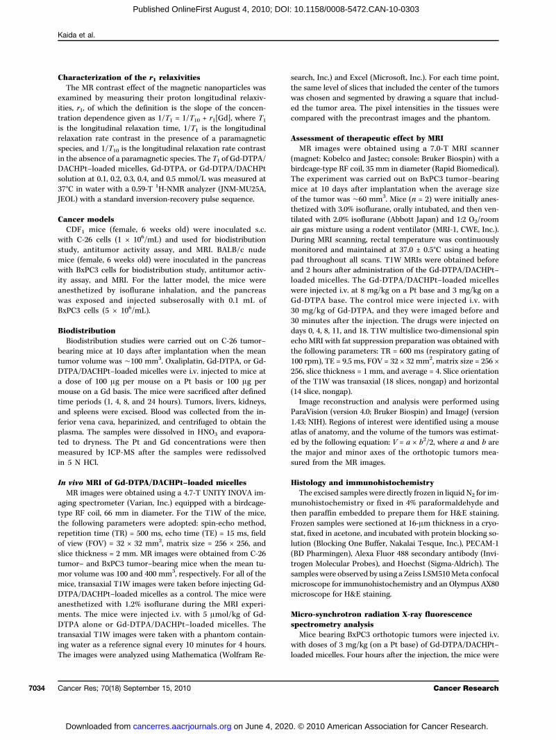
culatiothe ininjecteplatin(Fig. 3tumorthe camicellthe Pthours(C-26)Gd-DTthe tusities ocontrauncha
Gd-DTantituDACHreleasservedingly,the Ctestedtoringin a p(i.e., otal ad
DirecThe
tion oshowefor >4serveminissignalthan iof thesignalscopicthat rthe pomiddlrevealadeno(Fig. 4micellhigherAccorand 74 houThe
cellesthe mreduc(Fig. 5the mthat rDACHintensDACHaccumportinThe
studiethe elS, Cl,peaksscan.Pt) into eva
Kaida et al.
Cance7036
Dow
Published OnlineFirst August 4, 2010; DOI: 10.1158/0008-5472.CAN-10-0303
Gd-DTPA/DACHPt mixture was confirmed (Supple-ry Fig. S2).obtained micelles were 33 nm in diameter with a
w size distribution (polydispersity index = 0.067;). This diameter might be small enough for the mi-to avoid recognition by the reticuloendothelial sys-ass through the leaky vasculature of solid tumorsEPR effect, and attain deep tumor penetration (17).
mounts of DACHPt and Gd-DTPA incorporated in thees were found to be 0.42 mg DACHPt/mg polymer.04 mg Gd-DTPA/mg polymer, corresponding to 45%% of the carboxylic groups in PEG-b-P(Glu), respec-Moreover, the r1 of the micelles increased up tomol/L−1s−1, that is, ∼24-fold greater than Gd-DTPA(Fig. 2C).Gd-DTPA/DACHPt–loaded micelles did not releaseontents in distilled water (data not shown). However,physiologic conditions (i.e., 10 mmol/L PBS at 37°C),Pt and Gd-DTPA were released in a sustained mannerD, left). Moreover, the release of Gd-DTPA was consid-faster than that of DACHPt, probably due to strongerng between polymer and DACHPt than betweenPt and Gd-DTPA. In addition, the safe Gd-DTPA che-n this system might remain stable because no free Gd3+
tected in the released sample (Supplementary Fig. S3).radual drug release from Gd-DTPA/DACHPt–loadedes led to a reduction in the light scattering intensitymicelles (Fig. 2D, middle) due to the decreased densitymicellar cores. Accordingly, the light scattering inten-f the Gd-DTPA/DACHPt–loaded micelles underlogic conditions decreased to 20% in ∼60 hoursD, middle); however, the hydrodynamic diameter oficelles was maintained at ∼30 nm for >48 hoursD, right). The high stability of the micelles and pres-n of their hydrodynamic diameter are advantageousin vivo situation because the structural stability of
es is highly associated with their prolonged blood cir-n (11).
o performance of Gd-DTPA/DACHPt–loadedlesGd-DTPA/DACHPt–loaded micelles extended the cir-n of their cargo in the bloodstream, attaining ∼20% ofjected dose of DACHPt after 24 hours and >8% of thed dose of Gd-DTPA after 4 hours, whereas free oxali-and free Gd-DTPA were rapidly cleared from plasmaA). Moreover, the micelles delivered the drugs to solids due to the increased accumulation and retention atncer site because of the EPR effect. Accordingly, thees augmented the tumor accumulation 27.7 times fordrug at 24 hours, and >100 times for Gd-DTPA at 4
, in subcutaneous murine colon adenocarcinoma 26tumors (Fig. 3B) compared with oxaliplatin and freePA, resulting in high MRI contrast enhancement ofmor tissue (Fig. 3C). From the ratio of the signal inten-f tumor to muscle, the micelles showed to increase the
st, whereas the enhancement for Gd-DTPA was almostnged. Moreover, the elevated tumor accumulation ofelemetion p
r Res; 70(18) September 15, 2010
on June 4, 2020. © 2cancerres.aacrjournals.org nloaded from
PA/DACHPt–loaded micelles may also improve themor activity of the incorporated Pt drug becausePt complexes can exert their cytotoxicity after beinged from the Gd-DTPA/DACHPt–loaded micelles, as ob-in in vitro studies (Supplementary Table S1). Accord-the micelles showed strong antitumor effect against-26 tumor model (Supplementary Fig. S4). Thus, wethe potential of Gd-DTPA–loaded micelles for moni-the drug distribution, tumor imaging, and treatmentancreatic tumor model close to the clinical situationrthotopically inoculated BxPC3 human pancreatic duc-enocarcinoma tumor).
t detection and treatment of pancreatic cancerT1W T1-weighted MR images after i.v. administra-f the Gd-DTPA/DACHPt–loaded micelles clearlyd specific contrast enhancement at the tumor areahours (Fig. 4A and B). In contrast, we did not ob-any enhancement in the tumor region after the ad-tration of free Gd-DTPA (Fig. 4A and B), and theintensity was higher in the liver, kidney, or spleenn tumor as suggested from the tumor-to-organ ratiosMR intensity (Supplementary Table S2). Also, the
s in all organs decreased after 1 hour. The macro-observation of the orthotopic tumor-bearing mice
eceived Gd-DTPA/DACHPt–loaded micelles confirmedsition of every organ and the tumor (Fig. 4D, left ande), whereas the histologic study of the malignancyed the poorly differentiated histology of pancreaticcarcinoma, with thick fibrosis and low vascularizationD, right). The amount of Gd-DTPA delivered by thees in the orthotopic pancreatic tumor was seven timesthan the accumulation of free Gd-DTPA (Fig. 4C).
dingly, 3.5% of the total Gd dose from the micelles.2% of the total Pt dose had accumulated withinrs of administration.antitumor activity of Gd-DTPA/DACHPt–loaded mi-was also evaluated by MRI. Thus, the mice treated withicelles at 8 mg/kg on a Pt base achieved a significanttion in the volume of orthotopic BxPC3 tumorsA). Likewise, the weight of the pancreas at day 18 oficelle-treated animal was much lower than the miceeceived only Gd-DTPA (Fig. 5B). Moreover, Gd-DTPA/Pt–loaded micelles were shown to enhance the signality at the tumor region (Fig. 5C). Thus, Gd-DTPA/Pt–loaded micelle can be used to follow the micelleulation in the tumor and the tumor size by MRI, sup-g the theranostic concept.microdistribution of the drugs at the tumor site wasd using μ-SR-XRF on the pancreatic lesions. Besidesements traditionally present in animal tissue, such asK, Ca, Fe, Cu, Ni, and Zn, very distinct Pt-L and Gd-Lcan also be observed in the sum spectrum of the lineThus, the distribution of several atoms (Fe, K, Gd, andthe tissue sections of the whole pancreas was studiedluate the tissue properties and layout of the drugs. The
ntal mapping of Fe presents areas with high concentra-robably involving the vicinity of blood vessels and theCancer Research
010 American Association for Cancer Research.
distribpositi(Fig. 6tent wsibly ccofact
kinasewhich(31). Tareas
FigureDACHPclearanoxaliplamicellecompleand GdB, left, atumor aGd-DTPaccumutumorsor Gd-DC, left,transaxtumor aDACHP5 μmoltumor-tmicelle180 minenhancinjectiomicelleGd-DTPA.
Visible DDS for Diagnosis and Therapy of Solid Tumors
www.a
Dow
Published OnlineFirst August 4, 2010; DOI: 10.1158/0008-5472.CAN-10-0303
ution of heme proteins. Accordingly, the PECAM-1–ve area from the immunofluorescence microscopyA) showing the existence of endothelial cells is consis-ith this Fe-rich area (Fig. 6B). The K-rich regions pos-
orrespond to pancreatic cancer cells because K is aor required to obtain maximum activity of the pyruvateand Dand D
acrjournals.org
on June 4, 2020. © 2cancerres.aacrjournals.org nloaded from
, an enzyme involved in glycolytic energy production,has been observed in carcinoma tissue of the pancreashe Gd as well as the Pt atoms located at those K-richsuggest the selective tumor accumulation of Gd-DTPA
3. In vivo behavior of Gd-DTPA/t–loaded micelles. A, left, plasmace of Pt drugs after i.v. injection oftin and Gd-DTPA/DACHPt–loadeds; right, plasma clearance of Gdxes after i.v. injection of Gd-DTPA-DTPA/DACHPt–loaded micelles.ccumulation of Pt drugs in the C-26fter i.v. injection of oxaliplatin orA/DACHPt–loaded micelles; right,lation of Gd complexes in C-26after i.v. injection of Gd-DTPATPA/DACHPt–loaded micelles.in vivo MRI series of T1-weightedial slices of C-26 subcutaneousfter i.v. injection of Gd-DTPA/t–loaded micelles or Gd-DTPA at/kg Gd-DTPA. Right, top,o-muscle intensity ratio for thes and Gd-DTPA at 5, 60, and; bottom, relative intensityement in the tumor after i.v.n of Gd-DTPA/DACHPt–loadeds or Gd-DTPA at 5 μmol/kg
ACHPt. Moreover, the colocalization of the Gd-DTPAACHPt confirms the high potential of Gd-DTPA/
Cancer Res; 70(18) September 15, 2010 7037
010 American Association for Cancer Research.

DACHticanc
Discu
Pancancernomawherecannoenviroma, inaccumthe anitonea
putedpancrpredicwith plutionsmallThus,Gd-DTsuggestectionrecognfrom tThe
Figuretransaxi.v. injemicelleBALB/cafter excision with spleen and normal pancreas. Scale bar, 0.5 cm. Bottom, microscopic findings (H&E staining) of the pancreatic cancer (T) and normalpancrea
Kaida et al.
Cance7038
Dow
Published OnlineFirst August 4, 2010; DOI: 10.1158/0008-5472.CAN-10-0303
Pt–loaded micelles to assess the distribution of the an-er drug at the tumor site by MRI.
ssion
creatic cancer has one of the worst prognoses amongs (32). The high malignancy of pancreatic adenocarci-prompts the destruction of the surrounding tissue,as the lack of serous membrane in healthy pancreast prevent the dissemination of cancer cells. The micro-nment characteristics of the pancreatic adenocarcino-cluding hypovascularity and thick fibrosis, prevent theulation of drugs in the tumor tissue (33). Moreover,
tic tissue (P). Scale bar, 100 μm.
atomic position of the pancreas in the deep retroper-l space makes early detection difficult. Although com-
DACHaccum
r Res; 70(18) September 15, 2010
on June 4, 2020. © 2cancerres.aacrjournals.org nloaded from
tomography is widely used for the evaluation ofeatic carcinoma in the clinical setting, MRI may bettert the therapeutic efficacy and the prognosis in patientsancreatic cancer because of its superior contrast reso-of noncontour deforming lesions of the pancreas,liver metastases, and peritoneal disseminations (34).the outstanding contrast enhancement achieved byPA/DACHPt–loaded micelles on this tumor modelts the great potential of this modality for the clear de-of the lesions in the abdominal cavity and the facileition of the carcinomas of the pancreas as distincthe surrounding internal organs by MRI.exceptionally bright contrast achieved by Gd-DTPA/
4. In vivo behavior of Gd-DTPA/DACHPt–loaded micelles on an orthotopic pancreatic cancer (BxPC3). A, in vivo MRI series of T1-weightedial slices of mice after i.v. injection of Gd-DTPA/DACHPt–loaded micelles or Gd-DTPA at 5 μmol/kg. B, relative MRI intensity in each organ afterction of Gd-DTPA/DACHPt–loaded micelles at 5 μmol/kg Gd-DTPA or i.v. injection of Gd-DTPA at 5 μmol/kg. C, the Gd-DTPA/DACHPt–loadeds and Gd-DTPA accumulation in the BxPC3 tumor 4 h after i.v. administration (n = 4). D, top, macroscopic findings of orthotopic BxPC3-bearingnude mice after MRI acquisition. Scale bar, 1 cm. Pancreatic cancer (T), liver (L), kidney (K), and spleen (S). Middle, the pancreatic tumor
Pt–loaded micelles can be attributed to the enhancedulation of the micelles at the tumor site and to the
Cancer Research
010 American Association for Cancer Research.

augmethe mcellesinjectethe mthe rehigherthat thbindintion plation(36) supyridistructwateroptimthe foproba
of theleadined toof Gd-underThe
has bewith econtrof toxmacroThus,trastnephronly 2PAMA
FigureA, effecBxPC3macrosGd-DTP
Visible DDS for Diagnosis and Therapy of Solid Tumors
www.a
Dow
Published OnlineFirst August 4, 2010; DOI: 10.1158/0008-5472.CAN-10-0303
ntation of the relaxivity of the Gd-DTPA in the core oficelles. The amount of Gd-DTPA delivered by the mi-in the orthotopic pancreatic tumor was >3% of thed dose after 4 hours. Because the r1 of Gd-DTPA inicelles is 24 times higher than that of free Gd-DTPA,sulting contrast enhancement probably mimics a muchaccumulation level. In this regard, it has been reportede r1 of Gd-based MRI contrast agents increases afterg with polymers or proteins due to the flexibility reduc-er Gd molecule and the increase of the rotational corre-time (τR; ref. 35). Moreover, Livramento and colleaguesggested that an Fe/Gd chelate, a metallostar Fe{Gd2(bi-ne(diethylenetriaminetetraacetic acid)2(H2O)4)3}
4-
ure, showed a high relaxivity because the inner-spheremolecules presented an exchange rate (τm) close to theal value in addition to the increasing τR. In our system,
rmation of the Gd-DTPA/DACHPt–loaded micelles duringcopies of the excised pancreas after treatment with the micelles or Gd-DTPA. CA/DACHPt–loaded micelles. The tumor size was 89 mm3 at day 0 and 5 mm3 a
acrjournals.org
on June 4, 2020. © 2cancerres.aacrjournals.org nloaded from
τm in the hydrophobic environment at the micelle core,g to the increase in relaxivity. Further studies are need-establish the mechanism of the relaxivity enhancementDTPA/DACHPt–loaded micelles, and they are currentlyway in our laboratory.construction of macromolecular MRI contrast agentsen an attractive strategy to achieve diagnostic agentsxtended blood circulation. Nevertheless, for Gd-basedast agents, this approach could increase the riskicity due to the prolonged tissue exposure to thosemolecules and the potential release of Gd3+ ions.the accumulation of high-generation dendrimer con-agents in the healthy tissues might potentiate theotoxicity and hepatotoxicity risks (37). Accordingly,0% of the injected dose of a generation 4–basedM-Gd contrast agent was excreted from the body
the first 2 days, showing transient accumulation inbly combined an increase of the τR and the optimization the renal tubules. In contrast to this, the biodistribution of
5. In vivo antitumor activity of Gd-DTPA/DACHPt–loaded micelles on orthotopic pancreatic cancer model (BxPC3) assessed by volumetric MRI.t of Gd-DTPA/DACHPt–loaded micelles (8 mg/kg on Pt basis) and Gd-DTPA (30 mg/kg) injected i.v. at day 0, 4, 11 and 18 on the growth oftumors. B, left, weight of the whole pancreas for mice treated with the micelles or Gd-DTPA at day 18 on the antitumor experiment; right,
, MRI at days 0 and 18 of a tumor-bearing mouse treated witht day 18.
Cancer Res; 70(18) September 15, 2010 7039
010 American Association for Cancer Research.

Gd-DTmulatDTPAfrom tof lowrisk oThe
creaseto obtstage,DTPAassessstagesthe deuniforthereportincellesregimeeveryrouracrectalthe tuat the
Gd-DTplicatifunctiapplic
Discl
No p
Ackn
Weassistan
Grant
Thisvative RSocietyResearcdicine P
Theof pageaccorda
Refe1. Ino
mo2. Jon
1373. Ew
Figuresection hst, anbars, 1 al pan
Kaida et al.
Cance7040
Dow
Published OnlineFirst August 4, 2010; DOI: 10.1158/0008-5472.CAN-10-0303
PA/DACHPt–loaded micelles revealed minimal accu-ion of Gd-DTPA in normal tissues. Moreover, the Gd-released from the micelles probably is rapidly excretedhe body because of the relatively fast plasma clearance–molecular weight Gd-DTPA, thus eliminating thef undesired toxicity.real-time observation of drug distribution can in-the accuracy of treatment and enable practitionersain feedback on the therapeutic efficacy at an earlierand promptly adjust the treatment strategy. Gd-
/DACHPt–loaded micelles might be helpful for directlying the distribution of the anticancer drugs at earlyby MRI. In this study, the μ-XRF results showed thatlivered Gd-DTPA and DACHPt were colocalized andmly distributed within the pancreatic tumors, whereaswas no drug accumulation in healthy pancreas, sup-g the strong diagnostic and anticancer effect of the mi-(Fig. 6B, Pt and Gd). Moreover, the chemotherapyns are given in periodic cycles, for example, one cycle2 weeks during 12 weeks in FOLFOX (folinic acid, fluo-il, and oxaliplatin) regimen for the treatment of colo-cancer. By using Gd-DTPA/DACHPt–loaded micelles,
s 4 h after injection of the micelles. The cell nuclei were stained with Hoec00 μm. B, K, Fe, Pt, and Gd distribution in a tumor section including norm
mor size can be followed up in real-time by imaging Rece
es BL. Trastuzumab: hopes and realities. Lancet Oncol 2002;3:–44.er MS, Vooletich MT, Durand JB, et al. Reversibility of trastuzu-
maan
4. Scbomo
r Res; 70(18) September 15, 2010
on June 4, 2020. © 2cancerres.aacrjournals.org nloaded from
PA/DACHPt–loaded micelles will have significant im-ons in the design and development of advanced multi-onal nanomedicines with great potential for clinicalation as visible DDS.
osure of Potential Conflicts of Interest
otential conflicts of interest were disclosed.
owledgments
thank Sayaka Shibata and Teppei Nakahara for their technicalce on the MRI experiments.
Support
research was supported by Funding Program for World-Leading Inno-&D on Science and Technology (FIRST Program) from the Japanfor the Promotion of Science (JSPS) and Grants-in-Aid for Scientifich from the Japanese Ministry of Health, Labor, and Welfare (Nanome-roject).costs of publication of this article were defrayed in part by the paymentcharges. This article must therefore be hereby marked advertisement innce with 18 U.S.C. Section 1734 solely to indicate this fact.
d the blood vessels were marked with PECAM-1 antibody. Scalecreatic tissue determined by μ-SR-XRF. Scale bars, 100 μm.
ived 01/26/2010; revised 06/30/2010; accepted 07/09/2010; published
day of the drug administ ration. Consequently, the OnlineFirst 08/04/2010.rencesue A, Saijo Y, Maemondo M, et al. Severe acute interstitial pneu-nia and gefitinib. Lancet 2003;361:137–9.
b-related cardiotoxicity: new insights based on clinical coursed response to medical treatment. J Clin Oncol 2005;23:7820–6.
6. Intratumoral distribution of Gd-DTPA/DACHPt–loaded micelles in orthotopic BxPC3 tumors. A, immunofluorescence microscopy of tumor
appaticci FA, Skillings JR, Holden SN, et al. Arterial thromboem-lic events in patients with metastatic carcinoma treated with che-therapy and bevacizumab. J Natl Cancer Inst 2007;99:1232–9.
Cancer Research
010 American Association for Cancer Research.

5. Daing771
6. Tocar
7. PeNaNa
8. FeNa
9. DuRe
10. KaeryCri
11. Nisprode
12. Maage
13. Hative200
14. Matumdru
15. Mumatox15:
16. Grapator200
17. MapemuRe
18. HadrudoingMa
19. Kapavan200
20. Bustu200
21. Wistupo
22. Declin13
23. Mcfor12
24. Mcthe2:9
25. Patheres91
26. NaasLe
27. WegaRo
28. Catiopla20
29. GotriaingCh
30. Teing
31. VeTuca
32. Jem43
33. Soult
34. Micre
35. ZhP.co
36. LivHigba
Visible DDS for Diagnosis and Therapy of Solid Tumors
www.a
Dow
Published OnlineFirst August 4, 2010; DOI: 10.1158/0008-5472.CAN-10-0303
vis ME, Chen Z, Shin DM. Nanoparticle therapeutics: an emerg-treatment modality for cancer. Nat Rev Drug Discov 2008;7:–82.rchilin VP. Recent advances with liposomes as pharmaceuticalriers. Nat Rev Drug Discov 2005;4:145–60.er D, Karp JM, Hong S, Farokhzad OC, Margalit R, Langer R.nocarriers as an emerging platform for cancer therapy. Naturenotech 2007;2:751–60.rrari M. Cancer nanotechnology: opportunities and challenges.t Rev Cancer 2005;5:161–71.ncan R. Polymer conjugates as anticancer nanomedicines. Natv Cancer 2006;6:688–701.banov AV, Alakhov VY. Pluronic block copolymers in drug deliv-: from micellar nanocontainers to biological response modifiers.t Rev Ther Drug Carrier Syst 2002;19:1–73.hiyama N, Kataoka K. Current state, achievements, and futurespects of polymeric micelles as nanocarriers for drug and genelivery. Pharmacol Ther 2006;112:630–48.tsumura Y, Kataoka K. Preclinical and clinical studies of anticancernt-incorporating polymer micelles. Cancer Sci 2009;100:572–9.shizume H, Baluk P, Morikawa S, et al. Opening between defec-endothelial cells explain tumor vessel leakiness. Am J Pathol0;156:1363–80.eda H. The enhanced permeability and retention (EPR) effect inor vasculature: the key role of tumor-selective macromolecularg targeting. Adv Enzyme Regul 2001;41:189–207.ggia FM, Hainsworth JD, Jeffers S, et al. Phase II study of liposo-l doxorubicin in refractory ovarian cancer: antitumor activity andicity modification by liposomal encapsulation. J Clin Oncol 1997;987–93.dishar WJ, Tjulandin S, Davidson N, et al. Phase III trial of nano-rticle albumin-bound paclitaxel compared with polyethylated cas-oil-based paclitaxel in women with breast cancer. J Clin Oncol5;23:7794–803.tsumura Y, Maeda H. A new concept for macromolecular thera-utics in cancer chemotherapy: mechanism of tumoritropic accu-lation of proteins and the antitumor agent SMANCS. Cancers 1986;46:6387–92.maguchi T, Matsumura Y, Shirao K, et al. Phase I study of novelg delivery system, NK911, a polymer micelle encapsulatedxorubicin [abstract 571]. Proceedings of the 39th annual meet-of the American Society of Clinical Oncology (ASCO); 2003,y 31-June 3; Chicago, USA.to K, Hamaguchi T, Yasui H, et al. Phase I study of NK105, aclitaxel-incorporating micellar nanoparticle, in patients with ad-ced cancer. ASCO Annual Meeting Proceedings. J Clin Oncol6;24:2018.
rris HA III, Infante JR, Spigel DR, et al. A phase I dose-escalationdy of NK012. ASCO Annual Meeting Proceedings. J Clin Oncol8;26:2538.4437. Du
Re
acrjournals.org
on June 4, 2020. © 2cancerres.aacrjournals.org nloaded from
lson RH, Plummer R, Adam J, et al. Phase I and pharmacokineticdy of NC-6004, a new platinum entity of cisplatin-conjugatedlymer forming micelles. J Clin Oncol 2008;26:2573.nt R, Trudeau M, Pritchard KI, et al. Triple-negative breast cancer:ical features and patterns of recurrence. Clin Cancer Res 2007;:4429–34.Carthy JR, Weissleder R. Multifunctional magnetic nanoparticlestargeted imaging and therapy. Adv Drug Deliv Rev 2008;60:
41–51.Carthy JR, Jaffer FA, Weissleder R. A macrophage-targetedranostic nanoparticle for biomedical applications. Small 2006;83–7.n D, Caruthers SD, Hu G, et al. Ligand-directed nanobialys asranostic agent for drug delivery and manganese-based magneticonance imaging of vascular targets. J Am Chem Soc 2008;130:86–7.songkla N, Bey E, Ren J, et al. Multifunctional polymeric micellescancer-targeted, MRI-ultrasensitive drug delivery systems. Nanott 2006;6:2427–30.inmann HJ, Brasch RC, Press WR, Wesbey GE. Characteristics ofdolinium-DTPA complex: a potential NMR contrast agent. Am Jentgenol 1984;142:619–24.bral H, Nishiyama N, Okazaki S, Koyama H, Kataoka K. Prepara-n and biological properties of dichrolo(1,2-diaminocyclohexane)tinum(II) (DACHPt)-loaded polymeric micelles. J Control Release05;101:223–32.uin S, Winnik F. Quantitative assays of the amount of diethylene-minepentaacetic acid conjugated to water-soluble polymers us-isothermal titration calorimetry and colorimetry. Bioconjug
em 2001;12:372–7.rada Y, Goto S, Takimoto N, et al. Construction and commission-of BL37XU at SPring-8. AIP Conf Proc 2004;705:376–9.
ntrucci M, Cipolla A, Racchini C, Casadei R, Simoni P, Gullo L.mor M2-pyruvate kinase, a new metabolic marker for pancreaticncer. Dig Dis Sci 2004;49:1149–55.al A, et al. Cancer statistics, 2007. CA Cancer J Clin 2007;57:
–66.funi A, et al. Differential diagnosis of pancreatic tumors usingrasound contrast imaging. J Gastroenterol 2005;40:518–25.ller FH, Rini NJ, Keppke AL. MRI of adenocarcinoma of the pan-as. Am J Roentgenol 2006;187:W365–374.ang Z, Greenfield MT, Spiller M, McMurry TJ, Lauffer RB, CaravanMultilocus binding increases the relaxivity of protein-bound MRIntrast agents. Angew Chem 2005;117:6924–7.ramento JB, Toth E, Sour A, Borel A, Merbach AE, Ruloff R.h relaxivity confined to a small molecular space: a metallostar-sed potential MRI contrast agent. Angew Chem Int Ed 2005;:1480–4.
ncan R, Izzo L. Dendrimer biocompatibility toxicity. Adv Drug Delivv 2005;57:2215–37.Cancer Res; 70(18) September 15, 2010 7041
010 American Association for Cancer Research.
2010;70:7031-7041. Published OnlineFirst August 4, 2010.Cancer Res Sachiko Kaida, Horacio Cabral, Michiaki Kumagai, et al. Pancreatic Tumor ModelDirecting to Single-Platformed Diagnosis and Therapy of Visible Drug Delivery by Supramolecular Nanocarriers
Updated version
10.1158/0008-5472.CAN-10-0303doi:
Access the most recent version of this article at:
Material
Supplementary
http://cancerres.aacrjournals.org/content/suppl/2010/08/04/0008-5472.CAN-10-0303.DC1
Access the most recent supplemental material at:
Cited articles
http://cancerres.aacrjournals.org/content/70/18/7031.full#ref-list-1
This article cites 36 articles, 6 of which you can access for free at:
E-mail alerts related to this article or journal.Sign up to receive free email-alerts
Subscriptions
Reprints and
To order reprints of this article or to subscribe to the journal, contact the AACR Publications
Permissions
Rightslink site. Click on "Request Permissions" which will take you to the Copyright Clearance Center's (CCC)
.http://cancerres.aacrjournals.org/content/70/18/7031To request permission to re-use all or part of this article, use this link
on June 4, 2020. © 2010 American Association for Cancer Research.cancerres.aacrjournals.org Downloaded from
Published OnlineFirst August 4, 2010; DOI: 10.1158/0008-5472.CAN-10-0303