brain ventricles, cerebrospinal fluid, coverings and blood...
TRANSCRIPT

Brain ventricles, cerebrospinal fluid,
Coverings and blood vessels of the brain
MUDr. Veronika Němcová, CSc.

Cerbrospinal fluid
production – choroid plexus
absorbtion –arachnoid granulations
Ventricles and subarachnoidal space 140 ml
Daily 500 ml
Mechanic support of the brain(„it floats“)
Chemical communication in the CNS (neurons-
CSF-wall of ventricles– neurons)

Ventricles

Median aperture of the IV.ventricle 3= foramen Magendi
B) Lateral apertures of the IV.ventricle= Foramina LuschkaeLaterally in the tela choroidea ventriculi quarti
CSF comes from ventricles to subarachnoid space through:
A)

Frontal secion through the ventricles

Lateral ventricles, interventricular foramen , III. ventricle,
aqaeductus mesencephali, IV. ventricle, apertura mediana
+ aperturae laterales
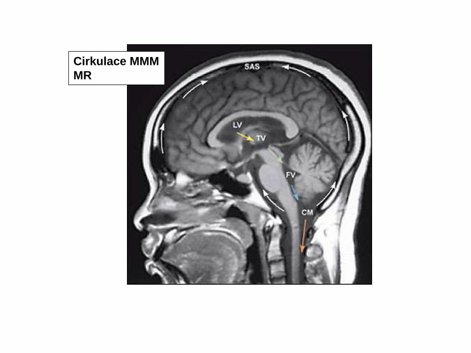
Cirkulace MMM
MR

Granulationes arachnoidales
absorbtion to sinus sagittalis superior

Cisternae
subarachnoidales
= cerebellomedularis
Other cisterns:
Cisterna fossae lateralis cerebri
Cisterna pontis
Cisterna laminae quadrigeminae
Cisterna corporis callosi
Between pia mater and arachnoid

Cisterna ambiens
Between quadrigeminal and interpeduncular cistern
http://www.medigraphic.com/pdfs/anaradmex/arm-2005/arm051f.pdf
Cisterna ambiens
Cisterna fossae lateralisCisterna laminae teminalis

T2
1 Cisterna cerebellomedullaris
2 Cisterna pontis
3 Cisterna interpeduncularis
5 Cisterna laminae quadrigeminae = cisterna superior
MRI – T2W
CSF is white

Cisterns
cerebellomedullary
chiasmatic
interpeduncular
ambiens
pontis
corporis callosi
subarachnoid space
medullae oblongatae
laminae terminalis

Ventricles and basal ganglia

Cornu frontale ventriculi lateralis
A
A B

Pars centralis and cornu temporale
Lamina affixa thalami
Tela choroidea between fornixem and ncl. caudatus
B

Dorsal aspect of ventricles
1- cornu frontale
2-cornu occipitale
3-fornix+ hippocampus
4-plexus choroideus VL
5-septum pellucidum
6- forceps ant
7-temporal lobe
8-cerebellum
9-forceps post

Lateral ventricle – dorsal aspect,
hippocampus
R
C
M

Temporal lobe medial aspect
C
R
V
D
parahippocampal gyrus
dentate gyrusCC
fibria fornicis (hippocampi)
collateral sulcus
subiculum

View of excised hippocampus and its cutout

hippocampus
amygdala
CC
commissura fornicis
calcar avis
R
C
L
M
Lateral ventricle -
temporal horn

Hippocampus
and fornix
Dorsal aspect

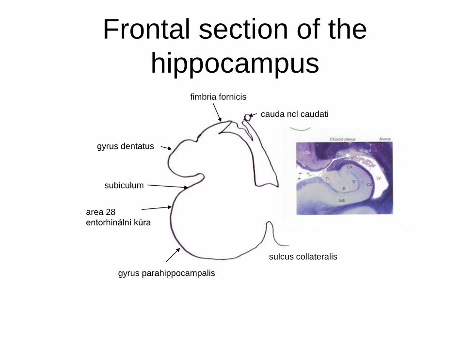
Frontal section of the
hippocampus
cauda ncl caudati
fimbria fornicis
gyrus dentatus
subiculum
area 28
entorhinální kůra
gyrus parahippocampalis
sulcus collateralis

???

hipp
amy
Sagittal section of the brain – silver impregnation
Pu GP
Cau


HIPPOCAMPUS

CC
S pell
Cau
For
stria terminalis
For
digitationes hip
Th
subiculum
gyrus dentatus
fimbria
cornu Amoniscommissura fornicis
CCeminentia collateralis
calcar avis
For
Cornu temporale and cornu occipitale ventriculi lateralis

Horizontal section through the interventricular foramen
III. ventricle
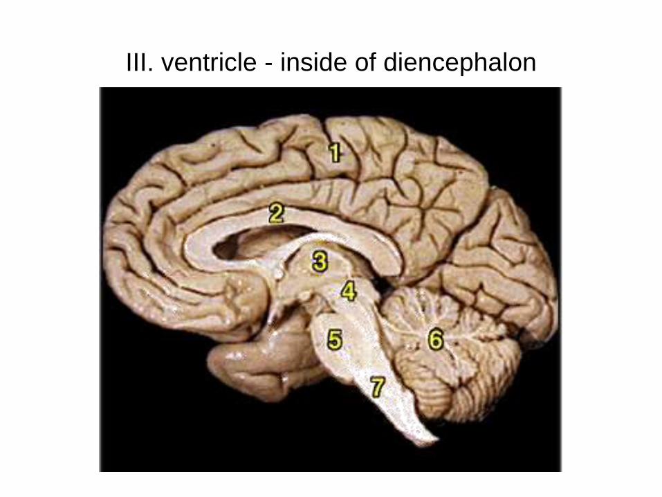
III. ventricle - inside of diencephalon

III. ventricle – sagittal fissure,
a roof of it is attached to stria medullaris thalami
stria medullaris thalami Fissura telodiencephalica

Horizontal section
Tela choroidea ventriculi tertii
Plexus choroideus
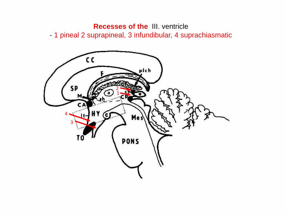
Recesses of the III. ventricle
- 1 pineal 2 suprapineal, 3 infundibular, 4 suprachiasmatic
4
3
21

III. ventricle
chiasma opticum
lamina terminalis
corpus mamillare
sulcus
hypothalamicus
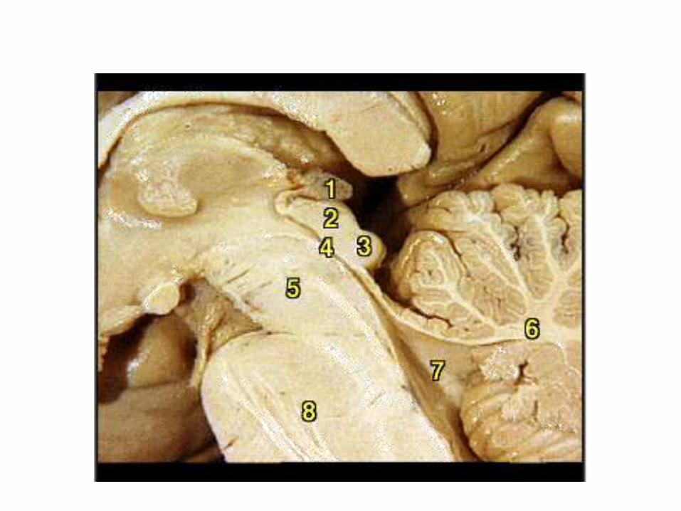
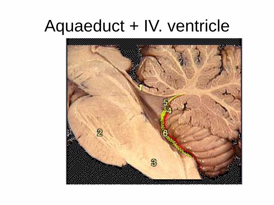
Aquaeduct + IV. ventricle

Circumventricular organs
area postrema
corpus pineale
neurohypophysis
eminentia
mediana
subfornical organ
vascular organ of the
lamina terminalis
subcommissural
organ
chemoreceptors
Blood-brain barrier is
permeable –
fenestrated capillars
Neurons of these organs
project to the hypothalamus

Circumventricular organs
in the rat brain
area postremaorganum subfornicaleorganum subcomissuraleeminentia mediananeurohypophysiscorpus pinealeorganum vasculosum laminae terminalis
chemoreception

MR – T2

MR – T2

MR – T2

Woman with headaches
• CT:
Heterogeneous
2 cm mass
within the
fourth ventricle
causing
hydrocephalus

Significant dilation of the bilateral lateral ventricles and third ventricles
secondary to the tumor filling the distended fourth ventricle and outlet
foramina

MRI

• MRI: Large mixed solid andcystic mass predominantly T1 isointense and T2 hyperintense) centered within the fourth ventricle with indistinct interface along the 4th ventricular floor. The small solid components of the intraventricular mass enhance avidly. A moderate degree of obstructive hydrocephalus is present, with significant dilation of the bilateral lateral ventricles and third ventricles secondary to the tumor filling the distended fourth ventricle and outlet foramina.

Ependymoma:
• Arise from ependymal cells or ependymal cell rests.
• > 60% are located in the fourth ventricle
• More common in children than adults
• "Soft" tumor which accommodates to shape of cisterns or ventricles and often extends into cerebellar pontine angle or cisterna magna.
• Heterogeneous on CT often with calcifications
• On MRI is T1 isointense and T2 hyperintense
• Generally contains enhancing portions, but variable
• Can be associated the NF2 (chromosome 22 defect)
• Most common presentation is child with headache and vomiting.
• Bimodal age distribution: 5 and 30.
• Treatment: surgical resection +/- chemo/radiation therapy
• 5 year survival 60-70%.
•

60-year woman with worsening cognitive
impairment and gait disturbance


Substantial enlargement of the 3rd, 4th, and lateral ventricles.
Relative normal appearance of sulci for age.
No evidence of substantial vascular pathology.

• Classical clinical triad of dementia, gait disturbance, and urinary incontinence is seen with normal pressure hydrocephalus.
• Symptoms result from distortion of white matter by distended ventricles.
• Patients commonly have a history of prior SAH or meningeal infection.
• Gradient between ventricular system and subarachnoid space due to incomplete subarachnoid block.
• Radiographic key: Diffuse ventriculomegaly out of proportion to sulcal prominence.
• Not a radiographic diagnosis. Diagnosis made by improvement of symptoms after shunting.
• Radioisotope cisternogram shows early entry into the lateral ventricles with persistence at 24-48 hours and delayed ascent to parasagittal regions.
• Flow void can be seen through the aqueduct of Sylvius on MR due to increased flow velocity
Normal pressure hydrocephalus

Normal pressure hydrocephalus
From decreased absorbtion
Trias: Dementia, gait abnormalities, urinary
incontinence
From the compression of the white matter
by enlarged ventricles
Often after subrachnoid hemorhage or
meningitis
The only treatable dementia
Sameš, Ústí nad Labem
Ventricle
valve
Abdominal cavity

Normal pressure hydrocephalus (NPH)
http://www.neurologiepropraxi.cz/pdfs/neu/2011/06/09.pdf
Upper figures
Typical MRI in NPH coronalplane T1WDilateted ventricles (*), flattened gyri and no sulci (oval) and increased Sylvianfissura (arrow).
Lower figuresdifuse dilatation ofsubarachnoid spaces(arrow) and enlarged ventricles
Brain atrophy

Coverings
Dura mater outermost layer
closely attached to the periosteum
Makes a wall of sinuses
Divides the hemispheres
Divides the cerebellum and occipital lobes
Divides subdural and epidural space


Cisternae
subarachnoidales
cerebellomedularis
Cisterna fossae lateralis cerebri
Cisterna pontis
Cisterna laminae quadrigeminae
Cisterna corporis callosi
ARACHNOID
thin avascular membrane
not reaching the deeper fissures
Divides subdural and
subarachnoidal space
PIA MATER
Vascular layer closlely attached to
the brain surface

Granulationes arachnoidales

Falx cerebri
overbridging
vein
periosteum SSSarachnoid
granulations
dura
mater
subdural
space
arachnoid
subarachnoid
space
pia mater
overbridging
veinSpaces:
Epidural (a.meningea media)
Subdural (ovebridging veins)
Subarachnoid CSF+ vessels

Repetition

Dura materArachnoideaPia mater
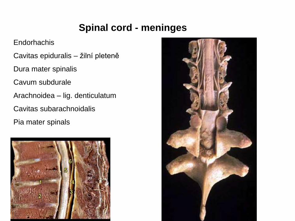
Spinal cord - meninges
Endorhachis
Cavitas epiduralis – žilní pleteně
Dura mater spinalis
Cavum subdurale
Arachnoidea – lig. denticulatum
Cavitas subarachnoidalis
Pia mater spinals

Spinal cord
meninges Denticulate
ligament

Sleeves ofdura mater
= root sleeves

Epidural
From middle
meningeal artery.
Subdural
From bridging veins
Subarachnoid
From Willis circuit
Intracranial hemorhage
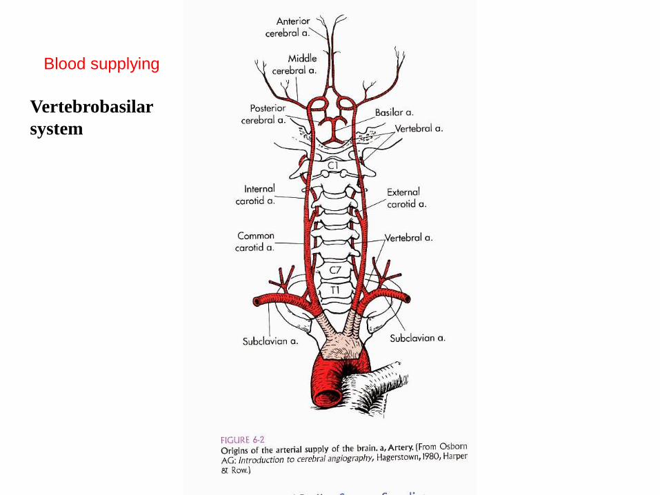
Vertebrobasilar
system
Blood supplying

Cévy cieculusBrain arteries
Willis circuit
• Communication betweenvertebral and a. carotisinterna systems
• Anterior and posteriorcommunicating arteriesallow blood to flowbetween both systems(PCA) or between rightand left vessels (ACA)

Willis circuit
ARTERIA CAROTIS
INTERNA
ARTERIA VERTEBRALIS
Arteriae centrales (peforatores)- groups:AnteromedialAnterolateralPosteromedialPosterolateral

Variets

CT – AG, 3-D

ACA occlusion –
contralateral plegia of
lower limb
PCA occlussion –
Visual field defect
- Contralateral homonymous hemianopsy
MCA occlusion
Contralateral hemiplegia more expressed on upper limbs
and face, can be aphasia


Cerebral arterial
territories
a.cerebri anterior
a.cerebri media
a. cerebri posterior
a. choroidea
anterior
a.cerebellaris
superior
a.cerebellaris
inferior posterior
a.cerebellaris
inferior anterior

Blood supplying of basal ganglia
arterie lenticulostriaticae

Blood supplying of basal ganglia, thalamus and capsula interna

Circulus arteriosus

Most common
diseases of brain
vessels

Circulus arteriosus Willisi –aneurysma
1
3
5
6
2
4
8
7 A. cerebelli sup.
9 A. cerebelli inf. ant.

Aneurysma lokalization

Aneurysma - treatment
Endovascular occlussionClip


Aneurysma – stent, recoiling

Intravascular coiling

A-V malformation
peroperačně

Cranial nerve origins and arteries
on the ventral part of the
brainstem

NMR – angoigraphy
1 - a.carotis interna
2 - a.vertebralis
3 - sinus cavernosus
4 - canalis caroticus
5 - a.cerebri anterior
6 - a.cerebri posterior

Thomas Willis
(1621–1675)


The home of Thomas Willis from 1657 to 1667
.
Oxford, Beam Hall

Thomas Willis
• Neuroanatomical terms coined by Willis
• Anterior commissure | Cerebellar peduncles | Claustrum | Corpus striatum | Inferior olives (corpora teretia) | Internal capsule | Medullary pyramids | Nervus ophthalmicus | The word 'neurology' | Optic thalamus | Spinal accessory nerve | Stria terminalis (taenia cornua) | Striatum | Vagus nerve
• Pathologies recognized by Willis
• Achalasia of the cardia (achalasia of the oesophagus) | Akathisia (restless legs syndrome, Ekbom's syndrome) | Symptoms of myasthenia gravis | Paracusis Willisii. Occurs in deaf patients whose hearing improves in the presence of noise, indicating osteosclerosis | Diabetes mellitus | Abnormalities of the brains of patients with congenital mental retardation | Unilateral degeneration of the cerebral peduncle in a case of long-standing unilateral paralysis | Symptoms of malaria | Distinctions between typhoid and puerperal fevers

5 Vena anastomotica sup.
(Trolard)
6 Vena anastomotica post.
(Labbé)
Superficial veins
Superior cerebral veins

Brain veins - % of thrombosis

Trombosis sinus sagittalis superior

Tromboses vv.
cerebri superiores


Deep cerebral
veins

superior sagittal sinus
internal cerebral
veins
vein of Labbé
sphenoparietal sinus
Cerebral Venous territories
„rough guide“

1. Epidural hemorhage
2. Subdural hemorhage
3. Subarachnoidal hemorhage

Epi
Subd
Subar

Spinal cord arteries
Yoshioka K et al. Radiographics 2003;23:1215-1225
©2003 by Radiological Society of North America
Artery of Adamkiewicz (a. radicularis
magna) from the a. intercostalis post.
at the level Th9–L1
a. iliolumbalis
lumbal artereries
aa. sacralis lateralis
vertebral art.
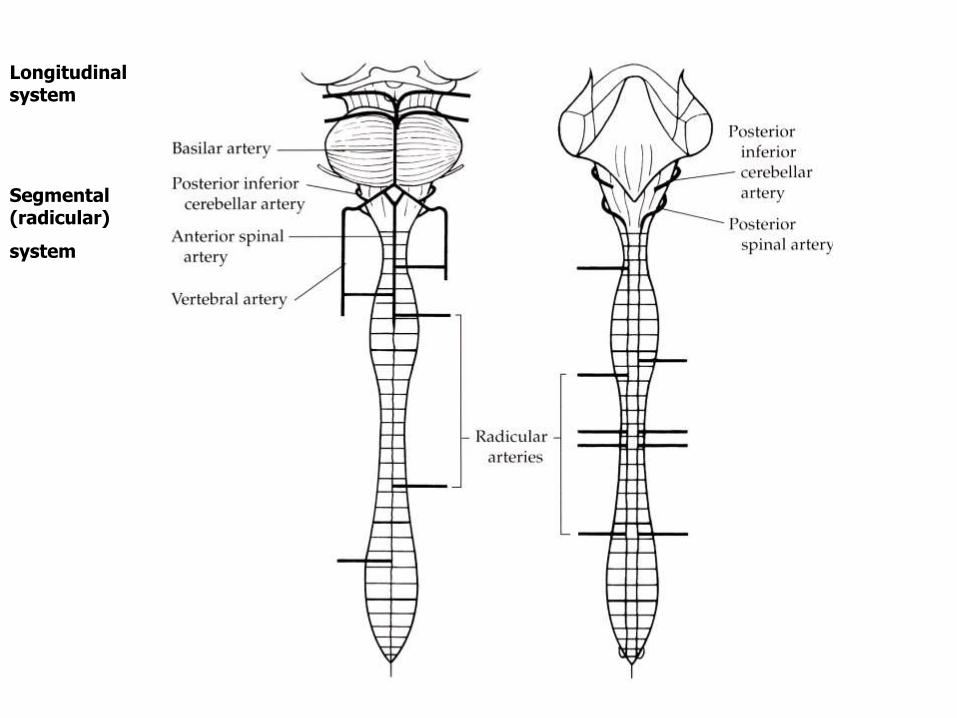
Longitudinal system
Segmental (radicular)
system

Spinal cord -arteries
vasocoronae
5 longitudinal truncs
r. spinalis
a. spinalis anterior
aa. spinales
posteriores

Vertebral veins

basivertebral veins
basivertebral vein
Anterior
external
vertebral
venous
plexus
Internal
vertebral
venous
plexus
Posterior
external
vertebral
venous
plexus

no valves
anastomoses
spreading of
infection and
cancer
Vertebral veins

Illustration of intradural-extradural venous anastomosis. Daniels after Netter.


Vertebral venous plexuses
• no valves, a lot of anastomoses
• anastamoses with venous plexus around sacrum and
pelvis
• 1) in the vertebral canal in the epidural space (plexus
venosi vertebrales interni)
• 2) outside the spine (plexus venosi vertebrales externi)
• 3) in the bodies of vertebrae (venae basivertebrales)
