bovine anaplasmosis results from infection with … · definition of the disease: bovine...
TRANSCRIPT
Definition of the disease: Bovine anaplasmosis results from infection with Anaplasma marginale.
A second species, A. centrale, has long been recognised and usually causes benign infections.
Anaplasma marginale is responsible for almost all outbreaks of clinical disease.
Anaplasma phagocytophilum and A. bovis, which infect cattle, have been recently included within
the genus but they are not reported to cause clinical disease. The organism is classified in the
genus Anaplasma belonging to the family Anaplasmataceae of the order Rickettsiales.
Description of the disease: Anaemia, jaundice and sudden death are characteristic signs of
anaplasmosis. Other signs include rapid loss of milk production and weight, but the clinical disease
can only be confirmed by identifying the organism. Once infected, cattle may remain carriers for life,
and identification of these animals depends on the detection of specific antibodies using serological
tests, or of rickettsial DNA using molecular amplification techniques. The disease is typically
transmitted by tick vectors, but mechanical transmission by biting insects or by needle can occur.
Identification of the agent: Microscopic examination of blood or organ smears stained with
Giemsa stain is the most common method of identifying Anaplasma in clinically affected animals. In
these smears, A. marginale organisms appear as dense, rounded, intraerythrocytic bodies
approximately 0.3–1.0 µm in diameter situated on or near the margin of the erythrocyte.
Anaplasma centrale is similar in appearance, but most of the organisms are situated toward the
centre of the erythrocyte. It can be difficult to differentiate A. marginale from A. centrale in a stained
smear, particularly with low levels of rickettsaemia. Commercial stains that give very rapid staining
of Anaplasma are available in some countries. Anaplasma phagocytophilum and A. bovis can only
be observed infecting granulocytes, mainly neutrophils.
It is important that smears be well prepared and free from foreign matter. Smears from live cattle
should preferably be prepared from blood drawn from the jugular vein or another large vessel. For
post-mortem diagnosis, smears should be prepared from internal organs (including liver, kidney,
heart and lungs) and from blood retained in peripheral vessels. The latter are particularly desirable
if post-mortem decomposition is advanced.
Serological tests: A competitive enzyme-linked immunosorbent assay (C-ELISA) has been
demonstrated to have good sensitivity in detecting carrier animals. Card agglutination is the next
most frequently used assay. The complement fixation test (CFT) is no longer considered a reliable
test for disease certification of individual animals due to variable sensitivity. Cross reactivity
between Anaplasma spp. can complicate interpretation of serological tests. In general, the C-ELISA
has the best specificity, with cross-reactivity described between A. marginale, A. centrale,
A. phagocytophilum and Ehrlichia spp. Alternatively, an indirect ELISA using the CFT with
modifications is a reliable test used in many laboratories and can be prepared in-house for routine
diagnosis of anaplasmosis.
Nucleic-acid-based tests have been used experimentally, and are capable of detecting the
presence of low-level infection in carrier cattle and tick vectors. A nested reaction is necessary to
identify low-level carriers using conventional polymerase chain reaction (PCR) and nonspecific
amplification can occur. Recently, real-time PCR assays with analytical sensitivity equivalent to
nested conventional PCR have been described.
Requirements for vaccines: Live vaccines are used in several countries to protect cattle against
A. marginale infection. A vaccine consisting of live A. centrale is most widely used and gives partial
protection against challenge with virulent A. marginale.
Anaplasma centrale vaccine is provided in chilled or frozen forms. Quality control is very important
as other blood-borne agents that may be present in donor cattle can contaminate vaccines and be
disseminated broadly. For this reason, frozen vaccine is recommended as it allows thorough post-
production quality control, which limits the risk of contamination with other pathogens.
Anaplasma centrale vaccine is not entirely safe. A practical recommendation is to restrict its use, as
far as possible, to calves, as nonspecific immunity will minimise the risk of some vaccine reactions
that may require treatment with tetracycline or imidocarb. Partial immunity develops in 6–8 weeks
and lasts for several years after a single vaccination. In countries where A. centrale is exotic, it
cannot be used as vaccine against A. marginale.
Outbreaks of bovine anaplasmosis are due to infection with Anaplasma marginale. Anaplasma centrale is capable of producing a moderate degree of anaemia, but clinical outbreaks in the field are extremely rare. New species of Anaplasma, A. phagocytophilum and A. bovis (Dumler et al., 2001), with a primary reservoir in rodents, have been reported to infect cattle, but do not cause clinical disease (Dreher et al., 2005; Hofmann-Lehmann et al., 2004).
The most marked clinical signs of anaplasmosis are anaemia and jaundice, the latter occurring late in the disease. Haemoglobinaemia and haemoglobinuria are not present, and this may assist in the differential diagnosis of anaplasmosis from babesiosis, which is often endemic in the same regions. The disease can only be confirmed, however, by identification of the organism.
Anaplasma marginale occurs in most tropical and subtropical countries, and in some more temperate regions. Anaplasma centrale was first described from South Africa. The organism has since been imported by other countries – including Australia and some countries in South America, South-East Asia and the Middle East – for use as a vaccine against A. marginale.
Anaplasma species were originally regarded as protozoan parasites, but further research showed they had no significant attributes to justify this description. Since the last major accepted revision of the taxonomy in 2001 (Dumler et al., 2001), the Family Anaplasmataceae (Order Rickettsiales) is now composed of four genera, Anaplasma, Ehrlichia, Neorickettsia, and Wolbachia. The genus Aegyptianellais is retained within the Family Anaplasmataceae as genus incertae sedis. The revised genus Anaplasma now contains Anaplasma marginale as the type species, A. phagocytophilum the agent of human granulocytic ehrlichiosis (formerly Ehrlichia phagocytophila and E. equi) A. platys, and A. bovis. Haemobartonella and Eperythrozoon are now considered most closely related to the mycoplasmas.
Anaplasma species are transmitted either mechanically or biologically by arthropod vectors. Reviews based on careful study of reported transmission experiments list up to 19 different ticks as capable of transmitting A. marginale (Kocan et al., 2004). These are: Argas persicus, Ornithodoros lahorensis, Dermacentor albipictus, D. andersoni, D. hunteri, D. occidentalis, D. variabilis, Hyalomma excavatum, H. rufipes, Ixodes ricinus, I. scapularis, Rhipicephalus annulatus (formerly Boophilus annulatus), R. bursa, R. calcaratus, R. decoloratus, R. evertsi, R. microplus, R. sanguineus and R. simus. However, the classification of several ticks in these reports has been questioned. Intrastadial or transstadial transmission is the usual mode, even in the one-host Rhipicephalus species. Male ticks may be particularly important as vectors; they can become persistently infected and serve as a reservoir for infection. Experimental demonstration of vector competence does not necessarily imply a role in transmission in the field. However, Rhipicephalus species are clearly important vectors of anaplasmosis in countries such as Australia and countries in Africa, and Latin America, and some species of Dermacentor are efficient vectors in the United States of America (USA).
Various other biting arthropods have been implicated as mechanical vectors, particularly in the USA. Experimental transmission has been demonstrated with a number of species of Tabanus (horseflies), and with mosquitoes of the genus Psorophora (Kocan et al., 2004). The importance of biting insects in the natural transmission of anaplasmosis appears to vary greatly from region to region. Anaplasma marginale also can be readily transmitted during vaccination against other diseases unless a fresh or sterilised needle is used for injecting each animal. Similar transmission by means of unsterilised surgical instruments has been described (Reinbold et al., 2010a).
The main biological vectors of A. centrale appear to be multihost ticks endemic in Africa, including R. simus. The common cattle tick (R. microplus) has not been shown to be a vector. This is of relevance where A. centrale is used as a vaccine in R. microplus-infested regions.
Anaplasma marginale infection has not been reported in humans. Thus, there is no risk of field or laboratory transmission to workers and laboratories working with A. marginale may operate at the lowest biosafety level, equivalent to BSL1.
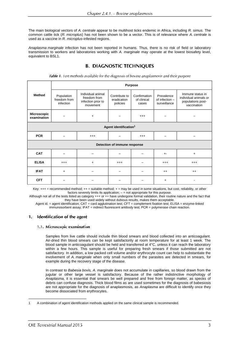
Method
Purpose
Population freedom from
infection
Individual animal freedom from
infection prior to movement
Contribute to eradication
policies
Confirmation of clinical
cases
Prevalence of infection – surveillance
Immune status in individual animals or
populations post-vaccination
Microscopic examination
– + – +++ – –
Agent identification1
PCR – +++ – +++ – –
Detection of immune response
CAT – – – – +- +
ELISA +++ + +++ – +++ +++
IFAT + – – – ++ ++
CFT – – – – + -
Key: +++ = recommended method; ++ = suitable method; + = may be used in some situations, but cost, reliability, or other factors severely limits its application; – = not appropriate for this purpose.
Although not all of the tests listed as category +++ or ++ have undergone formal validation, their routine nature and the fact that they have been used widely without dubious results, makes them acceptable.
Agent id. = agent identification; CAT = card agglutination test; CFT = complement fixation test; ELISA = enzyme-linked immunosorbent assay; IFAT = indirect fluorescent antibody test; PCR = polymerase chain reaction.
Samples from live cattle should include thin blood smears and blood collected into an anticoagulant. Air-dried thin blood smears can be kept satisfactorily at room temperature for at least 1 week. The blood sample in anticoagulant should be held and transferred at 4°C, unless it can reach the laboratory within a few hours. This sample is useful for preparing fresh smears if those submitted are not satisfactory. In addition, a low packed cell volume and/or erythrocyte count can help to substantiate the involvement of A. marginale when only small numbers of the parasites are detected in smears, for
example during the recovery stage of the disease.
In contrast to Babesia bovis, A. marginale does not accumulate in capillaries, so blood drawn from the jugular or other large vessel is satisfactory. Because of the rather indistinctive morphology of Anaplasma, it is essential that smears be well prepared and free from foreign matter, as specks of
debris can confuse diagnosis. Thick blood films as are used sometimes for the diagnosis of babesiosis are not appropriate for the diagnosis of anaplasmosis, as Anaplasma are difficult to identify once they become dissociated from erythrocytes.
1 A combination of agent identification methods applied on the same clinical sample is recommended.
Samples from dead animals should include air-dried thin smears from the liver, kidney, heart and lungs and from a peripheral blood vessel. The latter is particularly recommended should there be a significant delay before post-mortem examination because, under these circumstances, bacterial contamination of organ smears often makes identification of Anaplasma equivocal. Brain smears, which are useful for the diagnosis of some forms of babesiosis, are of no direct value for diagnosing anaplasmosis, but should be included for differential diagnosis where appropriate.
Blood from organs, rather than organ tissues per se, is required for smear preparation, as the aim is to be able to examine microscopically intact erythrocytes for the presence of Anaplasma. Organ-derived blood smears can be stored satisfactorily at room temperature for several days.
Both blood and organ smears can be stained in 10% Giemsa stain for approximately 30 minutes after fixation in absolute methanol for 1 minute. After staining, the smears are rinsed three or four times with tap water to remove excess stain, and are then air-dried. Conditions for Giemsa staining vary from laboratory to laboratory, but distilled water is not recommended for dilution of Giemsa stock. Water should be pH 7.2–7.4 to attain best resolution with Giemsa stain. Commercial stains that give very rapid staining of Anaplasma are available in some countries. Smears are examined under oil immersion at a magnification of ×700–1000.
Anaplasma marginale appear as dense, rounded and deeply stained intraerythrocytic bodies, approximately 0.3–1.0 µm in diameter. Most of these bodies are located on or near the margin of the erythrocyte. This feature distinguishes A. marginale from A. centrale, as in the latter most of the organisms have a more central location in the erythrocyte. However, particularly at low levels of rickettsaemia, differentiation of these two species in smears can be difficult. Appendages associated with the Anaplasma body have been described in some isolates of A. marginale (Kreier & Ristic, 1963; Stich et al., 2004).
The percentage of infected erythrocytes varies with the stage and severity of the disease. Maximum rickettsaemias in excess of 50% may occur with A. marginale. Multiple infections of individual erythrocytes are common during periods of high rickettsaemias.
The infection becomes visible microscopically 2–6 weeks following transmission. During the course of clinical disease, the rickettsaemia approximately doubles each day for up to about 10 days, and then decreases at a similar rate. Severe anaemia may persist for some weeks after the parasites have become virtually undetectable in blood smears. Following recovery from initial infection, cattle remain latently infected for life.
Nucleic-acid-based tests to detect A. marginale infection in carrier cattle have been developed
although not yet fully validated. The analytical sensitivity of polymerase chain reaction (PCR)-based methods has been estimated at 0.0001% infected erythrocytes, but at this level only a proportion of carrier cattle would be detected. A nested PCR has been used to identify A. marginale carrier cattle with a capability of identifying as few as 30 infected erythrocytes per ml of blood, well below the lowest levels in carriers. However, nested PCR poses significant quality control and specificity problems for routine use (Torioni De Echaide et al., 1998). Real-time PCR has also been described for identification of A. marginale (Carelli et al., 2007; Decaro et al., 2008; Reinbold et al., 2010b), and should be considered instead of the nested PCR. Two advantages of this technique, which uses a single closed tube for amplification and analysis, are reduced opportunity for amplicon contamination and a semi-quantitative assay result. Equipment needed for real-time PCR is expensive, requires preventive maintenance, and may be beyond the capabilities of some laboratories. Real-time PCR assays may target one of several genes (Carelli et al., 2007; Decaro et al., 2008), or 16S rRNA (Reinbold et al.,
2010b), and are reported to achieve a level of analytical sensitivity equivalent to nested conventional PCR (Carelli et al., 2007; Decaro et al., 2008; Reinbold et al., 2010b).
In general, unless animals have been treated or are at a very early stage of infection (<14 days), serology using the competitive enzyme-linked immunosorbent assay (C-ELISA), indirect ELISA (I-ELISA) or card agglutination test (CAT) (see below) may be the preferred methods of identifying infected animals in most laboratories. Anaplasma infections usually persist for the life of the animal. However, except for occasional small recrudescences, Anaplasma cannot readily be detected in blood smears after acute rickettsaemia and, even end-point PCR may not detect the presence of Anaplasma in blood samples from asymptomatic carriers. Thus, a number of serological tests have been developed with the aim of detecting persistently infected animals.
A feature of the serological diagnosis of anaplasmosis is the highly variable results with regard to both sensitivity and specificity reported for many of the tests from different laboratories. This is due at least in part to inadequate evaluation of the tests using significant numbers of known positive and negative animals. Importantly, the capacity of several assays to detect known infections of long-standing duration has been inadequately addressed. An exception is C-ELISA (see below), which has been validated using true positive and negative animals defined by nested PCR (Torioni De Echaide et al., 1998), and the card agglutination assay, for which relative sensitivity and specificity in comparison with the C-ELISA has been evaluated (Molloy et al., 1999). Therefore, while most of the
tests described in this section are useful for obtaining broad-based epidemiological data, caution is advised on their use for disease certification. The C-ELISA, I-ELISA and CAT are described in detail below.
It should be noted that there is a high degree of cross-reactivity between A. marginale and A. centrale, as well as cross-reactivity with both A. phagocytophilum and Ehrlichia spp. in serological tests (Al-Adhami et al., 2011; Dreher et al., 2005). While the infecting species can sometimes be identified using antigens from homologous and heterologous species, equivocal results are obtained on many occasions.
A C-ELISA using a recombinant antigen termed rMSP5 and MSP5-specific monoclonal antibody (MAb) has proven very sensitive and specific for detection of Anaplasma-infected animals (Hofmann-Lehmann et al., 2004; Reinbold et al., 2010b; Strik et al., 2007). All A. marginale strains tested, along with A. ovis and A. centrale, express the MSP5 antigen and induce antibodies against the immunodominant epitope recognised by the MSP5-specific MAb. A recent report suggests that antibodies from cattle experimentally infected with A. phagocytophilum will test positive in the C-ELISA (Dreher et al., 2005). However, in another study no cross-reactivity could be demonstrated, and the MAb used in the assay did not react with A. phagocytophilum MSP5 in direct binding assays (Strik et al., 2007). Cross reactivity has been demonstrated between A. marginale and Ehrlichia spp, in naturally and experimentally infected cattle (Al-Adhami et al, 2011). Earlier studies had shown that the C-ELISA was 100% specific using 261 known negative sera from a non-endemic region, detecting acutely infected cattle as early as 16 days after experimental tick or blood inoculation, and was demonstrated to detect cattle that have been experimentally infected as long as 6 years previously (Knowles et al., 1996). In detecting persistently infected cattle from an anaplasmosis-endemic region that were defined as true positive or negative using a nested PCR procedure, the rMSP5 C-ELISA had a sensitivity of 96% and a specificity of 95% (Torioni De Echaide et al., 1998).
Test results using the rMSP5 C-ELISA are available in less than 2.5 hours. A test kit available commercially contains specific instructions. In general, however, it is conducted as follows.
A 96-well microtitre plate coated with rMSP5 antigen,
A 96-well coated adsorption/transfer plate for serum adsorption to reduce background binding,
100×MAb/peroxidase conjugate,
10× wash solution and ready-to-use conjugate-diluting buffer,
Ready-to-use substrate and stop solutions,
Positive and negative controls
i) Add 70 µl of undiluted serum sample to the coated adsorption/transfer plate and incubate at room temperature for 30 minutes.
ii) Transfer 50 µl per well of the adsorbed serum to the rMSP5-coated plate and incubate at room temperature for 60 minutes.
iii) Discard the serum and wash the plate twice using diluted wash solution.
iv) Add 50 µl per well of the 1× diluted MAb/peroxidase conjugate to the rMSP5-coated plate, and incubate at room temperature for 20 minutes.
v) Discard the 1×diluted MAb/peroxidase conjugate and wash the plate four times using diluted wash solution.
vi) Add 50 µl per well of the substrate solution, cover the plate with foil, and incubate for 20 minutes at room temperature.
vii) Add 50 µl per well of stop solution to the substrate solution already in the wells and gently tap the sides of the plate to mix the wells.
viii) Immediately read the plate in the plate reader at 620 nm.
The mean optical density (OD) of the negative control must range from 0.40 to 2.10. The per cent inhibition of the positive control must be ≥30%.
The % inhibition is calculated as follows:
100 –
Sample OD × 100
= Per cent inhibition Mean negative control OD
Samples with <30% inhibition are negative. Samples with ≥30% inhibition are positive.
Specificity of the MSP5 C-ELISA may be increased by using a higher percentage inhibition cut-off value (Bradway et al., 2001); however the effect of this change on sensitivity has not been thoroughly evaluated.
Recently, an improved MSP5 C-ELISA was developed by replacing rMBP-MSP5 with rGST-MSP5 in addition to an improvement in the antigen-coating method by using a specific catcher system. The new rMSP5-GST C-ELISA was faster, simpler, had a higher specificity and an improved resolution compared with the rMSP5-MBP C-ELISA with MBP adsorption (Chung et al., 2014).
An I-ELISA was first developed using the CAT antigen (see below) and it can be implemented where the commercial C-ELISA is not available. Unlike the C-ELISA, most reagents, such as buffers and ready-to dissolve substrates, are available commercially in many countries. Any laboratory can prepare the antigen using local strains of A. marginale I-ELISA uses small amounts of serum and antigen, and the sensitivity and specificity of the test standardised with true positive and negative sera is as good as for the C-ELISA. As it can be prepared in each laboratory, only the general procedure is described here (Barry et al., 1986). For commercial kits, the manufacturer‟s instructions should be followed. In the case of in-house I-ELISA, refer to Barry et al. 1986. Initial bodies and membranes are obtained as for the complement fixation test (Rogers et al., 1964). This antigen is treated with 0.1% sodium dodecyl sulphate for 30 minutes prior to fixing the antigen to the microtitre plate. For each laboratory, the specific amount of antigen has to be adjusted to obtain the best reading and the least expenditure.
Test results using the I-ELISA are available in about 4 to 5 hours. It is conducted as follows:
A 96-well microtitre plate coated with crude A. marginale antigen,
PBS/Tween buffer, (PBS 0.1 M, pH 7.2, Tween 20 0.05%),
Blocking reagent (e.g. commercial dried skim milk)
Tris buffer 0.1M, MgCl2, 0.1M, NaCl .005 M, pH 9.8
Substrate p-Nitrophenyl phosphate disodium hexahydrate
Positive and negative controls.
i) Plates can be prepared ahead of time and kept under airtight conditions at –20°C
ii) Carefully remove the plastic packaging before using plates, being careful not to touch the bottom of them as this can distort the optical density reading.
iii) Remove the lid and deposit 200 l PBST20 solution in each well and incubate for 5 minutes at room temperature (RT).
iv) For one plate, dissolve 1.1 g of skim milk (blocking agent) in 22 ml of PBST20.
v) Remove the plate contents and deposit in each well, 200 µl of blocking solution put the lid and incubate for 60 minutes at 37°C.
vi) Wash the plate three times for 5 minutes with PBST20.
vii) Dilute all serum samples including controls 1/100 in PBST20 solution;
viii) Remove the contents of the plate and deposit 200 µl of diluted serum in each of the three wells for each dilution, starting with the positive and negative and blank controls.
ix) Incubate plate at 37°C covered for 60 minutes.
x) Wash three times as described in subsection vi.
xi) Dilute 1/1000 anti-IgG bovine alkaline phosphatase conjugate in PBST20 solution; Add 200 µl of the diluted conjugate per well; incubate the covered plate at 37°C for 60 minutes.
xii) Remove the lid and make three washes with PBST20.
xiii) Remove the contents of the plate and deposit 195 µl of 0.075% p-Nitrophenyl phosphate disodium hexahydrate in Tris buffer and incubate for 60 minutes at 37°C
ix) The reaction is quantified by a microplate reader spectrophotometer, adjusted to 405 nm wavelength. The data are expressed in optical density (OD).
Analysis of results should take into account the following parameters.
i) The mean value of the blank wells.
ii) The mean value of the positive wells with their respective standard deviations.
iii) The mean value of negative wells with their respective standard deviations.
iv) The mean value of the blank wells is subtracted from the mean of all the other samples if not automatically subtracted by the ELISA reader.
v) Control sera are titrated to give optical density values ranging from 0.90 to 1.50 for the positive and, 0.15 to 0.30 for the negative control
Positive values are those above the cut-off calculated value which is the sum of the average of the negative and two times the standard deviation.
For purposes of assessing the consistency of the test operator, the error “E” must also be estimated; this is calculated by determining the percentage represented by the standard deviation of any against their mean serum.
The advantages of the CAT are that it is sensitive, may be undertaken either in the laboratory or in the field, and gives a result within a few minutes. Nonspecific reactions may be a problem, and subjectivity in interpreting assay reactions can result in variability in test interpretation. In addition, the CAT antigen, which is a suspension of A. marginale particles, can be difficult to prepare and can vary from
batch to batch and laboratory to laboratory. Splenectomised calves are infected by intravenous inoculation with blood containing Anaplasma-infected erythrocytes. When the rickettsaemia exceeds 50%, the animal is exsanguinated, the infected erythrocytes are washed, lysed, and the erythrocyte ghosts and Anaplasma particles are pelleted. The pellets are sonicated, washed, and then
resuspended in a stain solution to produce the antigen suspension.
A test procedure that has been slightly modified from that originally described (Amerault & Roby, 1968; Amerault et al., 1972) is as follows, and is based on controlled conditions in a laboratory setting:
i) Ensure all test components are at a temperature of 25–26°C before use (this constant temperature is critical for the test).
ii) On each circle of the test card (a clear perspex/plastic or glass plate marked with circles that are 18 mm in diameter), place next to each other, but not touching, 10 µl of bovine
serum factor (BSF), 10 µl of test serum, and 5 µl of CAT antigen2. Negative and low positive control sera must be tested on each card.
BSF is serum from a selected animal with high known conglutinin level. If the conglutinin level is unknown, fresh serum from a healthy animal known to be free from Anaplasma can be used. The BSF must be stored at –70°C in small aliquots, a fresh aliquot being used each time the tests are performed. The inclusion of BSF improves the sensitivity of the test.
iii) Mix well with a glass stirrer. After mixing each test, wipe the stirrer with clean tissue to prevent cross-contamination.
iv) Place the test card in a humid chamber and rock at 100–110 rpm for 7 minutes.
v) Read immediately against a backlight. Characteristic clumping of the antigen (graded from +1 to +3) is considered to be a positive result. The test is considered to give a negative result when there is no characteristic clumping.
The complement fixation (CF) test has been used extensively for many years; however, it shows variable sensitivity (ranging from 20 to 60%), possibly reflecting differences in techniques for antigen production, and poor reproducibility. In addition, it has been demonstrated that the CF assay fails to detect a significant proportion of carrier cattle (Bradway et al., 2001). It is also uncertain as to whether or not the CF test can identify antibodies in acutely infected animals prior to other assays (Coetzee et al., 2007; Molloy et al., 1999). Therefore, the CF test is no longer recommended as a reliable assay for detecting infected animals.
Because of the limitations on the number of indirect fluorescent antibody (IFA) tests that can be performed daily by one operator, other serological tests are generally preferred to the IFA test. The IFA test is performed as described for bovine babesiosis in chapter 2.4.2, except that A. marginaIe infected blood is used for the preparation of antigen smears. A serious problem encountered with the test is nonspecific fluorescence. Antigen made from blood collected as soon as adequate rickettsaemia (5–10%) occurs is most likely to be suitable. Nonspecific fluorescence due to antibodies adhering to infected erythrocytes can be reduced by washing the erythrocytes in an acidic glycine buffer before antigen smears are prepared. Infected erythrocytes are washed twice in 0.1 M glycine buffer (pH 3.0, centrifuged at 1000 g for 15 minutes at 4°C) and then once in PBS, pH 7.4. Recently published data show that the IFA, like the C-ELISA, can cross react with other members of the Anaplasmataceae family (Al-Adhami et al., 2011).
Several immunisation methods have been used to protect cattle against anaplasmosis in countries where the disease is endemic, but none is ideal to date (McHardy, 1984). A review of A. marginale vaccines and antigens has been published (Kocan et al., 2003) Use of the less pathogenic A. centrale, which gives partial cross-protection against A. marginale, is the most widely accepted method, although not used in many countries where
the disease is exotic, including north America.
In this section, the production of live A. centrale vaccine is described. It involves infection of a susceptible, splenectomised calf and the use of its blood as a vaccine. Detailed accounts of the production procedure are available and reference should be made to these publications for details of the procedures outlined here (Bock et al., 2004; de Vos & Jorgensen, 1992; Pipano, 1995).
Guidelines for the production of veterinary vaccines are given in Chapter 1.1.8 Principles of veterinary vaccine production. The guidelines given here and in Chapter 1.1.8 are intended to be general in nature and may be supplemented by national and regional requirements.
2 The test as conducted in the USA and Mexico uses larger volumes of reagents: antigen (15 µl), serum (30 µl), and bovine
serum factor (30 µl), and a 4-minute reaction time (see step iv).

Anaplasma centrale vaccine can be provided in either frozen or chilled form depending on demand, transport networks, and the availability of liquid nitrogen or dry ice supplies. Frozen vaccine is recommended in most instances, as it allows for thorough post-production quality control of each batch. It is, however, more costly to produce and more difficult to transport than chilled vaccine. The risk of contamination makes post-production control essential, but may be prohibitively expensive.
Anaplasma centrale was isolated in 1911 in South Africa, and has been used as a vaccine in South America, Australia, Africa, the Middle East, and South-East Asia. It affords only partial, but adequate, protection in regions where the challenging strains are of moderate virulence (e.g. Australia) (Bock & de Vos, 2001). In the humid tropics where A. marginale appears to be a very virulent rickettsia, the protection afforded by A. centrale may be inadequate to prevent disease in some animals.
Anaplasma centrale usually causes benign infections, especially if used in calves under
9 months of age. Severe reactions following vaccination have been reported when adult cattle are inoculated. The suitability of an isolate of A. centrale as a vaccine can be determined by inoculating susceptible cattle, monitoring the subsequent reactions, and then challenging the animals and susceptible controls with a virulent local strain of A. marginale. Both safety and
efficacy can be judged by monitoring rickettsaemias in stained blood films and the depression of packed cell volumes of inoculated cattle during the vaccination and challenge reaction periods.
Infective material for preparing the vaccine is readily stored as frozen stabilates of infected blood in liquid nitrogen or dry ice. Dimethyl sulphoxide (DMSO) and polyvinylpyrrolidone M.W. 40,000 (Bock et al., 2004) are the recommended cryopreservatives, as they allow for intravenous administration after thawing of the stabilate. A detailed account of the freezing technique using DMSO is reported elsewhere (Mellorset al., 1982), but briefly involves the following: infected blood is collected, chilled to 4°C, and cold cryoprotectant (4 M DMSO in PBS) is added slowly with stirring to a final blood:protectant ratio of 1:1, to give a final concentration of 2 M DMSO. The entire dilution procedure is carried out in an ice bath and the diluted blood is dispensed into suitable containers (e.g. 5 mI cryovials), and frozen, as soon as possible, in the vapour phase of a liquid nitrogen container.
Evidence of purity of the A. centrale isolate can be determined by serological testing of paired sera from the cattle used in the safety test for possible contaminants that may be present (Bock et al., 2004; Pipano, 1997). Donor calves used to expand the seed for vaccine production should be examined for all blood-borne infections prevalent in the vaccine-producing country, including Babesia, Anaplasma, Ehrlichia, Theileria and Trypanosoma. This can be done by routine examination of stained blood films after splenectomy, and preferably also by serology. Any calves showing evidence of natural infections of any of these agents should be rejected. The absence of other infective agents should also be confirmed. These may include the agents of enzootic bovine leukosis, mucosal disease, infectious bovine rhinotracheitis, ephemeral fever, Akabane disease, bluetongue, foot and mouth disease, and rinderpest. The testing procedures will depend on the diseases prevalent in the country and the availability of tests, but should involve serology of paired sera at the very least and, in some cases, virus isolation, antigen, or DNA/RNA detection (Bock et al., 2004; Pipano, 1981; 1997).
i) Production of frozen vaccine
Quantities of the frozen stabilate (5–10 ml) are thawed by immersing the vials in water preheated to 40°C. The thawed material is kept on ice and used as soon as possible (within 30 minutes) to infect a susceptible, splenectomised calf by intravenous inoculation.
The rickettsaemia of the donor calf is monitored daily by examining stained films of jugular blood, and the blood is collected for vaccine production when suitable rickettsaemias are
reached. A rickettsaemia of 1 × 10⁸/ml (approximately 2% rickettsaemia in jugular blood) is the minimum required for production of vaccine as this is the dose to vaccinate a bovine. If a suitable rickettsaemia is not obtained, passage of the strain by subinoculation of 100–200 ml of blood to a second splenectomised calf may be necessary.
Blood from the donor is collected by aseptic jugular or carotid cannulation using heparin as an anticoagulant (5 International Units [IU] heparin/ml blood). The use of blood collection units for human use are also suitable and guaranty sterility and obviate the need to prepare glass flask that make the procedure more cumbersome.
In the laboratory, the infective blood is mixed in equal volumes with 3 M glycerol in PBS supplemented with 5 mM glucose at 37°C (final concentration of glycerol 1.5 M). The mixture is then equilibrated at 37°C for 30 minutes and dispensed into suitable containers (e.g. 5 ml cryovials). The vials are cooIed at approximately 10°C/minute in the vapour phase of liquid nitrogen and, when frozen, stored in the liquid phase (Bock et al., 2004).
DMSO can be used as a cryoprotectant in the place of glycerol. This is done in the same way as outlined for the preparation of seed stabilate (Mellors et al., 1982; Pipano, 1981).
If glycerolised vaccine is to be diluted, the diluent should consist of PBS with 1.5 M glycerol and 5 mM glucose (Jorgensen et al., 1989). Vaccine cryopreserved with DMSO should be diluted with diluent containing the same concentration of DMSO as in the original cryopreserved blood (Pipano et al., 1986).
ii) Production of chilled vaccine
Infective material for chilled vaccine is prepared in the same way as for frozen vaccine, but it must be issued and used as soon as possible after collection. The infective blood can be
diluted to provide 1 × 10⁷ parasites per dose of vaccine. A suitable diluent is 10% sterile bovine serum in a glucose/balanced salt solution containing the following quantities per litre: NaCI (7.00 g), MgCI2.6H2O (0.34 g), glucose (1.00 g), Na2HPO4(2.52 g),
KH2PO4(0.90 g), and NaHCO3(0.52 g).
If diluent is not available, acid citrate dextrose (20% [v/v]) or citrate phosphate dextrose (20% [v/v]) should be used as anticoagulant to provide the glucose necessary for survival of the organisms.
iii) Use of vaccine
In the case of frozen vaccine, vials should be thawed by immersion in water, preheated to 37°C to 40°C, and the contents mixed with suitable diluent to the required dilution. If glycerolised vaccine is prepared, it should be kept cool and used within 8 hours (Bock et al., 2004). If DMSO is used as a cryoprotectant, the prepared vaccine should be kept on ice and used within 15–30 minutes (Pipano, 1981). The vaccine is most commonly administered subcutaneously.
iv) Chilled vaccine should be kept refrigerated and used within 4–7 days of preparation.
The strain of A. centrale used in vaccine is of reduced virulence, but is not entirely safe. A
practical recommendation is, therefore, to limit the use of vaccine to calves, where nonspecific immunity will minimise the risk of vaccine reactions. When older animals have to be vaccinated, there is a risk of severe reactions. These reactions occur infrequently, but valuable breeding stock or pregnant animals obviously warrant close attention, and should be observed daily for 3 weeks post-vaccination. Clinically sick animals should be treated with oxytetracycline or imidocarb at dosages recommended by the manufacturers. Protective immunity develops in 6–8 weeks and usually lasts for several years.
Anaplasmosis and babesiosis vaccines are often used concurrently, but it is not advisable to use any other vaccines at the same time (Bock et al., 2004).
Anaplasma centrale cannot be cultured in vitro. No substrates or media other than buffers and diluents are used in vaccine production. DMSO or glycerol should be purchased from reputable companies.
i) Source and maintenance of vaccine donors
A source of calves free from natural infections of Anaplasma and other tick-borne diseases should be identified. If a suitable source is not available, it may be necessary to breed the calves under tick-free conditions specifically for the purpose of vaccine production.
The calves should be maintained under conditions that will prevent exposure to infectious diseases and to ticks and biting insects. In the absence of suitable facilities, the risk of contamination with the agents of infectious diseases present in the country involved should be estimated, and the benefits of local production of vaccine weighed against the possible adverse consequences of spreading disease (Bock et al., 2004).
ii) Surgery
Donor calves should be splenectomised to allow maximum yield of organisms for production of vaccine. This is best carried out in young calves and under general anesthesia.
iii) Screening of vaccine donors before inoculation
As for preparation of seed stabilate, donor calves for vaccine production should be examined for all blood-borne infections prevalent in the vaccine-producing country, including Babesia, Anaplasma, Ehrlichia, Theileria and Trypanosoma. This can be done by routine examination of stained blood films after splenectomy, and preferably also by serology. Any calves showing evidence of natural infections of any of these agents should be rejected. The absence of other infective agents should also be confirmed. These may include the agents of enzootic bovine leukosis, bovine viral diarrhoea, infectious bovine rhinotracheitis, ephemeral fever, Akabane disease, bluetongue, and foot and mouth disease. The testing procedures will depend on the diseases prevalent in the country and the availability of tests, but should involve serology of paired sera at the very least and, in some cases, virus isolation, antigen, or DNA/RNA detection (Bock et al., 2004; Pipano, 1981; 1997).
iv) Monitoring of rickettsaemias following inoculation
It is necessary to determine the concentration of rickettsia in blood being collected for vaccine. The rickettsial concentration can be estimated from the erythrocyte count and the rickettsaemia (percentage of infected erythrocytes).
v) Collection of blood for vaccine
All equipment should be sterilised before use (e.g. by autoclaving). Once the required rickettsaemia is reached, the blood is collected in heparin using strict aseptic techniques. This is best done if the calf is sedated and with the use of a closed-circuit collection system.
Up to 3 litres of heavily infected blood can be collected from a 6-month-old calf. If the calf is to live, the transfusion of a similar amount of blood from a suitable donor is indicated. Alternatively, the calf should be killed immediately after collection of the blood.
vi) Dispensing of vaccine
All procedures are performed in a suitable environment, such as a laminar flow cabinet, using standard sterile techniques. Use of a mechanical or magnetic stirrer will ensure thorough mixing of blood and diluent throughout the dispensing process. Penicillin (500,000 lU/litre) and streptomycin (370,000 µg/litre) are added to the vaccine at the time of dispensing.
The potency, safety and sterility of vaccine batches cannot be determined in the case of chilled vaccine, and specifications for frozen vaccine depend on the country involved. The following are the specifications for frozen vaccine produced in Australia.
i) Sterility and purity
Standard tests for sterility are employed for each batch of vaccine and diluent (see Chapter 1.1.9 Tests of biological materials for sterility and freedom from contamination).
The absence of contaminants is determined by doing appropriate serological testing of donor cattle, by inoculating donor lymphocytes into sheep and then monitoring them for evidence of viral infection, and by inoculating cattle and monitoring them serologically for infectious agents that could potentially contaminate the vaccine. Cattle inoculated during the test for potency (see Section C.2.2.4.iii) are suitable for the purpose. Depending on the country of origin of the vaccine, these agents include the causative organisms of enzootic bovine leukosis, infectious bovine rhinotracheitis, bovine viral diarrhoea, ephemeral fever, Akabane disease, Aino virus, bluetongue, parainfluenza, foot and mouth disease, lumpy skin disease, rabies, Rift Valley fever, contagious bovine pleuropneumonia, Jembrana disease, heartwater, pathogenic Theileria and Trypanosoma spp., Brucella abortus, Coxiella, and Leptospira (Bock et al., 2004; Pipano, 1981; 1997).Other pathogens to consider include the causal agents of bovine tuberculosis and brucellosis as they may spread through contaminated blood used for vaccine production. Most of these agents can be tested by means of specific PCR and there are many publications describing primers, and assay conditions for any particular disease.
ii) Safety
Vaccine reactions of the cattle inoculated in the test for potency (see Chapter 1.1.8 Principles of veterinary vaccine production) are monitored by measuring rickettsaemia and depression of packed cell volume. Only batches with pathogenicity levels equal to or lower than a predetermined standard are released for use.
iii) Potency
Vaccine is thawed and diluted 1/5 with a suitable diluent (Bock et al., 2004). The diluted vaccine is then incubated for 8 hours at 4°C, and five cattle are inoculated subcutaneously with 2 ml doses. The inoculated cattle are monitored for the presence of infections by examination of stained blood smears. All should become infected for a batch to be accepted. A batch proving to be infective is recommended for use at a dilution of 1/5 with isotonic diluent.
The strain of A. centrale used in vaccine is of reduced virulence, but is not entirely safe. A practical recommendation is, therefore, to limit the use of vaccine to calves, where nonspecific immunity will minimise the risk of vaccine reactions. When older animals have to be vaccinated, there is a risk of severe reactions. These reactions occur infrequently, but valuable breeding stock or pregnant animals obviously warrant close attention, and should be observed daily for 3 weeks post-vaccination. Clinically sick animals should be treated with oxytetracycline or imidocarb at dosages recommended by the manufacturers.
Anaplasma centrale is not infective to other species, and the vaccine is not considered to have other adverse environmental effects. The vaccine is not infective for humans. When the product is stored in liquid nitrogen, the usual precautions pertaining to the storage, transportation and handling of deep-frozen material applies.
Partial but long-lasting immunity results from one inoculation. There is no evidence that repeated vaccination will have a boosting effect. The vaccine is used for control of clinical anaplasmosis in endemic areas. It will not provide sterile immunity, and should not be used for eradication of A. marginale.
The vaccine can be kept for 5 years when stored in liquid nitrogen. Once thawed, it rapidly loses its potency. Thawed vaccine cannot be refrozen.

There are no vaccines based on biotechnology available for anaplasmosis.
AL-ADHAMI B., SCANDRETT W.B., LOVANOV V.A. & GAJADHAR A.A. (2011). Serological cross reactivity between Anaplasma marginale and Ehrlichia species in naturally and experimentally infected cattle. J. Vet. Diagn. Invest., 23, 1181–1188.
AMERAULT T.E. & ROBY T.O. (1968). A rapid card agglutination test for bovine anaplasmosis. J. Am. Vet. Med. Assoc., 153, 1828–1834.
AMERAULT T.E., ROSE J.E. & ROBY T.O. (1972). Modified card agglutination test for bovine anaplasmosis: evaluation with serum and plasma from experimental and natural cases of anaplasmosis. Proc. U.S. Anim. Health Assoc., 76, 736–744.
BARRY D.N., PARKER R.J., DE VOS A.J., DUNSTER P. & RODWELL B.J. (1986). A microplate enzyme-linked immunosorbent assay for measuring antibody to Anaplasma marginale in cattle serum. Aust. Vet. J., 63, 76–79.
BOCK R., JACKSON L., DE VOSA. & JORGENSEN W. (2004). Babesiosis of cattle. Parasitology,129, Suppl, S247–269.
BOCK R.E. & DE VOS A.J. (2001). Immunity following use of Australian tick fever vaccine: a review of the evidence. Aust. Vet. J., 79, 832–839.
BRADWAY D.S., TORIONI DE ECHAIDE S., KNOWLES D.P., HENNAGER S.G. & MCELWAIN T.F. (2001). Sensitivity and specificity of the complement fixation test for detection of cattle persistently infected with Anaplasma marginale. J. Vet. Diagn. Invest., 13, 79–81.
CARELLI G., DECARO N., LORUSSO A., ELIA G., LORUSSO E., MARI V., CECI L. & BUONAVOGLIA C. (2007). Detection and quantification of Anaplasma marginale DNA in blood samples of cattle by real-time PCR. Vet. Microbiol., 124,
107–114.
COETZEE J.F., SCHMIDT P.L., APLEY M.D., REINBOLD J.B. & KOCAN K.M. (2007). Comparison of the complement fixation test and competitive ELISA for serodiagnosis of Anaplasma marginale infection in experimentally infected steers. Am. J. Vet. Res., 68, 872–878.
CHUNG C.,
WILSON C., BANDARANAYAKA-MUDIYANSELAGE C.-B., KANG E., ADAMS D.S., KAPPMEYER L.S., KNOWLES
D.P., MCELWAIN T.F., EVERMANN J.F., UETI M.W., SCOLES G.A., LEE S.S. & MCGUIRE T.C. (2014). Improved diagnostic performance of a commercial Anaplasma antibody competitive enzyme-linked immunosorbent assay using recombinant major surface protein 5-glutathione S-transferase fusion protein as antigen. J. Vet. Diagn. Invest., 26, 61–71.
DECARO N., CARELLI G., LORUSSO E., LUCENTE M.S., GRECO G., LORUSSO A., RADOGNA A., CECI L. & BUONAVOGLIA C. (2008). Duplex real-time polymerase chain reaction for simultaneous detection and quantification of Anaplasma marginale and Anaplasma centrale. J. Vet. Diagn. Invest., 20, 606–611.
DE VOS A.J. & JORGENSEN W.K. (1992). Protection of cattle against babesiosis in tropical and subtropical countries with a live, frozen vaccine. In:Tick Vector Biology, Medical and Veterinary Aspects, Fivaz B.H., PetneyT.N. & Horak I.G., eds. Springer Verlag, Berlin, Germany, 159–174.
DREHER U.M., DE LA FUENTE J., HOFMANN-LEHMANN R., MELI M.K., PUSTERIA N., KOCAN K.M., WOLDEHIWET A., REGULA
G. & STAERK K.D.C. (2005). Serologic cross reactivity between Anaplasma marginale and Anaplasma phagocytophilum. Clin. Vaccine. Immunol., 12, 1177–1183.
DUMLER J.S., BARBET A.F., BEKKER C.P., DASCH G.A., PALMER G.H., RAY S.C., RIKIHISA Y. & RURANGIRWA F.R. (2001). Reorganization of genera in the Families Rickettsiaceae and Anaplasmataceae in the order Rickettsiales: unification of some species of Ehrlichia with Anaplasma, Cowdria with Ehrlichia, and Ehrlichia with Neorickettsia, descriptions of five new species combinations and designation of Ehrlichia equi and „HGE agent‟ as subjective synonyms of Ehrlichia phagocytophila. Int. J. Syst. Evol. Microbiol., 51, 2145–2165.
JORGENSEN W.K., DE VOS A.J. & DALGLIESH R.J. (1989). Infectivity of cryopreserved Babesia bovis, Babesia bigemina and Anaplasma centrale for cattle after thawing, dilution and incubation at 30°C. Vet. Parasitol., 31, 243–251.
KOCAN K.M., DE LA FUENTE J., BLOUIN E.F. & GARCIA-GARCIA J.C. (2004). Anaplasma marginale (Rickettsiales: Anaplasmataceae): recent advances in defining host-pathogen adaptations of a tick-borne rickettsia. Parasitology, 129, S285–S300.
KOCAN K.M., DE LA FUENTE J., GUGLIELMONE A.A. & MELENDÉZ R.D. (2003). Antigens and alternatives for control of Anaplasma marginale infection in cattle. Clin. Microbiol. Rev., 16, 698–712.
KNOWLES D., TORIONI DE ECHAIDE S., PALMER G., MCGUIRE T., STILLER D. & MCELWAIN T. (1996). Antibody against an Anaplasma marginale MSP5 epitope common to tick and erythrocyte stages identifies persistently infected cattle. J. Clin. Microbiol., 34, 2225–2230.
KREIER J.P. & RISTIC M. (1963). Anaplasmosis. X Morphological characteristics of the parasites present in the blood of calves infected with the Oregon strain of Anaplasma marginale. Am. J Vet. Res., 24, 676–687.
HOFMANN-LEHMANN R., MELI M.L., DREHER U.M., GÖNCZI E., DEPLAZES P., BRAUN U., ENGELS M., SCHÜPBACH J., JÖRGER K., THOMA R., GRIOT C., STÄRKK.D.C., WILLI B., SCHMIDT J., KOCAN K.M. & LUTZ H. (2004). Concurrent infections with vector-borne pathogens associated with fatal haemolytic anemia in a cattle herd in Switzerland. J. Clin. Microbiol., 42, 3775–3780.
MCHARDY N. (1984). Immunization against anaplasmosis: a review. Prev. Vet. Med., 2, 135–146.
MELLORS L.T., DALGLIESH R.J., TIMMS P., RODWELL B.J. & CALLOW L.L. (1982). Preparation and laboratory testing of a frozen vaccine containing Babesia bovis, Babesi abigemina and Anaplasma centrale. Res. Vet. Sci., 32, 194–197.
MOLLOY J.B., BOWLES P.M., KNOWLES D.P., MCELWAIN T.F., BOCK R.E., KINGSTON T.G., BLIGHT G.W. & DALGLIESH
R.J. (1999). Comparison of a competitive inhibition ELISA and the card agglutination test for detection of antibodies to Anaplasma marginale and Anaplasma centrale in cattle. Aust. Vet. J., 77, 245–249.
PIPANO E. (1981). Frozen vaccine against tick fevers of cattle. In: Xl International Congress on Diseases of Cattle, Haifa, Israel. Mayer E., ed. Bregman Press, Haifa, Israel, 678–681.
PIPANO E. (1995). Live vaccines against hemoparasitic diseases in livestock. Vet. Parasitol., 57, 213–231.
PIPANO E. (1997). Vaccines against hemoparasitic diseases in Israel with special reference to quality assurance. Trop. Anim. Health Prod., 29 (Suppl. 4), 86S–90S.
PIPANO E., KRIGEL Y., FRANK M., MARKOVICS A. & MAYER E. (1986). Frozen Anaplasma centrale vaccine against anaplasmosis in cattle. Br. Vet. J., 142, 553–556.
REINBOLD J.B., COETZEE J.F., HOLLIS L.C., NICKELL J.S., RIEGEL C.M., CHRISTOPHER J.A. & GANTA R.R. (2010a). Comparison of iatrogenic transmission of Anaplasma marginale in Holstein steers via needle and needle-free injection techniques. Am. J. Vet. Res., 71, 1178–1188.
REINBOLD J.B., COETZEE J.F., SIRIGIREDDY K.R. & GANTA R.R. (2010b). Detection of Anaplasma marginale and
A. phagocytophilum in bovine peripheral blood samples by duplex real-time reverse transcriptase PCR assay. J.
Clin. Microbiol., 48, 2424–2432.
ROGERS T.E., HIDALGO R.-J. & DIMOPOULLOS G.T. (1964). Immunology and serology of Anaplasma marginale. I. Fractionation of the complement-fixing antigen. J. Bacteriol., 88, 81–86.
STICH R.W., OLAH G.A., BRAYTON K.A., BROWN W.C., FECHHEIMER M., GREEN-CHURCH K., JITTAPALAPONG S., KOCAN
K.M., MCGUIRE T.C., RURANGIRWA F.R. & PALMER G.H. (2004).Identification of a novel Anaplasma marginale appendage-associated protein that localizes with actin filaments during intraerythrocytic infection. Infect Immun., 72, 7257–7264.
STIK N.I., ALLEMAN A.R., BARBET A.F., SORENSON H.L., WANSLEY H.L., GASCHEN F.P., LUCKSCHANDER N., WONG S., CHU F., FOLEY J.E., BJOERSDORFF A., STUEN S. & KNOWLES D.P. (2007). Characterization of Anaplasma phagocytophilum major surface protein 5 and the extent of its cross-reactivity with A. marginale. Clin. Vaccine Immunol., 14, 262–268.
TORIONI DE ECHAIDE S., KNOWLES D.P., MCGUIRE T.C., PALMER G.H., SUAREZ C.E. & MCELWAIN T.F. (1998). Detection of cattle naturally infected with Anaplasma marginale in a region of endemicity by nested PCR and a competitive enzyme-linked immunosorbent assay using recombinant major surface protein 5. J. Clin. Microbiol., 36, 777–782.
*
* *
NB: There is an OIE Reference Laboratory for Anaplasma sp.
(see Table in Part 4 of this Terrestrial Manual or consult the OIE Web site for the most up-to-date list: http://www.oie.int/en/our-scientific-expertise/reference-laboratories/list-of-laboratories/).
Please contact the OIE Reference Laboratory for any further information on diagnostic tests, reagents and vaccines for bovine anaplasmosis