bouveret’s syndrome case report and review of the literature
DESCRIPTION
TRANSCRIPT
Hindawi Publishing CorporationGastroenterology Research and PracticeVolume 2009, Article ID 914951, 4 pagesdoi:10.1155/2009/914951
Case Report
Bouveret’s Syndrome: Case Report and Review of the Literature
Iliana Doycheva,1 Alpna Limaye,1 Amitabh Suman,2 Christopher E. Forsmark,1
and Shahnaz Sultan3
1 Division of Gastroenterology, Hepatology, and Nutrition, College of Medicine, University of Florida, Gainesville, FL 32611, USA2 Division of Gastroenterology, Hepatology, and Nutrition, Malcom Randall Veterans Affairs Medical Center, College of Medicine,University of Florida, Gainesville, FL 32608, USA
3 Division of Gastroenterology, Hepatology, and Nutrition, Malcom Randall Veterans Affairs Medical Center,Rehabilitation Outcomes Research Center, College of Medicine, University of Florida, Gainesville, FL 32608, USA
Correspondence should be addressed to Shahnaz Sultan, [email protected]
Received 29 October 2008; Accepted 30 January 2009
Recommended by Peter Malfertheiner
Bouveret’s syndrome is defined as gastric outlet obstruction caused by duodenal impaction of a large gallstone which passesinto the duodenal bulb through a cholecystogastric or cholecystoduodenal fistula. Initial attempts at endoscopic retrieval with orwithout mechanical or extracorporeal lithotripsy should be performed as first-line treatment, though success rates with endoscopictreatment are variable. We describe a case of Bouveret’s Syndrome in an elderly patient that was successfully treated with endoscopicextraction combined with mechanical lithotripsy, and review the literature on this uncommon condition.
Copyright © 2009 Iliana Doycheva et al. This is an open access article distributed under the Creative Commons AttributionLicense, which permits unrestricted use, distribution, and reproduction in any medium, provided the original work is properlycited.
1. Introduction
The first published report of Bouveret’s syndrome (1896) isattributed to Leon Bouveret who reported on two patientswith this disease [1]. Since then, there have been several casereports of unique manifestations of Bouveret’s syndrome, aswell as reports of novel endoscopic treatment modalities.Bouveret’s syndrome tends to occur more commonly inwomen (65%) with a median age of 74.1 years at presentation[2]. Because it often presents in patients with advanced ageand multiple comorbidities, it is associated with a high rateof mortality. Therefore, endoscopic treatment should alwaysbe attempted in order to avoid surgery in these patients. Forpatients in whom endoscopic extraction has failed, simpleenterolithotomy, duodenotomy or gastrotomy, and stoneextraction can be performed [3].
2. Case Presentation
2.1. History. An 86-year-old Caucasian male was admittedwith a three-day history of nausea, vomiting, left lowerquadrant abdominal pain and three episodes of melena. He
had not consumed any food for the previous two days. Hehad a past medical history significant for bladder cancer,hypertension, peripheral vascular disease, and gallstones.
Two months prior to this presentation, the patienthad been admitted with elevated liver function tests.Abdominal computed tomography (CT) revealed gallbladderthickening with surrounding stranding, cholelithiasis, andcholedocholithiasis with a 7 mm stone in the common bileduct. On endoscopic retrograde cholangiopancreatography,a sphincterotomy was performed, three gallstones (the largestof which was 12 mm in diameter) were removed from thecommon bile duct, and a biliary stent was left in place. Therewere no complications and the patient was well until hepresented two months later.
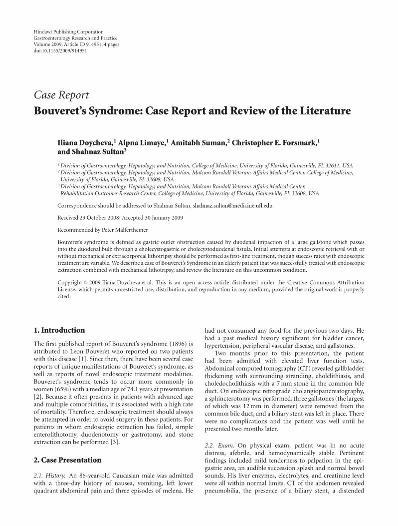
2.2. Exam. On physical exam, patient was in no acutedistress, afebrile, and hemodynamically stable. Pertinentfindings included mild tenderness to palpation in the epi-gastric area, an audible succession splash and normal bowelsounds. His liver enzymes, electrolytes, and creatinine levelwere all within normal limits. CT of the abdomen revealedpneumobilia, the presence of a biliary stent, a distended
2 Gastroenterology Research and Practice
kV
Figure 1: A gallstone in the duodenum and a distended stomachwas seen on CT scan.
stomach and a 3.3 cm hypodense oval object in the secondportion of the duodenum suggestive of Bouveret’s Syndrome(Figure 1).
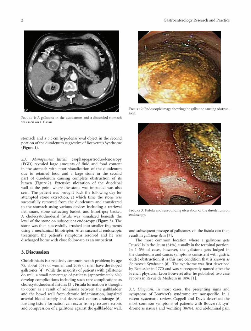
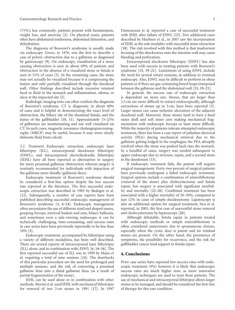
2.3. Management. Initial esophagogastroduodenoscopy(EGD) revealed large amounts of fluid and food contentin the stomach with poor visualization of the duodenumdue to retained food and a large stone in the secondpart of duodenum causing complete obstruction of itslumen (Figure 2). Extensive ulceration of the duodenalwall at the point where the stone was impacted was alsoseen. The patient was brought back the following day forattempted stone extraction, at which time the stone wassuccessfully removed from the duodenum and transferredto the stomach using various devices including a retrievalnet, snare, stone extracting basket, and lithotripsy basket.A cholecystoduodenal fistula was visualized beneath thelevel of the stone on subsequent endoscopy (Figure 3). Thestone was then successfully crushed into smaller fragmentsusing a mechanical lithotripter. After successful endoscopictreatment, the patient’s symptoms resolved and he wasdischarged home with close follow-up as an outpatient.
3. Discussion
Cholelithiasis is a relatively common health problem; by age75, about 35% of women and 20% of men have developedgallstones [4]. While the majority of patients with gallstonesdo well, a small percentage of patients (approximately 6%)develop complications including such rare complications ascholecystoduodenal fistulas [5]. Fistula formation is thoughtto occur as a result of adhesions between the gallbladderand the bowel wall from chronic inflammation, impairedarterial blood supply and decreased venous drainage [6].Ensuing fistula formation can occur from pressure necrosisand compression of a gallstone against the gallbladder wall,
Figure 2: Endoscopic image showing the gallstone causing obstruc-tion.
Figure 3: Fistula and surrounding ulceration of the duodenum onendoscopy.
and subsequent passage of gallstones via the fistula can thenresult in gallstone ileus [7].
The most common location where a gallstone gets“stuck” is in the ileum (84%), usually in the terminal portion.In 1–3% of cases, however, the gallstone gets lodged inthe duodenum and causes symptoms consistent with gastricoutlet obstruction; it is this rare condition that is known asBouveret’s Syndrome [8]. The syndrome was first describedby Beaussier in 1770 and was subsequently named after theFrench physician Leon Bouveret after he published two casereports in Revue de Medecin in 1896 [1].
3.1. Diagnosis. In most cases, the presenting signs andsymptoms of Bouveret’s syndrome are nonspecific. In arecent systematic review, Cappell and Davis described themost common symptoms of patients with Bouveret’s syn-drome as nausea and vomiting (86%), and abdominal pain
Gastroenterology Research and Practice 3
(71%); less commonly, patients present with hematemesis,weight loss, and anorexia [2]. On physical exam, patientsoften have abdominal tenderness, abdominal distention, anddehydration.
The diagnosis of Bouveret’s syndrome is usually madevia endoscopy. Grove, in 1976, was the first to describe acase of pyloric obstruction due to a gallstone as diagnosedby gastroscopy [9]. On endoscopy, visualization of a stonecausing obstruction is seen in about 69% of patients andobstruction in the absence of a visualized stone or fistula isseen in 31% of cases [2]. In the remaining cases, the stonemay not actually be visualized because it is compressing thelumen and only partially visualized through the duodenalwall. Other findings described include excessive retainedfood or fluid in the stomach and inflammation, edema, orulcer at the impacted site [2].
Radiologic imaging tests can often confirm the diagnosisof Bouveret’s syndrome. CT is diagnostic in about 60%of cases and is helpful in demonstrating the exact level ofobstruction, the biliary site of the duodenal fistula, and thestatus of the gallbladder [10, 11]. Approximately 15–25%of gallstones are isoattenuating and not well visualized onCT. In such cases, magnetic resonance cholangiopancreatog-raphy (MRCP) may be useful, because it may more clearlydelineate fluid from calculi.
3.2. Treatment. Endoscopic extraction, endoscopic laserlithotripsy (ILL), extracorporeal shockwave lithotripsy(ESWL), and intracorporeal electrohydraulic lithotripsy(IEHL) have all been reported as alternatives to surgeryfor more proximal gallstone obstruction whereas surgery isroutinely recommended for individuals with impaction ofthe gallstone more distally (gallstone ileus).
Endoscopic treatment of Bouveret’s syndrome shouldbe considered a first-line option despite the low successrate reported in the literature. The first successful endo-scopic extraction was described in 1985 by Bedogni et al.[12]. Subsequently, a number of case reports have beenpublished describing successful endoscopic management ofBouveret’s syndrome [3, 6–14]. Endoscopic managementoften necessitates the use of different sized and shaped snares,grasping forceps, retrieval baskets and nets, biliary balloons,and sometimes even a side-viewing endoscope; it can betechnically challenging, time-consuming, and success ratesin case series have been previously reportedly to be less than10% [3].
Endoscopic treatment, accompanied by lithotripsy usinga variety of different modalities, has been well described.There are several reports of intracorporeal laser lithotripsy(ILL) alone, and in combination with, ESWL [6, 14–16]. Thefirst reported successful use of ILL was in 1999 by Maiss etal. requiring a total of nine sessions [14]. The drawbacksof this particular procedure are the need for prolonged andmultiple sessions, and the risk of converting a proximalgallstone ileus into a distal gallstone ileus (as a result ofpartial fragmentation of the stone).
IEHL can be used alone or in combination with othermethods. Moriai et al. used IEHL with mechanical lithotripsyfor removal of two 3 cm stones in 1991 [17]. In 1997
Dumonceau et al. reported a case of successful treatmentwith IEHL after failure of ESWL [15]. Two additional casesdescribed by Huebner et al., in 2007 saw the incorporationof IEHL as the sole modality with successful stone extraction[18]. The risk involved with this method is that inadvertentfocusing of the shockwaves onto the intestine wall may causebleeding and perforation.
Extracorporeal shockwave lithotripsy (ESWL) has alsobeen used with success in treating patients with Bouveret’ssyndrome [15, 19–21]. Limitations of using ESWL includethe need for several return sessions, in addition to eventualendoscopy. Also, ESWL may be difficult to perform in obesepatients or if there are gas-containing bowel loops interposedbetween the gallstone and the abdominal wall [15, 19–21].
In general, the success rate of endoscopic extractionis dependent on stone size. Stones, that are larger than2.5 cm are more difficult to extract endoscopically, althoughextractions of stones up to 3 cm, have been reported [3].Larger stones can cause ischemic ulceration of the adjacentduodenal wall. Moreover, these stones tend to have a hardouter shell and soft inner core making mechanical frag-mentation with endoscopic forceps or laser more difficult.While the majority of patients tolerate attempted endoscopictreatment, there has been a case report of pulseless electricalactivity (PEA) during mechanical retrieval due to thegallstone getting lodged in the esophagus; the PEA abruptlyresolved when the stone was pushed back into the stomach.In a handful of cases, surgery was needed subsequent toupper endoscopy due to stricture, sepsis, and a second stonein the duodenum [13].
If endoscopic treatment fails, the patient will requiresurgical management. Forty-two percent of surgical patientshave previously undergone a failed endoscopic treatment.Surgical options include a combination of enterolithotomy(removal of the stone) plus cholecystectomy and fistularepair, but surgery is associated with significant morbid-ity and mortality [22–26]. Combined treatment has beenassociated with a higher mortality of 20–30%, compared tojust 12% in cases of simple duodenotomy. Laparoscopy isalso an additional option for surgical treatment; Sica et al.reported, in 2005, the first case of uneventful stone removaland cholecystectomy by laparoscopy [26].
Although debatable, fistula repair in patients treatedwith endoscopic methods or simple enterolithotomy isoften considered unnecessary due to spontaneous closure,especially when the cystic duct is patent and no residualstones are present. On the other hand, the persistence ofsymptoms, the possibility for recurrence, and the risk forgallbladder cancer lend support to fistula repair.
4. Conclusions
Prior case series have reported low success rates with endo-scopic treatment (9%) however it is likely that endoscopicsuccess rates are much higher now, as more innovativeendoscopic techniques are used to treat these patients. Theuse of mechanical and intracorporeal lithitripsy allows largerstones to be managed, and should be considered the first-lineof therapy for this rare condition.
4 Gastroenterology Research and Practice
References
[1] L. Bouveret, “Stenose du pylore adherent a la vesicule,” RevueMedicale (Paris), vol. 16, pp. 1–16, 1896.
[2] M. S. Cappell and M. Davis, “Characterization of Bouveret’ssyndrome: a comprehensive review of 128 cases,” The Ameri-can Journal of Gastroenterology, vol. 101, no. 9, pp. 2139–2146,2006.
[3] A. S. Lowe, S. Stephenson, C. L. Kay, and J. May, “Duodenalobstruction by gallstone (Bouveret’s syndrome): a review ofthe literature,” Endoscopy, vol. 37, no. 1, pp. 82–87, 2005.
[4] H. F. Newman and J. D. Northup, “The autopsy incidence ofgallstones,” Surgery, Gynecology & Obstetrics, vol. 109, no. 1,pp. 1–13, 1959.
[5] K. A. LeBlanc, L. H. Barr, and B. M. Rush, “Spontaneousbiliary enteric fistulas,” Southern Medical Journal, vol. 76, no.10, pp. 1249–1252, 1983.
[6] J. Langhorst, B. Schumacher, T. Deselaers, and H. Neuhaus,“Successful endoscopic therapy of a gastric outlet obstructiondue to a gallstone with intracorporeal laser lithotripsy: a caseof Bouveret’s syndrome,” Gastrointestinal Endoscopy, vol. 51,no. 2, pp. 209–213, 2000.
[7] P. J. Pickhardt, J. A. Friedland, D. S. Hruza, and A. J. Fisher,“CT, MR cholangiopancreatography, and endoscopy findingsin Bouveret’s syndrome,” American Journal of Roentgenology,vol. 180, no. 4, pp. 1033–1035, 2003.
[8] F. M. Frattaroli, D. Reggio, A. Guadalaxara, G. Illomei, D.Lomanto, and G. Pappalardo, “Bouveret’s syndrome: casereport and review of the literature,” Hepato-Gastroenterology,vol. 44, no. 16, pp. 1019–1022, 1997.
[9] O. Grove, “Acute pyloric obstruction by gallstone: report ofa case diagnosed by gastroscopy,” Gastrointestinal Endoscopy,vol. 22, no. 4, pp. 212–213, 1976.
[10] D. Tuney and C. Cimsit, “Bouveret’s syndrome: CT findings,”European Radiology, vol. 10, no. 11, pp. 1711–1712, 2000.
[11] S. Trubek, J. K. Bhama, and N. Lamki, “Radiological findingsin bouveret’s syndrome,” Emergency Radiology, vol. 8, no. 6,pp. 335–337, 2001.
[12] G. Bedogni, S. Contini, M. Meinero, C. Pedrazzoli, and G. C.Piccinini, “Pyloroduodenal obstruction due to a biliary stone(Bouveret’s syndrome) managed by endoscopic extraction,”Gastrointestinal Endoscopy, vol. 31, no. 1, pp. 36–38, 1985.
[13] J. Moschos, I. Pilpilidis, Z. Antonopoulos, et al., “Complicatedendoscopic management of Bouveret’s syndrome. A casereport and review,” Romanian Journal of Gastroenterology, vol.14, no. 1, pp. 75–77, 2005.
[14] J. Maiss, J. Hochberger, S. Muehldorfer, J. Keymling, E. G.Hahn, and H. T. Schneider, “Successful treatment of Bouveret’ssyndrome by endoscopic laserlithotripsy,” Endoscopy, vol. 31,no. 2, pp. S4–S5, 1999.
[15] J.-M. Dumonceau, M. Delhaye, J. Deviere, M. Baize, and M.Cremer, “Endoscopic treatment of gastric outlet obstructioncaused by a gallstone (Bouveret’s syndrome) after extracorpo-real shock-wave lithotripsy,” Endoscopy, vol. 29, no. 4, pp. 319–321, 1997.
[16] M. M. Alsolaiman, C. Reitz, A. T. Nawras, J. B. Rodgers, andB. J. Maliakkal, “Bouveret’s syndrome complicated by distalgallstone ileus after laser lithotropsy using Holmium: YAGlaser,” BMC Gastroenterology, vol. 2, article 15, pp. 1–4, 2002.
[17] T. Moriai, T. Hasegawa, M. Fuzita, A. Kimura, T. Tani, and I.Makino, “Successful removal of massive intragastric gallstonesby endoscopic electrohydraulic lithotripsy and mechanicallithotripsy,” American Journal of Gastroenterology, vol. 86, no.5, pp. 627–629, 1991.
[18] E. S. Huebner, S. DuBois, S. D. Lee, and M. D. Saunders,“Successful endoscopic treatment of Bouveret’s syndrome withintracorporeal electrohydraulic lithotripsy,” GastrointestinalEndoscopy, vol. 66, no. 1, pp. 183–184, 2007.
[19] C. Gemmel, U. Weickert, A. Eickhoff, D. Schilling, and J. F.Riemann, “Successful treatment of gallstone ileus (Bouveret’ssyndrome) by using extracorporal shock wave lithotripsy andargon plasma coagulation,” Gastrointestinal Endoscopy, vol. 65,no. 1, pp. 173–175, 2007.
[20] P. Ondrejka, “Bouveret’s Syndrome treated by a combinationof extracorporeal shock-wave lithotripsy (ESWL) and surgicalintervention,” Endoscopy, vol. 31, no. 9, p. 834, 1999.
[21] J. Holl, M. Sackmann, R. Hoffmann, et al., “Shock-wavetherapy of gastric outlet syndrome caused by a gallstone,”Gastroenterology, vol. 97, no. 2, pp. 472–474, 1989.
[22] J. C. Rodrıguez-Sanjuan, F. Casado, M. J. Fernandez, D. J.Morales, and A. Naranjo, “Cholecystectomy and fistula closureversus enterolithotomy alone in gallstone ileus,” British Journalof Surgery, vol. 84, no. 5, pp. 634–637, 1997.
[23] J. K. Bhama, J. W. Ogren, T. Lee, and W. E. Fisher, “Bouveret’ssyndrome,” Surgery, vol. 132, no. 1, pp. 104–105, 2002.
[24] R. M. Reisner and J. R. Cohen, “Gallstone ileus: a review of1001 reported cases,” American Surgeon, vol. 60, no. 6, pp.441–446, 1994.
[25] N. Zuegel, A. Hehl, F. Lindemann, and J. Witte, “Advantagesof one-stage repair in case of gallstone ileus,” Hepato-Gastroenterology, vol. 44, no. 13, pp. 59–62, 1997.
[26] G. S. Sica, P. Sileri, and A. L. Gaspari, “Laparoscopic treatmentof Bouveret’s syndrome presenting as acute pancreatitis,”Journal of the Society of Laparoendoscopic Surgeons, vol. 9, no.4, pp. 472–475, 2005.