bones the support system and more. skeletal cartilage contains no blood vessels or nerves contains...
TRANSCRIPT
BonesBones
The Support System and MoreThe Support System and More
Skeletal CartilageSkeletal Cartilage
Contains no blood vessels or nervesContains no blood vessels or nerves Surrounded by the perichondrium Surrounded by the perichondrium
(dense irregular connective tissue) (dense irregular connective tissue) that resists outward expansionthat resists outward expansion
Three types Three types HyalineHyaline ElasticElastic fibrocartilagefibrocartilage
Hyaline CartilageHyaline Cartilage
the most abundant skeletal cartilagethe most abundant skeletal cartilage Support, flexibility, and resilienceSupport, flexibility, and resilience
Present in these cartilages:Present in these cartilages: Articular – covers the ends of long bonesArticular – covers the ends of long bones Costal – connects the ribs to the sternumCostal – connects the ribs to the sternum Respiratory – makes up the larynx and Respiratory – makes up the larynx and
reinforces air passagesreinforces air passages Nasal – supports the noseNasal – supports the nose
Elastic CartilageElastic Cartilage
Similar to hyaline cartilage but Similar to hyaline cartilage but contains elastic fiberscontains elastic fibers
Found in:Found in: the external earthe external ear the epiglottisthe epiglottis
FibrocartilageFibrocartilage
Highly compressed with great tensile Highly compressed with great tensile strengthstrength
Contains collagen fibersContains collagen fibers Found in:Found in:
menisci of the kneemenisci of the knee intervertebral discsintervertebral discs
Growth of CartilageGrowth of Cartilage
Appositional Appositional cells in the perichondrium secrete matrix cells in the perichondrium secrete matrix
against the external face of existing cartilageagainst the external face of existing cartilage Interstitial Interstitial
lacunae-bound chondrocytes inside the lacunae-bound chondrocytes inside the cartilage divide and secrete new matrix, cartilage divide and secrete new matrix, expanding the cartilage from withinexpanding the cartilage from within
Calcification of cartilage occursCalcification of cartilage occurs During normal bone growthDuring normal bone growth During old ageDuring old age
Classification of Bones: By Classification of Bones: By ShapeShape
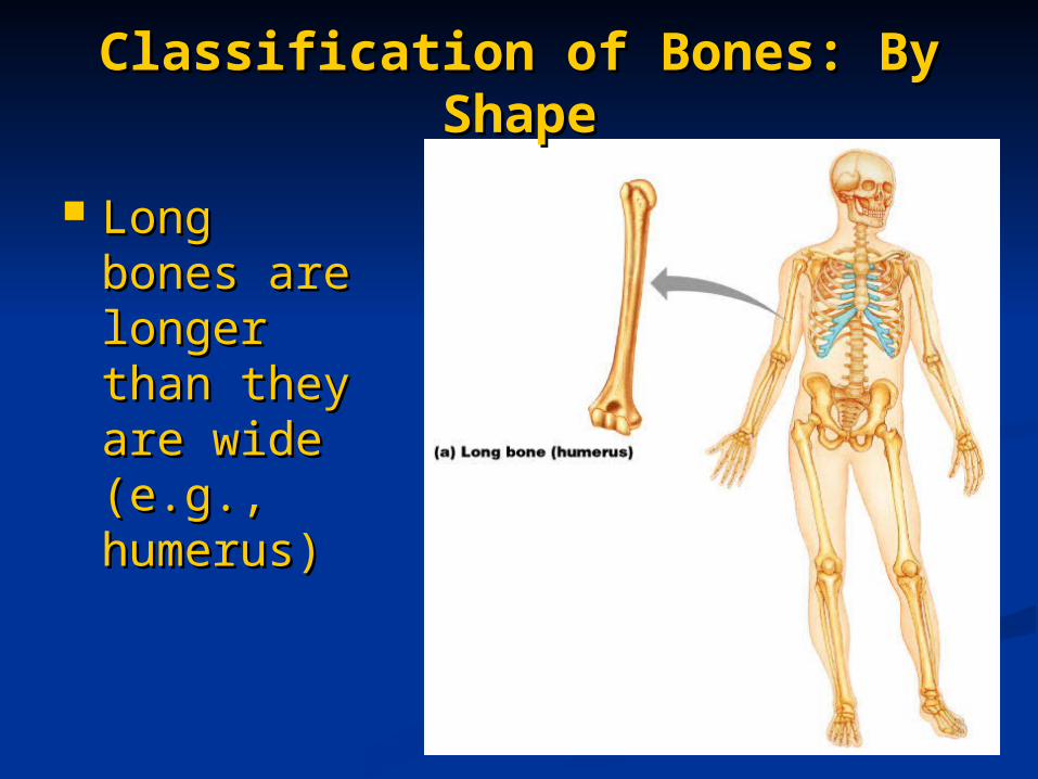
Long Long bones are bones are longer longer than they than they are wide are wide (e.g., (e.g., humerus)humerus)
Classification of Bones: Classification of Bones: By ShapeBy Shape
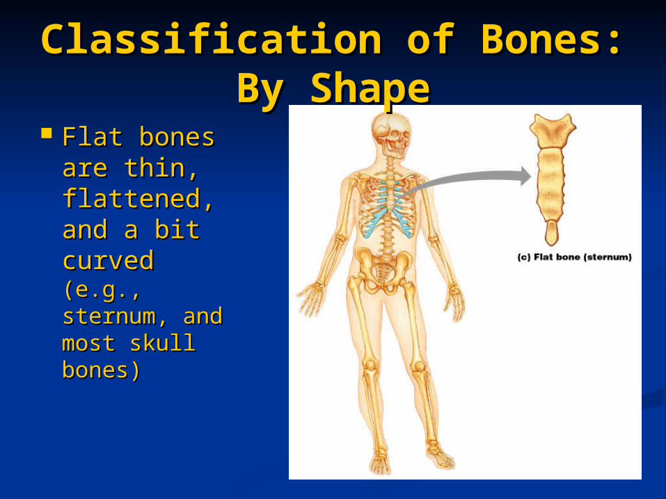
Flat bones Flat bones are thin, are thin, flattened, flattened, and a bit and a bit curved curved (e.g., (e.g., sternum, sternum, and most and most skull bones)skull bones)
Classification of Bones: By Classification of Bones: By ShapeShape
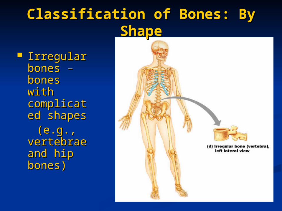
Irregular Irregular bones – bones – bones with bones with complicatecomplicated shapes d shapes
(e.g., (e.g., vertebrae vertebrae and hip and hip bones)bones)
Function of BonesFunction of Bones Support Support form the framework that supports the form the framework that supports the
body and cradles soft organsbody and cradles soft organs Protection – provide a protective case for Protection – provide a protective case for
the brain, spinal cord, and vital organsthe brain, spinal cord, and vital organs Movement – provide levers for musclesMovement – provide levers for muscles Mineral storage – reservoir for minerals, Mineral storage – reservoir for minerals,
especially calcium and phosphorusespecially calcium and phosphorus Blood cell formation – hematopoiesis Blood cell formation – hematopoiesis
occurs within the marrow cavities of bonesoccurs within the marrow cavities of bones
Gross Anatomy of Bones: Gross Anatomy of Bones: Bone TexturesBone Textures
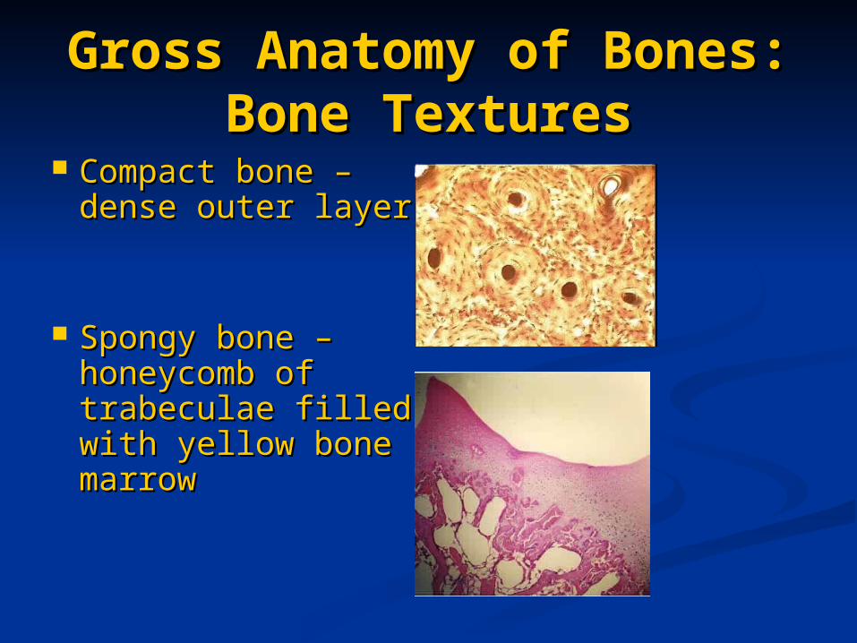
Compact bone – Compact bone – dense outer layerdense outer layer
Spongy bone – Spongy bone – honeycomb of honeycomb of trabeculae filled trabeculae filled with yellow bone with yellow bone marrowmarrow

Structure of Long BoneStructure of Long Bone
Figure 6.3
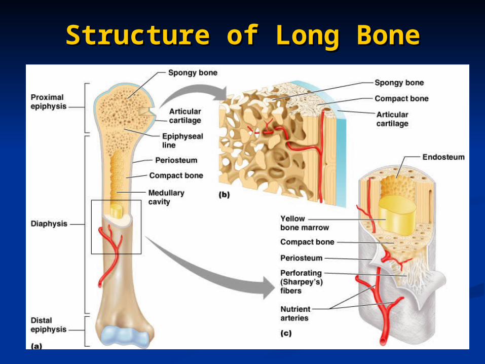
Structure of Long BoneStructure of Long Bone
Long bones consist of a diaphysis Long bones consist of a diaphysis and an epiphysisand an epiphysis
DiaphysisDiaphysis Tubular shaft that forms the axis of long Tubular shaft that forms the axis of long
bonesbones Composed of compact bone that Composed of compact bone that
surrounds the medullary cavitysurrounds the medullary cavity Yellow bone marrow (fat) is contained in Yellow bone marrow (fat) is contained in
the medullary cavitythe medullary cavity
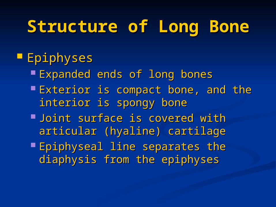
Structure of Long BoneStructure of Long Bone
EpiphysesEpiphyses Expanded ends of long bonesExpanded ends of long bones Exterior is compact bone, and the Exterior is compact bone, and the
interior is spongy boneinterior is spongy bone Joint surface is covered with articular Joint surface is covered with articular
(hyaline) cartilage(hyaline) cartilage Epiphyseal line separates the diaphysis Epiphyseal line separates the diaphysis
from the epiphysesfrom the epiphyses
Structure of Long BoneStructure of Long Bone
Figure 6.3
Bone MembranesBone Membranes Periosteum – double-layered protective Periosteum – double-layered protective
membranemembrane Outer fibrous layer is dense regular Outer fibrous layer is dense regular
connective tissueconnective tissue Inner osteogenic layer is composed of Inner osteogenic layer is composed of
osteoblasts and osteoclastsosteoblasts and osteoclasts Richly supplied with nerve fibers, blood, Richly supplied with nerve fibers, blood,
and lymphatic vessels, which enter the and lymphatic vessels, which enter the bone via nutrient foraminabone via nutrient foramina
Secured to underlying bone by Sharpey’s Secured to underlying bone by Sharpey’s fibersfibers
Endosteum – delicate membrane Endosteum – delicate membrane covering internal surfaces of bonecovering internal surfaces of bone
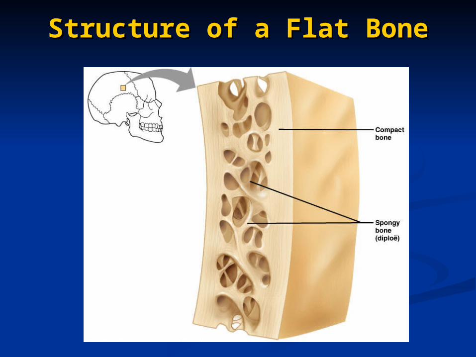
Structure of a Flat BoneStructure of a Flat Bone
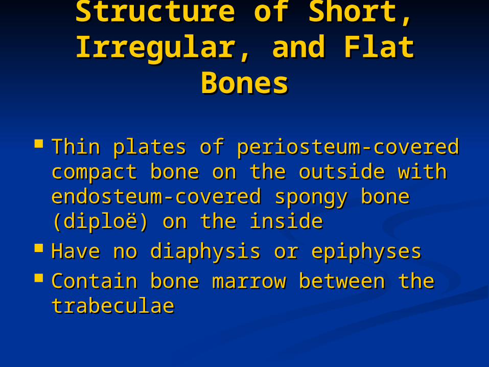
Structure of Short, Structure of Short, Irregular, and Flat BonesIrregular, and Flat Bones
Thin plates of periosteum-covered Thin plates of periosteum-covered compact bone on the outside with compact bone on the outside with endosteum-covered spongy bone endosteum-covered spongy bone (diploë) on the inside(diploë) on the inside
Have no diaphysis or epiphysesHave no diaphysis or epiphyses Contain bone marrow between the Contain bone marrow between the
trabeculaetrabeculae
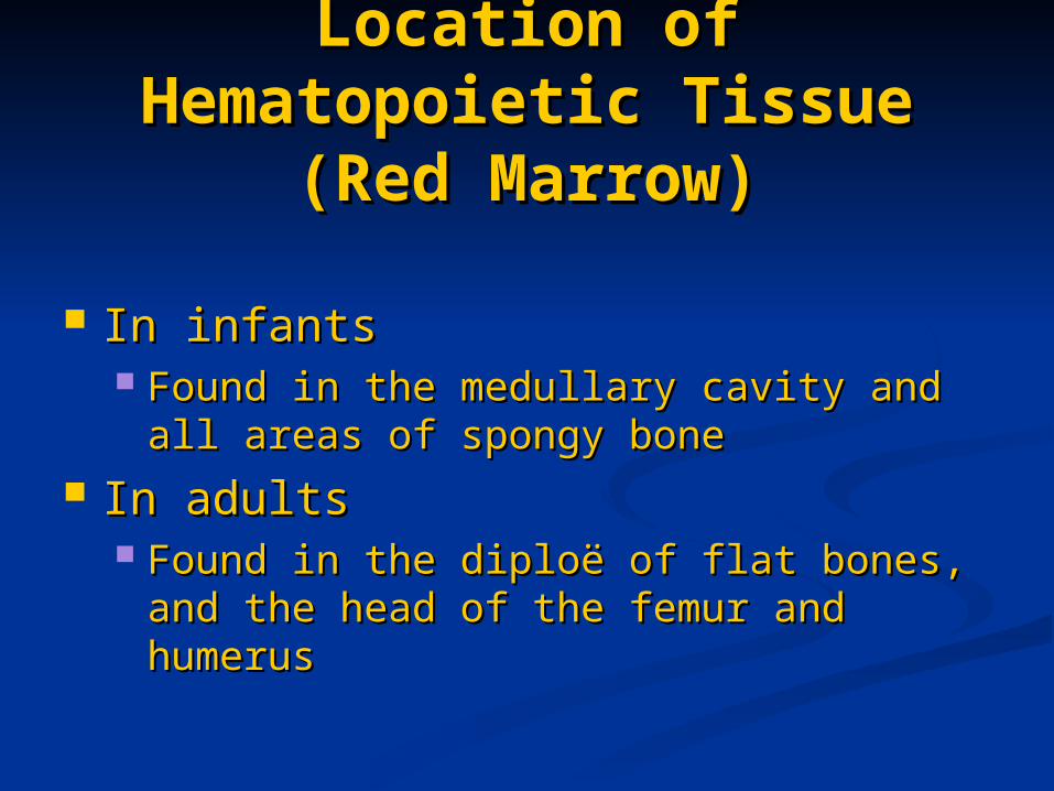
Location of Location of Hematopoietic Tissue Hematopoietic Tissue
(Red Marrow)(Red Marrow)
In infantsIn infants Found in the medullary cavity and all Found in the medullary cavity and all
areas of spongy bone areas of spongy bone In adultsIn adults
Found in the diploë of flat bones, and Found in the diploë of flat bones, and the head of the femur and humerusthe head of the femur and humerus
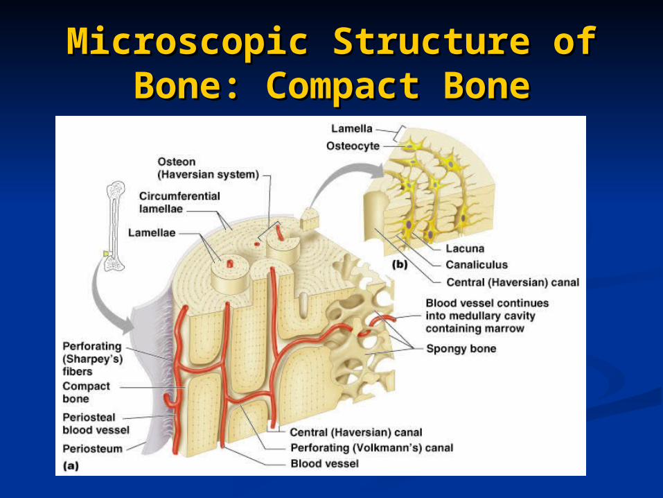
Microscopic Structure of Microscopic Structure of Bone: Compact BoneBone: Compact Bone
Cells of BoneCells of Bone
Osteoblasts – bone-forming cellsOsteoblasts – bone-forming cells Osteocytes – mature bone cellsOsteocytes – mature bone cells Osteoclasts – large cells that resorb Osteoclasts – large cells that resorb
or break down bone matrixor break down bone matrix
Chemical Composition of Chemical Composition of Bone: OrganicBone: Organic
Osteoid – unmineralized bone matrix Osteoid – unmineralized bone matrix composed of proteoglycans, composed of proteoglycans, glycoproteins, and collagenglycoproteins, and collagen
Chemical Composition of Chemical Composition of Bone: InorganicBone: Inorganic
Hydroxyapatites, or mineral saltsHydroxyapatites, or mineral salts Sixty-five percent of bone by massSixty-five percent of bone by mass Mainly calcium phosphatesMainly calcium phosphates Responsible for bone hardness and its Responsible for bone hardness and its
resistance to compressionresistance to compression
Developmental Aspects Developmental Aspects of Bonesof Bones
Mesoderm gives rise to embryonic Mesoderm gives rise to embryonic mesenchymal cells, which produce mesenchymal cells, which produce membranes and cartilages that form the membranes and cartilages that form the embryonic skeletonembryonic skeleton
The embryonic skeleton ossifies in a The embryonic skeleton ossifies in a predictable timetable that allows fetal age predictable timetable that allows fetal age to be easily determined from sonogramsto be easily determined from sonograms
At birth, most long bones are well ossified At birth, most long bones are well ossified (except for their epiphyses)(except for their epiphyses)
Developmental Aspects Developmental Aspects of Bonesof Bones
By age 25, nearly all bones are By age 25, nearly all bones are completely ossifiedcompletely ossified
In old age, bone resorption In old age, bone resorption predominatespredominates
A single gene that codes for vitamin A single gene that codes for vitamin D docking determines both the D docking determines both the tendency to accumulate bone mass tendency to accumulate bone mass early in life, and the risk for early in life, and the risk for osteoporosis later in lifeosteoporosis later in life
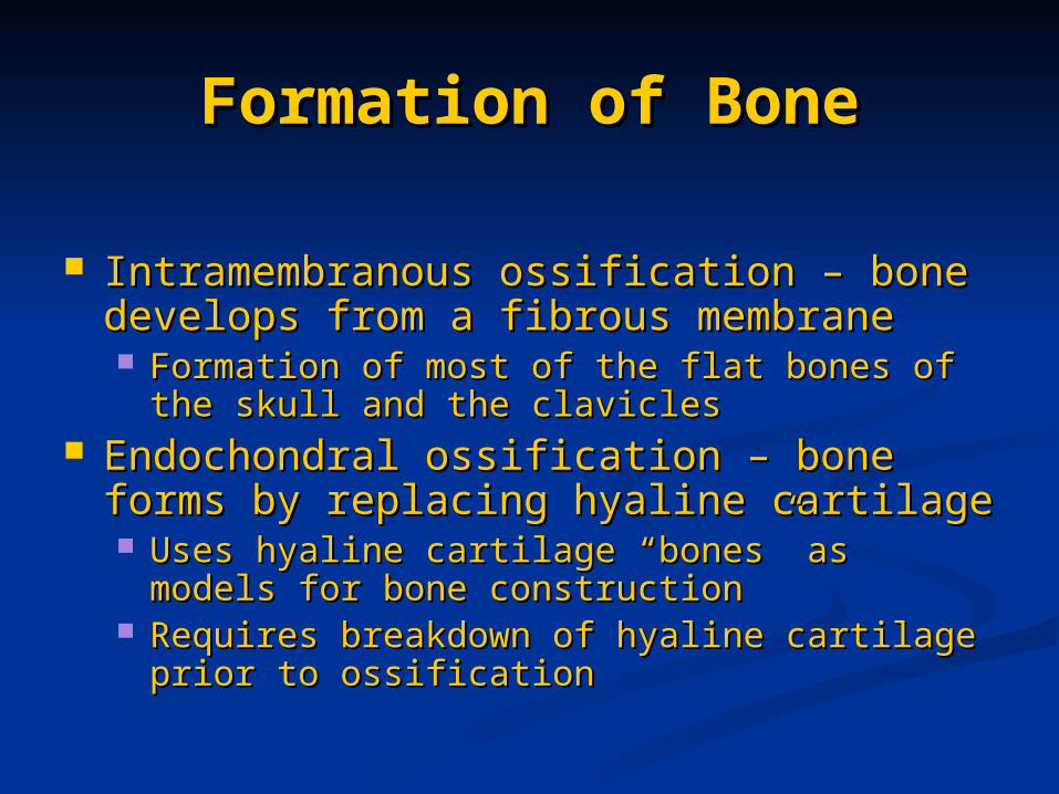
Formation of BoneFormation of Bone
Intramembranous ossification – bone Intramembranous ossification – bone develops from a fibrous membranedevelops from a fibrous membrane Formation of most of the flat bones of the skull Formation of most of the flat bones of the skull
and the claviclesand the clavicles Endochondral ossification – bone forms by Endochondral ossification – bone forms by
replacing hyaline cartilagereplacing hyaline cartilage Uses hyaline cartilage “bones” as models for Uses hyaline cartilage “bones” as models for
bone constructionbone construction Requires breakdown of hyaline cartilage prior Requires breakdown of hyaline cartilage prior
to ossificationto ossification
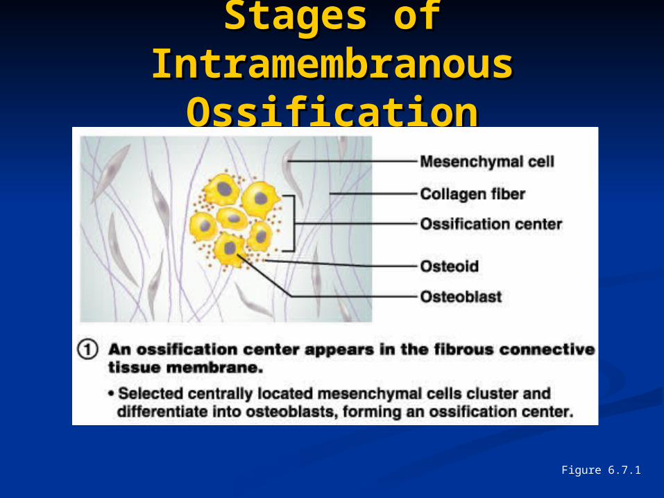
Stages of Stages of Intramembranous Intramembranous
OssificationOssification
Figure 6.7.1
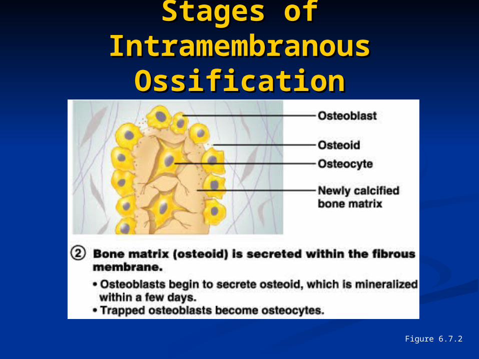
Stages of Stages of Intramembranous Intramembranous
OssificationOssification
Figure 6.7.2
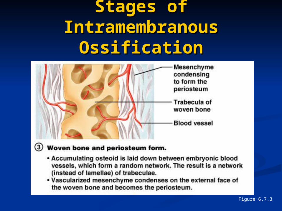
Stages of Stages of Intramembranous Intramembranous
OssificationOssification
Figure 6.7.3
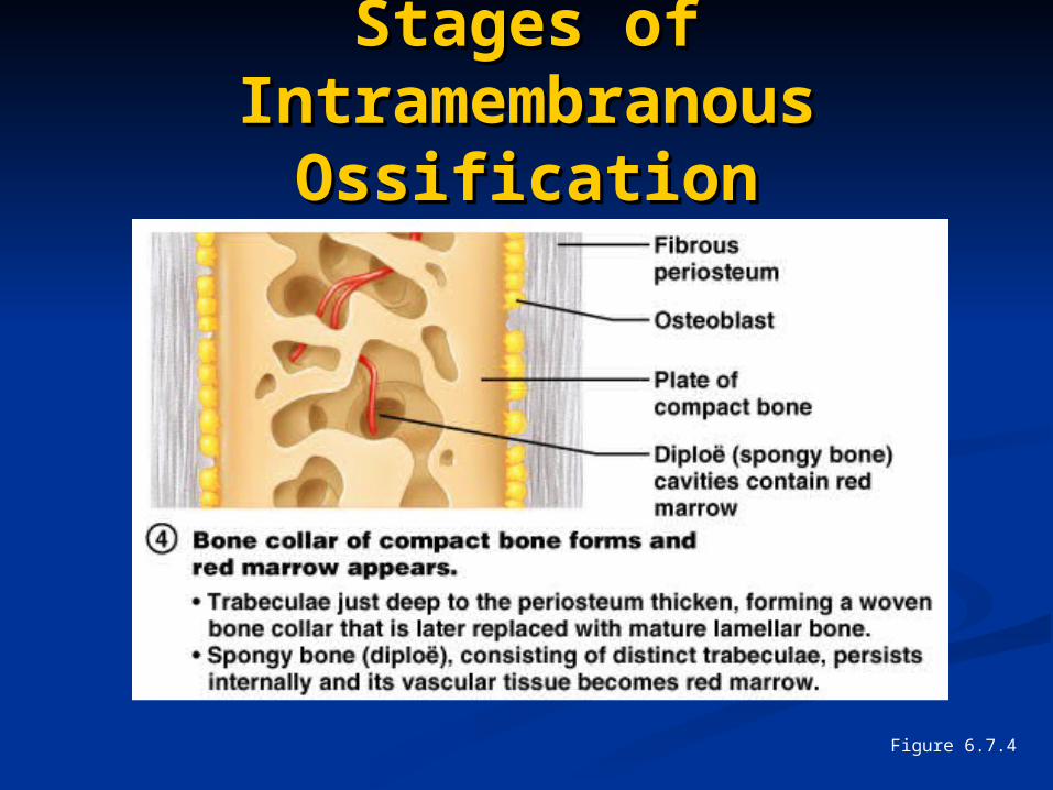
Stages of Stages of Intramembranous Intramembranous
OssificationOssification
Figure 6.7.4
Endochondral Endochondral OssificationOssification
Begins in the second month of Begins in the second month of developmentdevelopment
Uses hyaline cartilage “bones” as Uses hyaline cartilage “bones” as models for bone constructionmodels for bone construction
Requires breakdown of hyaline Requires breakdown of hyaline cartilage prior to ossificationcartilage prior to ossification
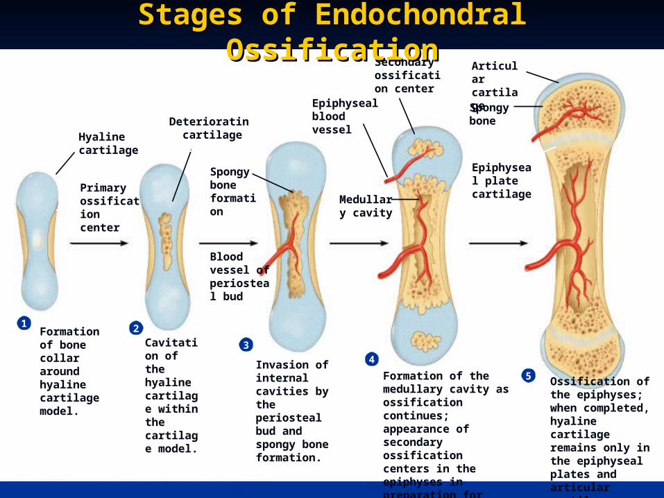
Formation of bone collar around hyaline cartilage model.
1 2
3
4
Cavitation of the hyaline cartilage within the cartilage model.
Invasion of internal cavities by the periosteal bud and spongy bone formation.
5 Ossification of the epiphyses; when completed, hyaline cartilage remains only in the epiphyseal plates and articular cartilages
Formation of the medullary cavity as ossification continues; appearance of secondary ossification centers in the epiphyses in preparation for stage 5.
Hyaline cartilage
Primary ossification center
Bone collar
Deteriorating cartilage matrix
Spongy bone formation
Blood vessel of periosteal bud
Secondary ossification center
Epiphyseal blood vessel
Medullary cavity
Epiphyseal plate cartilage
Spongy bone
Articular cartilage
Stages of Endochondral Stages of Endochondral OssificationOssification
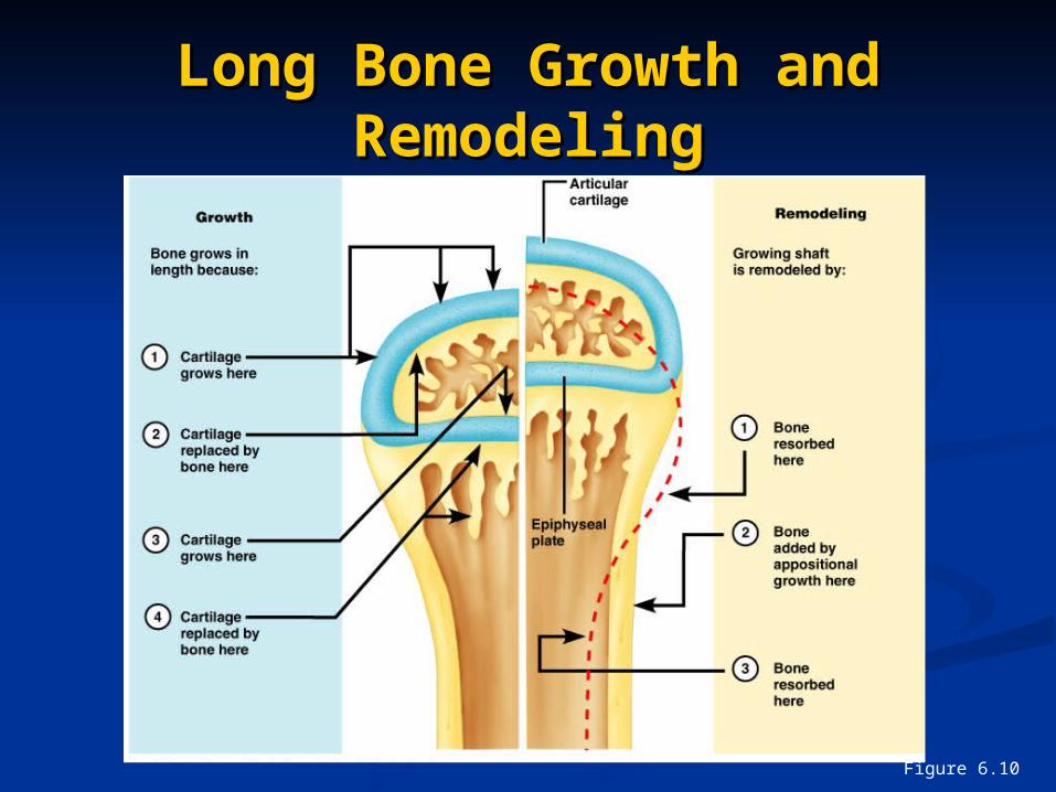
Long Bone Growth and Long Bone Growth and RemodelingRemodeling
Figure 6.10
Functional Zones in Long Functional Zones in Long Bone GrowthBone Growth
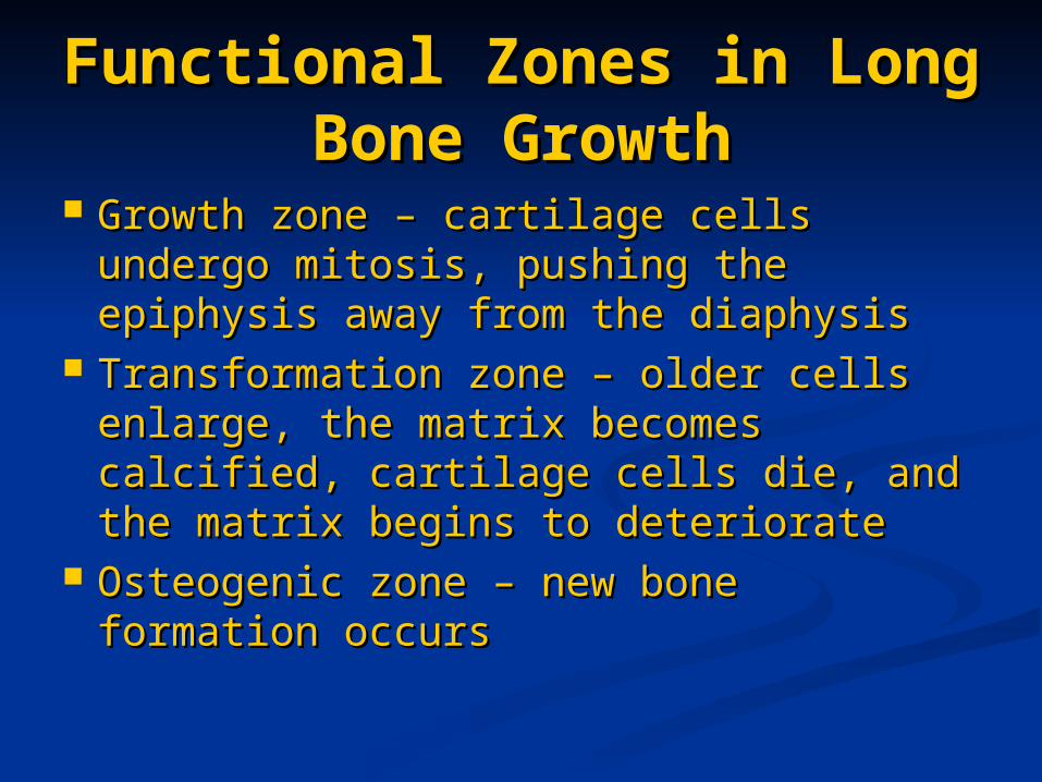
Growth zone – cartilage cells undergo Growth zone – cartilage cells undergo mitosis, pushing the epiphysis away mitosis, pushing the epiphysis away from the diaphysisfrom the diaphysis
Transformation zone – older cells Transformation zone – older cells enlarge, the matrix becomes enlarge, the matrix becomes calcified, cartilage cells die, and the calcified, cartilage cells die, and the matrix begins to deterioratematrix begins to deteriorate
Osteogenic zone – new bone Osteogenic zone – new bone formation occursformation occurs
Long Bone Growth and Long Bone Growth and RemodelingRemodeling
Growth in length – cartilage Growth in length – cartilage continually grows and is replaced by continually grows and is replaced by bone as shown bone as shown
Remodeling – bone is resorbed and Remodeling – bone is resorbed and added by appositional growth as added by appositional growth as shown in the next slide shown in the next slide
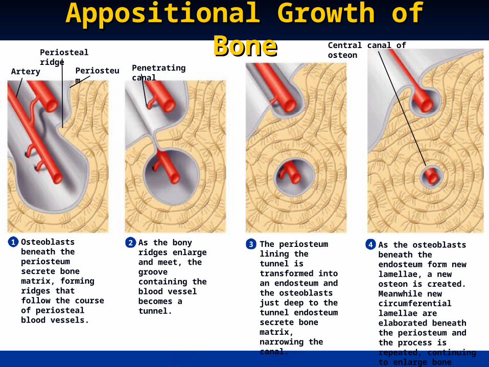
Osteoblasts beneath the periosteum secrete bone matrix, forming ridges that follow the course of periosteal blood vessels.
1 2 3 4As the bony ridges enlarge and meet, the groove containing the blood vessel becomes a tunnel.
The periosteum lining the tunnel is transformed into an endosteum and the osteoblasts just deep to the tunnel endosteum secrete bone matrix, narrowing the canal.
As the osteoblasts beneath the endosteum form new lamellae, a new osteon is created. Meanwhile new circumferential lamellae are elaborated beneath the periosteum and the process is repeated, continuing to enlarge bone diameter.
Artery Periosteum Penetrating canal
Central canal of osteonPeriosteal ridge
Appositional Growth of Appositional Growth of BoneBone
During infancy and childhood, epiphyseal During infancy and childhood, epiphyseal plate activity is stimulated by growth plate activity is stimulated by growth hormonehormone
During puberty, testosterone and estrogens: During puberty, testosterone and estrogens: Initially promote adolescent growth spurtsInitially promote adolescent growth spurts Cause masculinization and feminization of Cause masculinization and feminization of
specific parts of the skeletonspecific parts of the skeleton Later induce epiphyseal plate closure, ending Later induce epiphyseal plate closure, ending
longitudinal bone growth longitudinal bone growth
Hormonal Regulation of Hormonal Regulation of Bone Growth During Bone Growth During
YouthYouth
Bone DepositionBone Deposition Occurs where bone is injured or added strength is Occurs where bone is injured or added strength is
neededneeded Requires a diet rich in protein, vitamins C, D, and A, Requires a diet rich in protein, vitamins C, D, and A,
calcium, phosphorus, magnesium, and manganesecalcium, phosphorus, magnesium, and manganese Alkaline phosphatase is essential for mineralization Alkaline phosphatase is essential for mineralization
of boneof bone Sites of new matrix deposition are revealed by the:Sites of new matrix deposition are revealed by the:
Osteoid seam – unmineralized band of bone Osteoid seam – unmineralized band of bone matrixmatrix
Calcification front – abrupt transition zone Calcification front – abrupt transition zone between the osteoid seam and the older between the osteoid seam and the older mineralized bonemineralized bone
Bone ResorptionBone Resorption
Accomplished by osteoclastsAccomplished by osteoclasts Resorption bays – grooves formed by Resorption bays – grooves formed by
osteoclasts as they break down bone matrixosteoclasts as they break down bone matrix Resorption involves osteoclast secretion of:Resorption involves osteoclast secretion of:
Lysosomal enzymes that digest organic matrixLysosomal enzymes that digest organic matrix Acids that convert calcium salts into soluble Acids that convert calcium salts into soluble
formsforms Dissolved matrix is transcytosed across the Dissolved matrix is transcytosed across the
osteoclast’s cell where it is secreted into osteoclast’s cell where it is secreted into the interstitial fluid and then into the bloodthe interstitial fluid and then into the blood
Importance of Ionic Importance of Ionic Calcium in the BodyCalcium in the Body
Calcium is necessary for:Calcium is necessary for: Transmission of nerve impulsesTransmission of nerve impulses Muscle contractionMuscle contraction Blood coagulationBlood coagulation Secretion by glands and nerve cellsSecretion by glands and nerve cells Cell divisionCell division
Control of RemodelingControl of Remodeling
Two control loops regulate bone Two control loops regulate bone remodelingremodeling Hormonal mechanism maintains calcium Hormonal mechanism maintains calcium
homeostasis in the bloodhomeostasis in the blood Mechanical and gravitational forces Mechanical and gravitational forces
acting on the skeletonacting on the skeleton
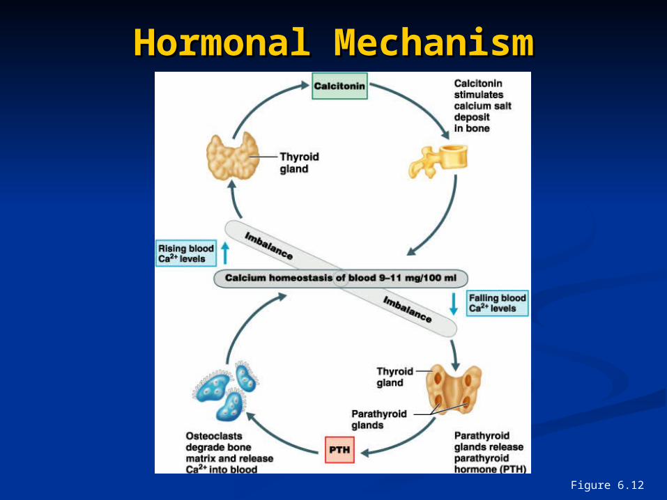
Hormonal MechanismHormonal Mechanism
Figure 6.12
Response to Mechanical Response to Mechanical StressStress
Wolff’s law – a bone grows or Wolff’s law – a bone grows or remodels in response to the forces or remodels in response to the forces or demands placed upon itdemands placed upon it
Observations supporting Wolff’s law Observations supporting Wolff’s law includeinclude Long bones are thickest midway along the Long bones are thickest midway along the
shaft (where bending stress is greatest)shaft (where bending stress is greatest) Curved bones are thickest where they are Curved bones are thickest where they are
most likely to bucklemost likely to buckle
Response to Mechanical Response to Mechanical StressStress
Trabeculae form along lines of stressTrabeculae form along lines of stress Large, bony projections occur where Large, bony projections occur where
heavy, active muscles attachheavy, active muscles attach
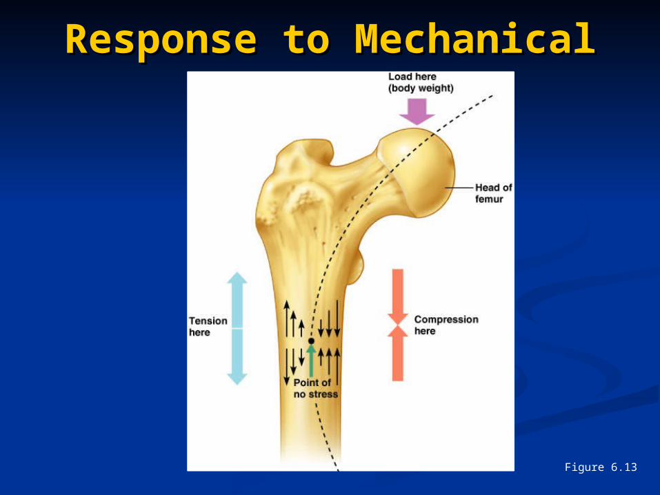
Response to Mechanical Response to Mechanical StressStress
Figure 6.13
Bone Fractures (Breaks)Bone Fractures (Breaks)
Classified by:Classified by: The position of the bone ends after The position of the bone ends after
fracturefracture The completeness of the breakThe completeness of the break The orientation of the bone to the long The orientation of the bone to the long
axisaxis Whether or not the bones ends Whether or not the bones ends
penetrate the skinpenetrate the skin
Types of Bone FracturesTypes of Bone Fractures
Nondisplaced – bone ends retain their Nondisplaced – bone ends retain their normal positionnormal position
Displaced – bone ends are out of normal Displaced – bone ends are out of normal alignmentalignment
Complete – bone is broken all the way Complete – bone is broken all the way throughthrough
Incomplete – bone is not broken all the Incomplete – bone is not broken all the way throughway through
Linear – the fracture is parallel to the long Linear – the fracture is parallel to the long axis of the boneaxis of the bone
Types of Bone FracturesTypes of Bone Fractures
Transverse – the fracture is Transverse – the fracture is perpendicular to the long axis of the perpendicular to the long axis of the bonebone
Compound (open) – bone ends Compound (open) – bone ends penetrate the skinpenetrate the skin
Simple (closed) – bone ends do not Simple (closed) – bone ends do not penetrate the skinpenetrate the skin
Common Types of Common Types of FracturesFractures
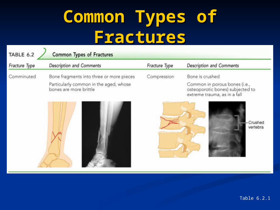
Comminuted – bone fragments into three Comminuted – bone fragments into three or more pieces; common in the elderlyor more pieces; common in the elderly
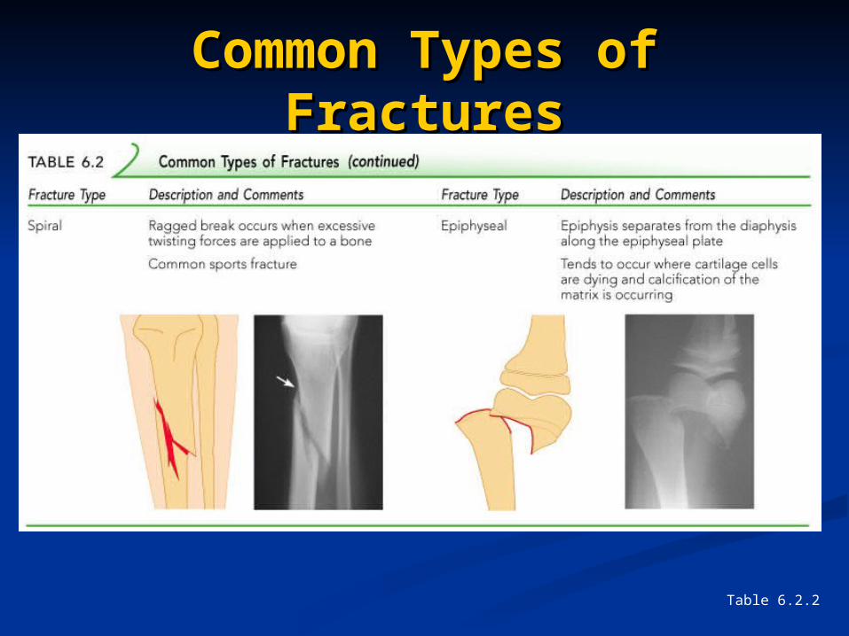
Spiral – ragged break when bone is Spiral – ragged break when bone is excessively twisted; common sports injuryexcessively twisted; common sports injury
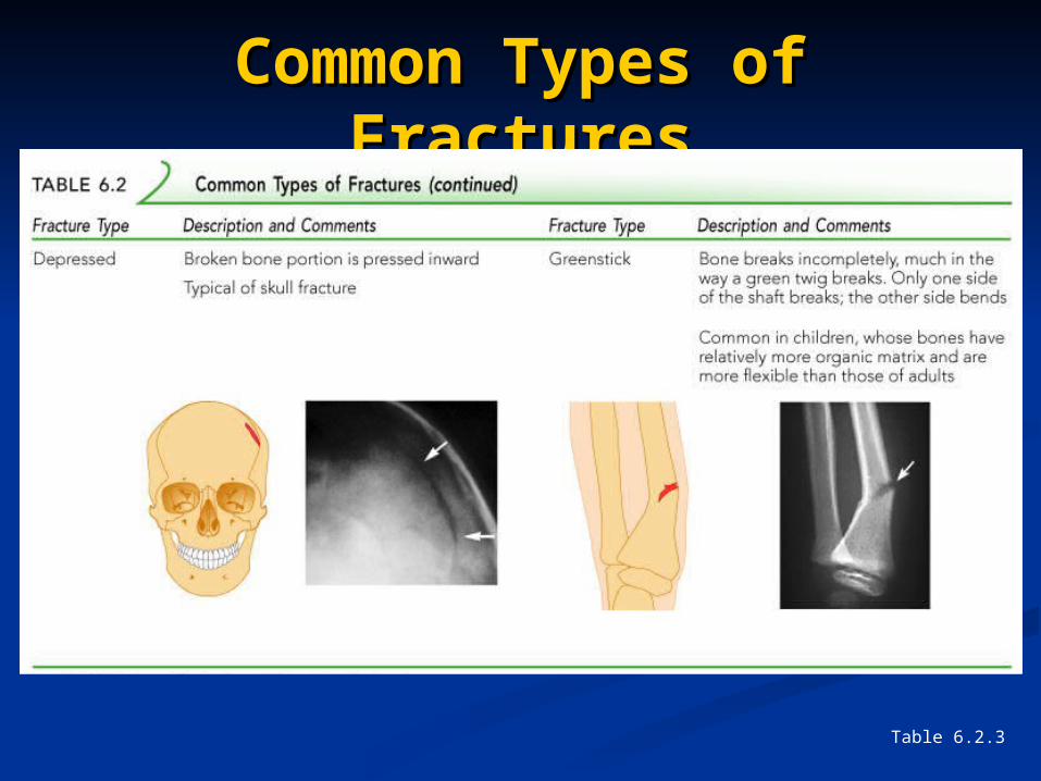
Depressed – broken bone portion pressed Depressed – broken bone portion pressed inward; typical skull fractureinward; typical skull fracture
Compression – bone is crushed; common Compression – bone is crushed; common in porous bonesin porous bones
Common Types of Common Types of FracturesFractures
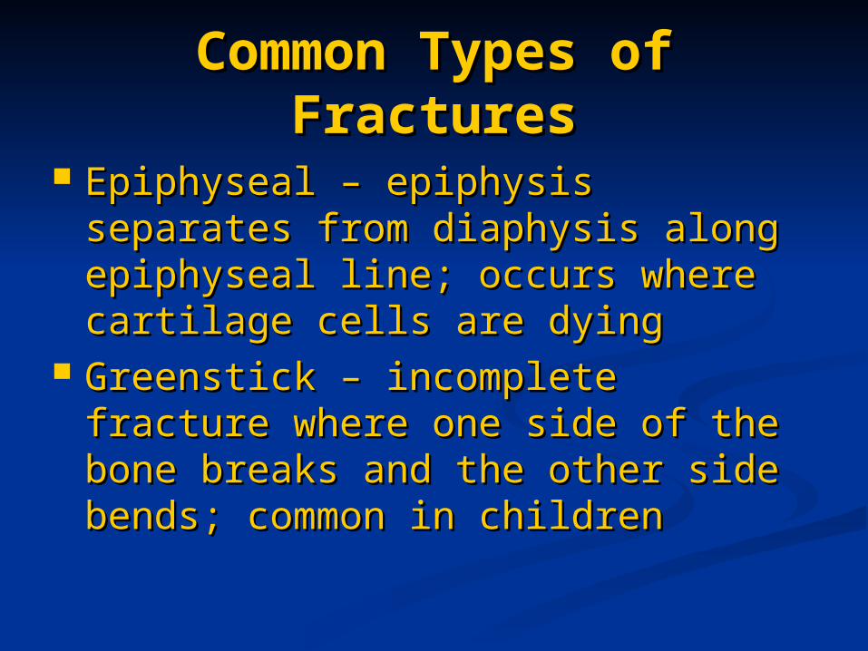
Epiphyseal – epiphysis separates Epiphyseal – epiphysis separates from diaphysis along epiphyseal line; from diaphysis along epiphyseal line; occurs where cartilage cells are occurs where cartilage cells are dyingdying
Greenstick – incomplete fracture Greenstick – incomplete fracture where one side of the bone breaks where one side of the bone breaks and the other side bends; common in and the other side bends; common in childrenchildren
Common Types of Common Types of FracturesFractures
Table 6.2.1
Common Types of Common Types of FracturesFractures
Table 6.2.2
Common Types of Common Types of FracturesFractures
Table 6.2.3
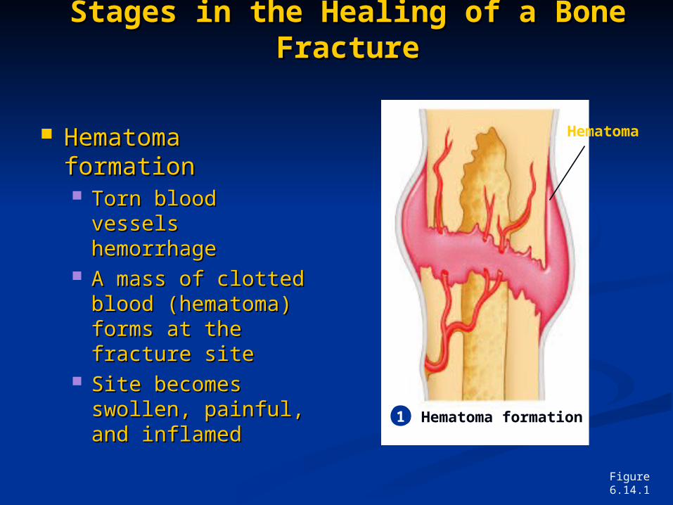
Stages in the Healing of a Bone Stages in the Healing of a Bone FractureFracture
Hematoma Hematoma formationformation Torn blood vessels Torn blood vessels
hemorrhagehemorrhage A mass of clotted A mass of clotted
blood (hematoma) blood (hematoma) forms at the forms at the fracture sitefracture site
Site becomes Site becomes swollen, painful, swollen, painful, and inflamedand inflamed
Figure 6.14.1
1
Hematoma
Hematoma formation
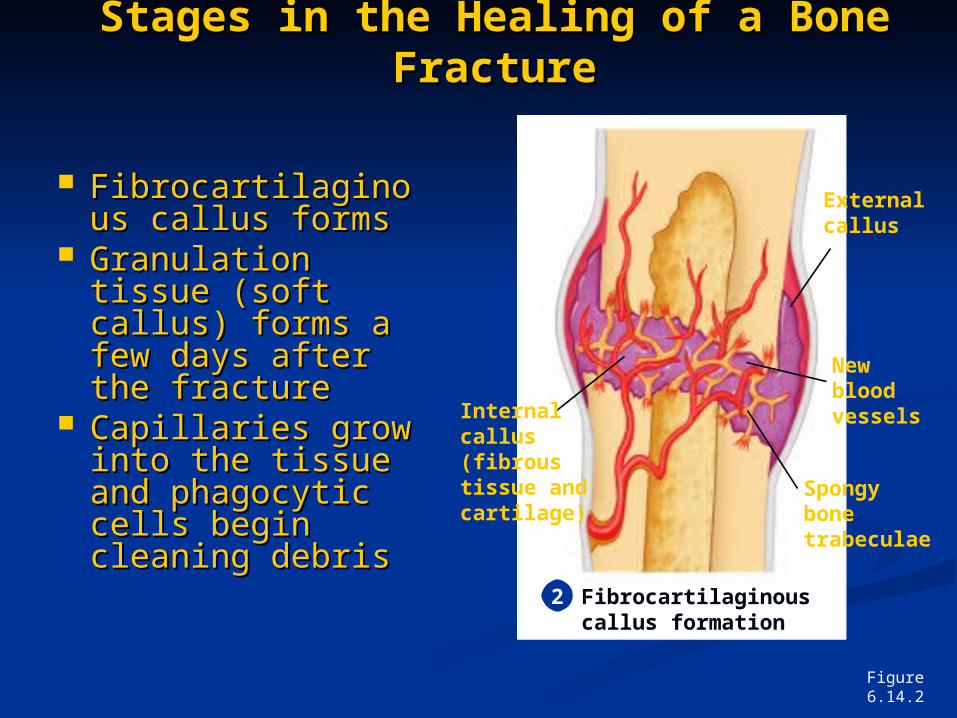
Stages in the Healing of a Bone Stages in the Healing of a Bone FractureFracture
FibrocartilaginouFibrocartilaginous callus formss callus forms
Granulation Granulation tissue (soft tissue (soft callus) forms a callus) forms a few days after few days after the fracturethe fracture
Capillaries grow Capillaries grow into the tissue into the tissue and phagocytic and phagocytic cells begin cells begin cleaning debriscleaning debris
Figure 6.14.2
2 Fibrocartilaginous callus formation
External callus
New blood vessels
Spongy bone trabeculae
Internal callus (fibrous tissue and cartilage)
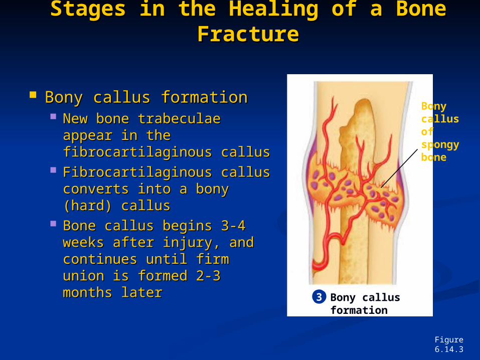
Stages in the Healing of a Bone Stages in the Healing of a Bone FractureFracture
Bony callus formationBony callus formation New bone trabeculae New bone trabeculae
appear in the appear in the fibrocartilaginous callusfibrocartilaginous callus
Fibrocartilaginous callus Fibrocartilaginous callus converts into a bony converts into a bony (hard) callus(hard) callus
Bone callus begins 3-4 Bone callus begins 3-4 weeks after injury, and weeks after injury, and continues until firm union continues until firm union is formed 2-3 months is formed 2-3 months laterlater
Figure 6.14.3
3 Bony callus formation
Bony callus of spongy bone
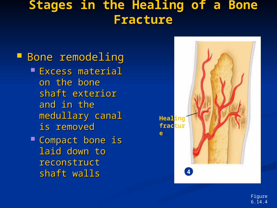
Stages in the Healing of a Bone Stages in the Healing of a Bone FractureFracture
Bone remodelingBone remodeling Excess material Excess material
on the bone shaft on the bone shaft exterior and in the exterior and in the medullary canal is medullary canal is removedremoved
Compact bone is Compact bone is laid down to laid down to reconstruct shaft reconstruct shaft wallswalls
Figure 6.14.4
4 Bone remodeling
Healing fracture
Homeostatic ImbalancesHomeostatic Imbalances
RicketsRickets Bones of children are inadequately Bones of children are inadequately
mineralized causing softened, weakened mineralized causing softened, weakened bonesbones
Bowed legs and deformities of the Bowed legs and deformities of the pelvis, skull, and rib cage are commonpelvis, skull, and rib cage are common
Caused by insufficient calcium in the Caused by insufficient calcium in the diet, or by vitamin D deficiencydiet, or by vitamin D deficiency
Homeostatic ImbalancesHomeostatic Imbalances
OsteoporosisOsteoporosis Group of diseases in which bone Group of diseases in which bone
reabsorption outpaces bone depositreabsorption outpaces bone deposit Spongy bone of the spine is most Spongy bone of the spine is most
vulnerablevulnerable Occurs most often in postmenopausal Occurs most often in postmenopausal
womenwomen Bones become so fragile that sneezing or Bones become so fragile that sneezing or
stepping off a curb can cause fracturesstepping off a curb can cause fractures