bones and skeletal tissues -...
TRANSCRIPT
# 105016 Cust: Benjamin Cummings/CA Au: Marieb Pg. No. 173 Title: Anatomy & Physiology Server: S4C
C/M/Y/KShort / Normal
DESIGN SERVICES OF
CARLISLEPublishing Services
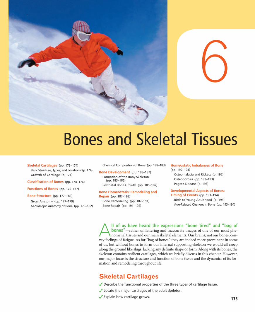
All of us have heard the expressions “bone tired” and “bag of bones”—rather unflattering and inaccurate images of one of our most phe-nomenal tissues and our main skeletal elements. Our brains, not our bones, con-
vey feelings of fatigue. As for “bag of bones,” they are indeed more prominent in some of us, but without bones to form our internal supporting skeleton we would all creep along the ground like slugs, lacking any definite shape or form. Along with its bones, the skeleton contains resilient cartilages, which we briefly discuss in this chapter. However, our major focus is the structure and function of bone tissue and the dynamics of its for-mation and remodeling throughout life.
Skeletal Cartilages Describe the functional properties of the three types of cartilage tissue.
Locate the major cartilages of the adult skeleton.
Explain how cartilage grows.
6Bones and Skeletal Tissues
Skeletal Cartilages (pp. 173–174)Basic Structure, Types, and Locations (p. 174)Growth of Cartilage (p. 174)
Classification of Bones (pp. 174–176)
Functions of Bones (pp. 176–177)
Bone Structure (pp. 177–183)
Gross Anatomy (pp. 177–179)Microscopic Anatomy of Bone (pp. 179–182)
Chemical Composition of Bone (pp. 182–183)
Bone Development (pp. 183–187)Formation of the Bony Skeleton
(pp. 183–185)Postnatal Bone Growth (pp. 185–187)
Bone Homeostasis: Remodeling and Repair (pp. 187–192)
Bone Remodeling (pp. 187–191)Bone Repair (pp. 191–192)
Homeostatic Imbalances of Bone (pp. 192–193)
Osteomalacia and Rickets (p. 192)Osteoporosis (pp. 192–193)Paget’s Disease (p. 193)
Developmental Aspects of Bones: Timing of Events (pp. 193–194)
Birth to Young Adulthood (p. 193)Age-Related Changes in Bone (pp. 193–194)
173
174 UNIT 2 Covering, Support, and Movement of the Body
6
# 105016 Cust: Benjamin Cummings/CA Au: Marieb Pg. No. 174 Title: Anatomy & Physiology Server: S4C
C/M/Y/KShort / Normal
DESIGN SERVICES OF
CARLISLEPublishing Services
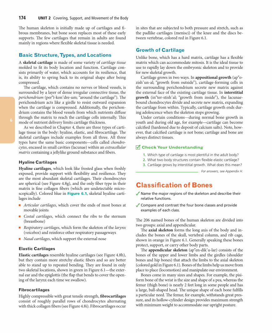
in sites that are subjected to both pressure and stretch, such as the padlike cartilages (menisci) of the knee and the discs be-tween vertebrae, colored red in Figure 6.1.
Growth of CartilageUnlike bone, which has a hard matrix, cartilage has a flexible matrix which can accommodate mitosis. It is the ideal tissue to use to rapidly lay down the embryonic skeleton and to provide for new skeletal growth.
Cartilage grows in two ways. In appositional growth (ap!o-zish"un-al; “growth from outside”), cartilage-forming cells in the surrounding perichondrium secrete new matrix against the external face of the existing cartilage tissue. In interstitial growth (in!ter-stish"al; “growth from inside”), the lacunae-bound chondrocytes divide and secrete new matrix, expanding the cartilage from within. Typically, cartilage growth ends dur-ing adolescence when the skeleton stops growing.
Under certain conditions—during normal bone growth in youth and during old age, for example—cartilage can become calcified (hardened due to deposit of calcium salts). Note, how-ever, that calcified cartilage is not bone; cartilage and bone are always distinct tissues.
Check Your Understanding
1. Which type of cartilage is most plentiful in the adult body? 2. What two body structures contain flexible elastic cartilage? 3. Cartilage grows by interstitial growth. What does this mean?
For answers, see Appendix H.
Classification of Bones Name the major regions of the skeleton and describe their relative functions.
Compare and contrast the four bone classes and provide examples of each class.
The 206 named bones of the human skeleton are divided into two groups: axial and appendicular.
The axial skeleton forms the long axis of the body and in-cludes the bones of the skull, vertebral column, and rib cage, shown in orange in Figure 6.1. Generally speaking these bones protect, support, or carry other body parts.
The appendicular skeleton (ap!en-dik"u-lar) consists of the bones of the upper and lower limbs and the girdles (shoulder bones and hip bones) that attach the limbs to the axial skeleton (colored gold in Figure 6.1). Bones of the limbs help us move from place to place (locomotion) and manipulate our environment.
Bones come in many sizes and shapes. For example, the pisi-form bone of the wrist is the size and shape of a pea, whereas the femur (thigh bone) is nearly 2 feet long in some people and has a large, ball-shaped head. The unique shape of each bone fulfills a particular need. The femur, for example, withstands great pres-sure, and its hollow-cylinder design provides maximum strength with minimum weight to accommodate our upright posture.
The human skeleton is initially made up of cartilages and fi-brous membranes, but bone soon replaces most of these early supports. The few cartilages that remain in adults are found mainly in regions where flexible skeletal tissue is needed.
Basic Structure, Types, and LocationsA skeletal cartilage is made of some variety of cartilage tissue molded to fit its body location and function. Cartilage con-sists primarily of water, which accounts for its resilience, that is, its ability to spring back to its original shape after being compressed.
The cartilage, which contains no nerves or blood vessels, is surrounded by a layer of dense irregular connective tissue, the perichondrium (per!ĭ-kon"dre-um; “around the cartilage”). The perichondrium acts like a girdle to resist outward expansion when the cartilage is compressed. Additionally, the perichon-drium contains the blood vessels from which nutrients diffuse through the matrix to reach the cartilage cells internally. This mode of nutrient delivery limits cartilage thickness.
As we described in Chapter 4, there are three types of carti-lage tissue in the body: hyaline, elastic, and fibrocartilage. The skeletal cartilages include examples from all three. All three types have the same basic components—cells called chondro-cytes, encased in small cavities (lacunae) within an extracellular matrix containing a jellylike ground substance and fibers.
Hyaline CartilagesHyaline cartilages, which look like frosted glass when freshly exposed, provide support with flexibility and resilience. They are the most abundant skeletal cartilages. Their chondrocytes are spherical (see Figure 4.8g), and the only fiber type in their matrix is fine collagen fibers (which are undetectable micro-scopically). Colored blue in Figure 6.1, skeletal hyaline carti-lages include■ Articular cartilages, which cover the ends of most bones at
movable joints■ Costal cartilages, which connect the ribs to the sternum
(breastbone)■ Respiratory cartilages, which form the skeleton of the larynx
(voicebox) and reinforce other respiratory passageways■ Nasal cartilages, which support the external nose
Elastic CartilagesElastic cartilages resemble hyaline cartilages (see Figure 4.8h), but they contain more stretchy elastic fibers and so are better able to stand up to repeated bending. They are found in only two skeletal locations, shown in green in Figure 6.1—the exter-nal ear and the epiglottis (the flap that bends to cover the open-ing of the larynx each time we swallow).
FibrocartilagesHighly compressible with great tensile strength, fibrocartilages consist of roughly parallel rows of chondrocytes alternating with thick collagen fibers (see Figure 4.8i). Fibrocartilages occur
Chapter 6 Bones and Skeletal Tissues 175
6
# 105016 Cust: Benjamin Cummings/CA Au: Marieb Pg. No. 175 Title: Anatomy & Physiology Server: S4C
C/M/Y/KShort / Normal
DESIGN SERVICES OF
CARLISLEPublishing Services
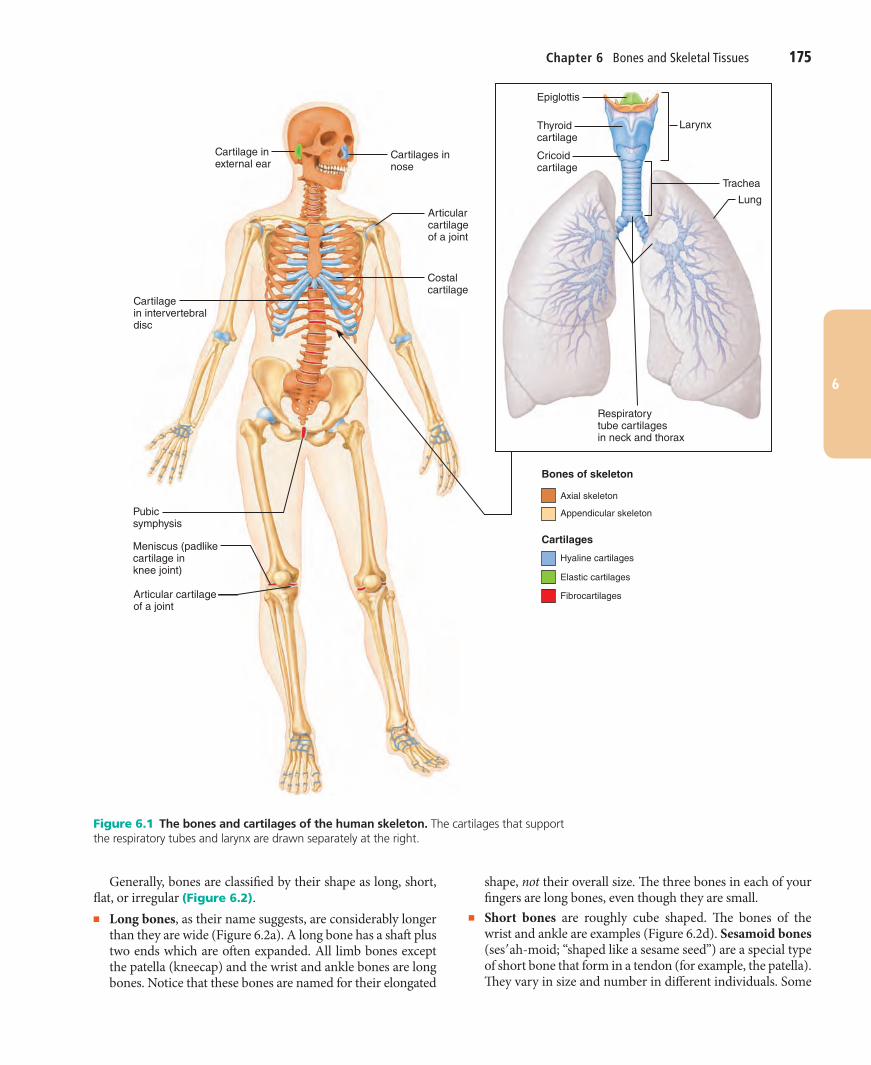
Generally, bones are classified by their shape as long, short, flat, or irregular (Figure 6.2).■ Long bones, as their name suggests, are considerably longer
than they are wide (Figure 6.2a). A long bone has a shaft plus two ends which are often expanded. All limb bones except the patella (kneecap) and the wrist and ankle bones are long bones. Notice that these bones are named for their elongated
shape, not their overall size. The three bones in each of your fingers are long bones, even though they are small.
■ Short bones are roughly cube shaped. The bones of the wrist and ankle are examples (Figure 6.2d). Sesamoid bones (ses"ah-moid; “shaped like a sesame seed”) are a special type of short bone that form in a tendon (for example, the patella). They vary in size and number in different individuals. Some
Axial skeleton
Appendicular skeleton
Hyaline cartilages
Elastic cartilages
Fibrocartilages
Cartilages
Bones of skeleton
Epiglottis
Larynx
Trachea
Cricoidcartilage
Lung
Respiratorytube cartilagesin neck and thorax
Thyroidcartilage
Cartilage inexternal ear
Cartilages innose
Articularcartilageof a joint
Costalcartilage
Cartilagein intervertebraldisc
Pubicsymphysis
Articular cartilageof a joint
Meniscus (padlikecartilage inknee joint)
Figure 6.1 The bones and cartilages of the human skeleton. The cartilages that support the respiratory tubes and larynx are drawn separately at the right.
176 UNIT 2 Covering, Support, and Movement of the Body
6
# 105016 Cust: Benjamin Cummings/CA Au: Marieb Pg. No. 176 Title: Anatomy & Physiology Server: S4C
C/M/Y/KShort / Normal
DESIGN SERVICES OF
CARLISLEPublishing Services
sesamoid bones act to alter the direction of pull of a tendon. The function of others is not known.
■ Flat bones are thin, flattened, and usually a bit curved. The sternum (breastbone), scapulae (shoulder blades), ribs, and most skull bones are flat bones (Figure 6.2c).
■ Irregular bones have complicated shapes that fit none of the preceding classes. Examples include the vertebrae and the hip bones (Figure 6.2b).
Check Your Understanding
4. What are the components of the axial skeleton? 5. Contrast the general function of the axial skeleton to that of
the appendicular skeleton. 6. What bone class do the ribs and skull bones fall into?
For answers, see Appendix H.
Functions of Bones List and describe seven important functions of bones.
Our bones perform seven important functions:■ Support. Bones provide a framework that supports the body
and cradles its soft organs. For example, bones of lower limbs act as pillars to support the body trunk when we stand, and the rib cage supports the thoracic wall.
■ Protection. The fused bones of the skull protect the brain. The vertebrae surround the spinal cord, and the rib cage helps protect the vital organs of the thorax.
■ Movement. Skeletal muscles, which attach to bones by ten-dons, use bones as levers to move the body and its parts. As a result, we can walk, grasp objects, and breathe. The design of joints determines the types of movement possible.
(a) Long bone (humerus)
(b) Irregular bone (vertebra), right lateral view (d) Short bone (talus)
(c) Flat bone (sternum)
Figure 6.2 Classification of bones on the basis of shape.
Chapter 6 Bones and Skeletal Tissues 177
6
# 105016 Cust: Benjamin Cummings/CA Au: Marieb Pg. No. 177 Title: Anatomy & Physiology Server: S4C
C/M/Y/KShort / Normal
DESIGN SERVICES OF
CARLISLEPublishing Services
■ Mineral and growth factor storage. Bone is a reservoir for minerals, most importantly calcium and phosphate. The stored minerals are released into the bloodstream in their ionic form as needed for distribution to all parts of the body. Indeed, “deposits” and “withdrawals” of minerals to and from the bones go on almost continuously. Additionally, mineral-ized bone matrix stores important growth factors.
■ Blood cell formation. Most blood cell formation, or hema-topoiesis (hem!ah-to-poi-e"sis), occurs in the red marrow cavities of certain bones.
■ Triglyceride (fat) storage. Fat, a source of energy for the body, is stored in bone cavities.
■ Hormone production. Bones produce osteocalcin, a hor-mone which not only helps regulate bone formation, but also protects against obesity, glucose intolerance, and diabetes mellitus. (Osteocalcin is discussed further in Chapter 16.)
Check Your Understanding
7. What is the functional relationship between skeletal muscles and bones?
8. What two types of substances are stored in bone matrix? 9. Describe two functions of a bone’s marrow cavities.
For answers, see Appendix H.
Bone Structure Describe the gross anatomy of a typical flat bone and a long bone. Indicate the locations and functions of red and yellow marrow, articular cartilage, periosteum, and endosteum.
Indicate the functional importance of bone markings.
Describe the histology of compact and spongy bone.
Discuss the chemical composition of bone and the advantages conferred by its organic and inorganic components.
Because they contain different types of tissue, bones are organs. (Recall that an organ contains several different tissues.) Although bone (osseous) tissue dominates bones, they also contain nervous tissue in their nerves, cartilage in their articular cartilages, fibrous connective tissue lining their cavities, and muscle and epithelial tissues in their blood vessels. We will consider bone structure at three levels: gross, microscopic, and chemical.
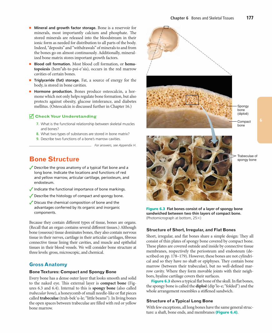
Gross AnatomyBone Textures: Compact and Spongy BoneEvery bone has a dense outer layer that looks smooth and solid to the naked eye. This external layer is compact bone (Fig-ures 6.3 and 6.4). Internal to this is spongy bone (also called trabecular bone), a honeycomb of small needle-like or flat pieces called trabeculae (trah-bek"u-le; “little beams”). In living bones the open spaces between trabeculae are filled with red or yellow bone marrow.
Structure of Short, Irregular, and Flat BonesShort, irregular, and flat bones share a simple design: They all consist of thin plates of spongy bone covered by compact bone. These plates are covered outside and inside by connective tissue membranes, respectively the periosteum and endosteum (de-scribed on pp. 178–179). However, these bones are not cylindri-cal and so they have no shaft or epiphyses. They contain bone marrow (between their trabeculae), but no well-defined mar-row cavity. Where they form movable joints with their neigh-bors, hyaline cartilage covers their surfaces.
Figure 6.3 shows a typical flat bone of the skull. In flat bones, the spongy bone is called the diploë (dip"lo-e; “folded”) and the whole arrangement resembles a stiffened sandwich.
Structure of a Typical Long BoneWith few exceptions, all long bones have the same general struc-ture: a shaft, bone ends, and membranes (Figure 6.4).
Compact bone
Trabeculae ofspongy bone
Spongy bone(diploë)
Figure 6.3 Flat bones consist of a layer of spongy bone sandwiched between two thin layers of compact bone. (Photomicrograph at bottom, 25# )
178 UNIT 2 Covering, Support, and Movement of the Body
6
# 105016 Cust: Benjamin Cummings/CA Au: Marieb Pg. No. 178 Title: Anatomy & Physiology Server: S4C
C/M/Y/KShort / Normal
DESIGN SERVICES OF
CARLISLEPublishing Services
lengthen the bone. The flared portion of the bone where the diaphysis and epiphysis meet, whether it is the epiphyseal plate or line, is sometimes called the metaphysis (meta $ between).
Membranes A glistening white, double-layered membrane called the periosteum (per!e-os"te-um; peri $ around, osteo $ bone) covers the external surface of the entire bone except the joint surfaces. The outer fibrous layer of the periosteum is dense irregular connective tissue. The inner osteogenic layer, abutting the bone surface, consists primarily of primitive stem cells, osteogenic cells, that give rise to all bone cells except bone-destroying cells. These cell types are described shortly.
The periosteum is richly supplied with nerve fibers and blood vessels, which pass through the shaft to enter the marrow cavity via nutrient foramina (fo-ra"me-nah; “openings”). Perforating (Sharpey’s) fibers—tufts of collagen fibers that extend from its fibrous layer into the bone matrix—secure the periosteum to
Diaphysis A tubular diaphysis (di-af"ĭ-sis; dia $ through, physis $ growth), or shaft, forms the long axis of the bone. It is constructed of a relatively thick collar of compact bone that surrounds a central medullary cavity (med"u-lar-e; “middle”), or marrow cavity. In adults, the medullary cavity contains fat (yellow marrow) and is called the yellow marrow cavity.
Epiphyses The epiphyses (e-pif"ĭ-sēz; singular: epiphysis) are the bone ends (epi $ upon). In many cases, they are broader than the diaphysis. An outer shell of compact bone forms the epiphysis exterior and their interior contains spongy bone. A thin layer of articular (hyaline) cartilage covers the joint surface of each epiphysis, cushioning the opposing bone ends during movement and absorbing stress.
Between the diaphysis and each epiphysis of an adult long bone is an epiphyseal line, a remnant of the epiphyseal plate, a disc of hyaline cartilage that grows during childhood to
Proximalepiphysis
(b)
(c)(a)
Yellowbone marrow
Endosteum
Epiphysealline
Articularcartilage
Periosteum
Spongy bone
Compact bone
Medullarycavity (linedby endosteum)
Compact bone
Compact bone
Periosteum
Perforating(Sharpey’s)fibers
Nutrientarteries
Diaphysis
Distalepiphysis
Endosteum
Figure 6.4 The structure of a long bone (humerus of arm). (a) Anterior view with bone sectioned frontally to show the interior at the proximal end. (b) Enlarged
view of spongy bone and compact bone of the epiphysis of (a). (For related images, see A Brief Atlas of the Human Body, Plates 20 and 21.) (c) Enlarged cross-sectional view
of the shaft (diaphysis) of (a). Note that the external surface of the diaphysis is covered by periosteum, but the articular surface of the epiphysis is covered with hyaline cartilage.
Chapter 6 Bones and Skeletal Tissues 179
6
# 105016 Cust: Benjamin Cummings/CA Au: Marieb Pg. No. 179 Title: Anatomy & Physiology Server: S4C
C/M/Y/KShort / Normal
DESIGN SERVICES OF
CARLISLEPublishing Services
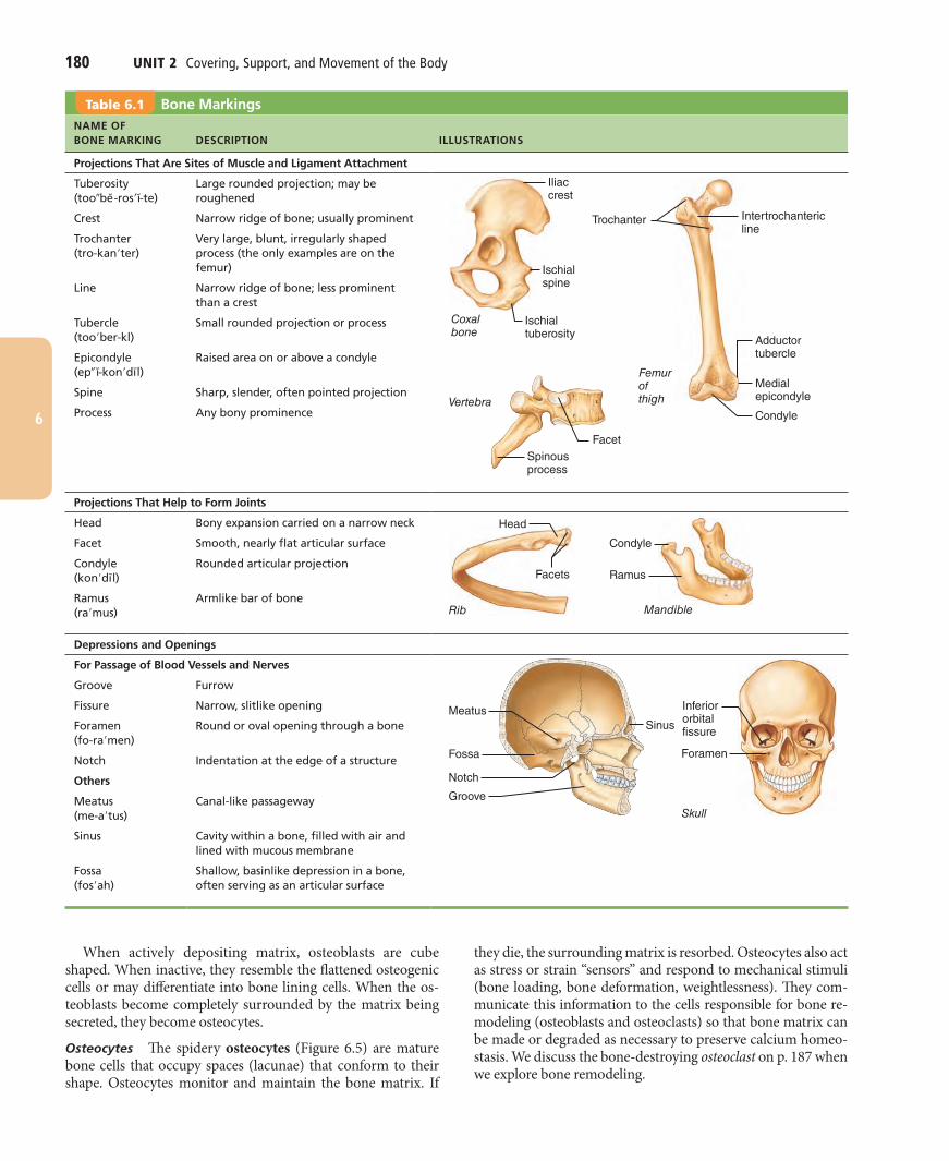
has distinguishing features and functions. In most cases, bone projections indicate the stresses created by muscles attached to and pulling on them or are modified surfaces where bones meet and form joints.
Bone markings that are depressions and openings include fossae (singular: fossa), sinuses, foramina (singular: foramen), and grooves. They usually allow nerves and blood vessels to pass. Table 6.1 describes the most important types of bone markings. Familiarize yourself with these terms because you will meet them again as identifying marks of the individual bones studied in the lab.
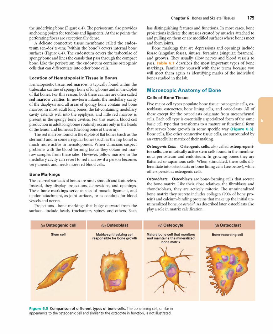
Microscopic Anatomy of BoneCells of Bone TissueFive major cell types populate bone tissue: osteogenic cells, os-teoblasts, osteocytes, bone lining cells, and osteoclasts. All of these except for the osteoclasts originate from mesenchymal cells. Each cell type is essentially a specialized form of the same basic cell type that transforms to a mature or functional form that serves bone growth in some specific way (Figure 6.5). Bone cells, like other connective tissue cells, are surrounded by an extracellular matrix of their making.
Osteogenic Cells Osteogenic cells, also called osteoprogeni-tor cells, are mitotically active stem cells found in the membra-nous periosteum and endosteum. In growing bones they are flattened or squamous cells. When stimulated, these cells dif-ferentiate into osteoblasts or bone lining cells (see below), while others persist as osteogenic cells.
Osteoblasts Osteoblasts are bone-forming cells that secrete the bone matrix. Like their close relatives, the fibroblasts and chondroblasts, they are actively mitotic. The unmineralized bone matrix they secrete includes collagen (90% of bone pro-tein) and calcium-binding proteins that make up the initial un-mineralized bone, or osteoid. As described later, osteoblasts also play a role in matrix calcification.
(a) Osteogenic cell (b) Osteoblast (c) Osteocyte
Stem cell Mature bone cell that monitorsand maintains the mineralized
bone matrix
Matrix-synthesizing cellresponsible for bone growth
(d) Osteoclast
Bone-resorbing cell
Figure 6.5 Comparison of different types of bone cells. The bone lining cell, similar in appearance to the osteogenic cell and similar to the osteocyte in function, is not illustrated.
the underlying bone (Figure 6.4). The periosteum also provides anchoring points for tendons and ligaments. At these points the perforating fibers are exceptionally dense.
A delicate connective tissue membrane called the endos-teum (en-dos"te-um; “within the bone”) covers internal bone surfaces (Figure 6.4). The endosteum covers the trabeculae of spongy bone and lines the canals that pass through the compact bone. Like the periosteum, the endosteum contains osteogenic cells that can differentiate into other bone cells.
Location of Hematopoietic Tissue in BonesHematopoietic tissue, red marrow, is typically found within the trabecular cavities of spongy bone of long bones and in the diploë of flat bones. For this reason, both these cavities are often called red marrow cavities. In newborn infants, the medullary cavity of the diaphysis and all areas of spongy bone contain red bone marrow. In most adult long bones, the fat-containing medullary cavity extends well into the epiphysis, and little red marrow is present in the spongy bone cavities. For this reason, blood cell production in adult long bones routinely occurs only in the heads of the femur and humerus (the long bone of the arm).
The red marrow found in the diploë of flat bones (such as the sternum) and in some irregular bones (such as the hip bone) is much more active in hematopoiesis. When clinicians suspect problems with the blood-forming tissue, they obtain red mar-row samples from these sites. However, yellow marrow in the medullary cavity can revert to red marrow if a person becomes very anemic and needs more red blood cells.
Bone MarkingsThe external surfaces of bones are rarely smooth and featureless. Instead, they display projections, depressions, and openings. These bone markings serve as sites of muscle, ligament, and tendon attachment, as joint surfaces, or as conduits for blood vessels and nerves.
Projections—bone markings that bulge outward from the surface—include heads, trochanters, spines, and others. Each
180 UNIT 2 Covering, Support, and Movement of the Body
6
# 105016 Cust: Benjamin Cummings/CA Au: Marieb Pg. No. 180 Title: Anatomy & Physiology Server: S4C
C/M/Y/KShort / Normal
DESIGN SERVICES OF
CARLISLEPublishing Services
they die, the surrounding matrix is resorbed. Osteocytes also act as stress or strain “sensors” and respond to mechanical stimuli (bone loading, bone deformation, weightlessness). They com-municate this information to the cells responsible for bone re-modeling (osteoblasts and osteoclasts) so that bone matrix can be made or degraded as necessary to preserve calcium homeo-stasis. We discuss the bone-destroying osteoclast on p. 187 when we explore bone remodeling.
When actively depositing matrix, osteoblasts are cube shaped. When inactive, they resemble the flattened osteogenic cells or may differentiate into bone lining cells. When the os-teoblasts become completely surrounded by the matrix being secreted, they become osteocytes.
Osteocytes The spidery osteocytes (Figure 6.5) are mature bone cells that occupy spaces (lacunae) that conform to their shape. Osteocytes monitor and maintain the bone matrix. If
Table 6.1 Bone MarkingsNAME OF BONE MARKING DESCRIPTION ILLUSTRATIONS
Projections That Are Sites of Muscle and Ligament Attachment
Tuberosity (too!be -ros"ı -te)
Large rounded projection; may be roughened
Crest Narrow ridge of bone; usually prominent
Trochanter (tro-kan"ter)
Very large, blunt, irregularly shaped process (the only examples are on the femur)
Line Narrow ridge of bone; less prominent than a crest
Tubercle (too"ber-kl)
Small rounded projection or process
Epicondyle (ep! ı -kon"dı l)
Raised area on or above a condyle
Spine Sharp, slender, often pointed projection
Process Any bony prominence
Projections That Help to Form Joints
Head Bony expansion carried on a narrow neck
Facet Smooth, nearly flat articular surface
Condyle (kon"dı l)
Rounded articular projection
Ramus (ra"mus)
Armlike bar of bone
Depressions and Openings
For Passage of Blood Vessels and Nerves
Groove Furrow
Fissure Narrow, slitlike opening
Foramen (fo-ra"men)
Round or oval opening through a bone
Notch Indentation at the edge of a structure
Others
Meatus (me-a"tus)
Canal-like passageway
Sinus Cavity within a bone, filled with air and lined with mucous membrane
Fossa (fos"ah)
Shallow, basinlike depression in a bone, often serving as an articular surface
Iliaccrest
Ischialspine
Ischialtuberosity
Coxalbone
Trochanter
Femurof thigh
Intertrochantericline
Adductortubercle
Condyle
MedialepicondyleVertebra
Spinousprocess
Facet
Head
Facets
Rib
Condyle
Ramus
Mandible
Sinus
Groove
Notch
Fossa
Meatus Inferiororbitalfissure
Foramen
Skull
Chapter 6 Bones and Skeletal Tissues 181
6
# 105016 Cust: Benjamin Cummings/CA Au: Marieb Pg. No. 181 Title: Anatomy & Physiology Server: S4C
C/M/Y/KShort / Normal
DESIGN SERVICES OF
CARLISLEPublishing Services
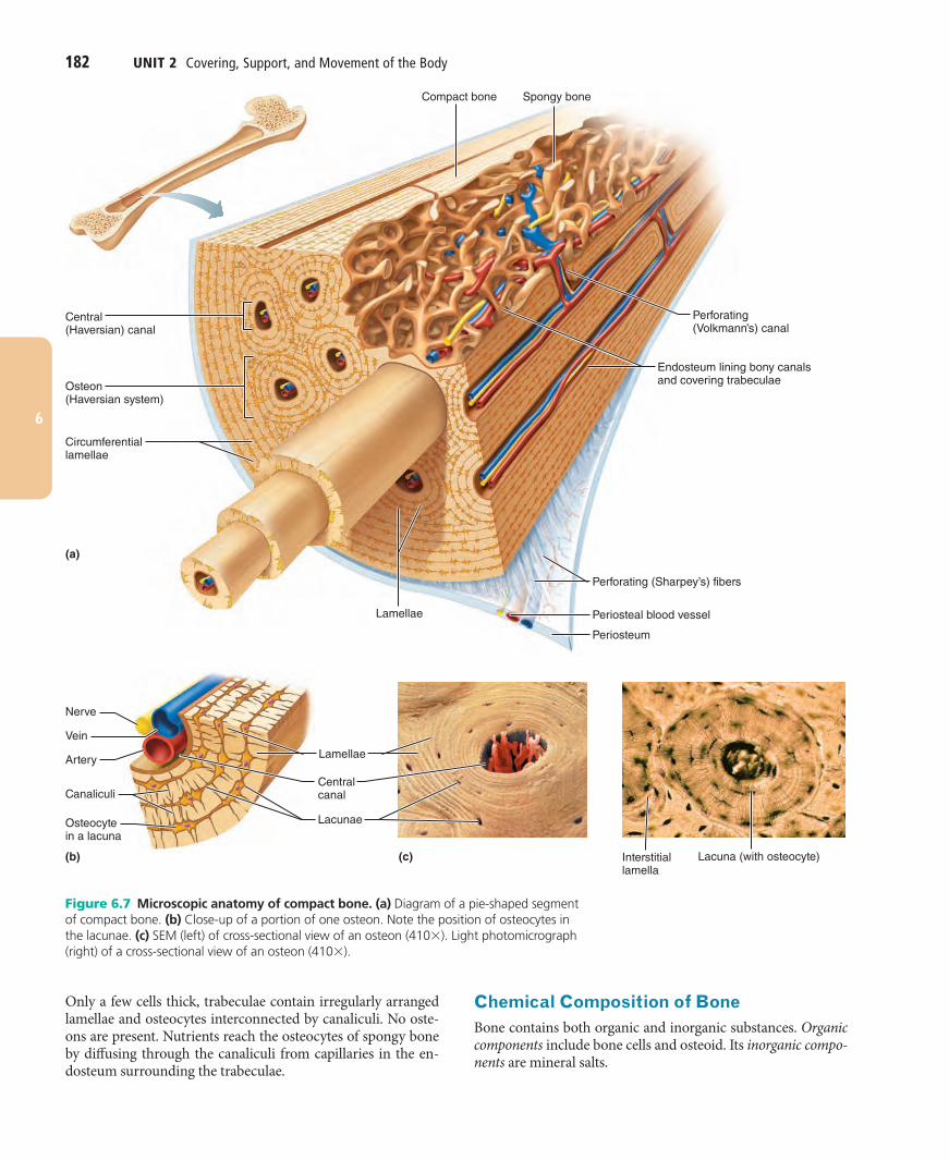
The manner in which canaliculi are formed is interesting. When bone is forming, the osteoblasts secreting bone matrix surround blood vessels and maintain contact with one another and local osteocytes by tentacle-like projections containing gap junctions. Then, as the newly secreted matrix hardens and the maturing cells become trapped within it, a system of tiny canals—the canaliculi filled with tissue fluid and containing the osteocyte extensions—is formed. The canaliculi tie all the osteocytes in a mature osteon together, allowing them to communicate and per-mitting nutrients and wastes to be relayed from one osteocyte to the next throughout the osteon. Although bone matrix is hard and impermeable to nutrients, its canaliculi and cell-to-cell relays (via gap junctions) allow bone cells to be well nourished.
Interstitial and Circumferential Lamellae Not all the lamellae in compact bone are part of complete osteons. Lying between intact osteons are incomplete lamellae called interstitial lamel-lae (in!ter-stish"al) (Figure 6.7c, right photomicrograph). They either fill the gaps between forming osteons or are remnants of osteons that have been cut through by bone remodeling (dis-cussed later). Circumferential lamellae, located just deep to the periosteum and just superficial to the endosteum, extend around the entire circumference of the diaphysis (Figure 6.7a) and effectively resist twisting of the long bone.
Spongy BoneIn contrast to compact bone, spongy bone looks like a poorly organized, even haphazard, tissue (see Figure 6.4 and Figure 6.3b). However, the trabeculae in spongy bone align precisely along lines of stress and help the bone resist stress. These tiny bone struts are as carefully positioned as the ca-bles on a suspension bridge.
Bone Lining Cells Bone lining cells are flat cells found on bone surfaces where bone remodeling is not going on. Like osteocytes, they are thought to help maintain the matrix. Bone lining cells on the external bone surface are also called periosteal cells, whereas those lining internal surfaces are called endosteal cells.
Osteoclasts Derived from the same hematopoietic stem cells that differentiate into macrophages, osteoclasts are giant multi-nucleate cells located at sites of bone resorption. When actively resorbing (breaking down) bone, the osteoclasts rest in a shal-low depression called a resorption bay and exhibit a distinctive ruffled border which directly contacts the bone. The deep plasma membrane infoldings of the ruffled border tremendously in-crease the surface area for enzymatically degrading the bones and seal off that area from the surrounding matrix.
Compact BoneAlthough compact bone looks solid, a microscope reveals that it is riddled with passageways that serve as conduits for nerves and blood vessels (see Figure 6.7).
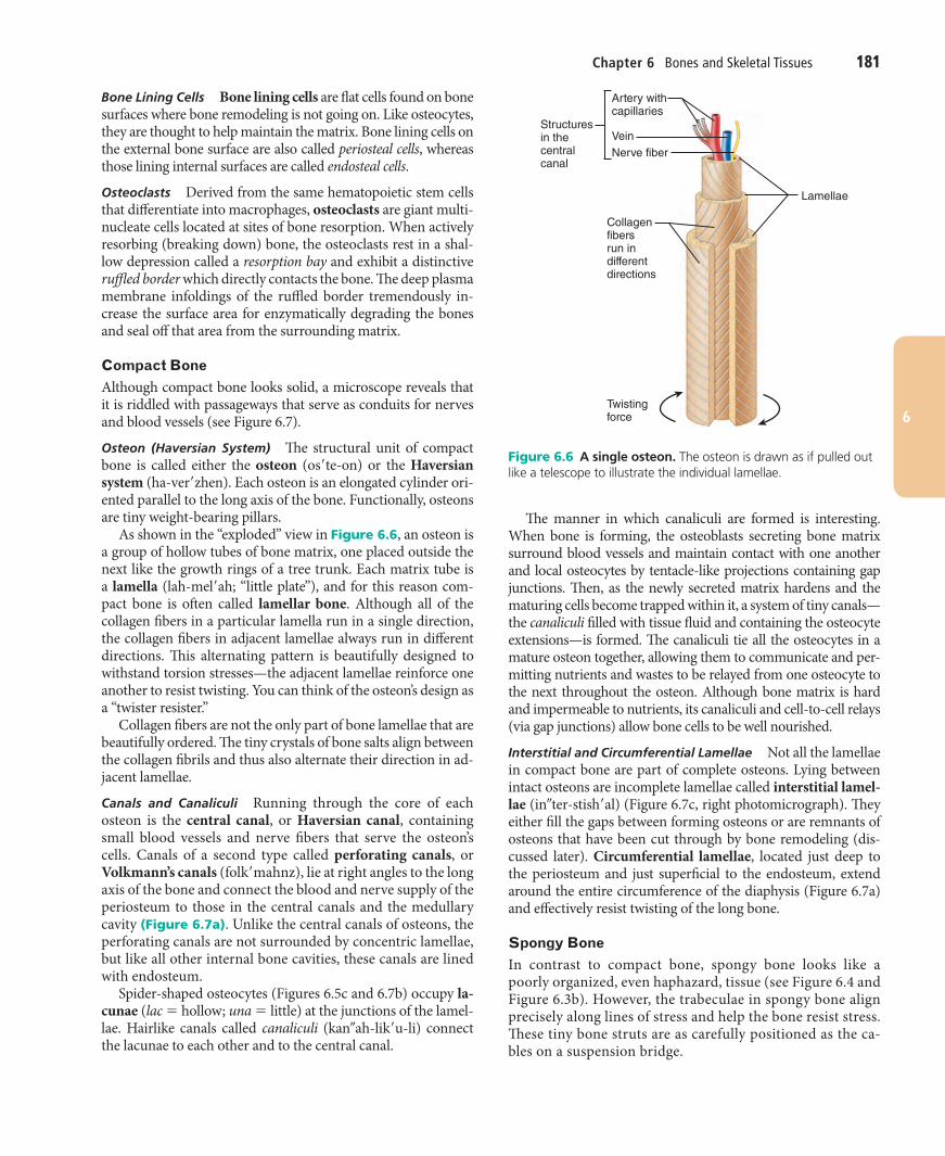
Osteon (Haversian System) The structural unit of compact bone is called either the osteon (os"te-on) or the Haversian system (ha-ver"zhen). Each osteon is an elongated cylinder ori-ented parallel to the long axis of the bone. Functionally, osteons are tiny weight-bearing pillars.
As shown in the “exploded” view in Figure 6.6, an osteon is a group of hollow tubes of bone matrix, one placed outside the next like the growth rings of a tree trunk. Each matrix tube is a lamella (lah-mel"ah; “little plate”), and for this reason com-pact bone is often called lamellar bone. Although all of the collagen fibers in a particular lamella run in a single direction, the collagen fibers in adjacent lamellae always run in different directions. This alternating pattern is beautifully designed to withstand torsion stresses—the adjacent lamellae reinforce one another to resist twisting. You can think of the osteon’s design as a “twister resister.”
Collagen fibers are not the only part of bone lamellae that are beautifully ordered. The tiny crystals of bone salts align between the collagen fibrils and thus also alternate their direction in ad-jacent lamellae.
Canals and Canaliculi Running through the core of each osteon is the central canal, or Haversian canal, containing small blood vessels and nerve fibers that serve the osteon’s cells. Canals of a second type called perforating canals, or Volkmann’s canals (folk"mahnz), lie at right angles to the long axis of the bone and connect the blood and nerve supply of the periosteum to those in the central canals and the medullary cavity (Figure 6.7a). Unlike the central canals of osteons, the perforating canals are not surrounded by concentric lamellae, but like all other internal bone cavities, these canals are lined with endosteum.
Spider-shaped osteocytes (Figures 6.5c and 6.7b) occupy la-cunae (lac $ hollow; una $ little) at the junctions of the lamel-lae. Hairlike canals called canaliculi (kan!ah-lik"u-li) connect the lacunae to each other and to the central canal.
Structuresin thecentralcanal
Artery withcapillaries
VeinNerve fiber
Lamellae
Collagenfibersrun indifferentdirections
Twistingforce
Figure 6.6 A single osteon. The osteon is drawn as if pulled out like a telescope to illustrate the individual lamellae.
182 UNIT 2 Covering, Support, and Movement of the Body
6
# 105016 Cust: Benjamin Cummings/CA Au: Marieb Pg. No. 182 Title: Anatomy & Physiology Server: S4C
C/M/Y/KShort / Normal
DESIGN SERVICES OF
CARLISLEPublishing Services
Chemical Composition of BoneBone contains both organic and inorganic substances. Organic components include bone cells and osteoid. Its inorganic compo-nents are mineral salts.
Only a few cells thick, trabeculae contain irregularly arranged lamellae and osteocytes interconnected by canaliculi. No oste-ons are present. Nutrients reach the osteocytes of spongy bone by diffusing through the canaliculi from capillaries in the en-dosteum surrounding the trabeculae.
(a)
(b) (c)
Compact bone Spongy bone
Endosteum lining bony canalsand covering trabeculae
Perforating (Volkmann’s) canal
Perforating (Sharpey’s) fibers
Periosteal blood vessel
Periosteum
Interstitial lamella
Lacunae
Lamellae
Lamellae
Nerve
Vein
Artery
Canaliculi
Osteocytein a lacuna
Circumferentiallamellae
Osteon(Haversian system)
Central(Haversian) canal
Centralcanal
Lacuna (with osteocyte)
Figure 6.7 Microscopic anatomy of compact bone. (a) Diagram of a pie-shaped segment of compact bone. (b) Close-up of a portion of one osteon. Note the position of osteocytes in the lacunae. (c) SEM (left) of cross-sectional view of an osteon (410# ). Light photomicrograph (right) of a cross-sectional view of an osteon (410# ).

Chapter 6 Bones and Skeletal Tissues 183
6
# 105016 Cust: Benjamin Cummings/CA Au: Marieb Pg. No. 183 Title: Anatomy & Physiology Server: S4C
C/M/Y/KShort / Normal
DESIGN SERVICES OF
CARLISLEPublishing Services
Organic ComponentsThe organic components of bone include its cells (osteogenic cells, osteoblasts, osteocytes, bone-lining cells, and osteoclasts) and osteoid (os"te-oid), the organic part of the matrix. Osteoid, which makes up approximately one-third of the matrix, includes ground substance (composed of proteoglycans and glycopro-teins) and collagen fibers, both of which are made and secreted by osteoblasts. These organic substances, particularly collagen, contribute both to a bone’s structure and to the flexibility and tensile strength that allow it to resist stretch and twisting.
Bone’s resilience is thought to come from sacrificial bonds in or between collagen molecules. These bonds stretch and break easily on impact, dissipating energy to prevent the force from rising to a fracture value. In the absence of continued or addi-tional trauma, most of the sacrificial bonds re-form.
Inorganic ComponentsThe balance of bone tissue (65% by mass) consists of inorganic hydroxyapatites (hi-drok!se-ap"ah-tītz), or mineral salts, largely calcium phosphates present as tiny, tightly packed, needle-like crystals in and around collagen fibers in the extracellular matrix. The crystals account for the most notable characteris-tic of bone—its exceptional hardness, which allows it to resist compression.
The proper combination of organic and inorganic matrix elements makes bone exceedingly durable and strong without being brittle. Healthy bone is half as strong as steel in resisting compression and fully as strong as steel in resisting tension.
Because of the mineral salts they contain, bones last long af-ter death and provide an enduring “monument.” In fact, skeletal remains many centuries old can still reveal the shapes and sizes of ancient peoples, the kinds of work they did, and many of the ailments they suffered, such as arthritis. Growth arrest lines, hori-zontal lines on long bones, provide visible proof of illness when the body uses nutrients to fight disease and the bones stop growing.
Check Your Understanding
10. Are crests, tubercles, and spines bony projections or depressions?
11. How does the structure of compact bone differ from that of spongy bone when viewed with the naked eye?
12. Which membrane lines the internal canals and covers the trabeculae of a bone?
13. Which component of bone—organic or inorganic—makes it hard?
14. Which cell has a ruffled border and acts to break down bone matrix?
For answers, see Appendix H.
Bone Development Compare and contrast intramembranous ossification and endochondral ossification.
Describe the process of long bone growth that occurs at the epiphyseal plates.
Ossification and osteogenesis (os!te-o-jen"ĕ-sis) are synonyms meaning the process of bone formation (os $ bone, genesis $ be-ginning). In embryos this process leads to the formation of the bony skeleton. Later another form of ossification known as bone growth goes on until early adulthood as the body increases in size. Bones are capable of growing thicker throughout life. However, ossification in adults serves mainly for bone remodeling and repair.
Formation of the Bony SkeletonBefore week 8, the skeleton of a human embryo is constructed entirely from fibrous membranes and hyaline cartilage. Bone tissue begins to develop at about this time and eventually re-places most of the existing fibrous or cartilage structures.■ In endochondral ossification (endo $ within, chondro $ car-
tilage), a bone develops by replacing hyaline cartilage. The resulting bone is called a cartilage, or endochondral, bone.
■ In intramembranous ossification, a bone develops from a fi-brous membrane and the bone is called a membrane bone.
The beauty of using flexible structures (membranes and carti-lages) to fashion the embryonic skeleton is that they can accom-modate mitosis. Were the early skeleton composed of calcified bone tissue from the outset, growth would be much more difficult.
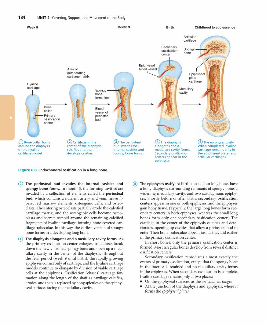
Endochondral OssificationExcept for the clavicles, essentially all bones below the base of the skull form by endochondral ossification (en!do-kon"dral). Beginning late in the second month of development, this pro-cess uses hyaline cartilage “bones” formed earlier as models, or patterns, for bone construction. It is more complex than in-tramembranous ossification because the hyaline cartilage must be broken down as ossification proceeds.
For example, the formation of a long bone typically be-gins in the center of the hyaline cartilage shaft at a region called the primary ossification center. First, blood vessels infiltrate the perichondrium covering the hyaline cartilage “bone,” converting it to a vascularized periosteum. As a re-sult of this change in nutrition, the underlying mesenchymal cells specialize into osteoblasts. The stage is now set for os-sification to begin (Figure 6.8):1 A bone collar forms around the diaphysis of the hyaline
cartilage model. Osteoblasts of the newly converted perios-teum secrete osteoid against the hyaline cartilage diaphysis, encasing it in a cuff or collar of bone called the periosteal bone collar.
2 Cartilage in the center of the diaphysis calcifies and then develops cavities. As the bone collar forms, chondrocytes within the shaft hypertrophy (enlarge) and signal the sur-rounding cartilage matrix to calcify. Then, because calcified cartilage matrix is impermeable to diffusing nutrients, the chondrocytes die and the matrix begins to deteriorate. This deterioration opens up cavities, but the bone collar stabi-lizes the hyaline cartilage model. Elsewhere, the cartilage remains healthy and continues to grow briskly, causing the cartilage model to elongate.
184 UNIT 2 Covering, Support, and Movement of the Body
6
# 105016 Cust: Benjamin Cummings/CA Au: Marieb Pg. No. 184 Title: Anatomy & Physiology Server: S4C
C/M/Y/KShort / Normal
DESIGN SERVICES OF
CARLISLEPublishing Services
5 The epiphyses ossify. At birth, most of our long bones have a bony diaphysis surrounding remnants of spongy bone, a widening medullary cavity, and two cartilaginous epiphy-ses. Shortly before or after birth, secondary ossification centers appear in one or both epiphyses, and the epiphyses gain bony tissue. (Typically, the large long bones form sec-ondary centers in both epiphyses, whereas the small long bones form only one secondary ossification center.) The cartilage in the center of the epiphysis calcifies and dete-riorates, opening up cavities that allow a periosteal bud to enter. Then bone trabeculae appear, just as they did earlier in the primary ossification center.
In short bones, only the primary ossification center is formed. Most irregular bones develop from several distinct ossification centers.
Secondary ossification reproduces almost exactly the events of primary ossification, except that the spongy bone in the interior is retained and no medullary cavity forms in the epiphyses. When secondary ossification is complete, hyaline cartilage remains only at two places:■ On the epiphyseal surfaces, as the articular cartilages■ At the junction of the diaphysis and epiphysis, where it
forms the epiphyseal plates
Hyalinecartilage
Area ofdeterioratingcartilage matrix
Epiphysealblood vessel
Spongyboneformation
Epiphysealplatecartilage
Secondaryossificationcenter
Bloodvessel ofperiostealbud
Medullarycavity
Articularcartilage
Childhood to adolescenceBirthWeek 9 Month 3
Spongybone
Bone collarPrimaryossificationcenter
1 2 3 4 5Bone collar forms around the diaphysis of the hyaline cartilage model.
Cartilage in the center of the diaphysis calcifies and then develops cavities.
The periosteal bud invades the internal cavities and spongy bone forms.
The diaphysis elongates and a medullary cavity forms. Secondary ossification centers appear in the epiphyses.
The epiphyses ossify. When completed, hyaline cartilage remains only in the epiphyseal plates and articular cartilages.
Figure 6.8 Endochondral ossification in a long bone.
3 The periosteal bud invades the internal cavities and spongy bone forms. In month 3, the forming cavities are invaded by a collection of elements called the periosteal bud, which contains a nutrient artery and vein, nerve fi-bers, red marrow elements, osteogenic cells, and osteo-clasts. The entering osteoclasts partially erode the calcified cartilage matrix, and the osteogenic cells become osteo-blasts and secrete osteoid around the remaining calcified fragments of hyaline cartilage, forming bone-covered car-tilage trabeculae. In this way, the earliest version of spongy bone forms in a developing long bone.
4 The diaphysis elongates and a medullary cavity forms. As the primary ossification center enlarges, osteoclasts break down the newly formed spongy bone and open up a med-ullary cavity in the center of the diaphysis. Throughout the fetal period (week 9 until birth), the rapidly growing epiphyses consist only of cartilage, and the hyaline cartilage models continue to elongate by division of viable cartilage cells at the epiphyses. Ossification “chases” cartilage for-mation along the length of the shaft as cartilage calcifies, erodes, and then is replaced by bony spicules on the epiphy-seal surfaces facing the medullary cavity.
Chapter 6 Bones and Skeletal Tissues 185
6
# 105016 Cust: Benjamin Cummings/CA Au: Marieb Pg. No. 185 Title: Anatomy & Physiology Server: S4C
C/M/Y/KShort / Normal
DESIGN SERVICES OF
CARLISLEPublishing Services
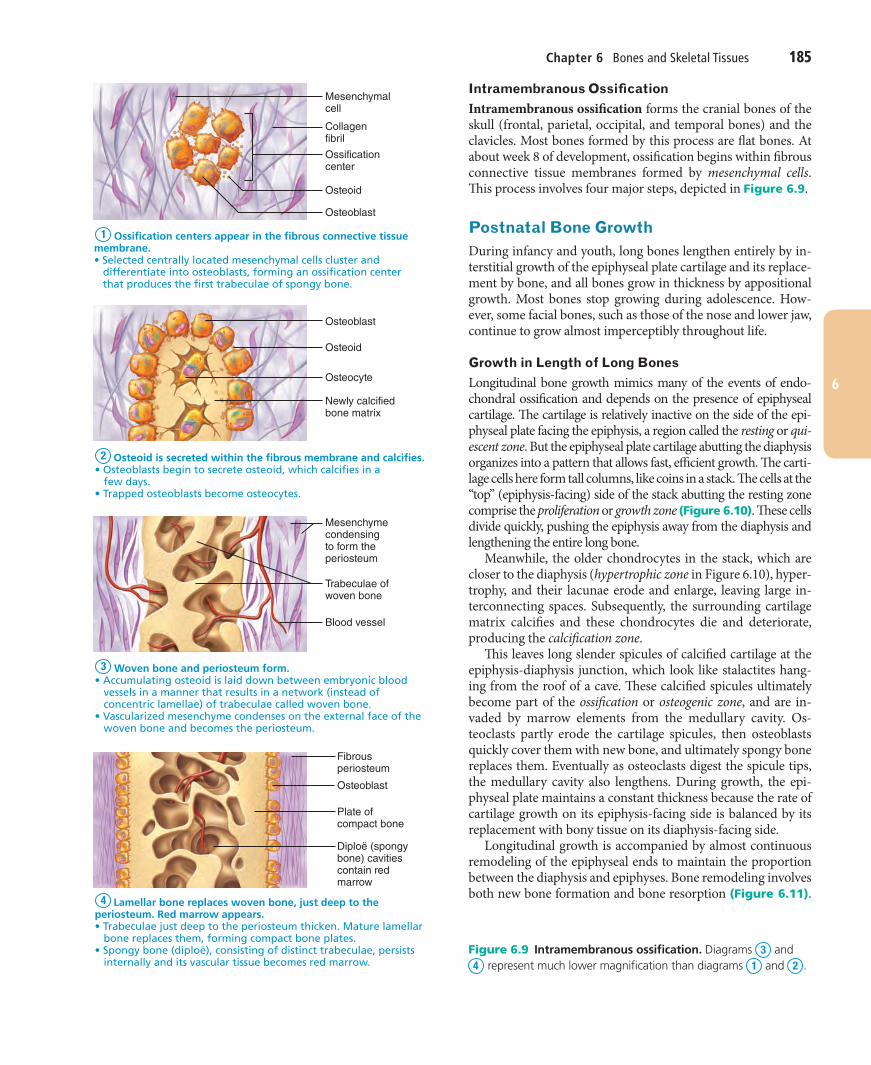
Intramembranous OssificationIntramembranous ossification forms the cranial bones of the skull (frontal, parietal, occipital, and temporal bones) and the clavicles. Most bones formed by this process are flat bones. At about week 8 of development, ossification begins within fibrous connective tissue membranes formed by mesenchymal cells. This process involves four major steps, depicted in Figure 6.9 .
Postnatal Bone GrowthDuring infancy and youth, long bones lengthen entirely by in-terstitial growth of the epiphyseal plate cartilage and its replace-ment by bone, and all bones grow in thickness by appositional growth. Most bones stop growing during adolescence. How-ever, some facial bones, such as those of the nose and lower jaw, continue to grow almost imperceptibly throughout life.
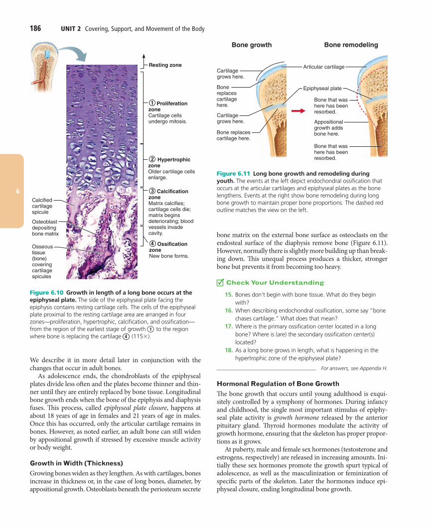
Growth in Length of Long BonesLongitudinal bone growth mimics many of the events of endo-chondral ossification and depends on the presence of epiphyseal cartilage. The cartilage is relatively inactive on the side of the epi-physeal plate facing the epiphysis, a region called the resting or qui-escent zone. But the epiphyseal plate cartilage abutting the diaphysis organizes into a pattern that allows fast, efficient growth. The carti-lage cells here form tall columns, like coins in a stack. The cells at the “top” (epiphysis-facing) side of the stack abutting the resting zone comprise the proliferation or growth zone (Figure 6.10). These cells divide quickly, pushing the epiphysis away from the diaphysis and lengthening the entire long bone.
Meanwhile, the older chondrocytes in the stack, which are closer to the diaphysis (hypertrophic zone in Figure 6.10), hyper-trophy, and their lacunae erode and enlarge, leaving large in-terconnecting spaces. Subsequently, the surrounding cartilage matrix calcifies and these chondrocytes die and deteriorate, producing the calcification zone.
This leaves long slender spicules of calcified cartilage at the epiphysis-diaphysis junction, which look like stalactites hang-ing from the roof of a cave. These calcified spicules ultimately become part of the ossification or osteogenic zone, and are in-vaded by marrow elements from the medullary cavity. Os-teoclasts partly erode the cartilage spicules, then osteoblasts quickly cover them with new bone, and ultimately spongy bone replaces them. Eventually as osteoclasts digest the spicule tips, the medullary cavity also lengthens. During growth, the epi-physeal plate maintains a constant thickness because the rate of cartilage growth on its epiphysis-facing side is balanced by its replacement with bony tissue on its diaphysis-facing side.
Longitudinal growth is accompanied by almost continuous remodeling of the epiphyseal ends to maintain the proportion between the diaphysis and epiphyses. Bone remodeling involves both new bone formation and bone resorption (Figure 6.11).
Mesenchymalcell
CollagenfibrilOssificationcenter
Osteoid
Osteoblast
Osteoid
Osteocyte
Newly calcifiedbone matrix
Osteoblast
Mesenchymecondensingto form theperiosteum
Blood vessel
Trabeculae ofwoven bone
Fibrousperiosteum
Osteoblast
Plate ofcompact bone
Diploë (spongybone) cavitiescontain redmarrow
Ossification centers appear in the fibrous connective tissue membrane.� Selected centrally located mesenchymal cells cluster and differentiate into osteoblasts, forming an ossification center that produces the first trabeculae of spongy bone.
Osteoid is secreted within the fibrous membrane and calcifies.�������� ������������������������������"������ ���'������ �������������� �����"�� $�����rapped osteoblasts become osteocytes.
Woven bone and periosteum form.����� � � ����������������� �����"�����"��������$��������������!���������� �� ������� ����� ������� ����"���������� ������ ��������������� ���� �������� ��� � ��� �����"�!�����������ascularized mesenchyme condenses on the external face of the ���"�!�������� ����������������������� ��
Lamellar bone replaces woven bone, just deep to the periosteum. Red marrow appears. ���� ��� � ��� ����������������������� ������������ � ���� ���� �� bone replaces them, forming compact bone plates.������$������������&���������������������������� ��� � ��������������������� ��$� �������! �� � ������ ��������������� ���".
4
3
2
1
Figure 6.9 Intramembranous ossification. Diagrams 3 and 4 represent much lower magnification than diagrams 1 and 2 .
186 UNIT 2 Covering, Support, and Movement of the Body
6
# 105016 Cust: Benjamin Cummings/CA Au: Marieb Pg. No. 186 Title: Anatomy & Physiology Server: S4C
C/M/Y/KShort / Normal
DESIGN SERVICES OF
CARLISLEPublishing Services
bone matrix on the external bone surface as osteoclasts on the endosteal surface of the diaphysis remove bone (Figure 6.11). However, normally there is slightly more building up than break-ing down. This unequal process produces a thicker, stronger bone but prevents it from becoming too heavy.
Check Your Understanding
15. Bones don’t begin with bone tissue. What do they begin with?
16. When describing endochondral ossification, some say “bone chases cartilage.” What does that mean?
17. Where is the primary ossification center located in a long bone? Where is (are) the secondary ossification center(s) located?
18. As a long bone grows in length, what is happening in the hypertrophic zone of the epiphyseal plate?
For answers, see Appendix H.
Hormonal Regulation of Bone GrowthThe bone growth that occurs until young adulthood is exqui-sitely controlled by a symphony of hormones. During infancy and childhood, the single most important stimulus of epiphy-seal plate activity is growth hormone released by the anterior pituitary gland. Thyroid hormones modulate the activity of growth hormone, ensuring that the skeleton has proper propor-tions as it grows.
At puberty, male and female sex hormones (testosterone and estrogens, respectively) are released in increasing amounts. Ini-tially these sex hormones promote the growth spurt typical of adolescence, as well as the masculinization or feminization of specific parts of the skeleton. Later the hormones induce epi-physeal closure, ending longitudinal bone growth.
We describe it in more detail later in conjunction with the changes that occur in adult bones.
As adolescence ends, the chondroblasts of the epiphyseal plates divide less often and the plates become thinner and thin-ner until they are entirely replaced by bone tissue. Longitudinal bone growth ends when the bone of the epiphysis and diaphysis fuses. This process, called epiphyseal plate closure, happens at about 18 years of age in females and 21 years of age in males. Once this has occurred, only the articular cartilage remains in bones. However, as noted earlier, an adult bone can still widen by appositional growth if stressed by excessive muscle activity or body weight.
Growth in Width (Thickness)Growing bones widen as they lengthen. As with cartilages, bones increase in thickness or, in the case of long bones, diameter, by appositional growth. Osteoblasts beneath the periosteum secrete
Calcifiedcartilagespicule
Osseoustissue (bone)coveringcartilagespicules
Resting zone
Osteoblastdepositingbone matrix
Proliferation zoneCartilage cells undergo mitosis.
Hypertrophic zoneOlder cartilage cells enlarge.
Ossification zoneNew bone forms.
Calcification zoneMatrix calcifies; cartilage cells die; matrix begins deteriorating; blood vessels invadecavity.
1
2
4
3
Figure 6.10 Growth in length of a long bone occurs at the epiphyseal plate. The side of the epiphyseal plate facing the epiphysis contains resting cartilage cells. The cells of the epiphyseal plate proximal to the resting cartilage area are arranged in four zones—proliferation, hypertrophic, calcification, and ossification—from the region of the earliest stage of growth 1 to the region where bone is replacing the cartilage 4 (115# ).
Bone growth Bone remodeling
Articular cartilage
Epiphyseal plate
Cartilage grows here.
Bone replaces cartilage here.
Cartilage grows here.
Bone that was here has been resorbed.
Bone that was here has been resorbed.
Appositionalgrowth addsbone here.Bone replaces
cartilage here.
Figure 6.11 Long bone growth and remodeling during youth. The events at the left depict endochondral ossification that occurs at the articular cartilages and epiphyseal plates as the bone lengthens. Events at the right show bone remodeling during long bone growth to maintain proper bone proportions. The dashed red outline matches the view on the left.

Chapter 6 Bones and Skeletal Tissues 187
6
# 105016 Cust: Benjamin Cummings/CA Au: Marieb Pg. No. 187 Title: Anatomy & Physiology Server: S4C
C/M/Y/KShort / Normal
DESIGN SERVICES OF
CARLISLEPublishing Services
is the product of the local concentrations of calcium and phos-phate (Pi) ions (the Ca2% ·Pi product) in the endosteal cavity. When the Ca2% ·Pi product reaches a certain level, tiny crystals of hydroxyapatite form spontaneously and catalyze further crystallization of calcium salts in the area. Other factors in-volved are matrix proteins that bind and concentrate calcium, and the enzyme alkaline phosphatase (shed in matrix vesicles by the osteoblasts), which is essential for mineralization. Once proper conditions are present, calcium salts are deposited all at once and with great precision throughout the “matured” matrix.
Bone ResorptionAs noted earlier, the giant osteoclasts accomplish bone re-sorption. Osteoclasts move along a bone surface, digging de-pressions or grooves as they break down the bone matrix. The ruffled border of the osteoclast clings tightly to the bone, seal-ing off the area of bone destruction and secreting lysosomal enzymes that digest the organic matrix and protons (H% ). The resulting acidic brew in the resorption bay converts the calcium salts into soluble forms that pass easily into solution. Osteoclasts may also phagocytize the demineralized matrix and dead os-teocytes. The digested matrix end products, growth factors, and dissolved minerals are then endocytosed, transported across the osteoclast (by transcytosis), and released at the opposite side. There they enter the interstitial fluid and then the blood.
When resorption of a given area of bone is completed, the osteoclasts undergo apoptosis. There is much to learn about os-teoclast activation, but PTH and proteins secreted by T cells of the immune system appear to be important.
Control of RemodelingRemodeling goes on continuously in the skeleton, regulated by genetic factors and two control loops that serve different “mas-ters.” One is a negative feedback hormonal loop that maintains Ca2% homeostasis in the blood. The other involves responses to mechanical and gravitational forces acting on the skeleton.
The hormonal feedback becomes much more meaningful when you understand calcium’s importance in the body. Ionic calcium is necessary for an amazing number of physiological processes, including transmission of nerve impulses, muscle contraction, blood coagulation, secretion by glands and nerve cells, and cell division.
The human body contains 1200–1400 g of calcium, more than 99% present as bone minerals. Most of the remainder is in body cells. Less than 1.5 g is present in blood, and the hor-monal control loop normally maintains blood Ca2% within the narrow range of 9–11 mg per dl (100 ml) of blood. Calcium is absorbed from the intestine under the control of vitamin D me-tabolites. The daily dietary calcium requirement is 400–800 mg from birth until age 10, and 1200–1500 mg from ages 11 to 24.
Hormonal Controls The hormonal controls primarily involve parathyroid hormone (PTH), produced by the parathyroid glands. To a much lesser extent calcitonin (kal!sĭ-to"nin), pro-duced by parafollicular cells (C cells) of the thyroid gland, may be involved.
Excesses or deficits of any of these hormones can result in ab-normal skeletal growth. For example, hypersecretion of growth hormone in children results in excessive height (gigantism), and deficits of growth hormone or thyroid hormone produce char-acteristic types of dwarfism.
Bone Homeostasis: Remodeling and Repair
Compare the locations and remodeling functions of the osteoblasts, osteocytes, and osteoclasts.
Explain how hormones and physical stress regulate bone remodeling.
Describe the steps of fracture repair.
Bones appear to be the most lifeless of body organs, and may even summon images of a graveyard. But as you have just learned, bone is a dynamic and active tissue, and small-scale changes in bone architecture occur continually. Every week we recycle 5–7% of our bone mass, and as much as half a gram of calcium may enter or leave the adult skeleton each day! Spongy bone is replaced every three to four years; compact bone, every ten years or so. This is fortunate because when bone remains in place for long periods more of the calcium salts crystallize (see description below) and the bone becomes more brittle—ripe conditions for fracture.
When we break bones—the most common disorder of bone homeostasis—they undergo a remarkable process of self-repair.
Bone RemodelingIn the adult skeleton, bone deposit and bone resorption occur at the surfaces of both the periosteum and the endosteum. To-gether, the two processes constitute bone remodeling. “Pack-ets” of adjacent osteoblasts and osteoclasts called remodeling units coordinate bone remodeling (with help from the stress-sensing osteocytes).
In healthy young adults, total bone mass remains constant, an indication that the rates of bone deposit and resorption are essentially equal. Remodeling does not occur uniformly, how-ever. For example, the distal part of the femur, or thigh bone, is fully replaced every five to six months, whereas its shaft is altered much more slowly.
Bone DepositAn osteoid seam—an unmineralized band of gauzy-looking bone matrix 10–12 micrometers (μm) wide—marks areas of new matrix deposits by osteoblasts. Between the osteoid seam and the older mineralized bone, there is an abrupt transition called the calcification front. Because the osteoid seam is always of constant width and the change from unmineralized to min-eralized matrix is sudden, it seems that the osteoid must mature for about a week before it can calcify.
The precise trigger for calcification is still controversial, but mechanical signals are definitely involved. One critical factor
188 UNIT 2 Covering, Support, and Movement of the Body
6
# 105016 Cust: Benjamin Cummings/CA Au: Marieb Pg. No. 188 Title: Anatomy & Physiology Server: S4C
C/M/Y/KShort / Normal
DESIGN SERVICES OF
CARLISLEPublishing Services
through an additional pathway mediated by the hypothalamus, which activates sympathetic nerves serving bones. However, the full scope of leptin’s bone-modifying activity in humans is still being worked out.
It is also evident that the brain, intestine, and skeleton have ongoing conversations that help regulate the balance between bone formation and destruction, with serotonin serving as a hormonal go-between. Serotonin is better known as a neu-rotransmitter that regulates mood and sleep, but most of the body’s serotonin is made in the gut (intestine) and the blood-brain barrier (see Chapter 12) bars it from entering the brain. The role of gut serotonin is still poorly understood. What is known is that when we eat, serotonin is secreted and circulated via the blood to the bones where it interferes with osteoblast ac-tivity. Reduction of bone turnover after eating may lock calcium in bone when new calcium is flooding into the bloodstream.
This is a troubling finding for those taking Prozac and other antidepressant drugs that inhibit serotonin uptake, making it more available to bone cells. Such patients have lower bone den-sity and suffer more fractures than people not taking these drugs.
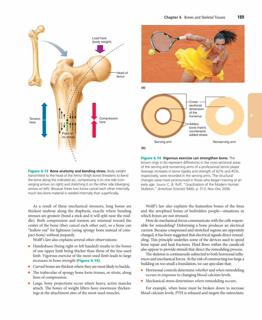
Response to Mechanical Stress The second set of controls regulating bone remodeling, bone’s response to mechanical stress (muscle pull) and gravity, keeps the bones strong where stressors are acting.
Wolff ’s law holds that a bone grows or remodels in response to the demands placed on it. The first thing to understand is that a bone’s anatomy reflects the common stresses it encoun-ters. For example, a bone is loaded (stressed) whenever weight bears down on it or muscles pull on it. This loading is usually off center and tends to bend the bone. Bending compresses the bone on one side and subjects it to tension (stretching) on the other (Figure 6.13).
When blood levels of ionic calcium decline, PTH is released (Figure 6.12). The increased PTH level stimulates osteoclasts to resorb bone, releasing calcium into blood. Osteoclasts are no respecters of matrix age: When activated, they break down both old and new matrix. As blood concentrations of calcium rise, the stimulus for PTH release ends. The decline of PTH reverses its effects and causes blood Ca2% levels to fall.
In humans, calcitonin appears to be a hormone in search of a function because its effects on calcium homeostasis are negligi-ble. When administered at pharmacological (abnormally high) doses, it does lower blood calcium levels temporarily.
These hormonal controls act to preserve blood calcium homeostasis, not the skeleton’s strength or well-being. In fact, if blood calcium levels are low for an extended time, the bones become so demineralized that they develop large, punched-out-looking holes. Thus, the bones serve as a storehouse from which ionic calcium is drawn as needed.
Homeostatic Imbalance 6.1Minute changes from the homeostatic range for blood calcium can lead to severe neuromuscular problems ranging from hyper-excitability (when blood Ca2% levels are too low) to nonrespon-siveness and inability to function (with high blood Ca2% levels). In addition, sustained high blood levels of Ca2% , a condition known as hypercalcemia (hi!per-kal-se"me-ah), can lead to un-desirable deposits of calcium salts in the blood vessels, kidneys, and other soft organs, which may hamper their function. ✚
Other hormones are also involved in modifying bone density and bone turnover. For example, leptin, a hormone released by adipose tissue, plays a role in regulating bone density. Best known for its effects on weight and energy balance (see pp. 940–941), in animal studies leptin appears to inhibit osteoblasts. It does so
Osteoclastsdegrade bonematrix and releaseCa2+ into blood.
Parathyroidglands
Thyroidgland
Parathyroidglands releaseparathyroidhormone (PTH).
StimulusFalling bloodCa2+ levels
PTH
Calcium homeostasis of blood: 9–11 mg/100 mlBALANCEBALANCE
IMBALANCE
IMBALANCE
Figure 6.12 Parathyroid hormone (PTH) control of blood calcium levels.
Chapter 6 Bones and Skeletal Tissues 189
6
# 105016 Cust: Benjamin Cummings/CA Au: Marieb Pg. No. 189 Title: Anatomy & Physiology Server: S4C
C/M/Y/KShort / Normal
DESIGN SERVICES OF
CARLISLEPublishing Services
Wolff ’s law also explains the featureless bones of the fetus and the atrophied bones of bedridden people—situations in which bones are not stressed.
How do mechanical forces communicate with the cells respon-sible for remodeling? Deforming a bone produces an electrical current. Because compressed and stretched regions are oppositely charged, it has been suggested that electrical signals direct remod-eling. This principle underlies some of the devices used to speed bone repair and heal fractures. Fluid flows within the canaliculi also appear to provide stimuli that direct the remodeling process.
The skeleton is continuously subjected to both hormonal influ-ences and mechanical forces. At the risk of constructing too large a building on too small a foundation, we can speculate that:■ Hormonal controls determine whether and when remodeling
occurs in response to changing blood calcium levels.■ Mechanical stress determines where remodeling occurs.
For example, when bone must be broken down to increase blood calcium levels, PTH is released and targets the osteoclasts.
As a result of these mechanical stressors, long bones are thickest midway along the diaphysis, exactly where bending stresses are greatest (bend a stick and it will split near the mid-dle). Both compression and tension are minimal toward the center of the bone (they cancel each other out), so a bone can “hollow out” for lightness (using spongy bone instead of com-pact bone) without jeopardy.
Wolff ’s law also explains several other observations:■ Handedness (being right or left handed) results in the bones
of one upper limb being thicker than those of the less-used limb. Vigorous exercise of the most-used limb leads to large increases in bone strength (Figure 6.14).
■ Curved bones are thickest where they are most likely to buckle.■ The trabeculae of spongy bone form trusses, or struts, along
lines of compression.■ Large, bony projections occur where heavy, active muscles
attach. The bones of weight lifters have enormous thicken-ings at the attachment sites of the most-used muscles.
Load here(body weight)
Head offemur
Compressionhere
Point ofno stress
Tensionhere
Figure 6.13 Bone anatomy and bending stress. Body weight transmitted to the head of the femur (thigh bone) threatens to bend the bone along the indicated arc, compressing it on one side (con-verging arrows on right) and stretching it on the other side (diverging arrows on left). Because these two forces cancel each other internally, much less bone material is needed internally than superficially.
Cross-sectionaldimensionof thehumerus
Addedbone matrixcounteractsadded stress
(b)
(a)
Serving arm Nonserving arm
Figure 6.14 Vigorous exercise can strengthen bone. The brown rings in (b) represent differences in the cross-sectional areas of the serving and nonserving arms of a professional tennis player. Average increases in bone rigidity and strength of 62% and 45%, respectively, were recorded in the serving arms. The structural changes were more pronounced in those who began training at an early age. Source: C. B. Ruff, “Gracilization of the Modern Human Skeleton,” American Scientist 94(6): p. 513, Nov–Dec 2006.
190 UNIT 2 Covering, Support, and Movement of the Body
6
# 105016 Cust: Benjamin Cummings/CA Au: Marieb Pg. No. 190 Title: Anatomy & Physiology Server: S4C
C/M/Y/KShort / Normal
DESIGN SERVICES OF
CARLISLEPublishing Services
Table 6.2 Common Types of FracturesFRACTURE TYPE DESCRIPTION AND COMMENTS
FRACTURE TYPE DESCRIPTION AND COMMENTS
Comminuted Bone fragments into three or more pieces.
Particularly common in the aged, whose bones are more brittle
Compression Bone is crushed.
Common in porous bones (i.e., osteoporotic bones) subjected to extreme trauma, as in a fall
Spiral Ragged break occurs when excessive twisting forces are applied to a bone.
Common sports fracture
Epiphyseal Epiphysis separates from the diaphysis along the epiphyseal plate.
Tends to occur where cartilage cells are dying and calcification of the matrix is occurring
Depressed Broken bone portion is pressed inward.
Typical of skull fracture
Greenstick Bone breaks incompletely, much in the way a green twig breaks. Only one side of the shaft breaks; the other side bends.
Common in children, whose bones have relatively more organic matrix and are more flexible than those of adults
Crushed vertebra
Chapter 6 Bones and Skeletal Tissues 191
6
# 105016 Cust: Benjamin Cummings/CA Au: Marieb Pg. No. 191 Title: Anatomy & Physiology Server: S4C
C/M/Y/KShort / Normal
DESIGN SERVICES OF
CARLISLEPublishing Services
but it takes much longer for large, weight-bearing bones and for bones of elderly people (because of their poorer circulation).
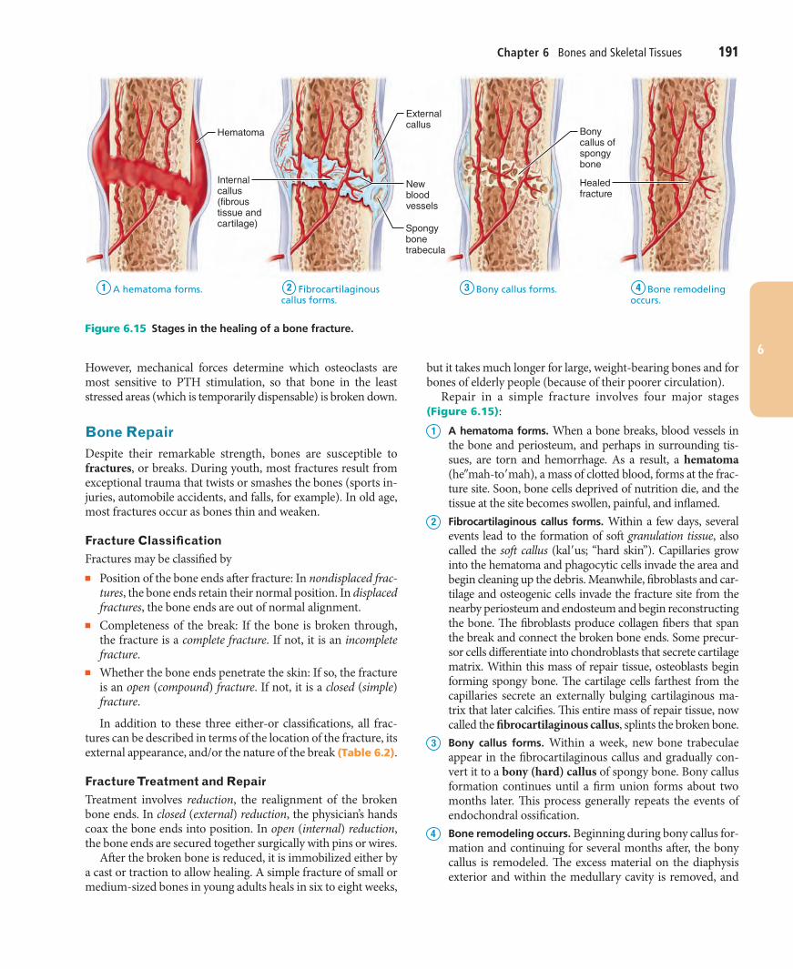
Repair in a simple fracture involves four major stages (Figure 6.15):1 A hematoma forms. When a bone breaks, blood vessels in
the bone and periosteum, and perhaps in surrounding tis-sues, are torn and hemorrhage. As a result, a hematoma (he!mah-to"mah), a mass of clotted blood, forms at the frac-ture site. Soon, bone cells deprived of nutrition die, and the tissue at the site becomes swollen, painful, and inflamed.
2 Fibrocartilaginous callus forms. Within a few days, several events lead to the formation of soft granulation tissue, also called the soft callus (kal"us; “hard skin”). Capillaries grow into the hematoma and phagocytic cells invade the area and begin cleaning up the debris. Meanwhile, fibroblasts and car-tilage and osteogenic cells invade the fracture site from the nearby periosteum and endosteum and begin reconstructing the bone. The fibroblasts produce collagen fibers that span the break and connect the broken bone ends. Some precur-sor cells differentiate into chondroblasts that secrete cartilage matrix. Within this mass of repair tissue, osteoblasts begin forming spongy bone. The cartilage cells farthest from the capillaries secrete an externally bulging cartilaginous ma-trix that later calcifies. This entire mass of repair tissue, now called the fibrocartilaginous callus, splints the broken bone.
3 Bony callus forms. Within a week, new bone trabeculae appear in the fibrocartilaginous callus and gradually con-vert it to a bony (hard) callus of spongy bone. Bony callus formation continues until a firm union forms about two months later. This process generally repeats the events of endochondral ossification.
4 Bone remodeling occurs. Beginning during bony callus for-mation and continuing for several months after, the bony callus is remodeled. The excess material on the diaphysis exterior and within the medullary cavity is removed, and
However, mechanical forces determine which osteoclasts are most sensitive to PTH stimulation, so that bone in the least stressed areas (which is temporarily dispensable) is broken down.
Bone RepairDespite their remarkable strength, bones are susceptible to fractures, or breaks. During youth, most fractures result from exceptional trauma that twists or smashes the bones (sports in-juries, automobile accidents, and falls, for example). In old age, most fractures occur as bones thin and weaken.
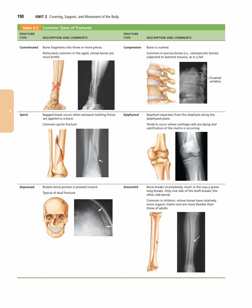
Fracture ClassificationFractures may be classified by■ Position of the bone ends after fracture: In nondisplaced frac-
tures, the bone ends retain their normal position. In displaced fractures, the bone ends are out of normal alignment.
■ Completeness of the break: If the bone is broken through, the fracture is a complete fracture. If not, it is an incomplete fracture.
■ Whether the bone ends penetrate the skin: If so, the fracture is an open (compound) fracture. If not, it is a closed (simple) fracture.
In addition to these three either-or classifications, all frac-tures can be described in terms of the location of the fracture, its external appearance, and/or the nature of the break (Table 6.2).
Fracture Treatment and RepairTreatment involves reduction, the realignment of the broken bone ends. In closed (external) reduction, the physician’s hands coax the bone ends into position. In open (internal) reduction, the bone ends are secured together surgically with pins or wires.
After the broken bone is reduced, it is immobilized either by a cast or traction to allow healing. A simple fracture of small or medium-sized bones in young adults heals in six to eight weeks,
Hematoma
Externalcallus
Bonycallus ofspongybone
Healedfracture
Newbloodvessels
Spongybonetrabecula
Internalcallus(fibroustissue andcartilage)
1 A hematoma forms. 2 Fibrocartilaginous callus forms.
3 Bony callus forms. 4 Bone remodeling occurs.
Figure 6.15 Stages in the healing of a bone fracture.
192 UNIT 2 Covering, Support, and Movement of the Body
6
# 105016 Cust: Benjamin Cummings/CA Au: Marieb Pg. No. 192 Title: Anatomy & Physiology Server: S4C
C/M/Y/KShort / Normal
DESIGN SERVICES OF
CARLISLEPublishing Services
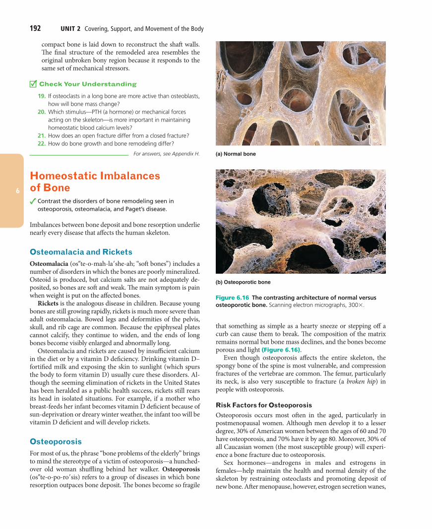
that something as simple as a hearty sneeze or stepping off a curb can cause them to break. The composition of the matrix remains normal but bone mass declines, and the bones become porous and light (Figure 6.16).
Even though osteoporosis affects the entire skeleton, the spongy bone of the spine is most vulnerable, and compression fractures of the vertebrae are common. The femur, particularly its neck, is also very susceptible to fracture (a broken hip) in people with osteoporosis.
Risk Factors for OsteoporosisOsteoporosis occurs most often in the aged, particularly in postmenopausal women. Although men develop it to a lesser degree, 30% of American women between the ages of 60 and 70 have osteoporosis, and 70% have it by age 80. Moreover, 30% of all Caucasian women (the most susceptible group) will experi-ence a bone fracture due to osteoporosis.
Sex hormones—androgens in males and estrogens in females—help maintain the health and normal density of the skeleton by restraining osteoclasts and promoting deposit of new bone. After menopause, however, estrogen secretion wanes,
compact bone is laid down to reconstruct the shaft walls. The final structure of the remodeled area resembles the original unbroken bony region because it responds to the same set of mechanical stressors.
Check Your Understanding
19. If osteoclasts in a long bone are more active than osteoblasts, how will bone mass change?
20. Which stimulus—PTH (a hormone) or mechanical forces acting on the skeleton—is more important in maintaining homeostatic blood calcium levels?
21. How does an open fracture differ from a closed fracture? 22. How do bone growth and bone remodeling differ?
For answers, see Appendix H.
Homeostatic Imbalances of Bone
Contrast the disorders of bone remodeling seen in osteoporosis, osteomalacia, and Paget’s disease.
Imbalances between bone deposit and bone resorption underlie nearly every disease that affects the human skeleton.
Osteomalacia and RicketsOsteomalacia (os!te-o-mah-la"she-ah; “soft bones”) includes a number of disorders in which the bones are poorly mineralized. Osteoid is produced, but calcium salts are not adequately de-posited, so bones are soft and weak. The main symptom is pain when weight is put on the affected bones.
Rickets is the analogous disease in children. Because young bones are still growing rapidly, rickets is much more severe than adult osteomalacia. Bowed legs and deformities of the pelvis, skull, and rib cage are common. Because the epiphyseal plates cannot calcify, they continue to widen, and the ends of long bones become visibly enlarged and abnormally long.
Osteomalacia and rickets are caused by insufficient calcium in the diet or by a vitamin D deficiency. Drinking vitamin D–fortified milk and exposing the skin to sunlight (which spurs the body to form vitamin D) usually cure these disorders. Al-though the seeming elimination of rickets in the United States has been heralded as a public health success, rickets still rears its head in isolated situations. For example, if a mother who breast-feeds her infant becomes vitamin D deficient because of sun-deprivation or dreary winter weather, the infant too will be vitamin D deficient and will develop rickets.
OsteoporosisFor most of us, the phrase “bone problems of the elderly” brings to mind the stereotype of a victim of osteoporosis—a hunched-over old woman shuffling behind her walker. Osteoporosis (os!te-o-po-ro"sis) refers to a group of diseases in which bone resorption outpaces bone deposit. The bones become so fragile
(b) Osteoporotic bone
(a) Normal bone
Figure 6.16 The contrasting architecture of normal versus osteoporotic bone. Scanning electron micrographs, 300# .
Chapter 6 Bones and Skeletal Tissues 193
6
# 105016 Cust: Benjamin Cummings/CA Au: Marieb Pg. No. 193 Title: Anatomy & Physiology Server: S4C
C/M/Y/KShort / Normal
DESIGN SERVICES OF
CARLISLEPublishing Services
along with reduced mineralization, causes a spotty weaken-ing of the bones. Late in the disease, osteoclast activity wanes, but osteoblasts continue to work, often forming irregular bone thickenings or filling the marrow cavity with Pagetic bone.
Paget’s disease may affect any part of the skeleton, but it is usually a localized condition. The spine, pelvis, femur, and skull are most often involved and become increasingly deformed and painful. It rarely occurs before age 40, and it affects about 3% of North American elderly people. Its cause is unknown, but a vi-rus may trigger it. Drug therapies include calcitonin (adminis-tered by a nasal inhaler), and the newer bisphosphonates, which have shown success in preventing bone breakdown.
Check Your Understanding
23. Which bone disorder is characterized by excessive deposit of weak, poorly mineralized bone?
24. What are three measures that may help to maintain healthy bone density?
25. What name is given to “adult rickets”?
For answers, see Appendix H.
Developmental Aspects of Bones: Timing of Events
Describe the timing and cause of changes in bone architecture and bone mass throughout life.
Bones are on a precise schedule from the time they form until death. The mesoderm germ layer gives rise to embryonic mesen-chymal cells, which in turn produce the membranes and cartilages that form the embryonic skeleton. These structures then ossify according to an amazingly predictable timetable that allows fetal age to be determined easily from either X rays or sonograms. Al-though each bone has its own developmental schedule, most long bones begin ossifying by 8 weeks after conception and have well-developed primary ossification centers by 12 weeks (Figure 6.17).
Birth to Young AdulthoodAt birth, most long bones of the skeleton are well ossified except for their epiphyses. After birth, secondary ossification centers develop in a predictable sequence. The epiphyseal plates persist and provide for long bone growth all through childhood and the sex hormone–mediated growth spurt at adolescence. By age 25, nearly all bones are completely ossified and skeletal growth ceases.
Age-Related Changes in BoneIn children and adolescents, bone formation exceeds bone re-sorption. In young adults, these processes are in balance, and in old age, resorption predominates. Despite the environmental factors (discussed earlier) that influence bone density, genet-ics still plays the major role in determining how much a per-son’s bone density will change over a lifetime. A single gene that codes for vitamin D’s cellular docking site helps determine both
and estrogen deficiency is strongly implicated in osteoporosis in older women.
Several other factors can contribute to osteoporosis:■ Petite body form■ Insufficient exercise to stress the bones■ A diet poor in calcium and protein■ Abnormal vitamin D receptors■ Smoking (which reduces estrogen levels)■ Hormone-related conditions such as hyperthyroidism, low
blood levels of thyroid-stimulating hormone, and diabetes mellitus
Osteoporosis can develop at any age as a result of immobility. It can also occur in males with prostate cancer who are being treated with androgen-suppressing drugs.
Treating OsteoporosisOsteoporosis has traditionally been treated with calcium and vi-tamin D supplements, weight-bearing exercise, and hormone (es-trogen) replacement therapy (HRT). Frustratingly, HRT slows the loss of bone but does not reverse it. Additionally, because of the increased risk of heart attack, stroke, and breast cancer associated with estrogen replacement therapy, it is a controversial treatment these days. Although not a substitute for HRT, estrogenic com-pounds in soy products (principally the isoflavones daidzein and genistein) offer a good addition or adjunct for some patients.
Newer drugs are available. Bisphosphonates decrease oste-oclast activity and number, and partially reverse osteoporosis in the spine. Selective estrogen receptor modulators (SERMs), such as raloxifene, dubbed “estrogen light,” mimic estrogen’s beneficial bone-sparing properties without targeting the uterus or breast. Additionally, statins, drugs used to lower cholesterol levels, have an unexpected side effect of increasing bone min-eral density up to 8% over four years. The monoclonal antibody drug denosumab significantly reduces fractures in men fighting prostate cancer and improves bone density in the elderly.
Preventing OsteoporosisHow can osteoporosis be prevented (or at least delayed)? The first requirement is to get enough calcium while your bones are still increasing in density (bones reach their peak density during early adulthood). Second, keep in mind that excessive intake of carbonated beverages and alcohol leaches minerals from bone and decreases bone density. Finally, get plenty of weight-bearing exercise (walking, jogging, tennis, etc.) throughout life. This will increase bone mass above normal values and provide a greater buffer against age-related bone loss.
Paget’s DiseaseOften discovered by accident when X rays are taken for some other reason, Paget’s disease (paj"ets) is characterized by ex-cessive and haphazard bone deposit and resorption. The newly formed bone, called Pagetic bone, is hastily made and has an abnormally high ratio of spongy bone to compact bone. This,
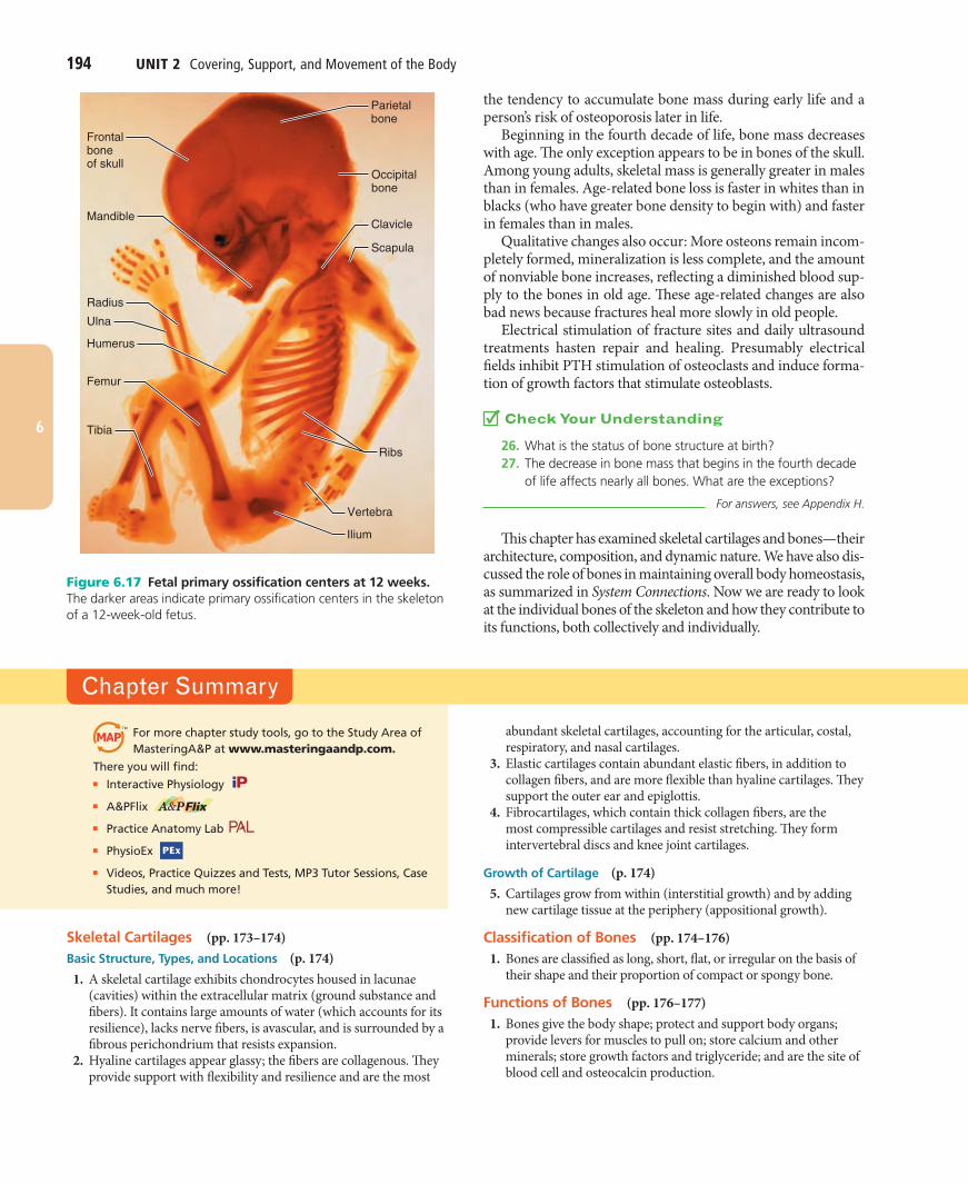
194 UNIT 2 Covering, Support, and Movement of the Body
6
# 105016 Cust: Benjamin Cummings/CA Au: Marieb Pg. No. 194 Title: Anatomy & Physiology Server: S4C
C/M/Y/KShort / Normal
DESIGN SERVICES OF
CARLISLEPublishing Services
the tendency to accumulate bone mass during early life and a person’s risk of osteoporosis later in life.
Beginning in the fourth decade of life, bone mass decreases with age. The only exception appears to be in bones of the skull. Among young adults, skeletal mass is generally greater in males than in females. Age-related bone loss is faster in whites than in blacks (who have greater bone density to begin with) and faster in females than in males.
Qualitative changes also occur: More osteons remain incom-pletely formed, mineralization is less complete, and the amount of nonviable bone increases, reflecting a diminished blood sup-ply to the bones in old age. These age-related changes are also bad news because fractures heal more slowly in old people.
Electrical stimulation of fracture sites and daily ultrasound treatments hasten repair and healing. Presumably electrical fields inhibit PTH stimulation of osteoclasts and induce forma-tion of growth factors that stimulate osteoblasts.
Check Your Understanding
26. What is the status of bone structure at birth? 27. The decrease in bone mass that begins in the fourth decade
of life affects nearly all bones. What are the exceptions?
For answers, see Appendix H.
This chapter has examined skeletal cartilages and bones—their architecture, composition, and dynamic nature. We have also dis-cussed the role of bones in maintaining overall body homeostasis, as summarized in System Connections. Now we are ready to look at the individual bones of the skeleton and how they contribute to its functions, both collectively and individually.
Parietalbone
Radius
Ulna
Humerus
Femur
Occipitalbone
Clavicle
Scapula
Ribs
Vertebra
Ilium
Tibia
Frontalboneof skull
Mandible
Figure 6.17 Fetal primary ossification centers at 12 weeks. The darker areas indicate primary ossification centers in the skeleton of a 12-week-old fetus.
Chapter Summary
For more chapter study tools, go to the Study Area of MasteringA&P at www.masteringaandp.com.
There you will find:■ Interactive Physiology ■ A&PFlix ■ Practice Anatomy Lab ■ PhysioEx
■ Videos, Practice Quizzes and Tests, MP3 Tutor Sessions, Case Studies, and much more!
Skeletal Cartilages (pp. 173–174)Basic Structure, Types, and Locations (p. 174) 1. A skeletal cartilage exhibits chondrocytes housed in lacunae
(cavities) within the extracellular matrix (ground substance and fibers). It contains large amounts of water (which accounts for its resilience), lacks nerve fibers, is avascular, and is surrounded by a fibrous perichondrium that resists expansion.
2. Hyaline cartilages appear glassy; the fibers are collagenous. They provide support with flexibility and resilience and are the most
abundant skeletal cartilages, accounting for the articular, costal, respiratory, and nasal cartilages.
3. Elastic cartilages contain abundant elastic fibers, in addition to collagen fibers, and are more flexible than hyaline cartilages. They support the outer ear and epiglottis.
4. Fibrocartilages, which contain thick collagen fibers, are the most compressible cartilages and resist stretching. They form intervertebral discs and knee joint cartilages.
Growth of Cartilage (p. 174) 5. Cartilages grow from within (interstitial growth) and by adding
new cartilage tissue at the periphery (appositional growth).
Classification of Bones (pp. 174–176) 1. Bones are classified as long, short, flat, or irregular on the basis of
their shape and their proportion of compact or spongy bone.
Functions of Bones (pp. 176–177) 1. Bones give the body shape; protect and support body organs;
provide levers for muscles to pull on; store calcium and other minerals; store growth factors and triglyceride; and are the site of blood cell and osteocalcin production.
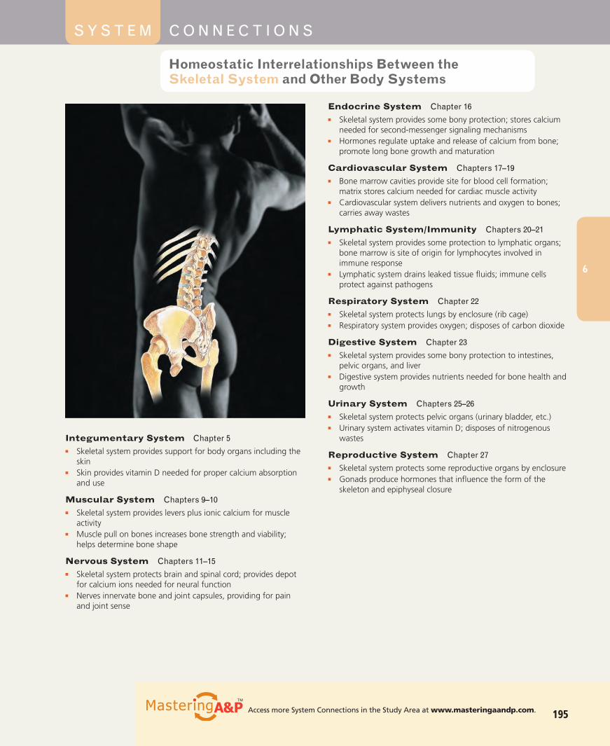
Chapter 6 Bones and Skeletal Tissues 195
# 105016 Cust: Benjamin Cummings/CA Au: Marieb Pg. No. 195 Title: Anatomy & Physiology Server: S4C
C/M/Y/KShort / Normal
DESIGN SERVICES OF
CARLISLEPublishing Services
Integumentary System Chapter 5■ Skeletal system provides support for body organs including the
skin■ Skin provides vitamin D needed for proper calcium absorption
and use
Muscular System Chapters 9–10■ Skeletal system provides levers plus ionic calcium for muscle
activity■ Muscle pull on bones increases bone strength and viability;
helps determine bone shape
Nervous System Chapters 11–15■ Skeletal system protects brain and spinal cord; provides depot
for calcium ions needed for neural function■ Nerves innervate bone and joint capsules, providing for pain
and joint sense
S Y S T E M C O N N E C T I O N S
Homeostatic Interrelationships Between the Skeletal System and Other Body Systems
Endocrine System Chapter 16■ Skeletal system provides some bony protection; stores calcium
needed for second-messenger signaling mechanisms■ Hormones regulate uptake and release of calcium from bone;
promote long bone growth and maturation
Cardiovascular System Chapters 17–19■ Bone marrow cavities provide site for blood cell formation;
matrix stores calcium needed for cardiac muscle activity■ Cardiovascular system delivers nutrients and oxygen to bones;
carries away wastes
Lymphatic System/Immunity Chapters 20–21■ Skeletal system provides some protection to lymphatic organs;
bone marrow is site of origin for lymphocytes involved in immune response
■ Lymphatic system drains leaked tissue fluids; immune cells protect against pathogens
Respiratory System Chapter 22■ Skeletal system protects lungs by enclosure (rib cage)■ Respiratory system provides oxygen; disposes of carbon dioxide
Digestive System Chapter 23■ Skeletal system provides some bony protection to intestines,
pelvic organs, and liver■ Digestive system provides nutrients needed for bone health and
growth
Urinary System Chapters 25–26■ Skeletal system protects pelvic organs (urinary bladder, etc.)■ Urinary system activates vitamin D; disposes of nitrogenous
wastes
Reproductive System Chapter 27■ Skeletal system protects some reproductive organs by enclosure■ Gonads produce hormones that influence the form of the
skeleton and epiphyseal closure
195Access more System Connections in the Study Area at www.masteringaandp.com.
6
196 UNIT 2 Covering, Support, and Movement of the Body
6
# 105016 Cust: Benjamin Cummings/CA Au: Marieb Pg. No. 196 Title: Anatomy & Physiology Server: S4C
C/M/Y/KShort / Normal
DESIGN SERVICES OF
CARLISLEPublishing Services
Bone Structure (pp. 177–183)Gross Anatomy (pp. 177–179) 1. Flat bones consist of two thin plates of compact bone enclosing a
diploë (spongy bone layer). Short and irregular bones resemble flat bones structurally.
2. A long bone is composed of a diaphysis (shaft) and epiphyses (ends). The medullary cavity of the diaphysis contains yellow marrow; the epiphyses contain spongy bone. The epiphyseal line is the remnant of the epiphyseal plate. Periosteum covers the diaphysis; endosteum lines inner bone cavities. Hyaline cartilage covers joint surfaces.
3. In adults, hematopoietic tissue (red marrow) is found within the diploë of flat bones and occasionally within the epiphyses of long bones. In infants, red marrow is also found in the medullary cavity.
4. Bone markings are important anatomical landmarks that reveal sites of muscle attachment, points of articulation, and sites of blood vessel and nerve passage.
Microscopic Anatomy of Bone (pp. 179–182) 5. There are five types of bone cells—osteogenic cells (bone stem
cells), osteoblasts (matrix-synthesizing cells), osteocytes (bone matrix maintenance cells), bone lining cells (line surfaces where no bone activity is ongoing), and osteoclasts (bone destruction cells).
6. The structural unit of compact bone, the osteon, consists of a central canal surrounded by concentric lamellae of bone matrix. Osteocytes, embedded in lacunae, are connected to each other and the central canal by canaliculi.
7. Spongy bone has slender trabeculae containing irregular lamellae, which enclose red marrow–filled cavities.
Chemical Composition of Bone (pp. 182–183) 8. Bone is composed of living cells and matrix. The extracellular
matrix includes osteoid, organic substances that are secreted by osteoblasts and give the bone tensile strength. Its inorganic (mineral) components, the hydroxyapatites (calcium salts), make bone hard.
Bone Development (pp. 183–187)Formation of the Bony Skeleton (pp. 183–185) 1. Intramembranous ossification forms the clavicles and most skull
bones. The ground substance of the bone matrix is deposited between collagen fibers within the fibrous membrane to form bone. Eventually, compact bone plates enclose the diploë.
2. Most bones are formed by endochondral ossification of a hyaline cartilage model. Osteoblasts beneath the periosteum secrete bone matrix on the cartilage model, forming the bone collar. As the cartilage model deteriorates, internal cavities open up, allowing periosteal bud entry. Bone matrix is deposited around the cartilage remnants but is later broken down.
Postnatal Bone Growth (pp. 185–187) 3. Long bones increase in length by interstitial growth of the
epiphyseal plate cartilage and its replacement by bone. 4. Appositional growth increases bone diameter/thickness.
Bone Homeostasis: Remodeling and Repair (pp. 187–192)Bone Remodeling (pp. 187–191) 1. Bone is continually deposited and resorbed in response to
hormonal and mechanical stimuli. Together these processes constitute bone remodeling.
2. An unmineralized osteoid seam appears at areas of new bone deposit; calcium salts are deposited a few days later.
3. Osteoclasts release lysosomal enzymes and acids on bone surfaces to be resorbed. The dissolved products are transcytosed to the opposite face of the osteoclast for release to the extracellular fluid.
4. The hormonal controls of bone remodeling serve blood calcium homeostasis. When blood calcium levels decline, PTH is released and stimulates osteoclasts to digest bone matrix, releasing ionic calcium. As blood calcium levels rise, PTH secretion declines.
5. Mechanical stress and gravity acting on the skeleton help maintain skeletal strength. Bones thicken, develop heavier prominences, or rearrange their trabeculae in sites where stressed.
Bone Repair (pp. 191–192) 6. Fractures are treated by open or closed reduction. The healing
process involves formation of a hematoma, a fibrocartilaginous callus, a bony callus, and bone remodeling, in succession.
Homeostatic Imbalances of Bone (pp. 192–193) 1. Imbalances between bone formation and resorption underlie all
skeletal disorders. 2. Osteomalacia and rickets occur when bones are inadequately
mineralized. The bones become soft and deformed. The most frequent cause is inadequate vitamin D.
3. Osteoporosis is any condition in which bone breakdown outpaces bone formation, causing bones to become weak and porous. Postmenopausal women are particularly susceptible.
4. Paget’s disease is characterized by excessive and abnormal bone remodeling.
Developmental Aspects of Bones: Timing of Events (pp. 193–194) 1. Osteogenesis is predictable and precisely timed. 2. Longitudinal long bone growth continues until the end of
adolescence. Skeletal mass increases dramatically during puberty and adolescence, when formation exceeds resorption.
3. Bone mass is fairly constant in young adulthood, but beginning in the 40s, bone resorption exceeds formation.
Multiple Choice/Matching(Some questions have more than one correct answer. Select the best answer or answers from the choices given.) 1. Which is a function of the skeletal system? (a) support,
(b) hematopoietic site, (c) storage, (d) providing levers for muscle activity, (e) all of these.
2. A bone with approximately the same width, length, and height is most likely (a) a long bone, (b) a short bone, (c) a flat bone, (d) an irregular bone.
3. The shaft of a long bone is properly called the (a) epiphysis, (b) periosteum, (c) diaphysis, (d) compact bone.
4. Sites of hematopoiesis include all but (a) red marrow cavities of spongy bone, (b) the diploë of flat bones, (c) medullary cavities in bones of infants, (d) medullary cavities in bones of a healthy adult.
Review Questions
Chapter 6 Bones and Skeletal Tissues 197
# 105016 Cust: Benjamin Cummings/CA Au: Marieb Pg. No. 197 Title: Anatomy & Physiology Server: S4C
C/M/Y/KShort / Normal
DESIGN SERVICES OF
CARLISLEPublishing Services
6
5. An osteon has (a) a central canal carrying blood vessels, (b) concentric lamellae, (c) osteocytes in lacunae, (d) canaliculi that connect lacunae to the central canal, (e) all of these.
6. The organic portion of matrix is important in providing all but (a) tensile strength, (b) hardness, (c) ability to resist stretch, (d) flexibility.
7. The flat bones of the skull develop from (a) areolar tissue, (b) hyaline cartilage, (c) fibrous connective tissue, (d) compact bone.
8. The remodeling of bone is a function of which cells? (a) chondrocytes and osteocytes, (b) osteoblasts and osteoclasts, (c) chondroblasts and osteoclasts, (d) osteoblasts and osteocytes.
9. Bone remodeling in adults is regulated and directed mainly by (a) growth hormone, (b) thyroid hormones, (c) sex hormones, (d) mechanical stress, (e) PTH.
10. Where within the epiphyseal plate are the dividing cartilage cells located? (a) nearest the shaft, (b) in the marrow cavity, (c) farthest from the shaft, (d) in the primary ossification center.
11. Wolff ’s law is concerned with (a) calcium homeostasis of the blood, (b) the shape of a bone being determined by mechanical stresses placed on it, (c) the electrical charge on bone surfaces.
12. Formation of the bony callus in fracture repair is followed by (a) hematoma formation, (b) fibrocartilaginous callus formation, (c) bone remodeling, (d) formation of granulation tissue.
13. The fracture type in which the bone ends are incompletely separated is (a) greenstick, (b) compound, (c) simple, (d) comminuted, (e) compression.
14. The disorder in which bones are porous and thin but bone composition is normal is (a) osteomalacia, (b) osteoporosis, (c) Paget’s disease.
Short Answer Essay Questions 15. Compare bone to cartilage tissue relative to its resilience, speed of
regeneration, and access to nutrients. 16. Describe in proper sequence the events of endochondral
ossification. 17. Osteocytes residing in lacunae of osteons of healthy compact bone
are located quite a distance from the blood vessels in the central canals, yet they are well nourished. How can this be explained?
18. As we grow, our long bones increase in diameter, but the thickness of the compact bone of the shaft remains relatively constant. Explain this phenomenon.
19. Describe the process of new bone formation in an adult bone. Use the terms osteoid seam and calcification front in your discussion.
20. Compare and contrast controls of bone remodeling exerted by hormones and by mechanical and gravitational forces, including the actual purpose of each control system and changes in bone architecture that might occur.
21. (a) During what period of life does skeletal mass increase dramatically? Begin to decline? (b) Why are fractures most common in elderly individuals? (c) Why are greenstick fractures most common in children?
22. Yolanda is asked to review a bone slide that her professor has set up under the microscope. She sees concentric layers surrounding a central cavity. Is this bone section taken from the diaphysis or the epiphyseal plate of the specimen?
Critical Thinking and Clinical Application Questions
1. Following a motorcycle accident, a 22-year-old man was rushed to the emergency room. X rays revealed a spiral fracture of his right tibia (main bone of the leg). Two months later, X rays revealed good bony callus formation. What is bony callus?
2. Mrs. Abbruzzo brought her 4-year-old daughter to the doctor, complaining that she didn’t “look right.” The child’s forehead was enlarged, her rib cage was knobby, and her lower limbs were bent and deformed. X rays revealed very thick epiphyseal plates. Mrs. Abbruzzo was advised to increase dietary amounts of vitamin D and milk and to “shoo” the girl outside to play in the sun. Considering the child’s signs and symptoms, what disease do you think she has? Explain the doctor’s instructions.
3. You overhear some anatomy students imagining out loud what their bones would look like if they had compact bone on the inside and spongy bone on the outside, instead of the other way around. You tell them that such imaginary bones would be poorly designed mechanically and would break easily. Explain your reason for saying this.
4. What would a long bone look like at the end of adolescence if bone remodeling did not occur?
5. Why do you think wheelchair-bound people with paralyzed lower limbs have thin, weak bones of the leg and thigh?
6. Jay Beckenstein went to weight-lifting camp in the summer between seventh and eighth grade. He noticed that the camp trainer put tremendous pressure on him and his friends to improve their strength. After an especially vigorous workout, Jay’s arm felt extremely sore and weak around the elbow. He went to the camp doctor, who took X rays and then told him that the injury was serious, for the “end of his upper arm bone was starting to twist off.” What had happened? Could the same thing happen to Jay’s 23-year-old sister, Trixie, who was also starting a program of weight lifting? Why or why not?
7. Old Norse stories tell of a famous Viking named Egil, who lived around 900 ad. His skull was greatly enlarged and misshapen, and the cranial bones were thickened (6 cm, more than 2 inches, thick). After he died, his skull was dug up and it withstood the blow of an ax without damage. In life, he had headaches from the pressure exerted by enlarged vertebrae on his spinal cord. So much blood was diverted to his bones to support their extensive remodeling that his fingers and toes always felt cold and his heart was damaged through overexertion. What bone disorder did Egil probably have?
Related Clinical TermsAchondroplasia (a-kon!dro-pla"ze-ah; a $ without; chondro $
cartilage; plasi $ mold, shape) A congenital condition involving defective cartilage and endochondral bone growth so that the limbs are too short but the membrane bones are of normal size; a type of dwarfism.
Bony spur Abnormal projection from a bone due to bony overgrowth; common in aging bones.
Ostealgia (os!te-al"je-ah; algia $ pain) Pain in a bone.Osteitis (os!te-i"tis; itis $ inflammation) Inflammation of bony
tissue.
AT T H E C L I N I C
198 UNIT 2 Covering, Support, and Movement of the Body
# 105016 Cust: Benjamin Cummings/CA Au: Marieb Pg. No. 198 Title: Anatomy & Physiology Server: S4C
C/M/Y/KShort / Normal
DESIGN SERVICES OF
CARLISLEPublishing Services
Remember Mrs. DeStephano? When we last heard about her she was being admitted for further studies. Relative to her skeletal system, the following
notes have been added to her chart.
■ Fracture of superior right tibia (shinbone of leg); skin lacerated; area cleaned and protruding bone fragments subjected to internal (open) reduction and casted
■ Nutrient artery of tibia damaged■ Medial meniscus (fibrocartilage disc) of right knee joint crushed;
knee joint inflamed and painful
Relative to these notes:
1. What type of fracture does Mrs. DeStephano have?
2. What problems can be predicted with such fractures and how are they treated?
3. What is internal reduction? Why was a cast applied?
4. Given an uncomplicated recovery, approximately how long should it take before Mrs. DeStephano has a good solid bony callus?
5. What complications might be predicted by the fact that the nutrient artery is damaged?
6. What new techniques might be used to enhance fracture repair if healing is delayed or impaired?
7. How likely is it that Mrs. DeStephano’s knee cartilage will regenerate? Why?
(Answers in Appendix H)
Case Study Skeletal System
Osteogenesis imperfecta Also called brittle bone disease, a disorder in which the bone matrix contains inadequate collagen, putting it at risk for shattering.
Osteomyelitis (os!te-o-mi!e -li"tis) Inflammation of bone and bone marrow caused by pus-forming bacteria that enter the body via a wound (e.g., compound bone fracture), or spread from an infection near the bone. Commonly affects the long bones, causing acute pain and fever. May result in joint stiffness, bone destruction, and shortening of a limb. Treatment involves antibiotics, draining any abscesses (local collections of pus), and removing dead bone fragments (which prevent healing).
Osteosarcoma (os!te-o-sar-ko"mah) A form of bone cancer typically arising in a long bone of a limb and most often in those 10–25 years of age. Grows aggressively, painfully eroding the bone;
tends to metastasize to the lungs and cause secondary lung tumors. Usual treatment is amputation of the affected bone or limb, followed by chemotherapy and surgical removal of any metastases. Survival rate is about 50% if detected early.
Pathologic fracture Fracture in a diseased bone involving slight (coughing or a quick turn) or no physical trauma. For example, a hip bone weakened by osteoporosis may break and cause the person to fall, rather than breaking because of the fall.
Traction (“pulling”) Placing sustained tension on a body region to keep the parts of a fractured bone in proper alignment. Also prevents spasms of skeletal muscles, which would separate the fractured bone ends or crush the spinal cord in the case of vertebral column fractures.
Related Clinical Terms (continued)
6