blood gas testing and other contemporary issues … · blood gas testing and other contemporary...
TRANSCRIPT
Blood Gas Testing and Other Contemporary
Issues in POCT in the Operating Room:
*Evaluating Two Models for Blood Gas Testing
*Need Better POC Methods for PTH
*Monitoring Clotting Status in the Bleeding Patient
John Toffaletti, PhD
Professor in Pathology
Director of Blood Gas Lab and Clinical Pediatric Lab
Duke Univ Medical Center
Chief of Clinical Chemistry
Durham VA Medical Center
Disclosures
• Receive research support from
Instrumentation Laboratory.
• Receive consultation fees from Roche
Diagnostics, Becton-Dickenson, and
Instrumentation Laboratory.
Objectives for Talk
• Evaluate different models of POC blood gas (etc) testing in the
operating rooms for costs, test menu, and test volumes.
• Describe the need for intra-operative PTH measurements.
• Describe an ideal POC testing device for PTH measurements.
• Describe the operation and coagulation parameters reported by
the ROTEM and TEG thromboelastography systems.
• Describe how ROTEM or TEG results guide therapy for bleeding
during open-heart surgery.
Factors That Promote Increase
In POC Testing
• Test panel or menu provides useful information.
• Testing requires minimal additional effort:
– Testing is rapid and convenient.
– Test ordering, billing, and documentation automatic
(connected to lab information system).
• Analyzer has reliable accuracy and precision:
– No puzzling results to investigate
– Results agree with laboratory results
• POC testing improves finances, outcomes,
and/or satisfaction of users/patients.
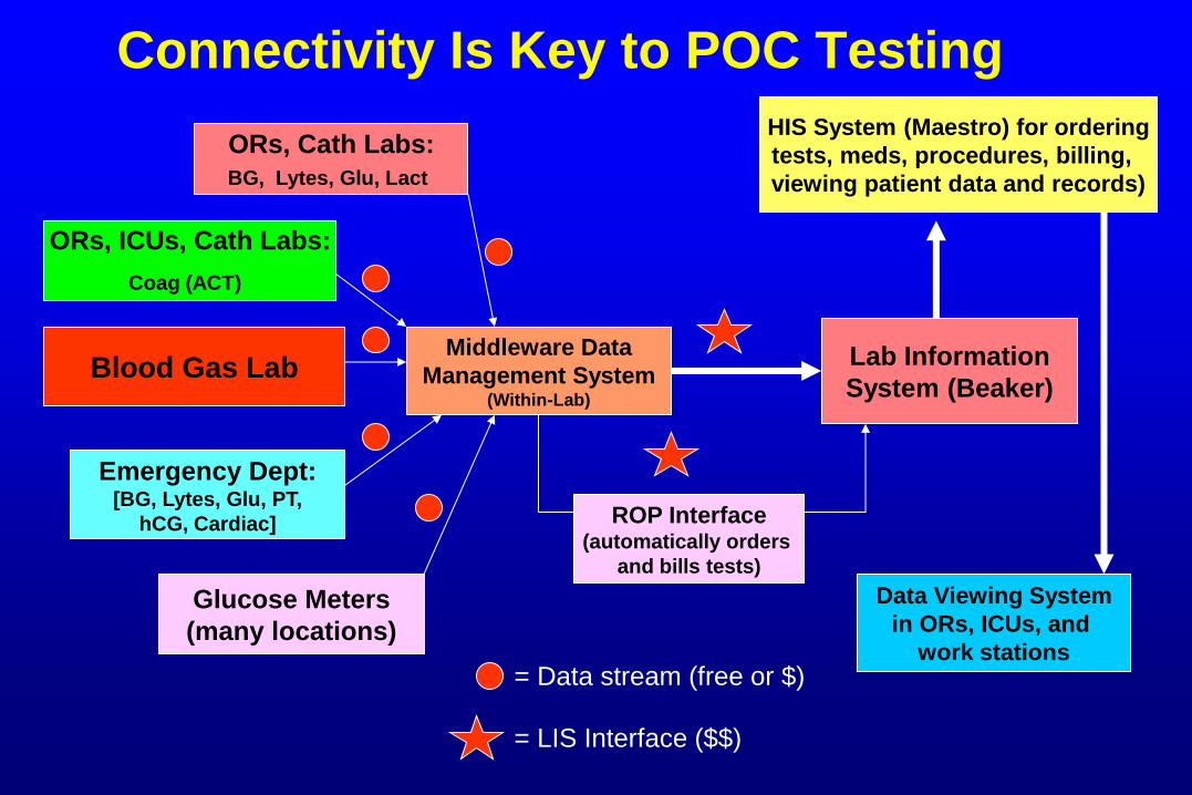
Connectivity Is Key to POC Testing
Middleware Data
Management System (Within-Lab)
Lab Information
System (Beaker)
ORs, Cath Labs:
BG, Lytes, Glu, Lact
Glucose Meters
(many locations)
ORs, ICUs, Cath Labs:
Coag (ACT)
Blood Gas Lab
Emergency Dept: [BG, Lytes, Glu, PT,
hCG, Cardiac]
Data Viewing System
in ORs, ICUs, and
work stations
ROP Interface (automatically orders
and bills tests)
= LIS Interface ($$)
= Data stream (free or $)
HIS System (Maestro) for ordering
tests, meds, procedures, billing,
viewing patient data and records)
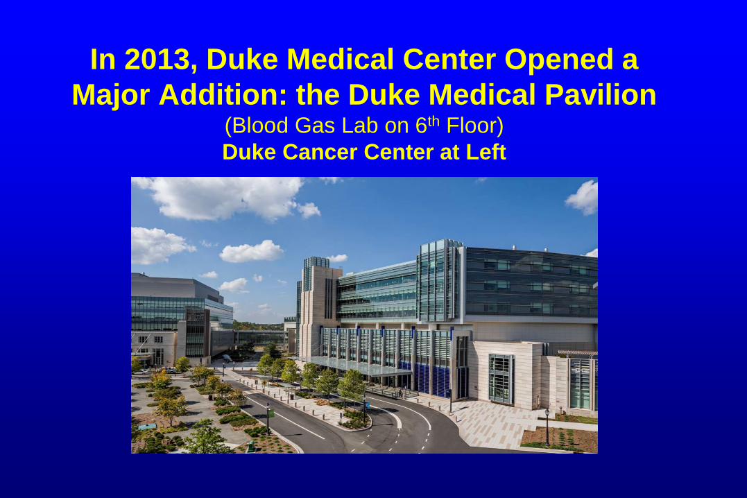
In 2013, Duke Medical Center Opened a
Major Addition: the Duke Medical Pavilion (Blood Gas Lab on 6th Floor)
Duke Cancer Center at Left
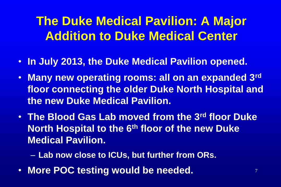
The Duke Medical Pavilion: A Major
Addition to Duke Medical Center
• In July 2013, the Duke Medical Pavilion opened.
• Many new operating rooms: all on an expanded 3rd
floor connecting the older Duke North Hospital and
the new Duke Medical Pavilion.
• The Blood Gas Lab moved from the 3rd floor Duke
North Hospital to the 6th floor of the new Duke
Medical Pavilion.
– Lab now close to ICUs, but further from ORs.
• More POC testing would be needed. 7

2013: New OR Locations and Estimated
POC Blood Gas Test Volumes
Old Locations New Locations Old Test Volumes New Test Volumes
OR 33 OR 33 3600/yr 1500/yr
OR 34 OR 34 3600 600
OR 35 OR 35 3600 900
OR 36 OR 36 3600 900
OR 37 OR 37 5000 3000
DMP OR 49 0 1200
DMP OR 50 0 2400-3000
DMP OR 51 0 900
DMP OR 54 0 900
DMP OR 55 0 2400-3000
DMP OR 56 0 2400
DMP OR 57 0 2400-3000
Pediatric ORs 0 2400
8
Evaluating Two Models of Point-of-
Care Blood Gas/Electrolyte/Etc.
Testing in Operating Room Areas
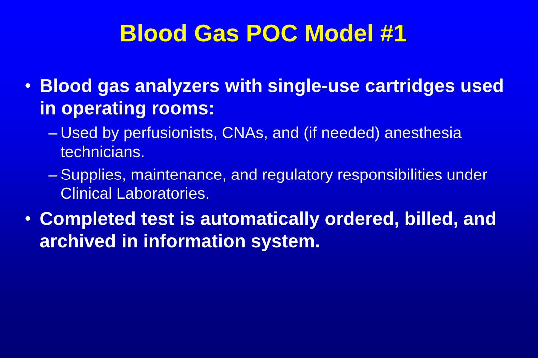
Blood Gas POC Model #1
• Blood gas analyzers with single-use cartridges used
in operating rooms:
– Used by perfusionists, CNAs, and (if needed) anesthesia
technicians.
– Supplies, maintenance, and regulatory responsibilities under
Clinical Laboratories.
• Completed test is automatically ordered, billed, and
archived in information system.

Blood Gas POC Model #2a (1985-2013)
• Blood gas analyzers with multi-use reagent packs
used in 4 (very busy) cardiac/thoracic operating
rooms and 1 peds cardiac cath lab:
– Used by perfusionists, anesthesia technicians, and cath lab
personnel.
– Maintenance, quality control, and regulatory responsibility are
under Clinical Laboratories.
• Completed test is automatically ordered, billed, and
archived in information system.
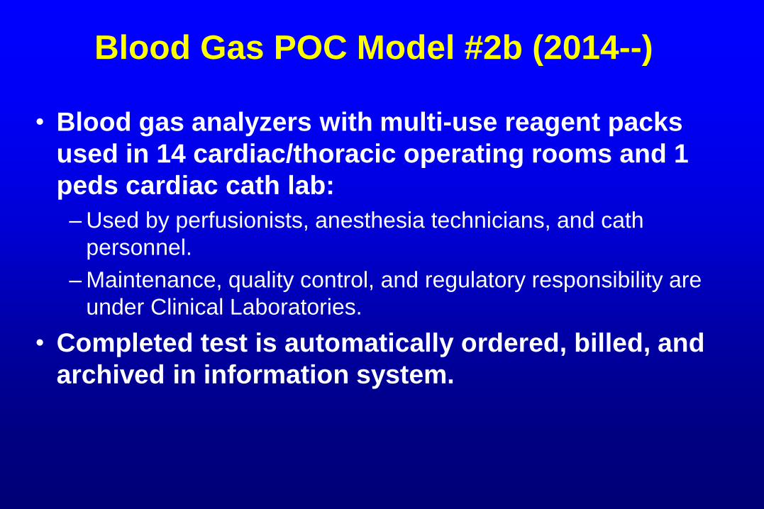
Blood Gas POC Model #2b (2014--)
• Blood gas analyzers with multi-use reagent packs
used in 14 cardiac/thoracic operating rooms and 1
peds cardiac cath lab:
– Used by perfusionists, anesthesia technicians, and cath
personnel.
– Maintenance, quality control, and regulatory responsibility are
under Clinical Laboratories.
• Completed test is automatically ordered, billed, and
archived in information system.
Disclaimer:
The following cost data are
approximate costs based on quotes
from manufacturers at different times
and different test volumes.
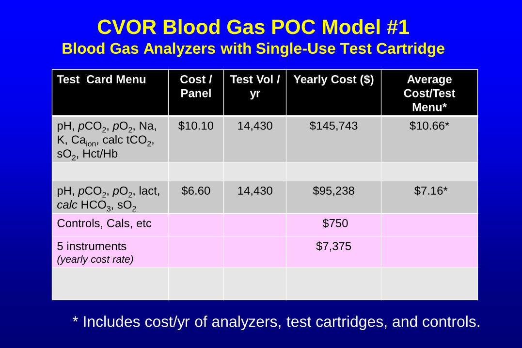
CVOR Blood Gas POC Model #1 Blood Gas Analyzers with Single-Use Test Cartridge
Test Card Menu Cost /
Panel
Test Vol /
yr
Yearly Cost ($) Average
Cost/Test
Menu*
pH, pCO2, pO2, Na,
K, Caion, calc tCO2,
sO2, Hct/Hb
$10.10 14,430 $145,743 $10.66*
pH, pCO2, pO2, lact,
calc HCO3, sO2
$6.60 14,430 $95,238 $7.16*
Controls, Cals, etc $750
5 instruments (yearly cost rate)
$7,375
* Includes cost/yr of analyzers, test cartridges, and controls.
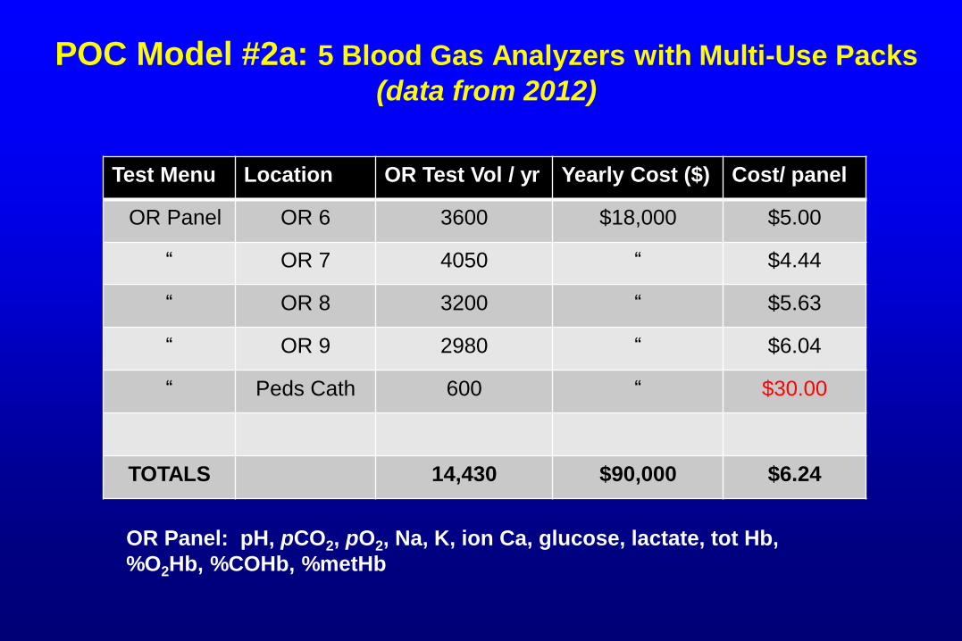
POC Model #2a: 5 Blood Gas Analyzers with Multi-Use Packs
(data from 2012)
Test Menu Location OR Test Vol / yr Yearly Cost ($) Cost/ panel
OR Panel OR 6 3600 $18,000 $5.00
“ OR 7 4050 “ $4.44
“ OR 8 3200 “ $5.63
“ OR 9 2980 “ $6.04
“ Peds Cath 600 “ $30.00
TOTALS 14,430 $90,000 $6.24
OR Panel: pH, pCO2, pO2, Na, K, ion Ca, glucose, lactate, tot Hb,
%O2Hb, %COHb, %metHb
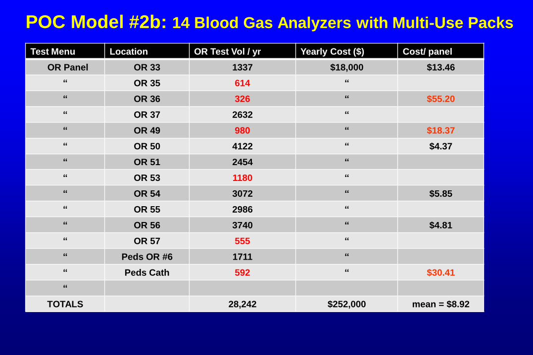
POC Model #2b: 14 Blood Gas Analyzers with Multi-Use Packs
Test Menu Location OR Test Vol / yr Yearly Cost ($) Cost/ panel
OR Panel OR 33 1337 $18,000 $13.46
“ OR 35 614 “
“ OR 36 326 “ $55.20
“ OR 37 2632 “
“ OR 49 980 “ $18.37
“ OR 50 4122 “ $4.37
“ OR 51 2454 “
“ OR 53 1180 “
“ OR 54 3072 “ $5.85
“ OR 55 2986 “
“ OR 56 3740 “ $4.81
“ OR 57 555 “
“ Peds OR #6 1711 “
“ Peds Cath 592 “ $30.41
“
TOTALS 28,242 $252,000 mean = $8.92

CVOR Blood Gas POC Model #1 Blood Gas Analyzers with Single-Use Test Cartridge
Test Card Menu Cost /
Panel
Test Vol /
yr
Yearly Cost ($) Average
Cost/Test
Menu*
pH, pCO2, pO2, Na,
K, Caion, calc tCO2,
sO2, Hct/Hb
$10.10 28,242 $285,244 $10.90*
pH, pCO2, pO2, lact,
calc HCO3, sO2
$6.60 28,242 $186,397 $7.40*
Controls, Cals, etc $2,000
14 instruments (yearly cost rate)
$20,645
* Includes cost/yr of analyzers, cartridges, and controls.
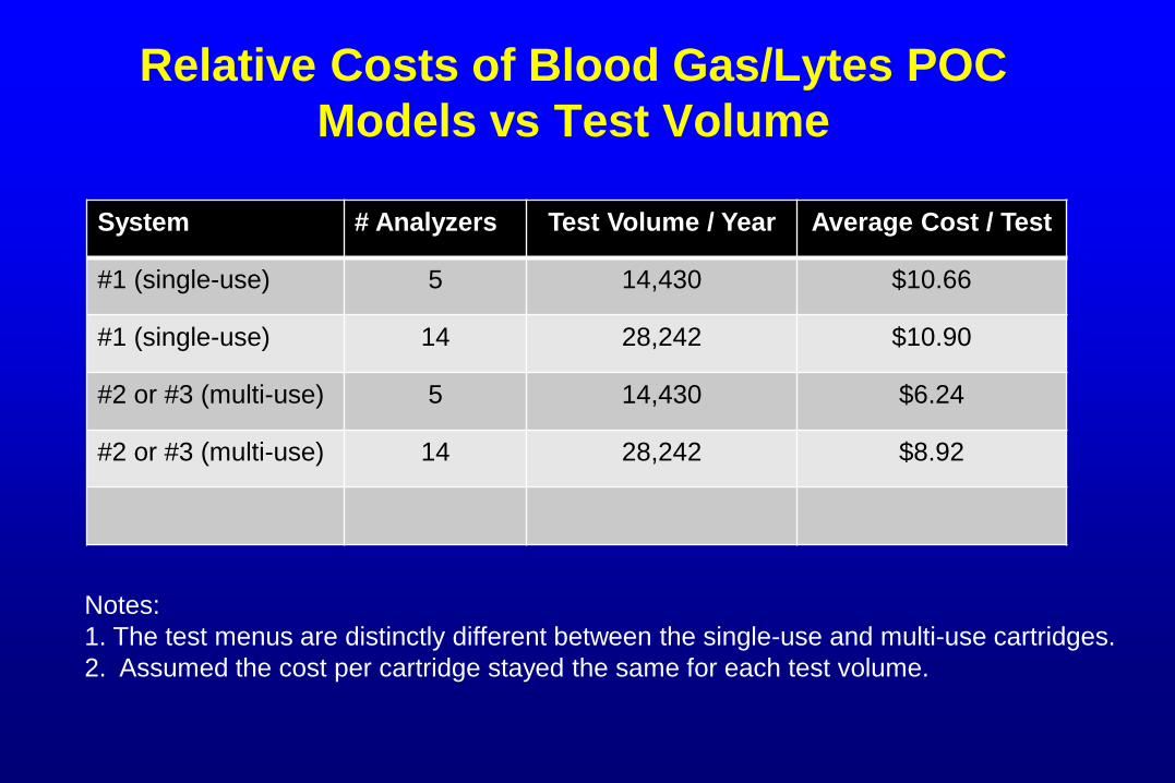
Relative Costs of Blood Gas/Lytes POC
Models vs Test Volume
System # Analyzers Test Volume / Year Average Cost / Test
#1 (single-use) 5 14,430 $10.66
#1 (single-use) 14 28,242 $10.90
#2 or #3 (multi-use) 5 14,430 $6.24
#2 or #3 (multi-use) 14 28,242 $8.92
Notes:
1. The test menus are distinctly different between the single-use and multi-use cartridges.
2. Assumed the cost per cartridge stayed the same for each test volume.

Pros/Cons of POC Model #1: Hand-held Single-Use Cartridge System
• Advantages
– Excellent portability
– Wider variety of tests available; ie. ACT, TnI
– Financially suited to lower volume settings
– Very good accuracy and reliability
• Disadvantages
– A complete critical care panel may require 2-3
cartridges (adds time and $/test)
– Cooximetry parameters are not measured
– Requires IQCP
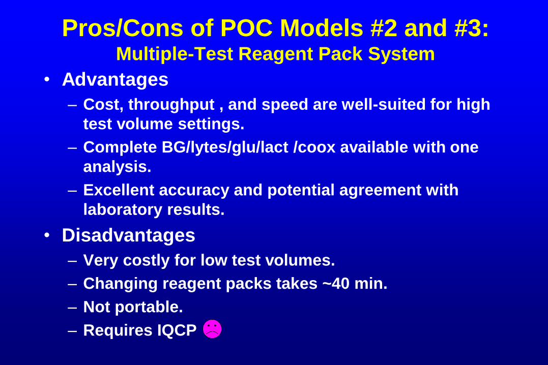
Pros/Cons of POC Models #2 and #3: Multiple-Test Reagent Pack System
• Advantages
– Cost, throughput , and speed are well-suited for high
test volume settings.
– Complete BG/lytes/glu/lact /coox available with one
analysis.
– Excellent accuracy and potential agreement with
laboratory results.
• Disadvantages
– Very costly for low test volumes.
– Changing reagent packs takes ~40 min.
– Not portable.
– Requires IQCP
Opportunities for Improved Assay
Devices for Intraoperative Parathyroid
Hormone (ioPTH) Measurements
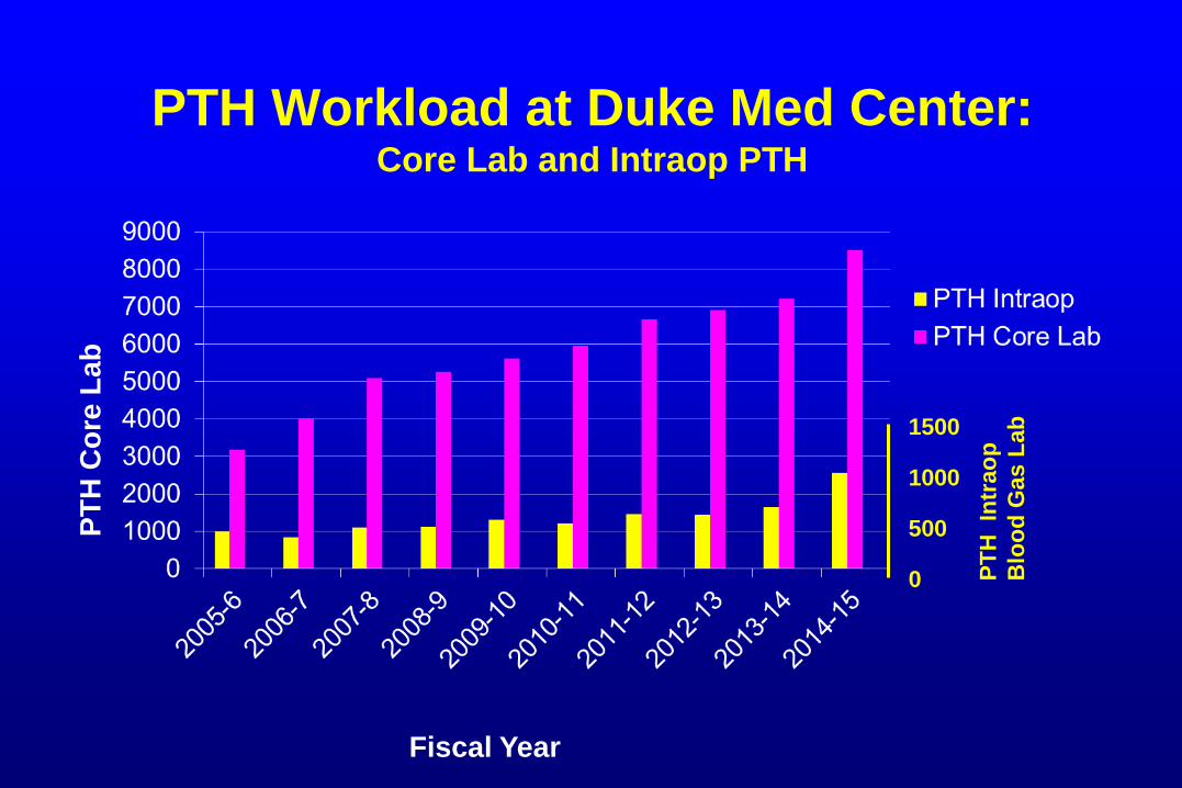
PTH Workload at Duke Med Center: Core Lab and Intraop PTH
Fiscal Year
PT
H C
ore
Lab
PT
H In
trao
p
Blo
od
Gas L
ab
1500
1000
500
0
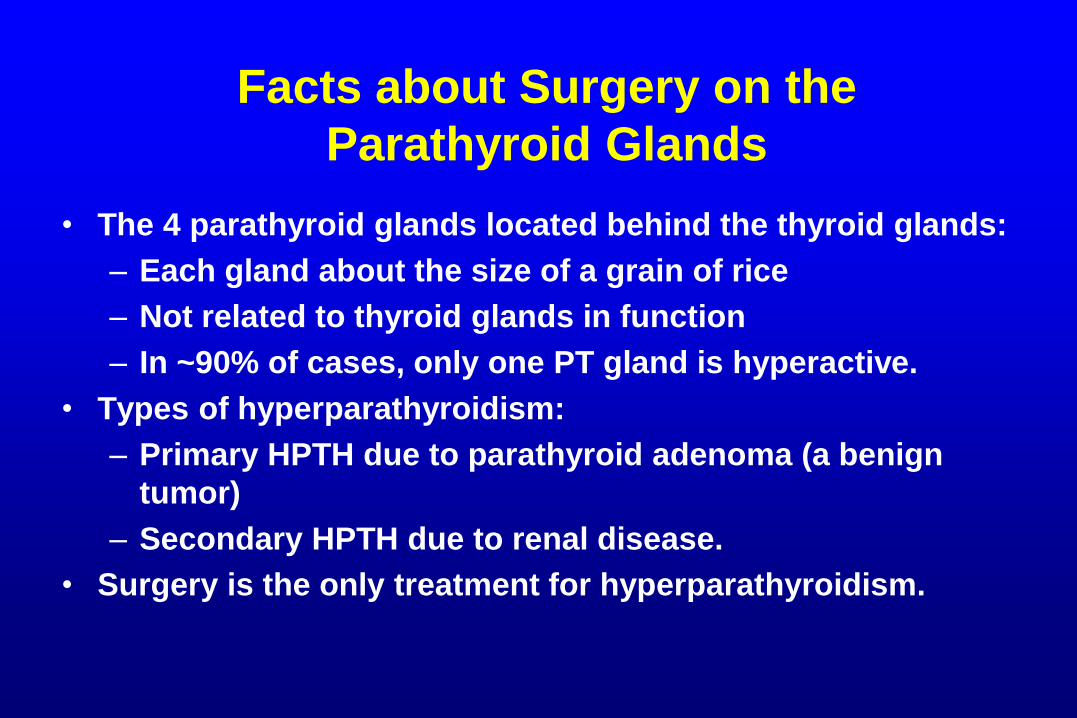
Facts about Surgery on the
Parathyroid Glands
• The 4 parathyroid glands located behind the thyroid glands:
– Each gland about the size of a grain of rice
– Not related to thyroid glands in function
– In ~90% of cases, only one PT gland is hyperactive.
• Types of hyperparathyroidism:
– Primary HPTH due to parathyroid adenoma (a benign
tumor)
– Secondary HPTH due to renal disease.
• Surgery is the only treatment for hyperparathyroidism.
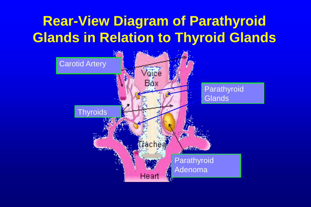
Rear-View Diagram of Parathyroid
Glands in Relation to Thyroid Glands
Parathyroid
Adenoma
Thyroids
Carotid Artery
Parathyroid
Glands
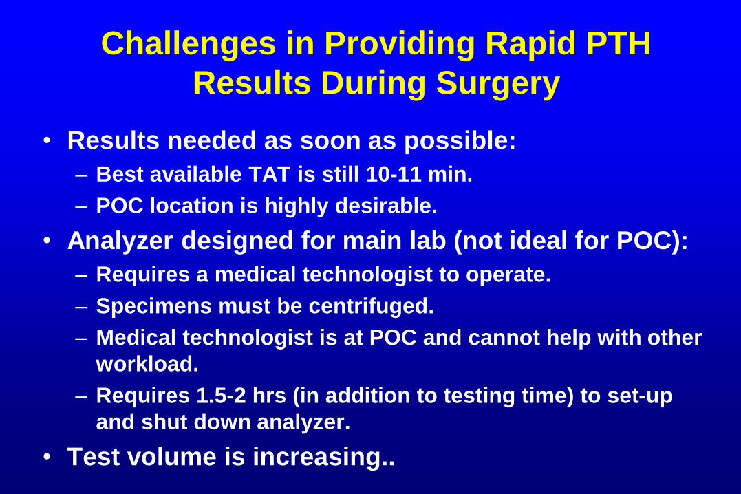
Challenges in Providing Rapid PTH
Results During Surgery
• Results needed as soon as possible:
– Best available TAT is still 10-11 min.
– POC location is highly desirable.
• Analyzer designed for main lab (not ideal for POC):
– Requires a medical technologist to operate.
– Specimens must be centrifuged.
– Medical technologist is at POC and cannot help with other
workload.
– Requires 1.5-2 hrs (in addition to testing time) to set-up
and shut down analyzer.
• Test volume is increasing..
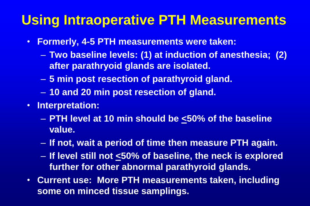
Using Intraoperative PTH Measurements
• Formerly, 4-5 PTH measurements were taken:
– Two baseline levels: (1) at induction of anesthesia; (2)
after parathryoid glands are isolated.
– 5 min post resection of parathyroid gland.
– 10 and 20 min post resection of gland.
• Interpretation:
– PTH level at 10 min should be <50% of the baseline
value.
– If not, wait a period of time then measure PTH again.
– If level still not <50% of baseline, the neck is explored
further for other abnormal parathyroid glands.
• Current use: More PTH measurements taken, including
some on minced tissue samplings.

Percent Change in Intraoperative PTH in
Patient with Two Enlarged Glands
Time After Gland Resection (min)
Perc
ent
of
Baselin
e P
TH
Valu
e
Point of Care 2007; 6: 253-260
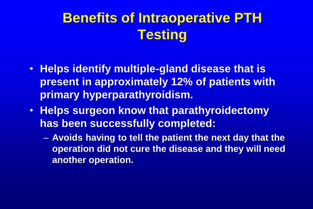
Benefits of Intraoperative PTH
Testing
• Helps identify multiple-gland disease that is
present in approximately 12% of patients with
primary hyperparathyroidism.
• Helps surgeon know that parathyroidectomy
has been successfully completed:
– Avoids having to tell the patient the next day that the
operation did not cure the disease and they will need
another operation.
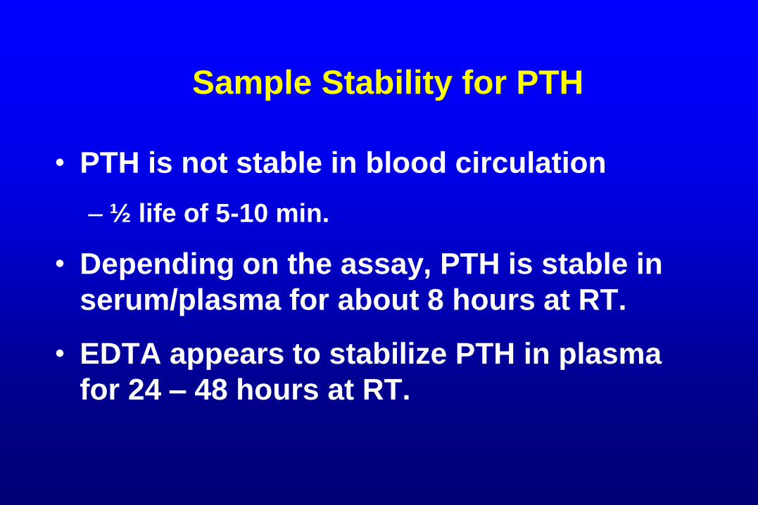
Sample Stability for PTH
• PTH is not stable in blood circulation
– ½ life of 5-10 min.
• Depending on the assay, PTH is stable in
serum/plasma for about 8 hours at RT.
• EDTA appears to stabilize PTH in plasma
for 24 – 48 hours at RT.

What Is Needed for an
Intraoperative PTH Test System?
• Small and portable analyzer.
• Easy test setup after days of non-use.
• No water or plumbing needed.
• No reagent preparation.
• Analysis on whole blood:
– Saves time AND you do not need a centrifuge.
• Results ASAP, but ideally in less than 10 min.
– Bench-type analyzer(s) has very few desirable features,
but assay takes about 11 min.

Reducing Transfusions in Cardiac OR by
Thromboelastography: TEG and ROTEM
• Cardiac surgery accounts for about 1/3 of all
intraoperative transfusions.
• Mortality correlates linearly with the number of
transfused blood products.
• When to give RBCs, platelets, fresh frozen
plasma (FFP), fibrinogen (as cryoprecipitate)?
Evaluating Need for
Fibrinogen/Cryoprecipitate in OR
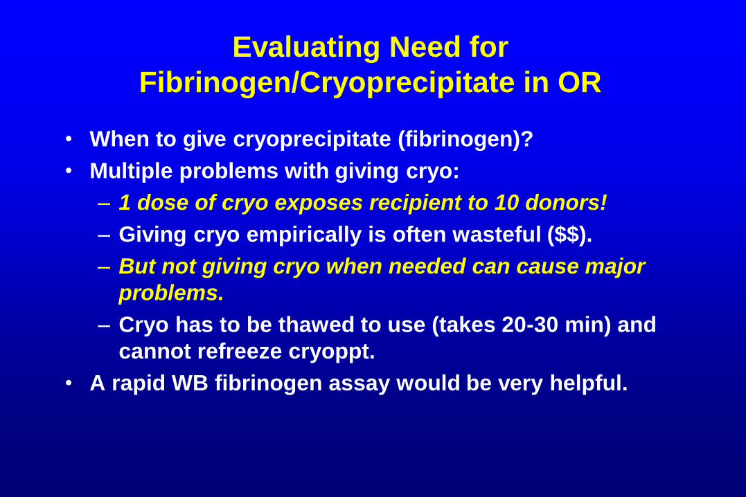
• When to give cryoprecipitate (fibrinogen)?
• Multiple problems with giving cryo:
– 1 dose of cryo exposes recipient to 10 donors!
– Giving cryo empirically is often wasteful ($$).
– But not giving cryo when needed can cause major
problems.
– Cryo has to be thawed to use (takes 20-30 min) and
cannot refreeze cryoppt.
• A rapid WB fibrinogen assay would be very helpful.
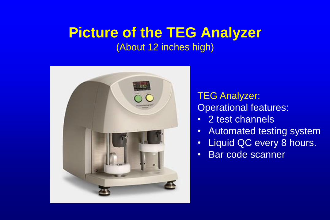
Picture of the TEG Analyzer (About 12 inches high)
TEG Analyzer:
Operational features:
• 2 test channels
• Automated testing system
• Liquid QC every 8 hours.
• Bar code scanner
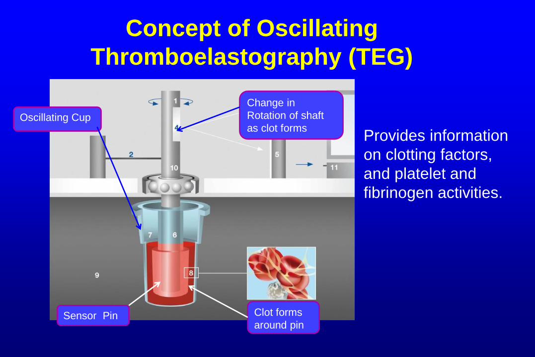
Concept of Oscillating
Thromboelastography (TEG)
Provides information
on clotting factors,
and platelet and
fibrinogen activities.
Oscillating Cup
Change in
Rotation of shaft
as clot forms
Sensor Pin Clot forms
around pin
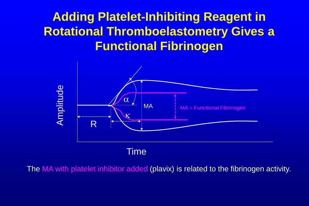
Adding Platelet-Inhibiting Reagent in
Rotational Thromboelastometry Gives a
Functional Fibrinogen
Time
Am
plit
ud
e
MA
R
The MA with platelet inhibitor added (plavix) is related to the fibrinogen activity.
k MA = Functional Fibrinogen
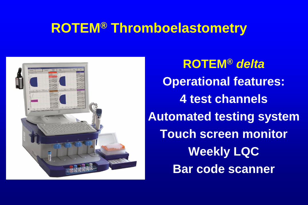
ROTEM® Thromboelastometry
ROTEM® delta
Operational features:
4 test channels
Automated testing system
Touch screen monitor
Weekly LQC
Bar code scanner
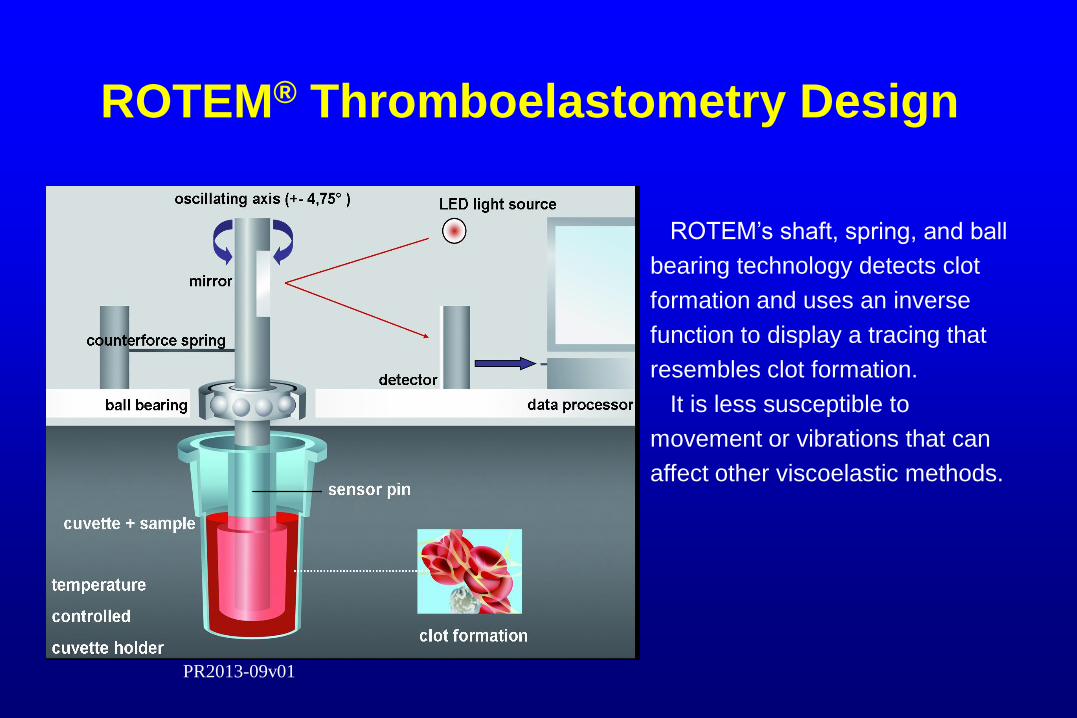
ROTEM® Thromboelastometry Design
ROTEM’s shaft, spring, and ball
bearing technology detects clot
formation and uses an inverse
function to display a tracing that
resembles clot formation.
It is less susceptible to
movement or vibrations that can
affect other viscoelastic methods.
PR2013-09v01
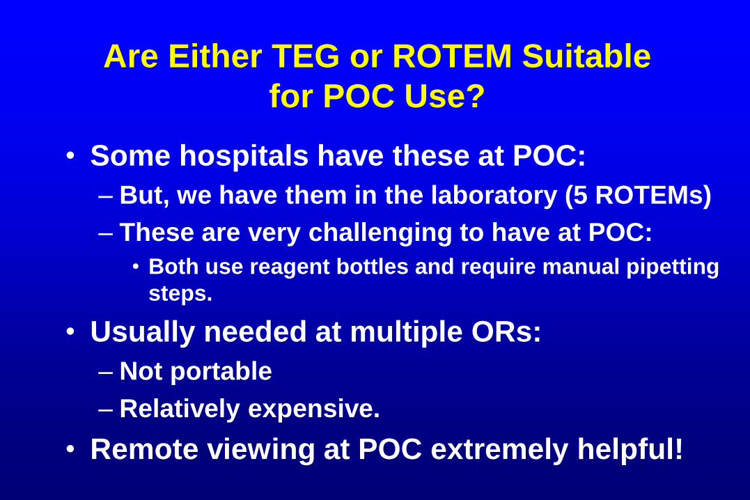
Are Either TEG or ROTEM Suitable
for POC Use?
• Some hospitals have these at POC:
– But, we have them in the laboratory (5 ROTEMs)
– These are very challenging to have at POC:
• Both use reagent bottles and require manual pipetting
steps.
• Usually needed at multiple ORs:
– Not portable
– Relatively expensive.
• Remote viewing at POC extremely helpful!
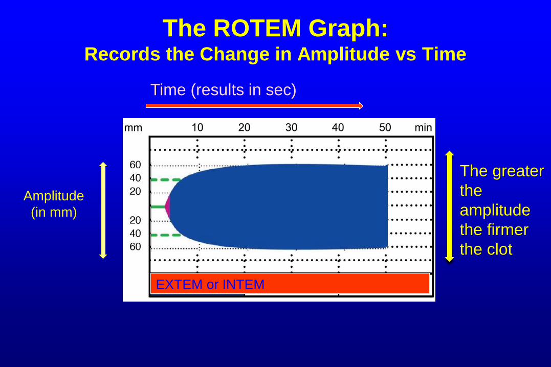
The ROTEM Graph: Records the Change in Amplitude vs Time
Amplitude
(in mm)
Time (results in sec)
The greater
the
amplitude
the firmer
the clot
EXTEM or INTEM
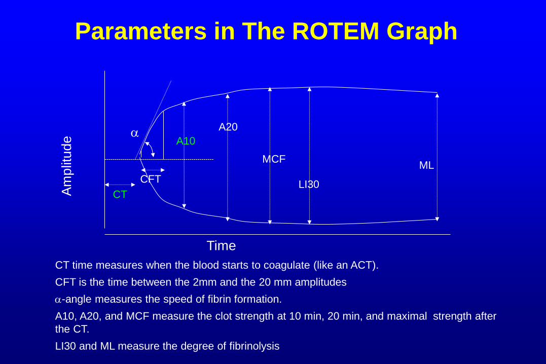
Parameters in The ROTEM Graph
Time
Am
plit
ud
e
MCF
CT
CT time measures when the blood starts to coagulate (like an ACT).
CFT is the time between the 2mm and the 20 mm amplitudes
-angle measures the speed of fibrin formation.
A10, A20, and MCF measure the clot strength at 10 min, 20 min, and maximal strength after
the CT.
LI30 and ML measure the degree of fibrinolysis
LI30 CFT
A10
A20
ML
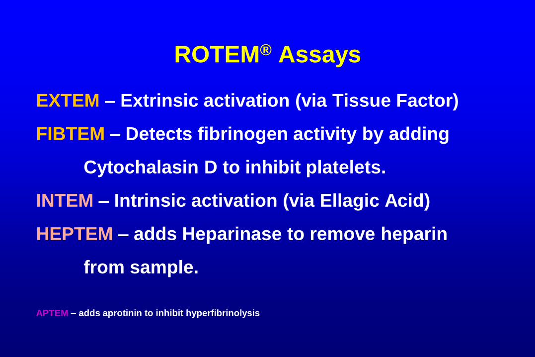
ROTEM® Assays
EXTEM – Extrinsic activation (via Tissue Factor)
FIBTEM – Detects fibrinogen activity by adding
Cytochalasin D to inhibit platelets.
INTEM – Intrinsic activation (via Ellagic Acid)
HEPTEM – adds Heparinase to remove heparin
from sample.
APTEM – adds aprotinin to inhibit hyperfibrinolysis
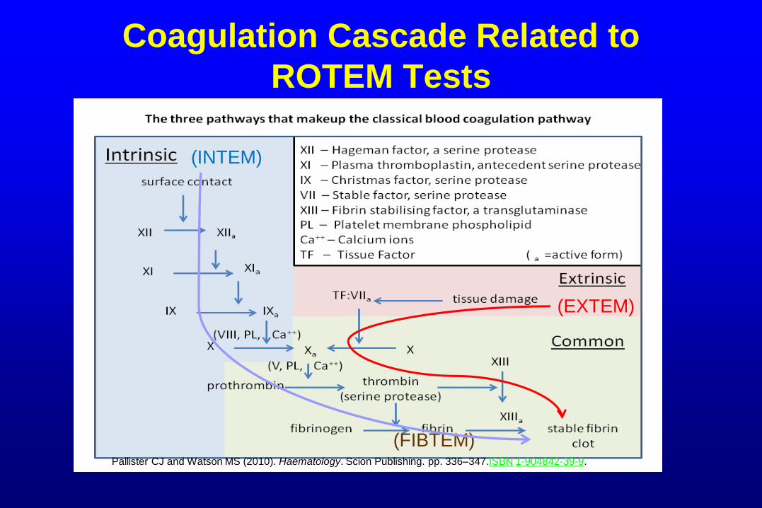
Coagulation Cascade Related to
ROTEM Tests
(INTEM)
(EXTEM)
(FIBTEM) Pallister CJ and Watson MS (2010). Haematology. Scion Publishing. pp. 336–347.ISBN 1-904842-39-9.
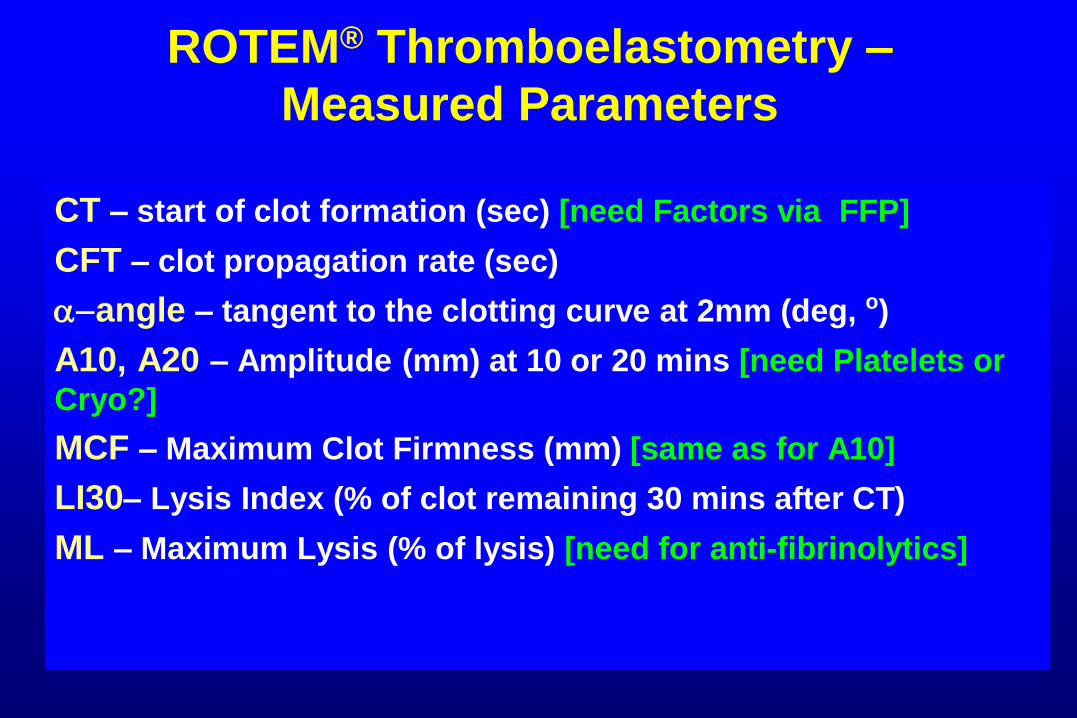
ROTEM® Thromboelastometry –
Measured Parameters
CT – start of clot formation (sec) [need Factors via FFP]
CFT – clot propagation rate (sec)
-angle – tangent to the clotting curve at 2mm (deg, o)
A10, A20 – Amplitude (mm) at 10 or 20 mins [need Platelets or
Cryo?]
MCF – Maximum Clot Firmness (mm) [same as for A10]
LI30– Lysis Index (% of clot remaining 30 mins after CT)
ML – Maximum Lysis (% of lysis) [need for anti-fibrinolytics]
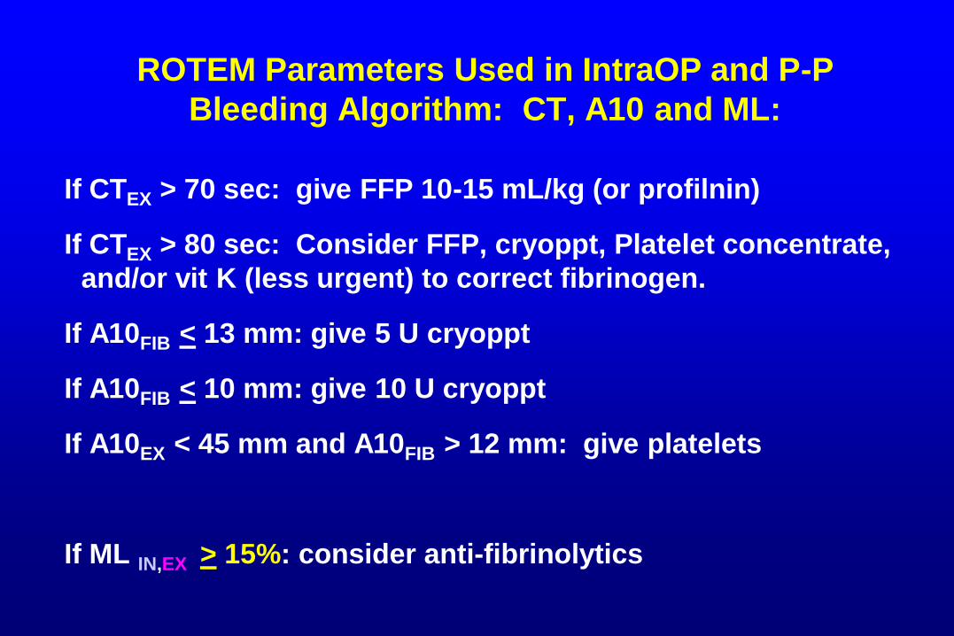
ROTEM Parameters Used in IntraOP and P-P
Bleeding Algorithm: CT, A10 and ML:
If CTEX > 70 sec: give FFP 10-15 mL/kg (or profilnin)
If CTEX > 80 sec: Consider FFP, cryoppt, Platelet concentrate,
and/or vit K (less urgent) to correct fibrinogen.
If A10FIB < 13 mm: give 5 U cryoppt
If A10FIB < 10 mm: give 10 U cryoppt
If A10EX < 45 mm and A10FIB > 12 mm: give platelets
If ML IN,EX > 15%: consider anti-fibrinolytics
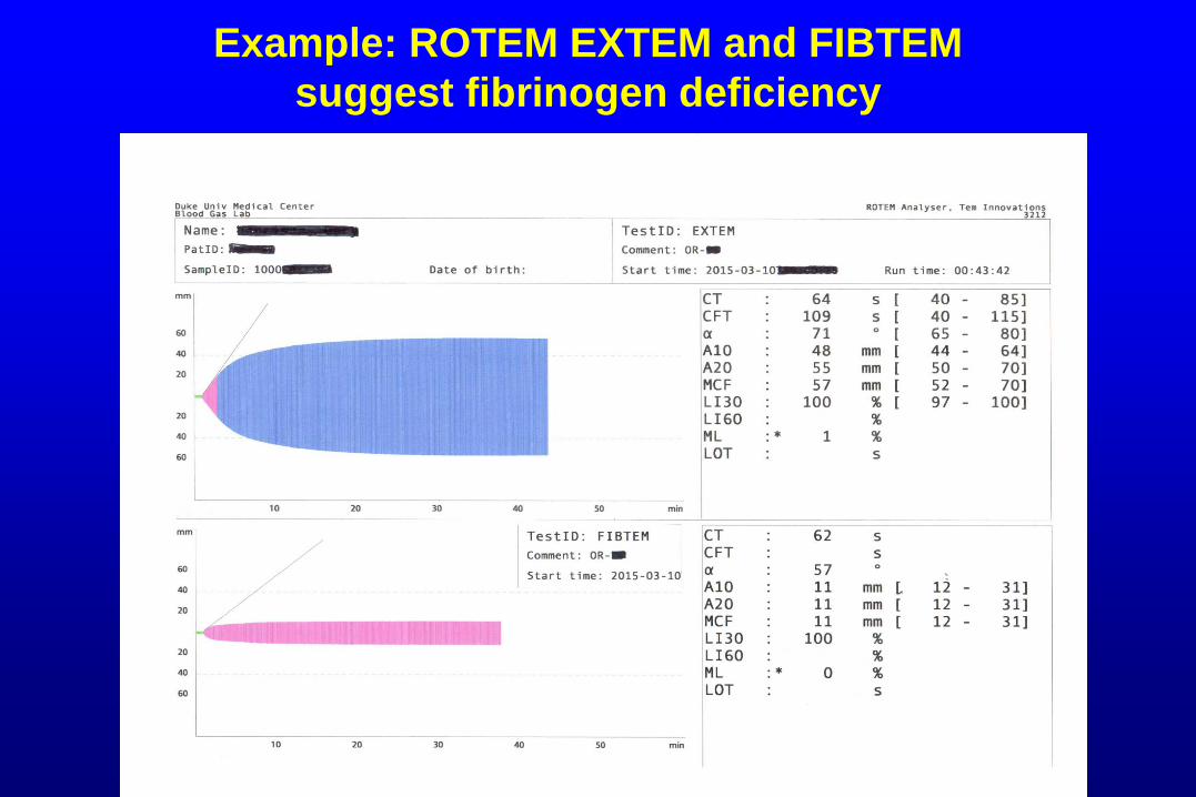
Example: ROTEM EXTEM and FIBTEM
suggest fibrinogen deficiency
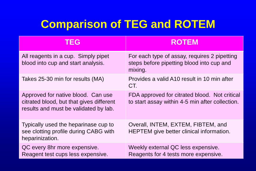
Comparison of TEG and ROTEM
TEG ROTEM
All reagents in a cup. Simply pipet
blood into cup and start analysis.
For each type of assay, requires 2 pipetting
steps before pipetting blood into cup and
mixing.
Takes 25-30 min for results (MA) Provides a valid A10 result in 10 min after
CT.
Approved for native blood. Can use
citrated blood, but that gives different
results and must be validated by lab.
FDA approved for citrated blood. Not critical
to start assay within 4-5 min after collection.
Typically used the heparinase cup to
see clotting profile during CABG with
heparinization.
Overall, INTEM, EXTEM, FIBTEM, and
HEPTEM give better clinical information.
QC every 8hr more expensive.
Reagent test cups less expensive.
Weekly external QC less expensive.
Reagents for 4 tests more expensive.
Actual, Totally True Incident (Years Ago) in
Our Point-of-Care Glucose Testing Program
• A lab person in the POCT program called a
caregiver about a result being an “outlier” on a
proficiency test sample.
• Caregiver heard this slightly differently and told
their supervisor: “The lab said I was an ‘out and
out liar’ on my [PT] result.”
• Moral: Effective communication is a must in POC
testing.