biosintesis de agnps, metodos amigables al ambiente
TRANSCRIPT
1. IntroductionNanoparticles (nano-scale particles = NSPs) are atomic or molecular aggregates with at least one dimension between
1 and 100nm [1,2], that can drastically modify their physico-chemical properties compared to the bulk material [3]. Nano-particles can be made from a fully variety of bulk materials and that they can expli cate their actions depending on both the chemical composition and on the size and/or shape of the particles [4]. Because of its smaller structure, they trigger the chemical activity due to their distinctive crystallographic nature that increases surface area, hence the scope of reactivity [5]. The advance technology accepts that the concept of interdisciplinary research in the areas of engineering and sciences leads to creation of environmentally acceptable “green process”, with special concern to nanoscience and nanotechnology. The for-mation of nanoparticles mediated by biological route is considered as better method than any other method because catalytic and functional information obtained under close to optimal conditions through action of enzymatic properties can help to understand the biochemical and molecular mechanisms of nanoparticles formation.
In nanotechnology, silver nanoparticles are the most promising one. Silver nanoparticles are nanoparticles of silver, i.e. silver particles size in range of between 1 nm and 100 and because of its nano size it have attracted intensive research in-terest. It is observed that silver nanoparticles do not affect living cells, so not able to provoke microbial resistance. It is be-lieved that Silver nanoparticles can attach to the cell wall and disturb cell-wall permeability and cellular respiration[6]. Silver containing particles also used in textile fabrics, as food additives, and in package and plastics to eliminate microorganisms. Because of such a wide range of applications, various methods concerning the fabrication of silver nanoparticles, as well as various silver-based compounds containing metallic silver (Ag0) have been developed [7]. The special attention towards the silver nanoparticles because of their strong antimicrobial activity either in metallic nature and nanoparticles form also, so it is found that silver nanoparticles has different applications to the environment and human. It has been well studied that a variety of biological sources are able to produce silver nanoparticles of different shapes and nature. Nanoparticle production and applications have been extensively studied; studies related to drug delivery, tissue engineering and bioMEMS have been undertaken for a great number of scientific publications and patents. Some uses of Silver Nanoparticles are mentioned below-• Minuteamountofsilverareparticularlyusedasdecontaminatingagentinwaterandpreventbiofilmformationinfoodcontact
surfaces[8].
• Theantimicrobialnatureofsilverionsplaysaprominentroleinfoodpackagingsystems[9].
• Silvernanoparticleshaveantibacterialpropertiesmediatedbysilverions[10].
• Itusedaspreservativeinfoodandvariousfoodrelatedproducts[11].
• Thesilvernanoparticlesarereportedtoshowbetterwoundhealingcapacity,bettercosmeticappearanceandscarlesshealing
Biosynthesis of silver nanoparticle by eco-friendly method
Richa L Karnani1 and Abhay Chowdhary2
1,2Department of Clinical Pathology, Haffkines Institute of Training, Research and Testing, Mumbai, [email protected]
Abstract Intherecenttime,synthesisofnanoparticleshasbeenthesubjectofalotofstudiesduetoitscommercialdemandsandwideap-
plicabilityinvariousareas.Nanotechnologyisanemergingfield,nanoparticlesishelpfulininvestigationandregulationatcelllevelinteractionbetweensyntheticandbiologicalmaterials.Inmanyareasofhumansciencethesematerialsaresuperiorandindispensableduetoitsuniquesizedependent.Generally,physical,mechanicalandchemicalmethodsinvolvedforthesynthesisofnanoparticles.Butthesemethodsareveryexpensiveandsomemethodsinvolveharmfulchemicals.Withtheaimofdevelopingclean,nontoxicandeco-friendlytechnologies,awiderangeofbiologicalsourceshasbeenusedfortheformationofparticle.Greenchemistryprocessesledtoeco-friendlymethodofsynthesisandsafeprocessascomparedtoothermethods.Inthisreview,wedescribeacheapanden-vironmentfriendlytechniqueforsynthesisofsilvernanoparticlesbygreenchemistryapproachedfromdifferentbiologicalsources.Theimportanceofthisstudyincludesthepreciseandspecificanalysisofsilvernanoparticles,biologicalsystemsthatmaysupportandrevolutionizetheartofsynthesisofnanoparticles.
Keywords: Silver Nanoparticles, Green chemistry
Vol:1 Issue:2 February 2013 Indian Journal of NanoScience
www.iseeadyar.org/indjns.html Research article 25
whentestedusingananimalmodel[12].
• TheFe3O4attachedsilvernanoparticlescanbeusedforthetreatmentofwaterandeasilyremovedusingmagneticfieldtoavoidcontaminationoftheenvironment[13].
• Environmental-friendlyantimicrobialnanopaintcanbedeveloped[14].
Thepresentreviewarticledrawsattentiontothecurrentknowledgeregardingthepotentialorganismsforbiosynthesisofsilvernanoparticlesandpresentsadatabasethatfutureresearcherscanbebasedon.
2. Biosynthesis of Silver NanoparticlesLiving cell ranges from prokaryotic to eukaryotic are typically 10 mm across. Many varieties of biological sources avail-
able in nature including bacteria, algae ,yeast ,fungi, lower plants and higher angiosperm plant products can all be involved for the synthesis of nanoparticles. These ambient biological systems provide excellent examples of nanophasic materials with highly optimized characteristics resulting from evolution over a long scale of time [15] and the synthesis of inorganic materi-als may occur either extracellularly or intracellularly [16].
In current research areas of nanotechnology, with the help of biological source it is a big challenge to develop reliable experimental protocols for the synthesis of nanoparticles over a range of chemical composition, size and synchronized mono-dispersity that should be non-toxic, clean and eco-friendly. Although many paper have been reported in the last few years [17,18,19], it is need to elaborate this technology in a consolidated manner with an approach that gives an overview of the current trend of research on the biosynthesis of different metal nanoparticles and their applications. The use of environmen-tally benign materials like plant extract [20], bacteria [21], fungi [22] and enzymes [23] for the synthesis of silver nanopar-ticles offer many benefits of ecofriendliness and suitability for pharmaceutical and other biomedical applications as they do not use toxic chemicals for the synthesis protocol. A chemical synthesis method involves presence of some toxic chemical absorbed on the surface that may have adverse effect in the medical applications. Green synthesis provides advancement over chemical and physical method as it is cheap, ecofriendly, can be scaled up for large scale synthesis very easily and in this method there is no need to use high pressure, energy, temperature and toxic chemicals [24]. Herein, we provide an overview of various reports of biological means of nanoparticle synthesis of desired characteristics.
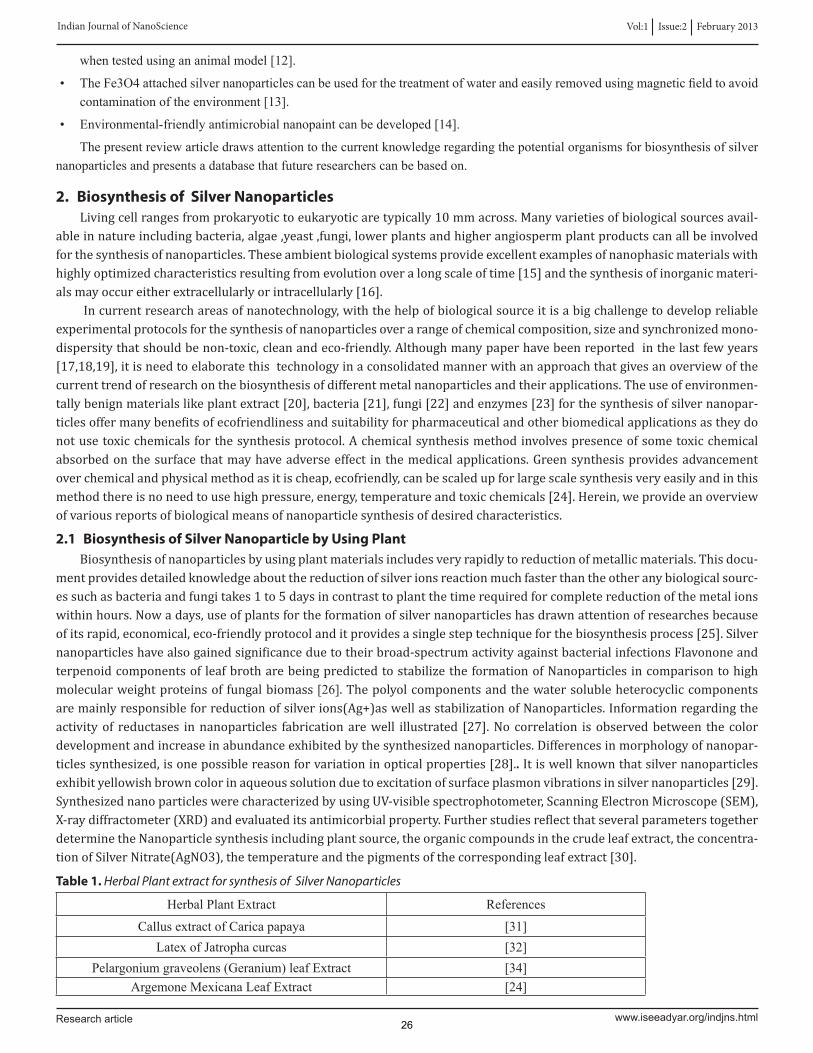
2.1 Biosynthesis of Silver Nanoparticle by Using PlantBiosynthesis of nanoparticles by using plant materials includes very rapidly to reduction of metallic materials. This docu-
ment provides detailed knowledge about the reduction of silver ions reaction much faster than the other any biological sourc-es such as bacteria and fungi takes 1 to 5 days in contrast to plant the time required for complete reduction of the metal ions within hours. Now a days, use of plants for the formation of silver nanoparticles has drawn attention of researches because of its rapid, economical, eco-friendly protocol and it provides a single step technique for the biosynthesis process [25]. Silver nanoparticles have also gained significance due to their broad-spectrum activity against bacterial infections Flavonone and terpenoid components of leaf broth are being predicted to stabilize the formation of Nanoparticles in comparison to high molecular weight proteins of fungal biomass [26]. The polyol components and the water soluble heterocyclic components are mainly responsible for reduction of silver ions(Ag+)as well as stabilization of Nanoparticles. Information regarding the activity of reductases in nanoparticles fabrication are well illustrated [27]. No correlation is observed between the color development and increase in abundance exhibited by the synthesized nanoparticles. Differences in morphology of nanopar-ticles synthesized, is one possible reason for variation in optical properties [28].. It is well known that silver nanoparticles exhibit yellowish brown color in aqueous solution due to excitation of surface plasmon vibrations in silver nanoparticles [29]. Synthesized nano particles were characterized by using UV-visible spectrophotometer, Scanning Electron Microscope (SEM), X-ray diffractometer (XRD) and evaluated its antimicorbial property. Further studies reflect that several parameters together determine the Nanoparticle synthesis including plant source, the organic compounds in the crude leaf extract, the concentra-tion of Silver Nitrate(AgNO3), the temperature and the pigments of the corresponding leaf extract [30].
Table 1. Herbal Plant extract for synthesis of Silver Nanoparticles
HerbalPlantExtract References
CallusextractofCaricapapaya [31]LatexofJatrophacurcas [32]
Pelargoniumgraveolens(Geranium)leafExtract [34]ArgemoneMexicanaLeafExtract [24]
Vol:1 Issue:2 February 2013 Indian Journal of NanoScience
www.iseeadyar.org/indjns.htmlResearch article 26
AlfalfaSproutsExtract [33]Caricapapaya(Papaya)FruitExtract [29]
Cinnamomumcamphora [35]Trianthemadecandra [36]Euphorbiahirta.L [37]
Partheniumleafextract [38]AloeveraplantExtract [39]
FruitextractofEmblicaofficinalis [40]RootsofMedicagosativa [33]HelianthusannusExtract [30]
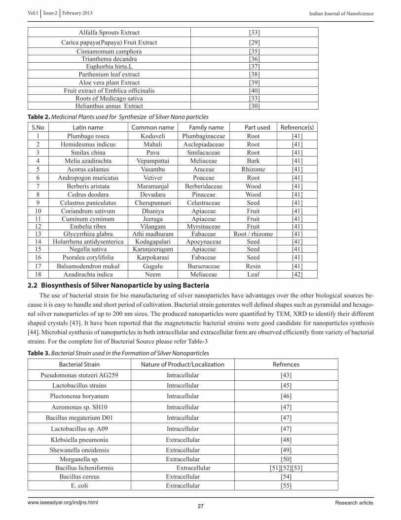
Table 2. Medicinal Plants used for Synthesize of Silver Nano particles
S.No Latin name Common name Family name Part used Reference(s)1 Plumbagorosea Koduveli Plumbaginaceae Root [41]2 Hemidesmusindicus Mahali Asclepiadaceae Root [41]3 Smilaxchina Pavu Smilacaceae Root [41]4 Meliaazadirachta Vepampattai Meliaceae Bark [41]5 Acoruscalamus Vasambu Araceae Rhizome [41]6 Andropogonmuricatus Vetiver Poaceae Root [41]7 Berberisaristata Maramanjal Berberidaceae Wood [41]8 Cedrusdeodara Devadaru Pinaceae Wood [41]9 Celastruspaniculatus Cherupunnari Celastraceae Seed [41]10 Coriandrumsativum Dhaniya Apiaceae Fruit [41]11 Cuminumcyminum Jeeraga Apiaceae Fruit [41]12 Embeliaribes Vilangam Myrsinaceae Fruit [41]13 Glycyrrhizaglabra Athimadhuram Fabaceae Root/rhizome [41]14 Holarrhenaantidysenterica Kodagapalari Apocynaceae Seed [41]15 Negellasativa Karunjeeragam Apiaceae Seed [41]16 Psoraleacorylifolia Karpokarasi Fabaceae Seed [41]17 Balsamodendronmukul Gugulu Burseraceae Resin [41]18 Azadirachtaindica Neem Meliaceae Leaf [42]
2.2 Biosynthesis of Silver Nanoparticle by using BacteriaTheuseofbacterialstrainforbiomanufacturingofsilvernanoparticleshaveadvantagesovertheotherbiologicalsourcesbe-
causeitiseasytohandleandshortperiodofcultivation.Bacterialstraingenerateswelldefinedshapessuchaspyramidalandhexago-nalsilvernanoparticlesofupto200nmsizes.TheproducednanoparticleswerequantifiedbyTEM,XRDtoidentifytheirdifferentshapedcrystals[43].Ithavebeenreportedthatthemagnetotacticbacterialstrainsweregoodcandidatefornanoparticlessynthesis[44].Microbialsynthesisofnanoparticlesinbothintracellularandextracellularformareobservedefficientlyfromvarietyofbacterialstrains.ForthecompletelistofBacterialSourcepleasereferTable-3
Table 3. Bacterial Strain used in the Formation of Silver Nanoparticles
Bacterial Strain Nature of Product/Localization Refrences
PseudomonasstutzeriAG259 Intracellular [43]Lactobacillusstrains Intracellular [45]Plectonemaboryanum Intracellular [46]
Aeromonassp.SH10 Intracellular [47]BacillusmegateriumD01 Intracellular [47]
Lactobacillussp.A09 Intracellular [47]
Klebsiellapneumonia Extracellular [48]Shewanellaoneidensis Extracellular [49]
Morganellasp. Extracellular [50]Bacilluslicheniformis Extracellular [51][52][53]
Bacilluscereus Extracellular [54]E.coli Extracellular [55]
Vol:1 Issue:2 February 2013 Indian Journal of NanoScience
www.iseeadyar.org/indjns.html Research article 27
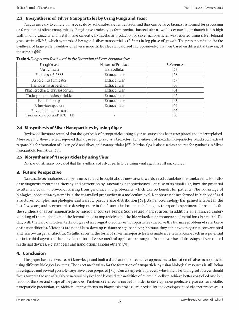
2.3 Biosynthesis of Silver Nanoparticles by Using Fungi and YeastFungusareeasytocultureonlargescalebysolidsubstratefermentationandthuscanbelargebiomassisformedforprocessing
orformationofsilvernanoparticles.Fungihavetendencytoformproductintracellularaswellasextracellularthoughithashighwallbindingcapacityandmetalintakecapacity.ExtracellularproductionofsilvernanoparticleswasreportedusingsilvertolerantyeaststrainMKY3,whichsynthesizedhexagonalsilvernanoparticles(2-5nm)inlogphaseofgrowth.Theproperconditionforthesynthesisoflargescalequantitiesofsilvernanoparticlesalsostandardizedanddocumentedthatwasbasedondifferentialthawingofthesamples[56].
Table 4. Fungus and Yeast used in the Formation of Silver Nanoparticles
Fungi/Yeast Nature of Product ReferencesVerticillium Intracellular [57]
Phomasp.3.2883 Extracellular [58]Aspergillusfumigates Extracellular [59]Trichodermaasperellum Extracellular [60]
Phaenerochaetechrysosporium Extracellular [61]Cladosporiumcladosporioides Extracellular [62]
Penicilliumsp. Extracellular [63]P.brevicompactum Extracellular [64]
Phytophthorainfestans - [65]FusariumoxysporumPTCC5115 - [66]
2.4 Biosynthesis of Silver Nanoparticles by using AlgaeReviewofliteraturerevealedthatthesynthesisofnanoparticlesusingalgaeassourcehasbeenunexploredandunderexploited.
Morerecently,therearefew,reportedthatalgaebeingusedasabiofactoryforsynthesisofmetallicnanoparticles.Mushroomextractresponsibleforformationofsilver,goldandsilver-goldnanoparticles[67].MarinealgaisalsousedasasourceforsynthesisinSilvernanoparticleformation[68].
2.5 Biosynthesis of Nanoparticles by using VirusReviewofliteraturerevealedthatthesynthesisofsilverparticlebyusingviralagentisstillunexplored.
3. Future PerspectiveNanoscale technologies can be improved and brought about new area towards revolutionizing the fundamentals of dis-
ease diagnosis, treatment, therapy and prevention by innovating nanomedicines. Because of its small size, have the potential to alter molecular discoveries arising from genomics and proteomics which can be benefit for patients. The advantage of biological production systems is in the controlled production at a molecular level. Nanoparticles are formed in highly defined structures, complex morphologies and narrow particle size distribution [69]. As nanotechnology has gained interest in the last few years, and is expected to develop more in the future, the foremost challenge is to expand experimental protocols for the synthesis of silver nanoparticle by microbial sources, Fungal Sources and Plant sources. In addition, an enhanced under-standing of the mechanism of the formation of nanoparticles and the bioreduction phenomenon of metal ions is needed. To-day, with the help of modern technologies of impregnation of silver nanoparticles can solve the burning problem of resistance against antibiotics. Microbes are not able to develop resistance against silver, because they can develop against conventional and narrow target antibiotics. Metallic silver in the form of silver nanoparticles has made a beneficial comeback as a potential antimicrobial agent and has developed into diverse medical applications ranging from silver based dressings, silver coated medicinal devices, e.g. nanogels and nanolotions among others [70].
4. ConclusionThispaperhasreviewedrecentknowledgeandbuiltadatabaseofbioreductiveapproachestoformationofsilvernanoparticles
usingdifferentbiologicalsystems.Theexactmechanismfortheformationofnanoparticlebyusingbiologicalresourcesisstillbeinginvestigatedandseveralpossiblewayshavebeenproposed[71].Currentaspectsofprocesswhichincludesbiologicalsourcesshouldfocustowardstheuseofhighlystructuredphysicalandbiosyntheticactivitiesofmicrobialcellstoachievebettercontrolledmanipu-lationofthesizeandshapeoftheparticles.Furthermoreeffectisneededinordertodevelopmoreproductiveprocessformetallicnanoparticleproduction.Inaddition,improvementsonbiogenesisprocessareneededforthedevelopmentofcheaperprocesses.It
Vol:1 Issue:2 February 2013 Indian Journal of NanoScience
www.iseeadyar.org/indjns.htmlResearch article 28
canbeconcludedthatinmicroorganismswhereproteins[72,73]andangiospermswherecarboxylicgroups,aminogroups,proteinsandcarbohydratesarepresentinthesourceextract,believedthatplayakeyroleinthebiosorptionandbioreductionprocessfortheformationofnanoparticles.Lotsofresearchworkstillneedtobeexecutedtounderstandtheeffectoftime,temperature,lightandotherparametersregardingthephytoformationofNanoparticles.
5. References1.BallP(2002)Naturalstrategiesforthemolecularengineer.Nanotechnol.13,15-28.
2.RocoMC(2003a)Broadersocietalissueofnanotechnology.J.NanoparticleRes.5,181-189
3.NelA,XiaT,MadlerLandLiN(2006)Toxicpotentialofmaterialsatthenanolevel.Sci.311,622-627
4.BrunnerTI,WickP,ManserP,SpohnP,GrassRN,LimbachLK,BruininkA,andStarkWJ(2006)Invitrocytotoxicityofoxidenanoparticles:comparisontoasbestos,silica,andeffectofparticlesolubility.Environ.Sci.Technol.40,4374-4381.
5.OsakaT,MatsunagaT,NakanishiT,ArakakiA,NiwaDandIidaH(2006)Synthesisofmagneticnanoparticlesandtheirapplica-tiontobioassays.Anal.Bioanal.Chem.384,593-600.
6.SinghM,SinghS,PrasadS,GambhirIS(2008)Nanotechnologyinmedicineandantibacterialeffectofsilvernanoparticles.DigestJ.Nanomaterials&Biostructures3,115-122.
7.DavidE,ElumalaiEK,PrasadTNVKV,VenkataKambalaandNagajyothiP.C.(2010)Greensynthesisofsilvernanoparticleus-ingEuphorbiahirtaLandtheirantifungalactivities.ArchivesofAppliedScienceResearch,2(6),76-81.
8.K.R.Sreekumari,Y.SatoandY.Kikuchi//Mater.Trans.4620051636.
9.R.Kumar,S.HowdleandH.Munsted//Biomed.Mater.Res.B75(2005)311.
10.C.N.Lok,C.M.Ho,R.Chen,Q.Y.He,W.Y.YuandH.Sun//J.Proteome.Res.5(2006)916.
11.A.GuptaandS.Silver//Nat.Biotechnol.16(1998)888.
12.J.Tian,K.K.Y.Wong,C.M.Ho,C.N.Lok,W.Y.YuandC.M.Che//Chem.Med.Chem.00(2006)171.
13.P.Gong,H.Li,X.He,K.Wang,J.HuandW.Tan//Nanotechnology18(2007)604.
14.A.Kumar,P.K.Vemula,P.M.AjayanandG.John//Nat.Mater.7(2008)236.
15.DicksonDPE,1999.Nanostructuredmagnetisminlivingsystems.JournalofMagnetismandMagneticMaterials203:46-49.
16.SenapatiS,MandalD,AhmadA,KhanMI,SastryM,KumarR,2004.Fungusmediatedsynthesisofsilvernanoparticles:anovelbiologicalapproach.IndianJournalofPhysicsA78:101-105.
17.Bhattacharya D, Rajinder G, 2005. Nanotechnology and potential of microorganisms. Critical Reviews in Biotechnology25:199-204.
18.MandalD,BolanderME,MukhopadhyayD,SarkarG,MukherjeeP,2006.Theuseofmicroorganismsfortheformationofmetalnanoparticlesandtheirapplication.AppliedMicrobiologyandBiotechnology69:485-492.
19.MohanpuriaP,NishaK,RanaNK,YadavSK,2008.Biosynthesisofnanoparticles:technologicalconceptsandfutureapplica-tions.JournalofNanoparticleResearch10:507–517.
20.Jain,D.,KumarDaima,S.,Kachhwaha,SandKothari,S.L.(2009)SynthesisofplantmediatedsilvernanoparticlesusingPa-payaFruitExtractAndEvaluationoftheirAntimicrobialActivities.DigestJournalofNanomaterialsandBiostructures,4(3),557-56.
21.Saifuddin,N.,Wong,C.WandYasumira,A.A.N.(2009)RapidBiosynthesisofsilvernanoparticlesusingculturesupernatantofbacteriawithmicrowaveirradiation.TheElectronicJournalofChemistry,6(1),61-70.
22.Verma,V.C.,Kharwa,R.NandGange,A.C.(2010)BiosynthesisofantimicrobialsilvernanoparticlesbytheendophyticfungusAspergillusclavatus.Journalofnanomedicine,5(1),33-40.
23.Willner, B., Basnar B andWillner B. (2007) Nanoparticle–enzyme hybrid systems for nanobiotechnology. FEBS Journal,274,302–309.
24.Singh,A.,Jain,D.,Upadhyay,M.K.,KhandelwalandVerma,H.N.(2010)GreensynthesisofsilvernanoparticlesusingAr-gemonemexicanaleafextractsandevaluationoftheirantimicrobialactivities.DigestJournalofNanomaterialsandBiostruc-tures,5,483-489.
25.HuangJ,LiQ,SunD,LuY,SuY,YangX,WangH,WangY,ShaoW,HeN,HongJ,ChenC,2007.BiosynthesisofsilverandgoldnanoparticlesbynovelsundriedCinnamomumcamphoraleaf.Nanotechnology18:105104-105115.
Vol:1 Issue:2 February 2013 Indian Journal of NanoScience
www.iseeadyar.org/indjns.html Research article 29
26.ShankarSS,RaiA,AhmadA,SastryMJ,2004.RapidsynthesisofAu,Ag,andbimetallicAucore–AgshellnanoparticlesusingNeem(Azadirachtaindica)leafbroth.JournalofColloidandInterfaceScience275:496-502.
27.KumarSA,AbyanehMK,GosaviSW,KulkarniSK,PasrichaR,AhmadA,KhanMI,2007.Nitratereductase-mediatedsynthe-sisofsilvernanoparticlesfromAgNO3.BiotechnologyLetters29:439-445.
28.XuH.andKällM,2002.Morphologyeffectsontheopticalpropertiesofsilvernanoparticles.JournalofNanoscienceandNanotechnology4:254-259
29.Jain,D.,KumarDaima,S.,Kachhwaha,SandKothari,S.L.(2009)SynthesisofplantmediatedsilvernanoparticlesusingPa-payaFruitExtractAndEvaluationoftheirAntimicrobialActivities.DigestJournalofNanomaterialsandBiostructures,4(3),557-56.
30.LeelaA.andVivekanandanM,2008.Tappingtheunexploitedplantresourcesforthesynthesisofsilvernanoparticles.AfricanJournalofBiotechnology7:3162-3165.
31.MudeN,IngleA,GadeA,RaiM,2009.SynthesisofsilvernanoparticlesusingcallusextractofCaricapapaya-AFirstReport.JournalofPlantBiochemistryandBiotechnology18:83-86.
32.BarH,BhuiDK,SahooGP,SarkarP,DeSP,MisraA,2009.GreensynthesisofsilvernanoparticlesusinglatexofJatrophacurcas.ColloidsandSurfacesA:PhysicochemicalandEngineeringAspects339:134-139.
33.Gardea-TorresdeyJL,GomezE,Peralta-VideaJR,ParsonsJG,TroianiH,Jose-YacamanM,2003.Alfalfasprouts:anaturalsourceforthesynthesisofsilvernanoparticles.Langmuir19:1357-1361.
34.ShankarSS,RaiA,AhmadA,SastryM,2004.Biosynthesisofsilverandgoldnanoparticlesfromextractsofdifferentpartsofthegeraniumplant.ApplicationsinNanotechnology1:69–77.
35.X.L.RenandF.Q.Tang//Acta.Chim.Sinica.60(2002)393.
36.Geethalakshmi,RandSarada,D.V.L.(2010)Synthesisofplant-mediatedsilvernanoparticlesusingTrianthemadecandraextractandevaluationoftheirantimicrobialactivities.InternationalJournalofEngineeringScienceandTechnology,2(5),76-81.
37.David,E.,Elumalai,EK.,Prasad,T.N.V.K.V.,VenkataKambalaandNagajyothi,P.C.(2010)Greensynthesisofsilvernanopar-ticleusingEuphorbiahirtaLandtheirantifungalactivities.ArchivesofAppliedScienceResearch,2(6),76-81.
38.Parashar,V.,Parashar,R.,Sharma,BandPandey,A.C.(2009)Partheniumleafextractmediatedsynthesisofsilvernanoparti-cles:anovelapproachtowardsweedutilization.DigestJournalofNanomaterialsandBiostructures,4,45-50.
39.ChandranSP,ChaudharyM,PasrichaR,AhmadA,SastryM,2006.NanotrianglesandsilvernanoparticlesusingAloeveraplantextractBiotechnologyProgramme22:577-583.
40.AnkamwarB,DamleC,AhmadA,SastryM,2005a.BiosynthesisofgoldandsilvernanoparticlesusingEmblicaofficinalisfruitextract, theirphasetransferandtransmetallationinanorganicsolution.JournalofNanoscienceandNanotechnology5:1665-1671.
41.S.Prashanth,I.MenakaR.Muthezhilan,NavinKumarSharma(2011)Synthesisofplant-mediatedsilvernanoparticlesusingme-dicinalplantextractandevaluationofitsantimicrobialactivities.InternationalJournalofEngineeringScienceandTechnology(IJEST)ISSN:0975-5462Vol.3No.8August20116250.
42.ShankarSS,RaiA,AhmadA,SastryMJ,2004.RapidsynthesisofAu,Ag,andbimetallicAucore–AgshellnanoparticlesusingNeem(Azadirachtaindica)leafbroth.JournalofColloidandInterfaceScience275:496-502.
43.T.Klaus,R.Joerger,E.OlssonandC.G.Granqvist//Proc.Natl.Acad.Sci.USA96(1999)13611.
44.D.SchulerandR.B.Frankel//Appl.Microbiol.Biotechnol.52(1999)464.
45.B.NairandT.Pradeep//Cryst.Growth.Des.2(2002)293.
46.M.LengkeandG.Southam//Geochim.Cosmochim.Acta70(2006a)3646.
47.F.U.Mouxing,L.I.Qingbiao,S.U.N.Daohua,L.U.Yinghua,H.E.Ning,D.E.N.G.Xu,WANGHuixuanandJialeHuang //Chinese.J.Chem.Eng.14(1)(2006)114.
48.A.R.Shahverdi,A.Fakhimi,H.R.ShahverdiandS.Minaian//Nanomedicine3(2007)168.
49.Y.Konishi,K.Ohno,N.Saitoh,T.NomuraandS.Nagamine//Trans.Mater.Res.Soc.Jpn.29(2004)2341.
50.R.P.Parikh,S.Singh,B.L.V.Prasad,M.S.Patole,M.SastryandY.S.Shouche//Chembiochem.9(2008)1415.
Vol:1 Issue:2 February 2013 Indian Journal of NanoScience
www.iseeadyar.org/indjns.htmlResearch article 30
51.K.Kalishwaralal,V.Deepak,S.Ramkumarpandian,H.NellaiahandG.Sangiliyandi//Mater.Lett.62(2008)4411.
52.R.Vaidyanathan,S.Gopalram,K.Kalishwaralal,V.Deepak,S.RamkumarpandianandS.Gurunathan//Colloids.Surf.B.75(2010)335.
53.K.Kalimuthu,R.Sureshbabu,D.Venkataraman,MohdBilalandS.Gurunathan//Colloid.Surf.B.65(2008)150.
54.M.M.GaneshBabuandP.Gunasekaran//Colloid.Surf.B.74(2009)191.
55.S.Gurunathana,K.Kalimuthu,R.Vaidyanathana,V.Deepak,S.Ramkumarpandiana,J.Muniyandi,H.NellaiahandSooHyunEom//Colloids.Surf.B.74(2009)328.
56.M.Kowshik,S.Ashtaputre,S.Kharrazi,W.Vogel,J.UrbanandS.K.Kulkarni//Nanotechnol.14(2003)95.
57.P.Mukherjee,A.Ahmad,D.Mandal,S.Senapati,S.R.SainkarandM.I.Khan//Angew.Chem.Int.Ed.40(2000)3585.
58.J.C.Chen,Z.H.LinandX.X.Ma//Lett.Appl.Microbiol.37(2003)105.
59.K.CBhainsaandS.F.D’souza//Colloids.Surf.B.47(2006)160.
60.P.Mukherjee,M.Roy,B.Mandal,G.DeyandJ.Ghatak//Nanotechnol.19(2008)75103.
61.N.Vigneshwaran,A.A.Kathe,P.V.Varadarajan,R.P.NachaneandR.H.Balasubramanya//Colloids.Surf.B.53(2006)55.
62.D.S.Balaji,S.Basavaraja,R.Deshpande,D.BedreMahesh,B.K.PrabhakaraandA.Venkataraman //Colloids.Surf.B.68(2009)88.
63.Z.Sadowski,I.H.Maliszewska,B.Grochowalska,I.PolowczykandT.Kozlecki//Mater.Sci.Poland.26(2008)419.
64.N.S.Shaligram,M.Bule,R.Bhambure,R.S.Singhal,K.SudheerKumarSingh,GeorgeSzakacsandAshokPandey//Process.Biochem.44(2009)939.
65.G.Thirumurugan,S.M.ShaheedhaandM.D.Dhanaraju//I.J.ChemTechResearch.1(2009)714.
66.M.Karbasian,S.M.Atyabi,S.D.Siadat,S.B.MomenandD.Norouzian//Am.J.Agric.BiologicalSci.3(2008)433.
67.DaizyPhilip//Spectrochimica.Acta.A.73(2009)374
68.Govindraju,K.,Kiruthiga,V.,GaneshKumar,VandSingaravelu,G.(2009),Extracellularsynthesisofsilvernanoparticlesbyamarinealga,SargassumwightiiGrevilliandtheirantibacterialeffects.JournalofNanoscienceandNanotechnology,9,5497-5501.
69.SharmaNC,SahiSV,NathS,ParsonsJG,Gardea-TorresdeyJL,PalT,2007.Synthesisofplant-mediatedgoldnanoparticlesandcatalyticroleofbiomatrix-embeddednanomaterials.EnvironmentalScienceandTechnology41:5137-5142.
70.RaiM,YadavA,GadeA,2009.Silvernanoparticlesasanewgenerationofantimicrobials.BiotechnologyAdvances27:76-83.
71.RaiM,YadavA,GadeA,2008.CurrenttrendsinphotosynthesisofmetalnanoparticlesCriticalReviewsinBiotechnology28:277-284.
72.DeplancheKandMacaskieLE,2008.BiorecoveryofgoldbyEscherichiacoliandDesulfovibriodesulfuricans.BiotechnologyandBioengineering99:1055-1064.
73.SanghiR.andVermaP,2009.Biomimeticsynthesisandcharacterisationofproteincappedsilvernanoparticles.Bioresourcetechnology100:501-504.
Vol:1 Issue:2 February 2013 Indian Journal of NanoScience
www.iseeadyar.org/indjns.html Research article 31