biomimetic preparation of elastomeric fibers with micro/nano structures on the surfaces
TRANSCRIPT

Chinese Materials Research Society
Progress in Natural Science: Materials International
Progress in Natural Science: Materials International 2012;22(5):493–501
1002-0071 & 2012http://dx.doi.org/1
nCorresponding au
E-mail addresses
Peer review under
Society.
www.elsevier.com/locate/pnsmiwww.sciencedirect.com
ORIGINAL RESEARCH
Biomimetic preparation of elastomeric fibers
with micro/nano structures on the surfaces
Quanyong Liua,n, Liqun Zhangc, Lei Jianga,b,n
aKey Laboratory of Bio-Inspired Smart Interfacial Science and Technology of Ministry of Education, School of Chemistry
and Environment, Beihang University, Beijing 100191, PR ChinabBeijing National Laboratory for Molecular Sciences (MNLMS), Key Laboratory of Organic Solids, Institute of Chemistry,
Chinese Academy of Sciences, Beijing 100190, PR ChinacState Key Laboratory of Organic–Inorganic Composites, Beijing University of Chemical Technology, Beijing 100029, PR China
Received 27 May 2012; accepted 4 August 2012
Available online 31 October 2012
KEYWORDS
Biomimetic preparation;
Elastomeric fiber;
Micro/nano structure;
Self assembly;
Spider silk
Chinese Materia0.1016/j.pnsc.201
thors. Tel.: þ86 10
: liu_quanyong@b
(L. Jiang).
responsibility of
Abstract Bioinspired by the spinning of spider silks, the biomimetic preparation of elastomeric fibers
with micro/nano structures on the surfaces was attempted, and as a result, three types of ultrafine full-
vulcanized powdered nitrile-butadiene rubber (UFPNBR)/thermoplastic polyurethane (TPU) fibers
were made. The first fiber was only decorated by the micron-sized grooves on the surface, and the
second fiber was dotted by both the micron-sized grooves and nanometer-sized spheres on the surface,
while the third fiber was helical and with a concave–convex surface. The biomimetic preparation mainly
consisted of four steps, and the formation mechanism was described as an integrated mechanism of
diffusion, coagulation, self assembly, and microphase separation. The micro/nano structures on the
fibers were controlled by changing the mass ratio of UFPNBR to TPU and environment of vertical
stretching. The UFPNBR/TPU elastomeric fibers were hoped to have an integrated function of
superhydrophobicity, self cleaning, and mechanical improvement of toughness or strength.
& 2012 Chinese Materials Research Society. Production and hosting by Elsevier Ltd. All rights reserved.
ls Research Society. Productio2.08.002
82316160, þ86 10 82621396.
uaa.edu.cn (Q.Y. Liu),
Chinese Materials Research
1. Introduction
Living beings in the nature have evolved over billions of years to
create various efficient functions, such as superhydrophobicity,
self cleaning, and mechanical reinforcement [1]. In these func-
tions, the native biomaterials with perfect micro/nano structures,
such as lotus leave, butterfly wing, and nacre, always play very
important roles [2–5]. When catching hold of the formation
principles of these native biomaterials and having an insight into
their structure–function relationships, that is to say, after
grasping the principles of bionics, we can start mimicking and
designing novel structural and functional materials.
n and hosting by Elsevier Ltd. All rights reserved.

Q. Liu et al.494
Spider silks, well-known native bioelastomer fibers fabri-
cated by the natural spider [6–8], have many outstanding
functions such as extraordinary mechanical properties [9–11],
excellent shape memory [12], high damping capacity [13],
directional water collection [14], certain biosorption ability
[15], and good biocompatibility [16]. All these functions are
chiefly attributed to two aspects. One is the unique spinning
process of spider silks, consisting of the gradual self-assembly
of silk proteins in vivo and subsequent stretching and drying
of protein silks in vitro [9,17], and the other is the micro/nano
structures of spider silks [18–21], such as the protein nano-
crystalline reinforced amorphous phase of dragline silks
[18,21] and the periodic helix of capture silks [14]. Spider silks
have been proposed for use in different fields such as space-
flight, military, building, agriculture, water gathering, biome-
dicine, etc [22,23]. The spider is sure to be a great engineer in
the nature, that is to say, ‘‘the spider wins the attention of
human beings because of his silk’’.
Mostly bioinspired by the micro/nano structures of spider
silks, researchers have designed many characteristic structural
and functional materials [24–30], and in these materials, three
kinds of bioinspired fibers have attracted more attentions from
us. The first one is the single-wall carbon nanotube/polyvinyl
alcohol fiber with extremely high strength and toughness,
being prepared by a modified coagulation-based fiber spinning
[24]. The second one is the artificial spider silk with periodic
spindle-knots and capable of driving tiny water drops
to move, being fabricated through immersing nylon fibers
into polymer solutions [29]. The third one is the poly(ethylene
glycol)/polystyrene fiber with bead-on-string heterostructures,
being manufactured by coaxial electrospinning and electro-
spraying [30]. In these designs, it is found that the produc-
tion method is very significant as well as the principle of
bionics.
Therefore, in this work, mainly bioinspired by the spinning
process of spider silks, we attempted to implement the
biomimetic preparation of elastomeric fibers with micro/nano
structures on the surfaces, and hoped the micro/nano struc-
tures on the surfaces of the fibers could be controlled. In the
biomimetic preparation, first of all, a thermoplastic polyur-
ethane (TPU) elastomer with outstanding hydrolysis resistance
and low temperature flexibility was chosen as the main
polymer, instead of silk protein, to prepare the elastomeric
fibers. Polyurethane elastomers are often used to prepare the
famous elastomeric fibers called Spandex, and usually show
many similar structural features to silk proteins, such as the
segmented molecular chains and abundant hydrogen bonds.
Second, the polymer nanoparticles, ultrafine full-vulcanized
powdered nitrile-butadiene rubbers (UFPNBR, about 100 nm
in diameter), were mixed into the TPU elastomers to prepare
the spinning dopes by a solvent-assistant processing method,
and the UFPNBR nanoparticles were expected, something like
the protein micelles (about 10–100 nm in diameter) formed in
the spinning of spider silks [17] and together with the TPU
macromolecules, to self assemble into distinctive micro/nano
structures on the surfaces of the elastomeric fibers. As a result,
three UFPNBR/TPU elastomeric fibers with different micro/
nano structures on the surfaces were successfully made, and
they, like lotus leave [31], butterfly wing [32], and spider silk,
might have an integrated function of superhydrophobicity, self
cleaning, and mechanical improving of toughness or strength
in the future.
2. Experimental
2.1. Materials
The TPU elastomer (Elastollans 1180A, 1.11 g cm�3 in density)
was obtained from the BASF group, presenting outstanding
hydrolysis resistance and low temperature flexibility. The nano-
particles of ultrafine full-vulcanized powdered nitrile-butadiene
rubber (UFPNBR, VP-401, about 100 nm in diameter, above
90% in gel fraction and 0.95–1.0 g cm�3 in density) were
achieved from SINOPEC Beijing Research Institute of Chemical
Industry. N, N-dimethyl formamide (above 99.5% in purity and
0.945–0.950 g cm�3 in density) and ethanol (above 99.7% in
purity and 0.887–0.889 g cm�3 in density) were obtained from
Beijing Chemical Works in China. All these materials and
reagents were used as received.
2.2. Preparation of the spinning dopes
First, the TPU solution with N, N-dimethyl formamide (DMF)
as solvents was prepared, and the solution concentration was
controlled at 0.2 g/mL (the ratio of TPU mass to DMF
volume). Second, a certain amount of UFPNBR nanoparticles
were taken and wetted by using a determined volume of ethanol
solvents and ultrasonically dispersed for 10 min in a reagent
bottle, and the mass ratios of UFPNBR to TPU were
controlled at 0/20, 1/20, 2/20, 3/20, 4/20, and 5/20. Third, the
wetted and dispersed UFPNBR nanoparticles, together with
the used ethanol, were poured into the prepared TPU solutions,
and the residual UFPNBR nanoparticles in the reagent bottle
were cleaned up by using a determined volume of DMF and
also poured into the TPU solutions. The volume of the secondly
used DMF was the same as the volume of the used ethanol, and
at that time the TPU solution concentration was changed from
initial 0.2 g/mL into 0.1 g/mL (the ratio of TPU mass to total
volume of DMF and ethanol). Fourth, the TPU solutions
including the UFPNBR nanoparticles were magnetically stirred
for 2 h to obtain the final homogeneous spinning dopes. The
related physical parameters of the spinning dopes were listed in
Table 1.
2.3. Preparation of the elastomeric fibers
The preparation process of the elastomeric fibers is illustrated
in Fig. 1. Firstly, the prepared spinning dopes were stored in a
container with piston and pressed very slowly into a artificial
duct (about 500 mm in inner diameter, and about 12 cm in
length). Secondly, the spinning dopes went through the duct
and flowed into the pure tap-water in a glass water tank,
immediately solidifying and spontaneously rising to form the
initial fibers. If the spinning dopes received a sudden rotating
force in the tap-water, they would helically rise to form the
initial helical fibers. Thirdly, after the initial fibers freely rose
from underwater to water surface and stayed for about 3 min
in the tap-water, they were taken out of the water tank and
received vertical stretching up to 5–6 times of the initial length
in another water tank full of tap-water or air, and after the
vertical stretching was kept for about 40 min, the stretched
fibers were prepared. Fourthly, the stretched fibers were placed
in a vacuum oven to be dried for about 12 h at 45 1C under
vacuum, and the final fibers were produced. After the initial

Table 1 Related physical parameters of the spinning dopes.
Mass ratio (UFPNBR/TPU) 0/20 1/20 2/20 3/20 4/20 5/20
m1 (g) 2 2 2 2 2 2
v1 (cm3) 1.802 1.802 1.802 1.802 1.802 1.802
m2 (g) 0 0.1 0.2 0.3 0.4 0.5
v2 (cm3) 0 0.103 0.205 0.308 0.410 0.513
m3 (g) 14.220 14.220 14.220 14.220 14.220 14.220
v3 (mL) 15 15 15 15 15 15
m4 (g) 4.440 4.440 4.440 4.440 4.440 4.440
v4 (mL) 5 5 5 5 5 5
c1 (g/mL) 0.100 0.105 0.110 0.115 0.120 0.125
c2 (g/g) 0.097 0.101 0.105 0.110 0.114 0.118
m (g) 20.66 20.76 20.86 20.96 21.06 21.16
v (cm3) 21.802 21.905 22.007 22.110 22.212 22.315
r (g/cm3) 0.9476 0.9477 0.9479 0.9480 0.9481 0.9482
Notes: m1, m2, m3, and m4 stood for the masses of TPU, UFNBR, DMF, and ethanol, respectively; v1, v2, v3, and v4 stood for the volumes
of TPU, UFNBR, DMF, and ethanol; the medium values of the supplied densities were used as the real densities of UFNBR, DMF, and
ethanol; c1, c2, m, v and r separately stood for the volume concentration, mass concentration, mass, volume and density of the spinning
dopes; c1¼(m1þm2)/(v3þv4), c2¼ (m1þm2)/(m1þm2þm3þm4), m¼m1þm2þm3þm4, v¼v1þv2þv3þv4, and r¼m/v.
Fig. 1 Preparation process of the elastomeric fibers.
Biomimetic preparation of elastomeric fibers with micro/nano structures on the surfaces 495
helical fibers were taken out of the tap-water, they were
directly dried in the same conditions without vertical stretch-
ing, and the final helical fibers were also made.
2.4. SEM observation on the elastomeric fibers
SEM observations on the surfaces of the elastomeric fibers
were performed with a field-emission scanning electron micro-
scopy (HITACHI-S4800, Japan). The specimens were coated
with gold before testing.
3. Results and discussion
3.1. Micro/nano structures on the surfaces of the
elastomeric fibers
Three kinds of elastomeric fibers with different micro/nano
structures on the surfaces were finally prepared, as shown
in Fig. 2. The first fiber (Fig. 2a and b, about 100 mmin diameter) was decorated only by micron-sized grooves
(below 8 mm in width) on the surface, and the micron-sized
grooves extended along the axis of the fiber. Moreover, some
nanometer-sized grooves were found in the micro-sized
grooves. The second fiber (Fig. 2c and d, about 100 mm in
diameter) was decorated by not only the micron-sized grooves
but also many nanometer-sized spheres (about 100 nm in
diameter), and the nanometer-sized spheres were homoge-
neously distributed on the surface of the fiber. The third fiber
(Fig. 2e and f) was a helical fiber (about 500 mm screw
diameter and screw pitch), presenting the concave–convex
structure on the surface.
Based on the fact that the micron-sized grooves always existed
on the surfaces of the first and second fibers no matter whether
the UFPNBR nanoparticles were added or not, it was thought
that the self assembly (also called as coagulation) of the TPU
macromolecules on the surfaces resulted in the formation of the
micro-sized grooves. Similarly, we also could judge the formation
of the concave–convex structures mainly originated from the self
assembly of the TPU macromolecules on the surface of the helical
fiber. The nanometer-sized spheres didnot exist on the surface of
the first fiber (mass ratio: 0/20), while they were homogeneously
distributed on the surface of the second fiber (mass ratio: 4/20);
moreover, the diameter of the nanometer-sized spheres was
similar to that of the UFPNBR nanoparticles, thus, the micro-
phase separation between the UFPNBR nanoparticles and TPU
macromolecules and the self assembly of the UFPNBR nano-
particles on the surface of the fiber were thought to jointly result
in the generation of the nanometer-sized spheres. Based on the
mixing method used for preparing the TPU spinning dopes with

Fig. 2 SEM images of the elastomeric fibers with different micro/nano structures. Image a and b showed the first fiber (mass ratio: 0/20,
scale bars: a—10 mm, b—100 nm). Image c and d displayed the second fiber (mass ratio: 4/20, scale bars: c—10 mm, d—100 nm). The
vertical stretching of the first and second fibers was conducted in the tap-water. Image e and f illustrated the helical fiber (mass ratio: 4/20,
scale bars: e—1 mm, f—10 mm).
Q. Liu et al.496
the UFPNBR nanoparticles, some of the UFPNBR nanoparticles
were sure to be embedded in the UFPNBR/TPU elastomeric
fibers.
Fig. 3 Spinning process of spider silks in three steps. In the first
step, the highly concentrated protein solutions were produced in
the lumen of silk glands. In the second step, the initial silk was
formed in the natural tapering spinning duct. In the third step, the
initial silk was stretched and dried in the air. Reproduced from
Ref. [20]with permission. Copyright 2008, Elsevier.
3.2. Biomimetic preparation and formation mechanism of the
elastomeric fibers
It was known that the spinning of spider silks mainly included
three steps, as shown in Fig. 3 [17,20,33]. First, highly
concentrated protein solutions (natural protein spinning dopes
with water as solvents, up to 0.50 g/mL in concentration) were
produced and stored in the lumen of silk glands. Then, the
protein solutions flowed into the natural tapering spinning
duct, and simultaneously started self-assembling to form an
initial silk (an initial silk fiber, only being partly solidified and
still containing many solvents) in the natural duct, accompa-
nied by the extraction of water solvents, ion exchange, shift of
pH, and shear force. Finally, the initial silk was extruded out
of the natural duct into the air, subsequently stretched and
dried in the air to remove the residual water, and the protein
nano-crystalline reinforced dragline silk or the periodically
helical capture silk was produced. Based on the described
spinning process of spider silks, the formation mechanism of
spider silks was usually considered as a simple self assembly
mechanism [17]: driven by hydrogen bonding and hydropho-
bic action, some silk proteins first self assembled into b-sheetsand further assembled into soft micelles (10–100 nm in
diameter); then, the soft micelles transformed into gel-like
states leading to metastable liquid crystalline structures with
increasing protein concentration; finally, triggers, such as the
extraction of water solvents, ion exchange, shift of pH, and
shear force, converted the gel states and liquid crystals into a
more stable b-sheet structure, and at the same time the initial
silk with micro/nano structures was formed. In the self
assembly, the diffusion of solvents and ions, coagulation of
silk proteins, and microphase separation between crystalline
phases and amorphous phases had also happened. Thus, more
accurately, the formation mechanism of spider silks was an
integrated mechanism of diffusion, coagulation, self assembly,
and microphase separation.
When the initial silk was formed in the natural spinning duct,
the self assembly and microphase separation processes had
happened. However, in the spinning of traditional polymer
fibers, the initial fibers were not formed in the artificial spinning
duct, and the self assembly and microphase separation of
macromolecules usually didn’t happen at that time. Therefore,

Biomimetic preparation of elastomeric fibers with micro/nano structures on the surfaces 497
in order to obtain the UFPNBR/TPU elastomeric fibers with
micro/nano structures on the surfaces, the key question in the
biomimetic preparation is how to activate the self assembly of
the UFPNBR nanoparticles and TPU macromolecules on the
surfaces of the fibers, and how to realize the microphase
separation between the TPU macromolecules and UFPNBR
nanoparticles on the surfaces of the fibers.
As shown in Fig. 1, the biomimetic preparation of the
UFPNBR/TPU elastomeric fibers could be divided into four
steps. In the first step, the TPU spinning dopes including the
UFPNBR nanoparticles were first stored in the container. As
shown in Table 1, the concentrations (c1¼0.1–0.125 g/mL,
and c2¼0.097–0.118 g/g) of the spinning dopes were lower
than the ones of the natural protein spinning dopes (0.50 g/mL)
and traditional polymer spinning dopes (wet spinning, 0.12–
0.20 g/g), so we specially adopted a solvent assistant processing
method [34,35] to make the spinning dopes fit for the spinning of
the elastomeric fibers. In other words, we added some ethanol
solvents into the TPU solutions to prepare the spinning dopes.
It is known that DMF is a good solvent of TPU, so the
adding of DMF will destroy the hydrogen bonding among
the TPU macromolecules and accelerate the dissolution of
the TPU macromolecules; while the ethanol, like the water, is
a poor solvent of TPU, so the adding of the ethanol solvents
will make the TPU macromolecules tend to be precipi-
tated from the TPU solutions. In conclusion, a few ethanol
solvents were first used to wet and disperse the UFPNBR
nanoparticles, and at the same time, the ethanol solvents
were also hoped, together with the UFPNBR nanoparticles,
to adjust the spinning dopes at a metastable gel-like state
which would be helpful for the formation of the elastomeric
fibers in the tap-water.
In the second step, the spinning dopes were slowly pressed
into the artificial duct. Obviously, the artificial duct was
completely different from the natural duct of spiders in
functions. In the artificial duct, the self assembly of and the
microphase separation between the TPU macromolecules and
UFPNBR nanoparticles didnot happen because there were no
triggers such as the extraction of solvents, ion exchange, shift
of pH, and shear force. Here, the artificial duct was only used
to guide the spinning dopes into the tap-water according to the
shape of the duct mouth.
In the third step, through the artificial duct, the spinning
dopes were slowly extruded into the pure tap-water, quickly
solidifying to form the initial fibers and freely rising onto the
tap-water. In the traditional wet spinning of polymer fibers,
water, usually containing other components (e.g. bi-component
and tri-component), is often only used as the coagulating agent,
and the formation mechanism of those polymer fibers is usually
thought as a simple diffusion–coagulation mechanism. In the
biomimetic preparation of the elastomeric fibers, the water was
not only used as the coagulating agent but also had other
functions, and the formation mechanism of the fibers was a bit
different from the formation mechanism of traditional polymer
fibers. When the spinning dopes entered into the tap-water, the
triggers such as the penetration of the water into the spinning
dopes, the extraction of the DMF and ethanol solvents into the
tap-water, and the confluence of the water and ethanol in the
spinning dopes, resulted in the quick coagulation of the TPU
macromolecules. At the same time, the buoyant force, originat-
ing from the lower density of the spinning dopes (r¼0.9476–0.9482 g/cm3, as shown in Table 1) than that of the
water (about 1 g/cm3), drove the solidifying spinning dopes to
form the initial fibers and rise onto the tap-water, and at that
time, the microphase separation between and the self assembly
of the TPU macromolecules and UFPNBR nanoparticles
started and resulted in the formation of micro/nano structures
on the surfaces of the initial fibers, which would be further
explained in the subsequent contents. Actually, the formation
process of the initial fibers in the tap-water seemed to be very
similar to that of the initial spider silks in the natural duct. That
was to say, a water-constructed duct, like the natural duct of
spiders, would be self-installed as soon as the spinning dopes
flowed into the tap-water. Thus, same as the formation
mechanism of spider silks, the formation mechanism of the
elastomeric fibers was also an integrated mechanism of diffu-
sion, coagulation, self assembly, and microphase separation.
In the fourth step, the initial fibers were taken out to receive
the vertical stretching in the pure tap-water or in the air and
subsequent drying, and the final elastomeric fibers were then
obtained. The vertical stretching in the tap-water or in the air
was hoped not only to make the initial fibers become small in
diameter but also to further perfect the formation of the initial
fibers. The subsequent drying was expected to remove the
residue solvents in the fibers and stabilize the micro/nano
structures formed on the fibers. The prepared initial helical
fibers were directly dried without vertical stretching in the tap-
water and in the air.
3.3. Control of the micro/nano structures on the elastomeric
fibers
Further observations showed that, when the vertical stretching
of the initial fibers was conducted in the tap-water or in
the air, the elastomeric fibers were always decorated by the
micron-sized grooves on the surfaces no matter whether the
UFPNBR nanoparticles were added or not. This hinted that
the formation of the micron-sized grooves was hardly influ-
enced by the mass ratios of UFPNBR to TPU. Two interest-
ing questions were noted that why the micron-sized grooves
extended along the axis of the fibers and why the surfaces of
the helical fibers presented the concave–convex structures. It
was thought that the free buoyant force in the formation of
the initial fibers and the subsequent vertical stretched force
were the main reasons resulting in an unbalanced axial self
assembly of the TPU macromolecules on the surfaces, and
then causing the formation of the micron-sized grooves. For
the helical fibers, the rotating force and buoyant force in the
formation of the initial helical fibers were considered as the
main reasons resulting in an unbalanced and irregular self
assembly of the TPU macromolecules on the surfaces, and
then causing the formation of the concave–convex structures.
As a result, the force modes during the fibers’ formation
greatly influenced the micro/nano structures on the fibers.
However, when the UFPNBR nanoparticles were added
and the vertical stretching of the initial fibers was conducted in
the tap-water, the elastomeric fibers were only sometimes
decorated by the nanometer-sized spheres on the surfaces, as
shown in Fig. 4. When the mass ratio was higher than 3/20,
the nanometer-sized spheres existed on the fibers (Fig. 4d–f),
and the size of the spheres became bigger with the increase of
the mass ratios from 3/20 to 5/20. Furthermore, the
nanometer-sized spheres were not simply adsorbed on the

Fig. 4 SEM images of the surfaces of the elastomeric fibers whose vertical stretching was conducted in the tap-water. Mass ratios:
a—0/20, b—1/20, c—2/20, d—3/20, e—4/20, f—5/20. Scale bars: 100 nm.
Q. Liu et al.498
surfaces of the fibers, while they had been partly covered by
the TPU macromolecules (this was proved by the fact that the
size of the nanometer-sized spheres on the fibers became
bigger with the increase of the mass ratios, which meant that
they were covered by more TPU macromolecules as the mass
ratios decreased from 5/20 to 3/20). That was to say, the
observed shape of a nanometer-sized sphere on the fibers was
only the partial shape of the whole sphere. This hinted that the
interaction between the nanometer-sized spheres and fiber
surfaces was strong so that the spheres would be difficult to be
washed away when the fibers were placed in the water.
A notable question was why the nanometer-sized spheres on
the fibers were partly covered by the TPU macromolecules? It
was known that the UFPNBR nanoparticles (crosslinked rub-
bers) could be swelled by the DMF solvents so that some TPU
macromolecules would enter into the UFPNBR nanoparticles to
form interpenetrating polymer networks. That was to say, the
UFPNBR nanoparticles would present strong interaction and
good compatibility with the TPU macromolecules; furthermore,
the hydrophilicity of the UFPNBR nanoparticles was usually
very weak, so the movement of the UFPNBR nanoparticles in
the interfaces between the tap-water and fibers was weak. Thus,
when the initial fibers were formed and subsequently stretched in
the tap-water, the UFPNBR nanoparticles could self assemble to
form the nanometer-sized spheres on the fibers, but they were
only partly separated from the TPU macromolecules on the
surfaces. When the mass ratio was lower than 2/20, the
nanometer-sized spheres didnot appear on the fibers (Fig. 4a–c).
Why didnot the nanometer-sized spheres exist on the fibers
when the mass ratio was low (r2/20), while the spheres
existed when the mass ratio of UFPNBR to TPU was high
(Z3/20)? Why did the size of the spheres on the fibers become
bigger with the increase of the mass ratio from 3/20 to 5/20? In
order to well explain the two questions, the vertical stretching
of the initial fibers was also conducted in the air, and then the
obtained final fibers were observed by SEM, as shown in
Fig. 5.
As displayed in Fig. 5a, all the fibers were also decorated by
the micron-sized grooves on the surfaces, and the diameters of
these fibers were also about 100 mm. This hinted that the self
assembly of the TPU macromolecules to form the micron-
sized grooves on the fibers was hardly influenced by the
environments of vertical stretching such as in the tap-water
and in the air. Thus, the self assembly of the TPU macro-
molecules to form the micron-sized grooves was thought to
have happened on the surfaces of the initial fibers, and the
formed micron-sized grooves were kept in the subsequent
vertical stretching.
When the mass ratios were 1/20 and 2/20, opposite to the
previous fibers (Fig. 4b and c), the present fibers (Fig. 5b and c)
were decorated by the nanometer-sized spheres on the
surfaces. It was thought that, when only a few UFPNBR
nanoparticles were added, the whole interaction between the
UFPNBR nanoparticles and TPU macromolecules (originat-
ing from the high specific surface area of the nanoparticles and
the hydrogen bonding between the UFPNBR nanoparticles
and TPU macromolecules) was relatively weak, and the
viscosity of the spinning dopes was also relatively low, so
the phase separation between the UFPNBR nanoparticles and
TPU macromolecules and the self assembly of the UFPNBR
nanoparticles on the fibers were relatively easy to happen
during the formation of the initial fibers, and further activated
under the vertical stretching in the tap-water; but at that time,
the inner TPU macromolecules of the initial fibers were
also relatively easy to self assemble onto the surfaces under
the vertical stretching in the tap-water with the continuous
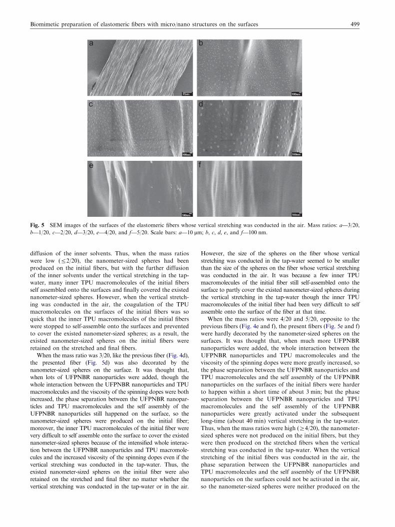
Fig. 5 SEM images of the surfaces of the elastomeric fibers whose vertical stretching was conducted in the air. Mass ratios: a—3/20,
b—1/20, c—2/20, d—3/20, e—4/20, and f—5/20. Scale bars: a—10 mm; b, c, d, e, and f—100 nm.
Biomimetic preparation of elastomeric fibers with micro/nano structures on the surfaces 499
diffusion of the inner solvents. Thus, when the mass ratios
were low (r2/20), the nanometer-sized spheres had been
produced on the initial fibers, but with the further diffusion
of the inner solvents under the vertical stretching in the tap-
water, many inner TPU macromolecules of the initial fibers
self assembled onto the surfaces and finally covered the existed
nanometer-sized spheres. However, when the vertical stretch-
ing was conducted in the air, the coagulation of the TPU
macromolecules on the surfaces of the initial fibers was so
quick that the inner TPU macromolecules of the initial fibers
were stopped to self-assemble onto the surfaces and prevented
to cover the existed nanometer-sized spheres; as a result, the
existed nanometer-sized spheres on the initial fibers were
retained on the stretched and final fibers.
When the mass ratio was 3/20, like the previous fiber (Fig. 4d),
the presented fiber (Fig. 5d) was also decorated by the
nanometer-sized spheres on the surface. It was thought that,
when lots of UFPNBR nanoparticles were added, though the
whole interaction between the UFPNBR nanoparticles and TPU
macromolecules and the viscosity of the spinning dopes were both
increased, the phase separation between the UFPNBR nanopar-
ticles and TPU macromolecules and the self assembly of the
UFPNBR nanoparticles still happened on the surface, so the
nanometer-sized spheres were produced on the initial fiber;
moreover, the inner TPU macromolecules of the initial fiber were
very difficult to self assemble onto the surface to cover the existed
nanometer-sized spheres because of the intensified whole interac-
tion between the UFPNBR nanoparticles and TPU macromole-
cules and the increased viscosity of the spinning dopes even if the
vertical stretching was conducted in the tap-water. Thus, the
existed nanometer-sized spheres on the initial fiber were also
retained on the stretched and final fiber no matter whether the
vertical stretching was conducted in the tap-water or in the air.
However, the size of the spheres on the fiber whose vertical
stretching was conducted in the tap-water seemed to be smaller
than the size of the spheres on the fiber whose vertical stretching
was conducted in the air. It was because a few inner TPU
macromolecules of the initial fiber still self-assembled onto the
surface to partly cover the existed nanometer-sized spheres during
the vertical stretching in the tap-water though the inner TPU
macromolecules of the initial fiber had been very difficult to self
assemble onto the surface of the fiber at that time.
When the mass ratios were 4/20 and 5/20, opposite to the
previous fibers (Fig. 4e and f), the present fibers (Fig. 5e and f)
were hardly decorated by the nanometer-sized spheres on the
surfaces. It was thought that, when much more UFPNBR
nanoparticles were added, the whole interaction between the
UFPNBR nanoparticles and TPU macromolecules and the
viscosity of the spinning dopes were more greatly increased, so
the phase separation between the UFPNBR nanoparticles and
TPU macromolecules and the self assembly of the UFPNBR
nanoparticles on the surfaces of the initial fibers were harder
to happen within a short time of about 3 min; but the phase
separation between the UFPNBR nanoparticles and TPU
macromolecules and the self assembly of the UFPNBR
nanoparticles were greatly activated under the subsequent
long-time (about 40 min) vertical stretching in the tap-water.
Thus, when the mass ratios were high (Z4/20), the nanometer-
sized spheres were not produced on the initial fibers, but they
were then produced on the stretched fibers when the vertical
stretching was conducted in the tap-water. When the vertical
stretching of the initial fibers was conducted in the air, the
phase separation between the UFPNBR nanoparticles and
TPU macromolecules and the self assembly of the UFPNBR
nanoparticles on the surfaces could not be activated in the air,
so the nanometer-sized spheres were neither produced on the

Mass ratio
4/20, 5/20
The initial fiber in 1st stage
1/20 , 2/20
Vertical stretchingin the tap-water
Vertical stretchingin the air
3/20
Vertical stretchingin the tap-water
Vertical stretchingin the air
The stretched fiber in 2nd stage
Drying
FiberGroove
Nanoparticle
Embedded nanoparticle
1/20, 2/20, 3/20
Mass ratio
The final fiber in 3rd stage
Fig. 6 Three-stage formation sketch map of the elastomeric fibers with micro/nano structures on the surfaces in the mass ratios of 1/20,
2/20, 3/20, 4/20, and 5/20.
Q. Liu et al.500
initial fibers nor on the stretched and final fibers. Now, we
could further explain why the size of the nanometer-sized
spheres on the fibers (Fig. 4d–f) became bigger with the
increase of the mass ratio from 3/20 to 5/20. The cause was
that, as the mass ratio increased, the produced nanometer-
sized spheres on the surfaces were more difficult to be further
covered by the inner TPU macromolecules of the fibers during
the vertical stretching in the tap-water.
Based on all the above analysis, the formation sketch map of
the UFPNBR/TPU elastomeric fibers with micro/nano struc-
tures on the surfaces could be described as three stages, as
shown in Fig. 6. In the first stage, the initial fibers were formed
in the tap-water and presented two kinds of micro/nano
structures on the surfaces. When the mass ratios were 1/20,
2/20, and 3/30, the initial fibers were decorated by the micron-
sized grooves and nanometer-sized spheres on the surfaces; but
when the mass ratios were 4/20 and 5/20, the initial fibers were
decorated only by the micron-sized grooves on the surfaces, and
the UFPNBR nanoparticles were almost completely embedded
by the TPU macromolecules. In the second stage, after the
initial fibers received vertical stretching in the tap-water and in
the air, some of the stretched fibers presented the micro/nano
structures different from those of the corresponding initial
fibers. When the vertical stretching was conducted in the tap-
water, the stretched fibers in the mass ratios of 1/20 and 2/20
were only decorated by the micron-sized grooves on the
surfaces, and the existed nanometer-sized spheres on the initial
fibers were completely covered by the TPU macromolecules,
while the stretched fibers in the mass ratios of 3/20, 4/20, and 5/
20 were decorated by both the micron-sized grooves and
nanometer-sized spheres on the surfaces. When the vertical
stretching was conducted in the air, the stretched fibers in the
mass ratios of 1/20, 2/20, and 3/20 were decorated by both
the micron-sized grooves and nanometer-sized spheres on the
surfaces, while the stretched fibers in the mass ratios of 4/20 and
5/20 were only decorated by the micron-sized grooves on the
surfaces because the UFPNBR nanoparticles were still comple-
tely embedded by the TPU macromolecules. In the third stage,
the stretched fibers were dried, and the produced micro/nano
structures including the micron-sized grooves and nanometer-
sized spheres on the stretched fibers were kept and stabilized on
the final fibers. It needed to be emphasized that all the produced
nanometer-sized spheres on the fibers had been partly covered
by the TPU macromolecules.
In summary, the micro/nano structures on the initial fibers
could be controlled by changing the mass ratios of UFPNBR
to TPU. When the micro/nano structures on the initial fibers
had been formed, the micro/nano structures on the final fibers
could be further manipulated by altering the environments of
vertical stretching (in the tap-water or in the air). In the
control of the micro/nano structures on the elastomeric fibers,
it should be specially recognized that the tap-water always
plays a very important role.
4. Conclusions
In this study, the biomimetic preparation of the UFPNBR/
TPU elastomeric fibers was attempted, and three elastomeric
fibers with different micro/nano structures on the surfaces
were finally fabricated. The first fiber was only decorated by
the micron-sized grooves, and the second fiber was dotted by
both the micron-sized grooves and nanometer-sized spheres,
while the third fiber was a helical fiber with concave–convex
structures. The biomimetic preparation of the fibers was
chiefly divided into four steps, a bit different from the three-
step spinning process of spider silks; but the formation
mechanism of the fibers was almost same as that of spider
Biomimetic preparation of elastomeric fibers with micro/nano structures on the surfaces 501
silks, the integrated mechanism of diffusion, coagulation, self
assembly, and microphase separation. The micro/nano struc-
tures on the fibers could be controlled by changing the mass
ratios of UFPNBR to TPU and the environments of vertical
stretching (in the tap-water or in the air). The existed
nanometer-sized spheres on the fibers had been partly covered
by the TPU macromolecules, which suggested that the inter-
action between the nanometer-sized spheres and fiber surfaces
was so strong that the nanometer-sized spheres would be
difficult to be washed away when the fibers were used in the
water. The prepared fibers were hoped, simultaneously like
lotus leave, butterfly wing, and spider silk, to have the
integrate functions of superhydrophobicity, self cleaning,
and mechanical improvement of toughness or strength. For
example, on the one hand, the coexistence of the micron-sized
grooves and nanometer-sized spheres on the surfaces would
endow the fibers and their textiles (being made of the fibers)
with superhydrophobicity, and at the same time the micron-
sized grooves would be helpful to drive the formed water-
drops on the surface to spontaneously roll down along the axis
of the fibers, further resulting in the self cleaning. On the other
hand, the existing UFPNBR nanoparticles in the fibers might
improve the mechanical properties of the fibers such as the
toughness and strength because the UFPNBR nanoparticles
were flexible and presented high specific surface area.
Acknowledgments
This work was supported by the National Science Foundation
for Young Scientists of China (51003003) and the National Basic
Research Program of China (2010CB934700, 2009CB930404,
and 2007CB936403). Authors thank Prof. Jinliang Qiao and
Prof. Xiaohong Zhang for supplying the nanoparticles of
ultrafine full-vulcanized powdered nitrile-butadiene rubbers.
References
[1] L. Eadie, T.K. Ghosh, Journal of Royal Society Interface 8 (59)
(2011) 761–775.
[2] K.S. Liu, L. Jiang, Nano Today 6 (2) (2011) 155–175.
[3] G.M. Luz, J.F. Mano, Composites Science and Technology 70
(13) (2010) 1777–1788.
[4] H.Y. Erbil, A.L. Demirel, Y. Avci, O. Mert, Science 299 (5611)
(2003) 1377–1380.
[5] E. Munch, M.E. Launey, D.H. Alsem, E. Saiz, A.P. Tomsia,
R.O. Ritchie, Science 322 (5907) (2008) 1516–1520.
[6] A. Miserez, S. ScottWasko, C.F. Carpenter, J.H. Waite, Nature
Materials 8 (11) (2009) 910–916.
[7] F. Vollrath, D. Porter, Soft Matter 2 (5) (2006) 377–385.
[8] Q.Y. Liu, L. Jiang, R. Shi, L.Q. Zhang, Progress in Polymer
Science 37 (5) (2012) 715–765.
[9] M. Heim, D. Keerl, T. Scheibel, Angewandte Chemie—
International Edition 48 (20) (2009) 3584–3596.
[10] Y. Liu, Z.Z. Shao, F. Vollrath, Nature Materials 4 (12) (2005)
901–905.
[11] J. Perez-Rigueiro, M. Elices, G.V. Guinea, Polymer 44 (13) (2003)
3733–3736.
[12] O. Emile, A. Le Floch, F. Vollrath, Nature 440 (7084) (2006) 621.
[13] S.P. Kelly, A. Sensenig, K.A. Lorentz, T.A. Blackledge, Zoology
114 (4) (2011) 233–238.
[14] Y.M. Zheng, H. Bai, Z.B. Huang, X.L. Tian, F.Q. Nie, Y. Zhao,
J. Zhai, L. Jiang, Nature 463 (7281) (2010) 640–643.
[15] L. Pelit, F.N. Ertas, A.E. Eroglu, T. Shahwan, H. Tural,
Bioresource Technology 102 (19) (2011) 8807–8813.
[16] R.V. Lewis, Chemical Reviews 106 (9) (2006) 3762–3774.
[17] J.A. Kluge, O. Rabotyagova, G.G. Leisk, D.L. Kaplan, Trends in
Biotechnology 26 (5) (2008) 244–251.
[18] M. Heim, L. Romer, T. Scheibel, Chemical Society Reviews 39
(1) (2010) 156–164.
[19] L. Eisoldt, A. Smith, T. Scheibel, Materials Today 14 (3) (2011)
80–86.
[20] J.G. Hardy, L.M. Romer, T.R. Scheibel, Polymer 49 (20) (2008)
4309–4327.
[21] S. Kubik, Angewandte Chemie-International Edition 41 (15)
(2002) 2721–2723.
[22] F.G. Omenetto, D.L. Kaplan, Science 329 (5991) (2010) 528–531.
[23] Q.Y. Liu, L. Jiang, Chemical Journal of Chinese Universities 31
(6) (2010) 1065–1071.
[24] A.B. Dalton, S. Collins, E. Munoz, J.M. Razal, V.H. Ebron, J.P.
Ferraris, J.N. Coleman, B.G. Kim, R.H. Baughman, Nature 423
(6941) (2003) 703.
[25] S.M. Liff, N. Kumar, G.H. McKinley, Nature Materials 6 (1)
(2007) 76–83.
[26] Q.J. Wu, M. Henriksson, X. Liu, L.A. Berglund, Biomacromo-
lecules 8 (12) (2007) 3687–3692.
[27] P. Podsiadlo, E.M. Arruda, E. Kheng, A.M. Waas, J. Lee, K.
Critchley, M. Qin, E. Chuang, A.K. Kaushik, H.S. Kim, Y. Qi,
S.T. Noh, N.A. Kotov, ACS Nano 3 (6) (2009) 1564–1572.
[28] S.M. Lee, E. Pippel, U. Gosele, C. Dresbach, Y. Qin, C.V.
Chandran, T. Brauniger, G. Hause, M. Knez, Science 324 (5926)
(2009) 488–492.
[29] H. Bai, X.L. Tian, Y.M. Zheng, J. Ju, Y. Zhao, L. Jiang,
Advanced Materials 22 (48) (2010) 5521–5525.
[30] X.L. Tian, H. Bai, Y.M. Zheng, L. Jiang, Advanced Functional
Materials 21 (8) (2011) 1398–1402.
[31] L. Feng, S.H. Li, Y.S. Li, H.J. Li, L.J. Zhang, J. Zhai, Y.L. Song,
B.Q. Liu, L. Jiang, D.B. Zhu, Advanced Materials 14 (24) (2002)
1857–1860.
[32] P. Vukusic, J.R. Sambles, Nature 424 (6950) (2003) 852–855.
[33] F. Vollrath, D.P. Knight, Nature 410 (6828) (2001) 541–548.
[34] Q.Y. Liu, J.Y. Wu, T.W. Tan, L.Q. Zhang, D.F. Chen, W. Tian,
Polymer Degradation and Stability 94 (9) (2009) 1427–1435.
[35] Q.Y. Liu, Q.H. Hu, L.Q. Zhang, Acta Materiae Compositae
Sinica 28 (1) (2011) 1–7.