biomaterials science - tissueeng.nettissueeng.net/lab/papers/structural reinforcement of...
TRANSCRIPT
BiomaterialsScience
PAPER
Cite this: Biomater. Sci., 2014, 2, 703
Received 9th September 2013,Accepted 14th October 2013
DOI: 10.1039/c3bm60210a
www.rsc.org/biomaterialsscience
Structural reinforcement of cell-laden hydrogelswith microfabricated three dimensional scaffolds†
Chaenyung Cha,a,b Pranav Soman,c,e Wei Zhu,c Mehdi Nikkhah,a,b
Gulden Camci-Unal,a,b Shaochen Chen*c and Ali Khademhosseini*a,b,d
Hydrogels commonly used in tissue engineering are mechanically soft and thus often display structural
weakness. Herein, we introduce a strategy for enhancing the structural integrity and fracture toughness of
cell-laden hydrogels by incorporating a three-dimensional (3D) microfabricated scaffold as a structural
element. Digital micromirror device projection printing (DMD-PP) system, a rapid prototyping technology
which employs a layer-by-layer stereolithographic approach, was utilized to efficiently fabricate 3D
scaffolds made from photocrosslinkable poly(ethylene glycol) diacrylate (PEGDA). The scaffold was in-
corporated into a photocrosslinkable gelatin hydrogel by placing it in a pre-gel solution, and inducing
in situ hydrogel formation. The resulting scaffold-reinforced hydrogels demonstrated a significant
increase in ultimate stress and provided structural support for mechanically weak hydrogels. In addition,
the scaffold did not affect the rigidity of hydrogels, as it was not involved in the crosslinking reaction to
form the hydrogel. Therefore, the presented approach could avoid inadvertent and undesired changes in
the hydrogel rigidity which is a known regulator of cellular activities. Furthermore, the biocompatibility of
scaffold-reinforced hydrogels was confirmed by evaluating the viability and proliferation of encapsulated
fibroblasts. Overall, the strategy of incorporating 3D scaffolds into hydrogels as structural reinforcements
presented in this study will be highly useful for enhancing the mechanical toughness of hydrogels for
various tissue engineering applications.
1. Introduction
Hydrogels are widely used as scaffold materials for tissueengineering applications, because their structure, a crosslinkednetwork of polymers with high fluid content and elasticity,closely mimics native extracellular matrices (ECM) and thereforeprovides a suitable microenvironment for cells and tissues.1–4
Various strategies have been employed to control the biochemi-cal and mechanical properties of the hydrogels. For example,ECM proteins (e.g. collagen, laminin and fibronectin)5–7 or their
functional peptide sequences (e.g. RGD peptide)8–10 are chemi-cally incorporated into hydrogels to induce cell adhesion to thehydrogel surface. The rigidity of hydrogels is often modulatedby controlling the crosslinking density.11,12
In order to emulate the natural biomechanical environmentof the cells, the hydrogel rigidity is often controlled to matchthe inherent softness of native ECM.13–15 However, due to thestructural weakness of hydrogels, they are easily broken andoften display a high degree of swelling. As a result, handlinghydrogels becomes challenging, and their original structure anddimensions often do not remain intact over time. There arevarious reinforcement strategies to improve the toughness of thehydrogels. For example, a secondary polymeric network is intro-duced to strengthen the hydrogels (e.g. formation of interpene-trating networks).16,17 In addition, nanostructures areincorporated into the polymeric network to create compositehydrogels (e.g. clay, minerals, polymeric and metal nano-spheres).18,19 However, these approaches often result in changesin rigidity and diffusion properties of hydrogels, which influencethe cellular phenotypes.20,21 Similarly, it has been shown thatnanostructures could elicit non-specific responses from cells.22
Therefore, it is desirable to employ a strategy that only enhancesthe structural integrity and fracture resistance of hydrogelswithout inadvertently influencing their cell responsiveness.
†Electronic supplementary information (ESI) available: Supporting figuresassociated with this article are available in ESI. See DOI: 10.1039/c3bm60210a
aDivision of Biomedical Engineering, Department of Medicine, Brigham and Women’s
Hospital, Harvard Medical School, Cambridge, MA 02139, USA.
E-mail: [email protected]; Fax: +1-617-768-8202; Tel: +1-617-388-9271bHarvard-MIT Division of Health Sciences and Technology, Massachusetts Institute
of Technology, Cambridge, MA 02139, USAcDepartment of Nanoengineering, University of California, San Diego, La Jolla,
CA 92093, USA. E-mail: [email protected]; Fax: +1-858-534-9553;
Tel: +1-858-822-7856dWyss Institute for Biologically Inspired Engineering, Harvard University, Boston,
MA 02115, USAeDepartment of Biomedical and Chemical Engineering, Syracuse University, Syracuse,
NY 13244, USA
This journal is © The Royal Society of Chemistry 2014 Biomater. Sci., 2014, 2, 703–709 | 703
Publ
ishe
d on
13
Nov
embe
r 20
13. D
ownl
oade
d on
10/
08/2
015
18:4
0:48
.
View Article OnlineView Journal | View Issue
Here, we present an approach to enhance the structuralintegrity and toughness of hydrogels by introducing a 3D poly-meric scaffold designed to act as a structural framework toreinforce the hydrogels. This approach was inspired by theendoskeletal system of vertebrate species, which has evolvednot only to provide structural support and protection for tissuestructures, but also to guide their overall shape.23 Therefore,we hypothesized that the presence of a solid scaffold wouldsupport the structural integrity and increase the fracturestrength of soft cell-laden hydrogels without affecting theirrigidity. The scaffold made of poly(ethylene glycol)diacrylate(PEGDA) was developed by digital micromirror device projec-tion printing (DMD-PP), a rapid prototyping stereolithographytechnique which allows for a highly efficient fabrication ofthree dimensional (3D) structures in micro-scale dimensions.24–27
The concentration of PEGDA was varied to control the flexi-bility of the scaffolds. Then, the scaffold was immersed in apre-gel solution containing methacrylated gelatin (GelMA) anda photoinitiator, followed by UV irradiation to fabricate thescaffold-reinforced hydrogels. The mechanical properties of thescaffold-reinforced GelMA hydrogels were evaluated by measur-ing elastic moduli and ultimate stress, and were comparedwith those of pure GelMA hydrogels to evaluate the reinforcingeffect of the PEGDA scaffold. Furthermore, fibroblasts wereencapsulated within the scaffold-reinforced hydrogels andtheir viability and proliferation were evaluated to assess theeffect of scaffolds on the cellular viability and proliferation.
2. Experimental section2.1. Synthesis of methacrylated gelatin (GelMA)
Gelatin (10 g, Sigma Aldrich) and 4-dimethylaminopyridine(0.5 g, Sigma Aldrich) were dissolved in dimethyl sulfoxide(90 mL, Fisher) at 50 °C. Then, glycidyl methacrylate (4 mL,Sigma Aldrich) was slowly added to the solution, and themixture was continuously stirred at 50 °C for 48 hours underdry N2. The mixture was dialyzed against deionized (DI) water,and lyophilized to obtain the product. The chemical conju-gation of methacrylate to gelatin was confirmed by 1H-NMR(ESI Fig. 1†).
2.2. Fabrication of PEGDA scaffolds with digital micromirrordevice projection printing (DMD-PP)
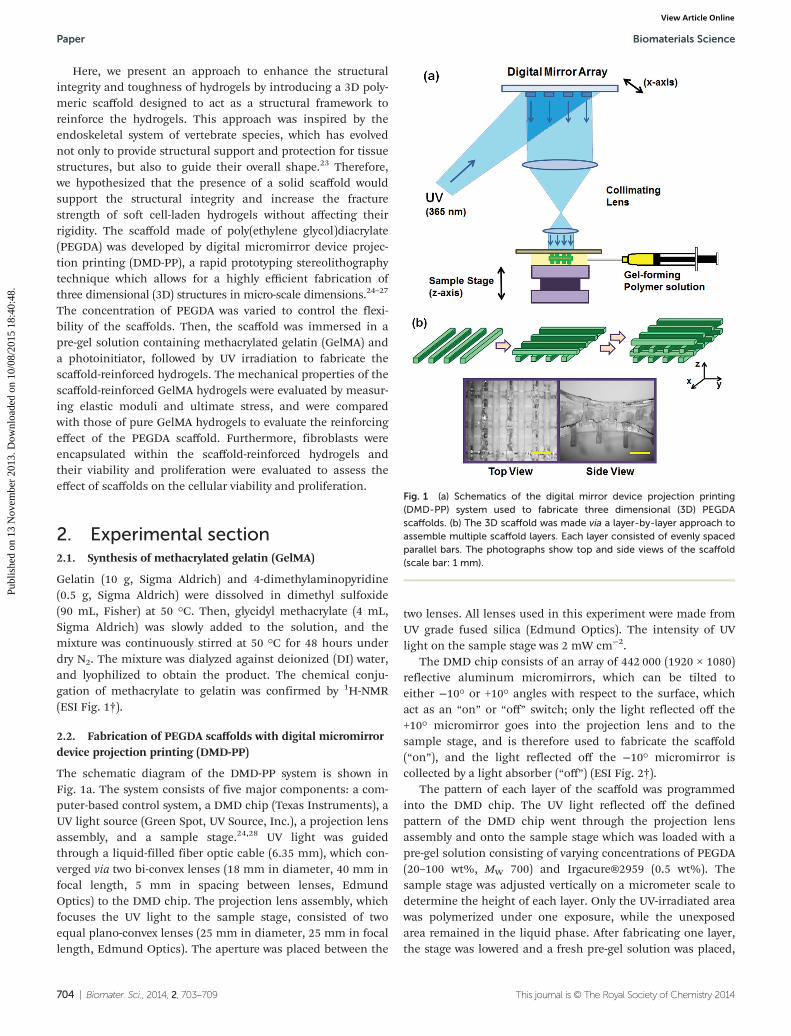
The schematic diagram of the DMD-PP system is shown inFig. 1a. The system consists of five major components: a com-puter-based control system, a DMD chip (Texas Instruments), aUV light source (Green Spot, UV Source, Inc.), a projection lensassembly, and a sample stage.24,28 UV light was guidedthrough a liquid-filled fiber optic cable (6.35 mm), which con-verged via two bi-convex lenses (18 mm in diameter, 40 mm infocal length, 5 mm in spacing between lenses, EdmundOptics) to the DMD chip. The projection lens assembly, whichfocuses the UV light to the sample stage, consisted of twoequal plano-convex lenses (25 mm in diameter, 25 mm in focallength, Edmund Optics). The aperture was placed between the
two lenses. All lenses used in this experiment were made fromUV grade fused silica (Edmund Optics). The intensity of UVlight on the sample stage was 2 mW cm−2.
The DMD chip consists of an array of 442 000 (1920 × 1080)reflective aluminum micromirrors, which can be tilted toeither −10° or +10° angles with respect to the surface, whichact as an “on” or “off” switch; only the light reflected off the+10° micromirror goes into the projection lens and to thesample stage, and is therefore used to fabricate the scaffold(“on”), and the light reflected off the −10° micromirror iscollected by a light absorber (“off”) (ESI Fig. 2†).
The pattern of each layer of the scaffold was programmedinto the DMD chip. The UV light reflected off the definedpattern of the DMD chip went through the projection lensassembly and onto the sample stage which was loaded with apre-gel solution consisting of varying concentrations of PEGDA(20–100 wt%, MW 700) and Irgacure®2959 (0.5 wt%). Thesample stage was adjusted vertically on a micrometer scale todetermine the height of each layer. Only the UV-irradiated areawas polymerized under one exposure, while the unexposedarea remained in the liquid phase. After fabricating one layer,the stage was lowered and a fresh pre-gel solution was placed,
Fig. 1 (a) Schematics of the digital mirror device projection printing(DMD-PP) system used to fabricate three dimensional (3D) PEGDAscaffolds. (b) The 3D scaffold was made via a layer-by-layer approach toassemble multiple scaffold layers. Each layer consisted of evenly spacedparallel bars. The photographs show top and side views of the scaffold(scale bar: 1 mm).
Paper Biomaterials Science
704 | Biomater. Sci., 2014, 2, 703–709 This journal is © The Royal Society of Chemistry 2014
Publ
ishe
d on
13
Nov
embe
r 20
13. D
ownl
oade
d on
10/
08/2
015
18:4
0:48
. View Article Online
followed by UV exposure to fabricate the next layer on top ofthe previous one. These steps were repeated sequentially todevelop the desired 3D scaffold.
2.3. Fabrication of scaffold-reinforced hydrogels
The scaffold was first placed in a custom-made mold madefrom PDMS elastomer (Sylgard® 184, Dow Corning). The pre-gel solution containing GelMA (5–10 wt%) and Irgacure® 2959(0.1 wt%) was placed in the mold. The solution readily pene-trated into the scaffold, and covered the entire mold. Then, UVirradiation was applied to induce polymerization to formhydrogels (2 minutes, output power of 4.8 mW cm−2, Omni-Cure® S2000) (ESI Fig. 3†). The resulting scaffold-reinforcedhydrogel was taken out of the mold, and placed in phosphatebuffered saline (PBS, pH 7.4) for further characterization.The overall dimensions of the scaffold-reinforced hydrogelswere 8 mm × 8 mm × 2.5 mm. The GelMA hydrogel withoutthe scaffold was also fabricated as a control.
Scanning electron microscopy (SEM) was used to character-ize the detailed morphology of the scaffold-reinforced hydro-gels. A sample was frozen in liquid nitrogen, and fractured toexpose the cross-section. Then the sample was dried vialyophilization, sputter-coated with gold (2 nm thickness, IBS/TM200S, VCR Group, Inc.), and visualized under a SEM(Quanta 200 FEG, FEI™) under high vacuum.
2.4. Evaluation of mechanical properties
The mechanical properties of the scaffolds, hydrogels, andscaffold-reinforced hydrogels were evaluated by measuringstress–strain curves via uniaxial compression at the rate of1 mm min−1 until they were completely fractured, using amechanical testing system (Model 5943, Instron®).12,29 Theelastic modulus of each sample was calculated from the slopeof a stress–strain curve at the first 10% strain where the curvewas linear. Ultimate stress was determined as the maximumstress before the scaffold-reinforced hydrogel became fractured.
Cyclic uniaxial compression tests were performed on thePEGDA scaffolds to further characterize their mechanicalstrengths. Briefly, each scaffold was compressed (‘loading’)and decompressed (‘unloading’) at the rate of 1 mm min−1 for5 times continuously, and the stress–strain curve for bothloading and unloading was recorded (Model 5943, Instron®).
2.5. Cell studies
NIH-3T3 fibroblasts were suspended in the pre-gel solution(cell density: 2 × 10⁶ cells per mL). Then, the scaffold-reinforced hydrogel was prepared as described above to encap-sulate the cells. The constructs were incubated in the cell-culture media (Dulbecco’s modified Eagle medium sup-plemented with 10% fetal bovine serum and 1% penicillin/streptomycin, all purchased from Invitrogen) at 37 °C with 5%CO2 throughout the culture period.
To determine the viability of encapsulated cells, eachsample was taken at a designated time point and the cellswere fluorescently labeled with calcein-AM and ethidiumhomodimer-1 to identify live (green fluorescence) and dead
(red fluorescence) cells, respectively (LIVE/DEAD® Viability/Cytotoxicity Assay kit, Invitrogen), and then visualized using afluorescence microscope (Eclipse Ti, Nikon). The viability wasreported as the percentage of live cells from the total numberof cells. To evaluate the actin cytoskeleton organization of cellswithin the hydrogel constructs, the cells were stained withAlexa Fluor®488-phalloidin (Invitrogen) and visualized usingthe fluorescence microscope.
3. Results and discussion3.1. Microfabrication of PEGDA scaffolds
DMD-PP was used to fabricate 3D scaffolds which would beused as the structural framework for hydrogels (Fig. 1a). Thisrapid prototyping stereolithographic method utilizes a DMDchip which allows for the fabrication of micropatternedscaffolds by controlling the photocrosslinkable area withswitchable micromirrors (ESI Fig. 2†). Each UV exposurereflected from the DMD chip to a gel-forming solution resultsin a single layer of scaffold. This photocrosslinking step wasrepeated on top of the previous layer to ultimately fabricate amulti-layered 3D scaffold. By controlling the DMD chip toadjust the photocrosslinked area, the architecture of thescaffold could be easily controlled on a micrometer scale.Herein, the scaffold made of photocrosslinked PEGDA con-sisted of four layers, each consisting of evenly spaced (1 mm)parallel bars having 200 μm width, 400 μm height, and 7 mmlength (Fig. 1b). Each layer was aligned perpendicular to theprevious one, so the inner space of the scaffold was connectedthroughout the structure; this ensures that the pre-gel solutioncan penetrate into the entire scaffold.
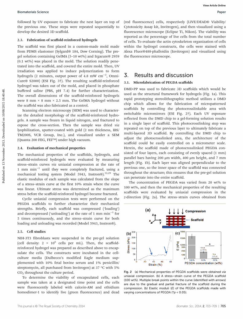
The concentration of PEGDA was varied from 20 wt% to100 wt%, and then the mechanical properties of the resultingscaffolds were evaluated by uniaxial compression in thez-direction (Fig. 2a). The stress–strain curves obtained from
Fig. 2 (a) Mechanical properties of PEGDA scaffolds were obtained viauniaxial compression. (b) A stress–strain curve of the PEGDA scaffold(100 wt%). Multiple break points within the curve (identified with arrows)are due to the gradual and partial fracture of the scaffold during thecompression. (b) Elastic moduli (E) of the PEGDA scaffolds made withvarying concentrations of PEGDA (*p < 0.05).
Biomaterials Science Paper
This journal is © The Royal Society of Chemistry 2014 Biomater. Sci., 2014, 2, 703–709 | 705
Publ
ishe
d on
13
Nov
embe
r 20
13. D
ownl
oade
d on
10/
08/2
015
18:4
0:48
. View Article Online
the uniaxial compression showed several breaks with increas-ing strain, due to gradual and partial breakage of the scaffolds(Fig. 2b, ESI Fig. 4†). Elastic modulus, which was determinedfrom the slope of the initial linear region of the stress–straincurve, could be controlled in a wide range from 15.5 kPa to1.6 MPa, demonstrating that the rigidity of the scaffold couldbe conveniently controlled by concentration of gel-formingpolymers (Fig. 2c). Similar stress–strain profiles were obtainedwhen the scaffolds were compressed in the y-direction (ESIFig. 5†). However, the ultimate stress values for more flexible50 wt% and 20 wt% PEGDA scaffolds were higher than thoseobtained in the z-direction, likely due to the enhanced abilityof the scaffold to bend along the longer axis (y-direction) thanthe shorter axis (z-direction).
The mechanical strengths of the PEGDA scaffolds werefurther characterized by cyclic uniaxial compression (Fig. 3).The scaffolds were subjected to continuous loading andunloading for 5 cycles, and their stress–strain curves wereobtained. There were no significant losses in stress valueswhen they were repeatedly compressed below the initial breakstrain (<15%, Fig. 3a–c (left)). Even when they were compressedbeyond their initial break strain, there were only smalldecreases in stress values (Fig. 3a–c (right)). These resultsfurther demonstrated their mechanical strengths and theirefficacy as structural elements to reinforce soft hydrogels.
3.2. Fabrication of scaffold-reinforced hydrogels
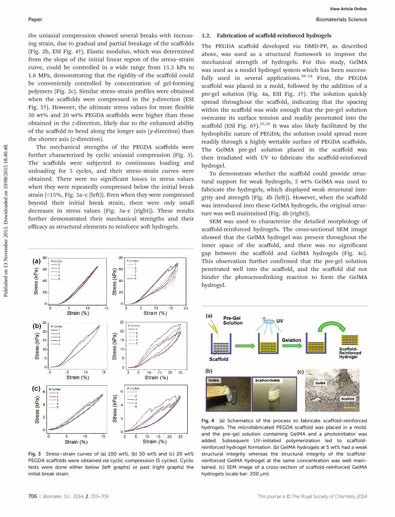
The PEGDA scaffold developed via DMD-PP, as describedabove, was used as a structural framework to improve themechanical strength of hydrogels. For this study, GelMAwas used as a model hydrogel system which has been success-fully used in several applications.30–34 First, the PEGDAscaffold was placed in a mold, followed by the addition of apre-gel solution (Fig. 4a, ESI Fig. 3†). The solution quicklyspread throughout the scaffold, indicating that the spacingwithin the scaffold was wide enough that the pre-gel solutionovercame its surface tension and readily penetrated into thescaffold (ESI Fig. 6†).35,36 It was also likely facilitated by thehydrophilic nature of PEGDA; the solution could spread morereadily through a highly wettable surface of PEGDA scaffolds.The GelMA pre-gel solution placed in the scaffold wasthen irradiated with UV to fabricate the scaffold-reinforcedhydrogel.
To demonstrate whether the scaffold could provide struc-tural support for weak hydrogels, 5 wt% GelMA was used tofabricate the hydrogels, which displayed weak structural inte-grity and strength (Fig. 4b (left)). However, when the scaffoldwas introduced into these GelMA hydrogels, the original struc-ture was well maintained (Fig. 4b (right)).
SEM was used to characterize the detailed morphology ofscaffold-reinforced hydrogels. The cross-sectional SEM imageshowed that the GelMA hydrogel was present throughout theinner space of the scaffold, and there was no significantgap between the scaffold and GelMA hydrogels (Fig. 4c).This observation further confirmed that the pre-gel solutionpenetrated well into the scaffold, and the scaffold did nothinder the photocrosslinking reaction to form the GelMAhydrogel.
Fig. 3 Stress–strain curves of (a) 100 wt%, (b) 50 wt% and (c) 20 wt%PEGDA scaffolds were obtained via cyclic compression (5 cycles). Cyclictests were done either below (left graphs) or past (right graphs) theinitial break strain.
Fig. 4 (a) Schematics of the process to fabricate scaffold-reinforcedhydrogels. The microfabricated PEGDA scaffold was placed in a mold,and the pre-gel solution containing GelMA and a photoinitiator wasadded. Subsequent UV-initiated polymerization led to scaffold-reinforced hydrogel formation. (b) GelMA hydrogels at 5 wt% had a weakstructural integrity whereas the structural integrity of the scaffold-reinforced GelMA hydrogel at the same concentration was well main-tained. (c) SEM image of a cross-section of scaffold-reinforced GelMAhydrogels (scale bar: 200 μm).
Paper Biomaterials Science
706 | Biomater. Sci., 2014, 2, 703–709 This journal is © The Royal Society of Chemistry 2014
Publ
ishe
d on
13
Nov
embe
r 20
13. D
ownl
oade
d on
10/
08/2
015
18:4
0:48
. View Article Online
3.3. Mechanical properties of scaffold-reinforced hydrogels
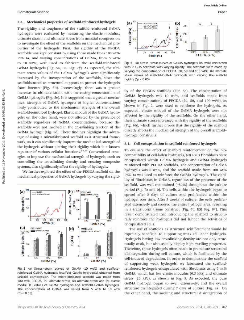
The rigidity and toughness of the scaffold-reinforced GelMAhydrogels were evaluated by measuring the elastic modulus,ultimate strain, and ultimate stress from uniaxial compressionto investigate the effect of the scaffolds on the mechanical pro-perties of the hydrogels. First, the rigidity of the PEGDAscaffolds was kept constant by using those made from 100 wt%PEGDA, and varying concentrations of GelMA, from 5 wt%to 10 wt%, were used to fabricate the scaffold-reinforcedGelMA hydrogels (Fig. 5a, ESI Fig. 7†). As expected, the ulti-mate stress values of the GelMA hydrogels were significantlyincreased by the incorporation of the scaffolds, since thescaffolds acted as structural supports to protect the hydrogelsfrom fracture (Fig. 5b). Interestingly, there was a greaterincrease in ultimate strain with increasing concentration ofGelMA hydrogels (Fig. 5c). It is suggested that a greater mecha-nical strength of GelMA hydrogels at higher concentrationslikely contributed to the mechanical strength of the overallscaffold-reinforced hydrogel. Elastic moduli of the GelMA hydro-gels, on the other hand, were not affected by the presence ofscaffolds regardless of GelMA concentrations, because thescaffolds were not involved in the crosslinking reaction of theGelMA hydrogel (Fig. 5d). These findings highlight the advan-tage of using a microfabricated scaffold as a structural frame-work, as it can significantly improve the mechanical strength ofthe hydrogels without altering their rigidity which is a knownregulator of various cellular functions.14,15 Conventional strat-egies to improve the mechanical strength of hydrogels, such ascontrolling the crosslinking density and creating compositesystems, also significantly affect the rigidity of hydrogels.
We further explored the effect of the PEGDA scaffold on themechanical properties of GelMA hydrogels by varying the rigid-
ity of the PEGDA scaffolds (Fig. 6a). The concentration ofGelMA hydrogels was 10 wt%, and scaffolds made fromvarying concentrations of PEGDA (20, 50, and 100 wt%), asshown in Fig. 2, were used to reinforce the hydrogels. Asexpected, elastic moduli of the GelMA hydrogels were notaffected by the rigidity of the scaffolds. On the other hand,their ultimate stress increased with the rigidity of the scaffolds(Fig. 6b), which further proves that the rigidity of the scaffolddirectly affects the mechanical strength of the overall scaffold–hydrogel constructs.
3.4. Cell encapsulation in scaffold-reinforced hydrogels
To evaluate the effect of scaffold reinforcement on the bio-compatibility of cell-laden hydrogels, NIH-3T3 fibroblasts wereencapsulated within GelMA hydrogels and GelMA hydrogelsreinforced with PEGDA scaffolds. The concentration of GelMAhydrogels was 8 wt%, and the scaffold made from 100 wt%PEGDA was used to reinforce the GelMA hydrogels. The viabi-lity of fibroblasts in GelMA, regardless of the presence of thescaffold, was well maintained (>80%) throughout the cultureperiod (Fig. 7a and b). The cells within the hydrogels began tospread after 3 days of culture and proliferated within thehydrogel over time. After 3 weeks of culture, the cells prolifer-ated extensively and covered the entire hydrogel area, resultingin a translucent tissue construct (Fig. 7c, ESI Fig. 8†). Thisresult demonstrated that introducing the scaffold to structu-rally reinforce the hydrogels did not hinder the activities ofencapsulated cells.
The use of scaffolds as structural reinforcement would beespecially beneficial to supporting weak cell-laden hydrogels.Hydrogels having low crosslinking density are not only struc-turally weak, but also usually display high swelling properties.Therefore, those hydrogels often result in premature structuraldisintegration during cell culture, which is facilitated by thecell-induced degradation. In order to demonstrate the scaffoldof supporting weak hydrogels, we fabricated the scaffold-reinforced hydrogels encapsulated with fibroblasts using 5 wt%GelMA, which has low elastic modulus (0.3 kPa) and ultimatestress (20 kPa), as shown in Fig. 5. As expected, the pureGelMA hydrogel began to swell extensively, and the overallstructure disintegrated during 7 days of culture (Fig. 8a). Onthe other hand, the swelling and structural disintegration of
Fig. 5 (a) Stress–strain curves of GelMA (10 wt%) and scaffold-reinforced GelMA hydrogels (scaffold-GelMA hydrogels) obtained fromuniaxial compression. The microfabricated scaffold was made from100 wt% PEGDA. (b) Ultimate stress, (c) ultimate strain and (d) elasticmoduli (E) values of GelMA hydrogels and scaffold-GelMA hydrogels.The concentration of GelMA was varied from 5 wt% to 10 wt%(*p < 0.05).
Fig. 6 (a) Stress–strain curves of GelMA hydrogels (10 wt%) reinforcedwith PEGDA scaffolds with varying rigidity. The scaffolds were made byvarying the concentration of PEGDA (20, 50 and 100 wt%). (b) Ultimatestress values of scaffold-GelMA hydrogels with varying the scaffoldrigidity (*p < 0.05).
Biomaterials Science Paper
This journal is © The Royal Society of Chemistry 2014 Biomater. Sci., 2014, 2, 703–709 | 707
Publ
ishe
d on
13
Nov
embe
r 20
13. D
ownl
oade
d on
10/
08/2
015
18:4
0:48
. View Article Online
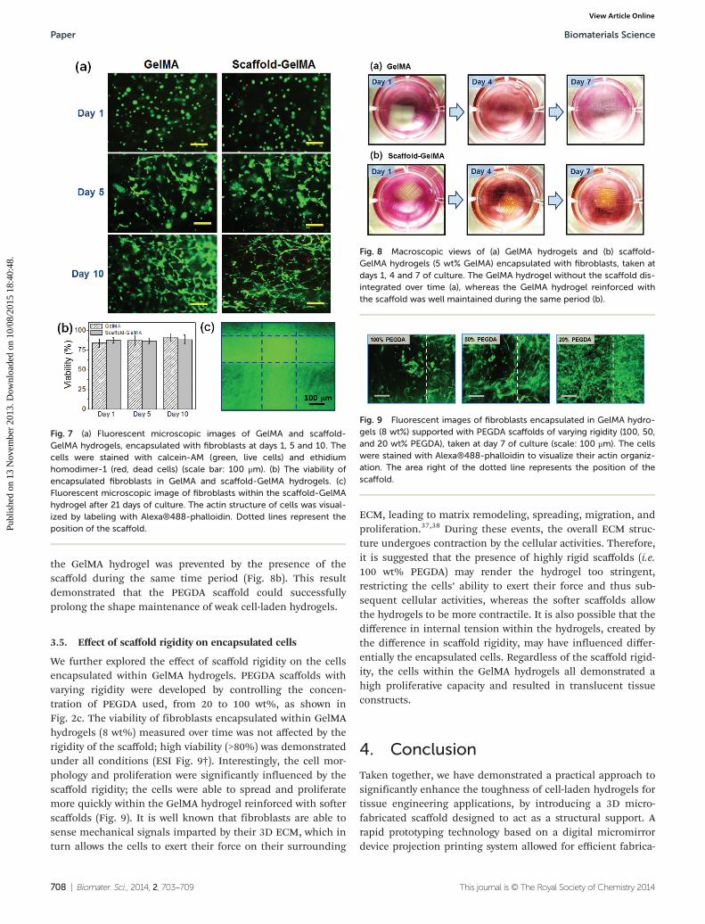
the GelMA hydrogel was prevented by the presence of thescaffold during the same time period (Fig. 8b). This resultdemonstrated that the PEGDA scaffold could successfullyprolong the shape maintenance of weak cell-laden hydrogels.
3.5. Effect of scaffold rigidity on encapsulated cells
We further explored the effect of scaffold rigidity on the cellsencapsulated within GelMA hydrogels. PEGDA scaffolds withvarying rigidity were developed by controlling the concen-tration of PEGDA used, from 20 to 100 wt%, as shown inFig. 2c. The viability of fibroblasts encapsulated within GelMAhydrogels (8 wt%) measured over time was not affected by therigidity of the scaffold; high viability (>80%) was demonstratedunder all conditions (ESI Fig. 9†). Interestingly, the cell mor-phology and proliferation were significantly influenced by thescaffold rigidity; the cells were able to spread and proliferatemore quickly within the GelMA hydrogel reinforced with softerscaffolds (Fig. 9). It is well known that fibroblasts are able tosense mechanical signals imparted by their 3D ECM, which inturn allows the cells to exert their force on their surrounding
ECM, leading to matrix remodeling, spreading, migration, andproliferation.37,38 During these events, the overall ECM struc-ture undergoes contraction by the cellular activities. Therefore,it is suggested that the presence of highly rigid scaffolds (i.e.100 wt% PEGDA) may render the hydrogel too stringent,restricting the cells’ ability to exert their force and thus sub-sequent cellular activities, whereas the softer scaffolds allowthe hydrogels to be more contractile. It is also possible that thedifference in internal tension within the hydrogels, created bythe difference in scaffold rigidity, may have influenced differ-entially the encapsulated cells. Regardless of the scaffold rigid-ity, the cells within the GelMA hydrogels all demonstrated ahigh proliferative capacity and resulted in translucent tissueconstructs.
4. Conclusion
Taken together, we have demonstrated a practical approach tosignificantly enhance the toughness of cell-laden hydrogels fortissue engineering applications, by introducing a 3D micro-fabricated scaffold designed to act as a structural support. Arapid prototyping technology based on a digital micromirrordevice projection printing system allowed for efficient fabrica-
Fig. 7 (a) Fluorescent microscopic images of GelMA and scaffold-GelMA hydrogels, encapsulated with fibroblasts at days 1, 5 and 10. Thecells were stained with calcein-AM (green, live cells) and ethidiumhomodimer-1 (red, dead cells) (scale bar: 100 μm). (b) The viability ofencapsulated fibroblasts in GelMA and scaffold-GelMA hydrogels. (c)Fluorescent microscopic image of fibroblasts within the scaffold-GelMAhydrogel after 21 days of culture. The actin structure of cells was visual-ized by labeling with Alexa®488-phalloidin. Dotted lines represent theposition of the scaffold.
Fig. 8 Macroscopic views of (a) GelMA hydrogels and (b) scaffold-GelMA hydrogels (5 wt% GelMA) encapsulated with fibroblasts, taken atdays 1, 4 and 7 of culture. The GelMA hydrogel without the scaffold dis-integrated over time (a), whereas the GelMA hydrogel reinforced withthe scaffold was well maintained during the same period (b).
Fig. 9 Fluorescent images of fibroblasts encapsulated in GelMA hydro-gels (8 wt%) supported with PEGDA scaffolds of varying rigidity (100, 50,and 20 wt% PEGDA), taken at day 7 of culture (scale: 100 μm). The cellswere stained with Alexa®488-phalloidin to visualize their actin organiz-ation. The area right of the dotted line represents the position of thescaffold.
Paper Biomaterials Science
708 | Biomater. Sci., 2014, 2, 703–709 This journal is © The Royal Society of Chemistry 2014
Publ
ishe
d on
13
Nov
embe
r 20
13. D
ownl
oade
d on
10/
08/2
015
18:4
0:48
. View Article Online
tion of 3D scaffolds in microscale dimensions. The scaffold-reinforced hydrogels demonstrated significantly enhancedstructural integrity and fracture toughness of the hydrogelwithout inadvertently affecting the rigidity of the hydrogel, aknown regulator of cell behavior. In addition, the biocompat-ibility of scaffold-reinforced hydrogels was confirmed by evalu-ating the viability and proliferation of encapsulated cells.Therefore, we expect that the strategy of using 3D scaffolds forhydrogel reinforcement will be highly useful for significantlyenhancing the mechanical strength of cell-laden hydrogels forvarious tissue engineering applications.
Acknowledgements
The work was supported by funds from the National ScienceFoundation (CAREER: DMR 0847287), the Presidential EarlyCareer Award for Scientists and Engineers (PECASE), and theNational Institutes of Health (HL092836, DE021468, AR05837,EB012597, and HL099073).
Notes and references
1 N. A. Peppas, J. Z. Hilt, A. Khademhosseini and R. Langer,Adv. Mater., 2006, 18, 1345.
2 B. V. Slaughter, S. S. Khurshid, O. Z. Fisher,A. Khademhosseini and N. A. Peppas, Adv. Mater., 2009, 21,3307.
3 N. Annabi, J. W. Nichol, X. Zhong, C. Ji, S. Koshy,A. Khademhosseini and F. Dehghani, Tissue Eng. Part B,2012, 16, 371.
4 A. S. Hoffman, Adv. Drug Delivery Rev., 2002, 54, 3.5 C. R. Nuttelman, D. J. Mortisen, S. M. Henry and
K. S. Anseth, J. Biomed. Mater. Res., 2001, 57, 217.6 M. Rafat, F. Li, P. Fagerholm, N. S. Lagali, M. A. Watsky,
R. Munger, T. Matsuura and M. Griffith, Biomaterials, 2008,29, 3960.
7 S. Hou, Q. Xu, W. Tian, F. Cui, Q. Cai, J. Ma and I.-S. Lee,J. Neurosci. Methods, 2005, 148, 60.
8 D. L. Hern and J. A. Hubbell, J. Biomed. Mater. Res., 1998,39, 266.
9 F. Yang, C. G. Williams, D.-A. Wang, H. Lee, P. N. Mansonand J. Elisseeff, Biomaterials, 2005, 26, 5991.
10 U. Hersel, C. Dahmen and H. Kessler, Biomaterials, 2003,24, 4385.
11 K. Y. Lee, J. A. Rowley, P. Eiselt, E. M. Moy, K. H. Bouhadirand D. J. Mooney, Macromolecules, 2000, 33, 4291.
12 C. Cha, R. H. Kohman and H. Kong, Adv. Funct. Mater.,2009, 19, 3056.
13 G. D. Prestwich, Organogenesis, 2008, 4, 42.14 B. Bhana, R. K. Iyer, W. L. K. Chen, R. Zhao, K. L. Sider,
M. Likhitpanichkul, C. A. Simmons and M. Radisic, Bio-technol. Bioeng., 2010, 105, 1148.
15 A. J. Engler, S. Sen, H. L. Sweeney and D. E. Discher, Cell,2006, 126, 677.
16 J.-Y. Sun, X. Zhao, W. R. K. Illeperuma, O. Chaudhuri,K. H. Oh, D. J. Mooney, J. J. Vlassak and Z. Suo, Nature,2012, 489, 133.
17 H. Shin, B. D. Olsen and A. Khademhosseini, Biomaterials,2012, 33, 3143.
18 P. Schexnailder and G. Schmidt, Colloid Polym. Sci., 2009,287, 1.
19 A. K. Gaharwar, S. M. Mihaila, A. Swami, A. Patel, S. Sant,R. L. Reis, A. P. Marques, M. E. Gomes andA. Khademhosseini, Adv. Mater., 2013, 25, 3329.
20 E. C. Muniz and G. Geuskens, Macromolecules, 2001, 34,4480.
21 K. Haraguchi and T. Takehisa, Adv. Mater., 2002, 14, 1120.22 K. Unfried, C. Albrecht, L.-O. Klotz, A. V. Mikecz,
S. Grether-Beck and R. P. F. Schins, Nanotoxicology, 2007,1, 52.
23 L. Gilbert, The Skeletal System, The Rosen PublishingGroup, New York, 2001.
24 Y. Lu, G. Mapili, G. Suhali, S. Chen and K. Roy, J. Biomed.Mater. Res. A, 2006, 77A, 396.
25 D. Y. Fozdar, P. Soman, J. W. Lee, L.-H. Han and S. Chen,Adv. Funct. Mater., 2011, 21, 2712.
26 P. Soman, J. A. Kelber, J. W. Lee, T. N. Wright,K. S. Vecchio, R. L. Klemke and S. Chen, Biomaterials, 2012,33, 7064.
27 P. Zorlutuna, N. Annabi, G. Camci-Unal, M. Nikkhah,J. M. Cha, J. W. Nichol, A. Manbachi, H. Bae, S. Chen andA. Khademhosseini, Adv. Mater., 2012, 24, 1782.
28 P. Soman, B. D. Tobe, J. Lee, A. M. Winquist, I. Singec,K. Vecchio, E. Snyder and S. Chen, Biomed. Microdevices,2012, 14, 829.
29 C. Cha, J. H. Jeong, J. Shim and H. Kong, Acta Biomater.,2011, 7, 3719.
30 J. W. Nichol, S. T. Koshy, H. Bae, C. M. Hwang, S. Yamanlarand A. Khademhosseini, Biomaterials, 2010, 31, 5536.
31 Y.-C. Chen, R.-Z. Lin, H. Qi, Y. Yang, H. Bae, J. M. Melero-Martin and A. Khademhosseini, Adv. Funct. Mater., 2012,22, 2027.
32 M. Nikkhah, N. Eshak, P. Zorlutuna, N. Annabi,M. Castello, K. Kim, A. Dolatshahi-Pirouz, F. Edalat,H. Bae, Y. Yang and A. Khademhosseini, Biomaterials,2012, 33, 9009.
33 R.-Z. Lin, Y.-C. Chen, R. Moreno-Luna, A. Khademhosseiniand J. M. Melero-Martin, Biomaterials, 2013, 34, 6785.
34 M. Kharaziha, M. Nikkhah, S.-R. Shin, N. Annabi,N. Masoumi, A. K. Gaharwar, G. Camci-Unal andA. Khademhosseini, Biomaterials, 2013, 34, 6355.
35 C. P. Huang, J. Lu, H. Seon, A. P. Lee, L. A. Flanagan,H.-Y. Kim, A. J. Putnam and N. L. Jeon, Lab Chip, 2009, 9,1740.
36 H. Cho, H.-Y. Kim, J. Y. Kang and T. S. Kim, J. Colloid Inter-face Sci., 2007, 306, 379.
37 E. Cukierman, R. Pankov and K. M. Yamada, Curr. Opin.Cell Biol., 2002, 14, 633.
38 F. Grinnell, Trends Cell Biol., 2003, 13, 264.
Biomaterials Science Paper
This journal is © The Royal Society of Chemistry 2014 Biomater. Sci., 2014, 2, 703–709 | 709
Publ
ishe
d on
13
Nov
embe
r 20
13. D
ownl
oade
d on
10/
08/2
015
18:4
0:48
. View Article Online