biokimia darah
DESCRIPTION
frTRANSCRIPT

BLOOD BLOOD BIOCHEMISTRYBIOCHEMISTRY
Disusun Oleh:Disusun Oleh:
dr. Husnil Kadri, M.Kesdr. Husnil Kadri, M.Kes
Biochemistry Departement Biochemistry Departement
Medical Faculty Of Andalas University Medical Faculty Of Andalas University
PadangPadang

BIOSINTESIS HEMOGLOBIN(PORFIRIN)
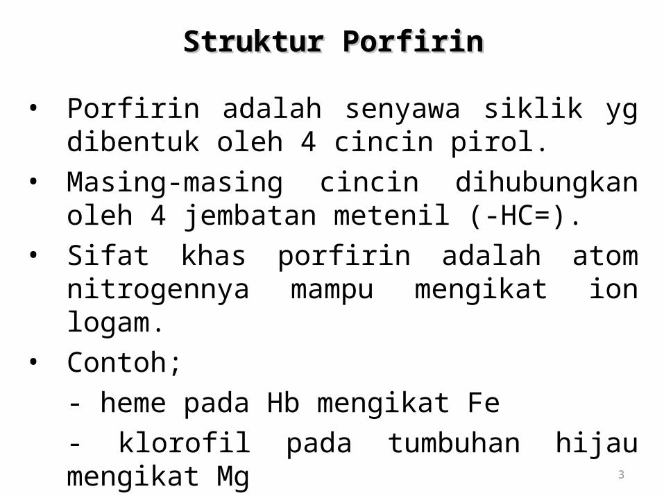
Struktur PorfirinStruktur Porfirin
• Porfirin adalah senyawa siklik yg dibentuk oleh 4 cincin pirol.
• Masing-masing cincin dihubungkan oleh 4 jembatan metenil (-HC=).
• Sifat khas porfirin adalah atom nitrogennya mampu mengikat ion logam.
• Contoh; - heme pada Hb mengikat Fe- klorofil pada tumbuhan hijau mengikat Mg
3
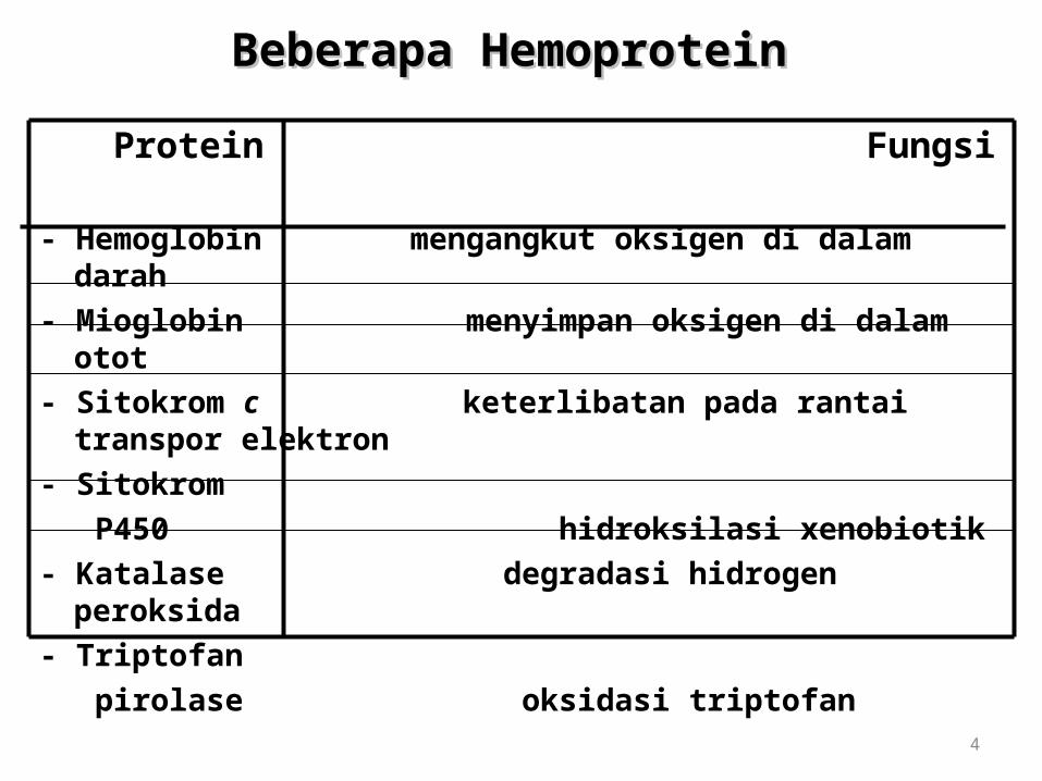
Beberapa Hemoprotein Beberapa Hemoprotein
Protein Fungsi
- Hemoglobin mengangkut oksigen di dalam darah- Mioglobin menyimpan oksigen di dalam otot- Sitokrom c keterlibatan pada rantai transpor elektron- Sitokrom P450 hidroksilasi xenobiotik- Katalase degradasi hidrogen peroksida- Triptofan pirolase oksidasi triptofan
4

Sintesis Heme di MitokondriaSintesis Heme di Mitokondria
• 85% sintesis heme terjadi dalam sel pembentuk eritrosit pada sumsum tulang
• Heme disintesis dari suksinil KoA + glisin.• Piridoksal fosfat diperlukan untuk mengaktifkan
glisin.
5

Sintesis Heme di Mitokondria Sintesis Heme di Mitokondria
6

Pengaturan Sintesis Heme Pengaturan Sintesis Heme
• Enzim regulator adalah ALA-sintase.
• Heme bertindak sebagai regulator negatif (umpan balik negatif) sintesis enzim ALA- sintase.
• Jika heme meningkat, maka sintesis ALA-sintase akan menurun.
7

PorfiriaPorfiria
• Merupakan gangguan genetik biosintesis heme.• Umumnya autosomal dominan, kecuali porfiria
eritropoitik kongenital.• Gejala;
- nyeri abdomen- gangguan neuropsikiatri- fotosensitifitas kulit- bila berat = prototipe manusia srigala
8

Fungsi Utama DarahFungsi Utama Darah
1. Respirasi;
pengangkutan O2 dan CO2
2. Nutrisi; pengangkutan hasil absorpsi usus
3. Ekskresi; pengangkutan sisa metabolik ke ginjal,
paru-paru, kulit, & usus
Fungsi Utama DarahFungsi Utama Darah
4. Keseimbangan asam-basa
5. Keseimbangan air; antara sirkulasi darah dan jaringan
6. Pengaturan suhu tubuh
7. Pertahanan terhadap infeksi; oleh sel darah putih & antibodi

Fungsi Utama DarahFungsi Utama Darah
8. Pengangkutan hormon &
pengaturan metabolisme
9. Pengangkutan metabolit
10. Koagulasi

Components of Whole Blood
Withdraw blood and place in tube
1 2 Centrifuge
Plasma(55% of whole blood)
Formed elements
Buffy coat:leukocyctes and platelets(<1% of whole blood)
Erythrocytes(45% of whole blood)
• Hematocrit • Males: 47% ± 5%
• Females: 42% ± 5%
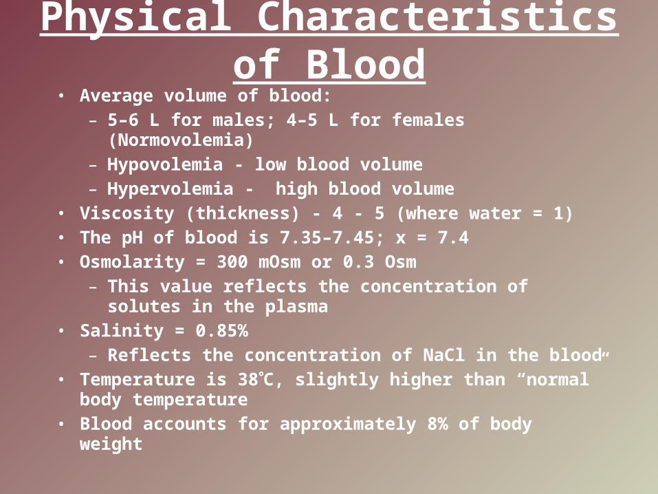
Physical Characteristics of Blood
• Average volume of blood:– 5–6 L for males; 4–5 L for females (Normovolemia)– Hypovolemia - low blood volume– Hypervolemia - high blood volume
• Viscosity (thickness) - 4 - 5 (where water = 1)• The pH of blood is 7.35–7.45; x = 7.4• Osmolarity = 300 mOsm or 0.3 Osm
– This value reflects the concentration of solutes in the plasma
• Salinity = 0.85%– Reflects the concentration of NaCl in the blood
• Temperature is 38C, slightly higher than “normal” body temperature
• Blood accounts for approximately 8% of body weight
Components of Blood
–55% plasma
– 45% cells
• 99% RBCs
• < 1% WBCs and platelets
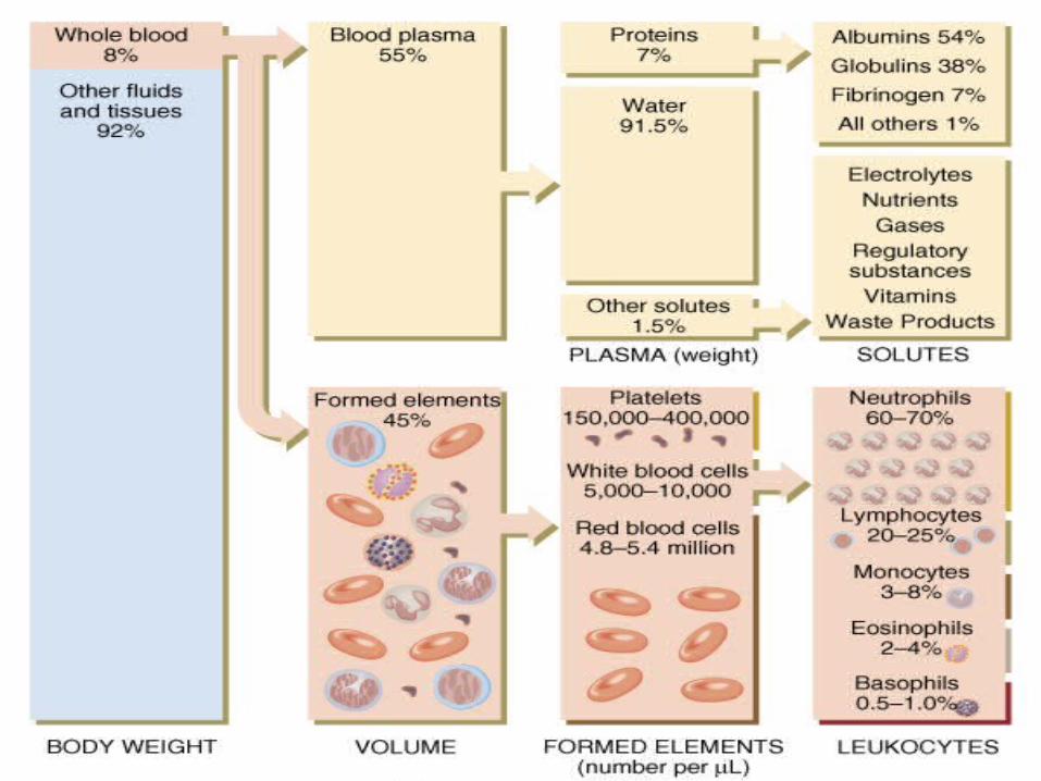

Blood Plasma
• Blood plasma components:– Water = 90-92%– Proteins = 6-8%– Organic nutrients – glucose, carbohydrates,
amino acids– Electrolytes – sodium, potassium, calcium,
chloride, bicarbonate – Nonprotein nitrogenous substances – lactic acid,
urea, creatinine– Respiratory gases – oxygen and carbon dioxide
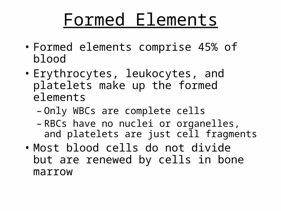
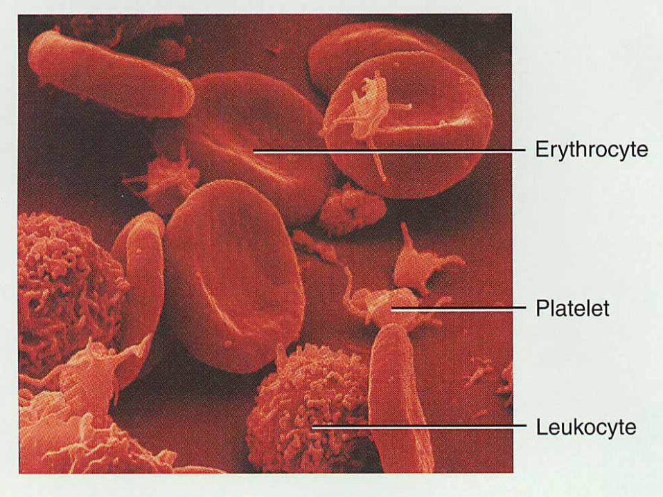
Formed Elements
• Formed elements comprise 45% of blood
• Erythrocytes, leukocytes, and platelets make up the formed elements– Only WBCs are complete cells– RBCs have no nuclei or organelles, and
platelets are just cell fragments
• Most blood cells do not divide but are renewed by cells in bone marrow

Erythrocytes (RBCs)• Biconcave disc
– Folding increases surface area (30% more surface area)– Plasma membrane contains spectrin
• Give erythrocytes their flexibility• Anucleate, no centrioles, no organelles
– End result - no cell division– No mitochondria means they generate ATP anaerobically
• Prevents consumption of O2 being transported
• Filled with hemoglobin (Hb) - 97% of cell contents– Hb functions in gas transport
• Hb + O2 HbO2 (oxyhemoglobin)• Most numerous of the formed elements
– Females: 4.3–5.2 million cells/cubic millimeter– Males: 5.2–5.8 million cells/cubic millimeter

Erythrocytes (RBCs)
Figure 17.3
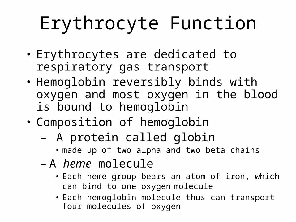
Erythrocyte Function
• Erythrocytes are dedicated to respiratory gas transport
• Hemoglobin reversibly binds with oxygen and most oxygen in the blood is bound to hemoglobin
• Composition of hemoglobin– A protein called globin
• made up of two alpha and two beta chains
– A heme molecule• Each heme group bears an atom of iron, which can bind to
one oxygen molecule• Each hemoglobin molecule thus can transport four
molecules of oxygen
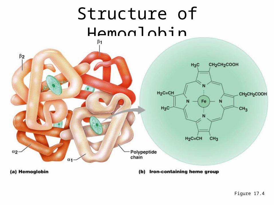
Structure of Hemoglobin
Figure 17.4
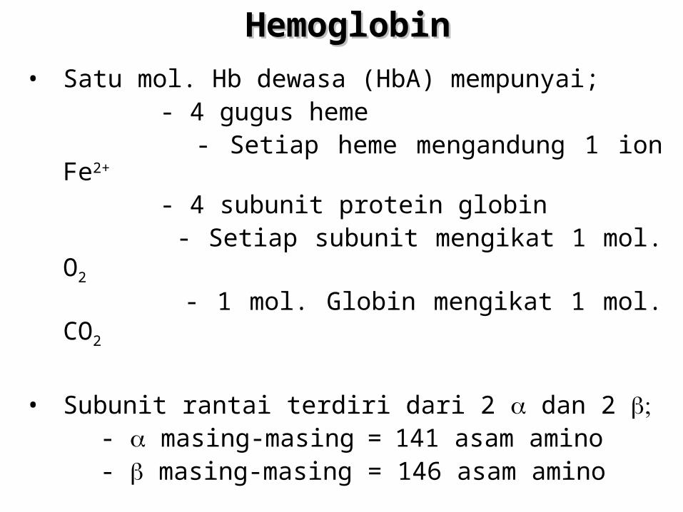
HemoglobinHemoglobin
• Satu mol. Hb dewasa (HbA) mempunyai; - 4 gugus heme - Setiap heme mengandung 1 ion Fe2+
- 4 subunit protein globin
- Setiap subunit mengikat 1 mol. O2
- 1 mol. Globin mengikat 1 mol. CO2
• Subunit rantai terdiri dari 2 dan 2 - masing-masing=141 asam amino - masing-masing = 146 asam amino
HemoglobinHemoglobin
• Oxyhemoglobin – hemoglobin bound to oxygen– Oxygen loading takes place in the lungs
• Deoxyhemoglobin – hemoglobin after oxygen diffuses into tissues (reduced Hb)
• Carbaminohemoglobin – hemoglobin bound to carbon dioxide
– Carbon dioxide loading takes place in the tissues
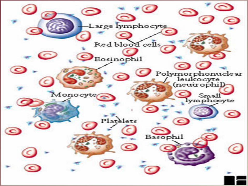
WBC Anatomy and Types
• All WBCs (leukocytes) have a nucleus and no hemoglobin
• Granular or agranular classification based on presence of cytoplasmic granules made visible by staining– granulocytes are neutrophils, eosinophils or
basophils– agranulocytes are monocyes or lymphocytes
Differential WBC Count
• Detection of changes in numbers of circulating WBCs (percentages of each type)– indicates infection, poisoning, leukemia,
chemotherapy, parasites or allergy reaction
• Normal WBC counts– neutrophils 60-70% (up if bacterial infection)– lymphocyte 20-25% (up if viral infection)– monocytes 3 - 8 % (up if fungal/viral infection)– eosinophil 2 - 4 % (up if parasite or allergy reaction)– basophil <1% (up if allergy reaction or hypothyroid)

Neutrophils (Granulocyte)
• Polymorphonuclear Leukocytes or Polys• Nuclei = 2 to 5 lobes connected by thin strands
– older cells have more lobes– young cells called band cells because of
horseshoe shaped nucleus (band)
• Fine, pale lilac practically invisible granules • Diameter is 10-12 microns • 60 to 70% of circulating WBCs

Eosinophils (Granulocyte)
• Nucleus with 2 or 3 lobes connected by a thin strand
• Large, uniform-sized granules stain orange-red with acidic dyes– do not obscure the nucleus
• Diameter is 10 to 12 microns
• 2 to 4% of circulating WBCs

Basophils (Granulocyte)
• Large, dark purple, variable-sized granules stain with basic dyes– obscure the nucleus
• Irregular, s-shaped, bilobed nuclei
• Diameter is 8 to 10 microns
• Less than 1% of circulating WBCs
Lymphocyte (Agranulocyte)
• Dark, oval to round nucleus
• Cytoplasm sky blue in color– amount varies from rim of blue to normal
amount
• Small cells 6 - 9 microns in diameter
• Large cells 10 - 14 microns in diameter– increase in number during viral infections
• 20 to 25% of circulating WBCs

LymphocytesLymphocytes
– B cells - responsible for humoral immunity– T cells - responsible for cell mediated
immunity
• B cells responsible for production of antibodies – Receptor matches antigen– Cells multiply– Antibodies
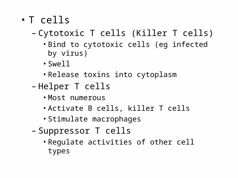
• T cells– Cytotoxic T cells (Killer T cells)
• Bind to cytotoxic cells (eg infected by virus)• Swell• Release toxins into cytoplasm
– Helper T cells• Most numerous• Activate B cells, killer T cells• Stimulate macrophages
– Suppressor T cells• Regulate activities of other cell types

Monocyte (Agranulocyte)• Nucleus is kidney or horse-shoe shaped• Largest WBC in circulating blood
– does not remain in blood long before migrating to the tissues– differentiate into macrophages
• fixed group found in specific tissues– alveolar macrophages in lungs– kupffer cells in liver
• wandering group gathers at sites of infection
• Diameter is 12 - 20 microns• Cytoplasm is a foamy blue-gray • 3 to 8% o circulating WBCs
UNSUR SELULAR DALAM RESPON IMUNUNSUR SELULAR DALAM RESPON IMUN
1. Jalur limfoid yang membentuk limfosit dan subsetnya
2. Jalur mieloid yang membentuk sel-sel fagosit mononuklear & polimorfonuklear (PMN).
PMN terdiri dari:
neutrofil, eosinofil, basofil

• Platelets are fragments of mega-karyocytes
• Platelets function in the clotting mechanism by forming a temporary plug that helps seal breaks in blood vessels
Platelets
Protein PlasmaProtein Plasma
- Bagian utama unsur padat dalam plasma.
- Konsentrasi total protein plasma + 7-7,5 g/dl.
- Berbagai protein plasma dapat dipisahkan menurut karakteristik kelarutannya.
- Metode pemisahan tsb antara lain;
1. Salting-out (Na2SO4 23%, dll)
2. Elektroforesis
Protein PlasmaProtein Plasma
1. Sebagian besar disintesis di hepar.2. Umumnya disintesis sbg preprotein pada
poliribosom terikat membran. Preprotein akan mengalami modifikasi pascatranslasi.
3. Hampir semuanya berupa glikoprotein, kecuali albumin.
4. Bersifat polimorfisme (ciri bawaan pd populasi dgn sedikitnya 2 macam fenotipe).contoh; gol. Darah ABO
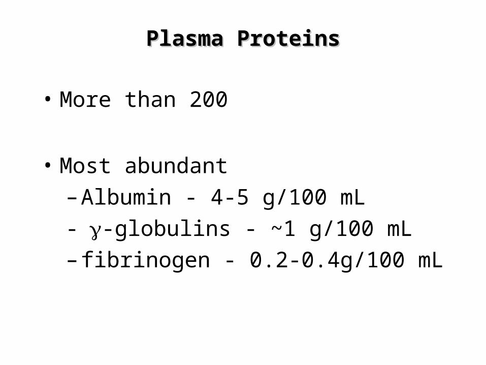
Plasma ProteinsPlasma Proteins
• More than 200
• Most abundant
– Albumin - 4-5 g/100 mL
--globulins - ~1 g/100 mL
– fibrinogen - 0.2-0.4g/100 mL

AlbuminAlbumin
- Merupakan protein utama dalam plasma.
- Mempertahankan 75-80% tekanan osmotik.
- Berfungsi mengikat berbagai macam ligand, seperti; asam lemak bebas, Ca, Cu, Zn, hormon steroid, bilirubin, metheme

AlbuminAlbumin
- Albumin juga dapat mengikat obat-an,
seperti; sulfonamid, penisilin-G,
dikumarol, aspirin
- Penyakit hepar akan memperlihatkan rasio albumin/globulin yang menurun.

TransferinTransferin
• Adalah 1-globulin berbentuk glikoprotein yang disintesis di hepar.
• Berfungsi sebagai alat transpor besi (Fe3+) untuk dibawa ke jaringan.
• Jika besi tidak diikat oleh transferin, maka akan menjadi prooksidan.
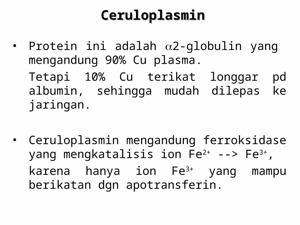
CeruloplasminCeruloplasmin
• Protein ini adalah 2-globulin yang mengandung 90% Cu plasma.
Tetapi 10% Cu terikat longgar pd albumin, sehingga mudah dilepas ke jaringan.
• Ceruloplasmin mengandung ferroksidase yang mengkatalisis ion Fe2+ --> Fe3+,
karena hanya ion Fe3+ yang mampu berikatan dgn apotransferin.
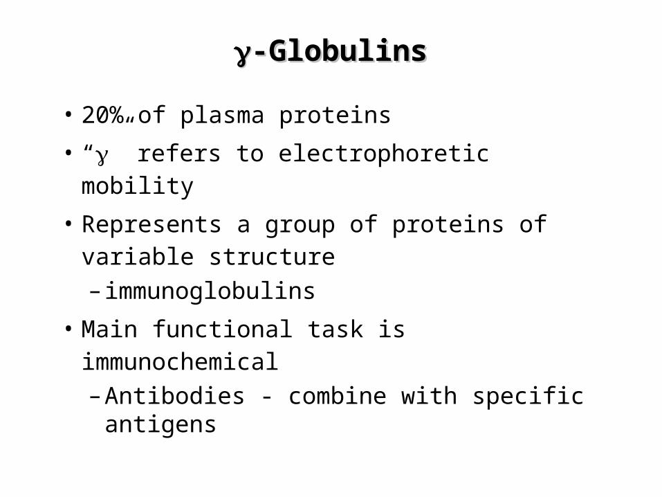
-Globulins-Globulins
• 20% of plasma proteins
• “” refers to electrophoretic mobility
• Represents a group of proteins of variable structure
– immunoglobulins
• Main functional task is immunochemical
– Antibodies - combine with specific antigens

Imunoglobulin PlasmaImunoglobulin Plasma
• Disintesis dalam sel plasma.
• Sel plasma adalah turunan Sel- yang mensintesis dan mensekresikan imuno- globulin sebagai respon terhadap pajanan berbagai antigen.
• Semua imunoglobulin mengandung paling kurang 2 rantai ringan dan 2 rantai berat.
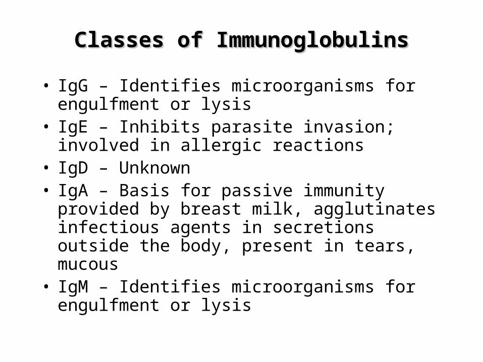
Classes of ImmunoglobulinsClasses of Immunoglobulins
• IgG – Identifies microorganisms for engulfment or lysis
• IgE – Inhibits parasite invasion; involved in allergic reactions
• IgD – Unknown• IgA – Basis for passive immunity provided by
breast milk, agglutinates infectious agents in secretions outside the body, present in tears, mucous
• IgM – Identifies microorganisms for engulfment or lysis
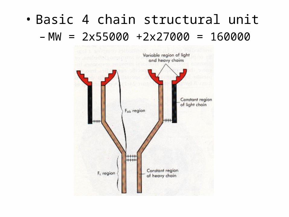
• Basic 4 chain structural unit– MW = 2x55000 +2x27000 = 160000


HaptoglobinHaptoglobin
• Merupakan glikoprotein plasma yang mengikat hemoglobin ekstrakorpuskular.
• Membentuk komplek Hb-Hp (Hemoglobin-Haptoglobin).
• Hb ekstrakorpuskular merupakan hasil penguraian + 10% Hb yang dilepas ke dlm sirkulasi.

Kepustakaan
• Marks, DB., Marks, AD., Smith CM. 1996. Basic medical biochemistry: a clinical approach. Dalam: B.U. Pendit, penerjemah. Biokimia Kedokteran Dasar: Sebuah Pendekatan Klinis. Eds. J. Suyono., V. Sadikin., L.I. Mandera. Jakarta: EGC, 2000: 612 - 4.
• Murray, RK. 2003. Porfirin dan pigmen empedu. Dalam: Andry Hartono, penerjemah. Harper’s Biochemistry. 25th ed. Eds. R.K. Murray, D.K. Granner, P.A. Mayes, V.W. Rodwell. McGraw-Hill Companies, New York: 342 - 9.
• Schumm, DE. 1992. Essentials of biochemistry. Dalam: Moch. Sadikin, penerjemah. Intisari Biokimia. Jakarta: Bina Aksara, 1993: 147.
50
KepustakaanKepustakaan
• Harbut, C. 150 Blood. Download 19-10-2010. http://www.cerritos.edu/charbut/AP150/lec_otl/150%20Blood.ppt
• Rand, ML., Murray, RK. 2003. Protein plasma, imunoglobulin, dan pembekuan darah. Dalam: Andry Hartono, penerjemah. Harper’s Biochemistry. 25th ed. Eds. R.K. Murray, D.K. Granner, P.A. Mayes, V.W. Rodwell. McGraw-Hill Companies, New York: 702 - 11.
• Simpson, S. Chapter 19 Blood. Download 19-10-2010.
• Sheardown, H. Blood Biochemistry. McMaster University. Download 20-05-2007.