biochemistry and evolution of anaerobic energy …involved in fes cluster biogenesis (162, 199). in...
TRANSCRIPT
Biochemistry and Evolution of Anaerobic Energy Metabolism in Eukaryotes
Miklós Müller,a Marek Mentel,b Jaap J. van Hellemond,c Katrin Henze,d Christian Woehle,d Sven B. Gould,d Re-Young Yu,d
Mark van der Giezen,e Aloysius G. M. Tielens,c and William F. Martind
The Rockefeller University, New York, New York, USAa; Department of Biochemistry, Faculty of Natural Sciences, Comenius University, Bratislava, Slovakiab; Department of
Medical Microbiology and Infectious Diseases, Erasmus University Medical Center, Rotterdam, Netherlandsc; Institute of Molecular Evolution, University of Düsseldorf,
Düsseldorf, Germanyd; and Biosciences, College of Life and Environmental Sciences, University of Exeter, Exeter, United Kingdome
INTRODUCTION . . . . . . . . . . . . . . . . . . . . . . . . . . . . . . . . . . . . . . . . . . . . . . . . . . . . . . . . . . . . . . . . . . . . . . . . . . . . . . . . . . . . . . . . . . . . . . . . . . . . . . . . . . . . . . . . . . . . . . . . . . . . . . . . . . . . . . . . . . . .444A FEW DISTINCTIONS . . . . . . . . . . . . . . . . . . . . . . . . . . . . . . . . . . . . . . . . . . . . . . . . . . . . . . . . . . . . . . . . . . . . . . . . . . . . . . . . . . . . . . . . . . . . . . . . . . . . . . . . . . . . . . . . . . . . . . . . . . . . . . . . . . . . . .447
Functional and Environmental Anaerobiosis . . . . . . . . . . . . . . . . . . . . . . . . . . . . . . . . . . . . . . . . . . . . . . . . . . . . . . . . . . . . . . . . . . . . . . . . . . . . . . . . . . . . . . . . . . . . . . . . . . . . . . . . . . . . .447Redox Balance through Respiration and Fermentation . . . . . . . . . . . . . . . . . . . . . . . . . . . . . . . . . . . . . . . . . . . . . . . . . . . . . . . . . . . . . . . . . . . . . . . . . . . . . . . . . . . . . . . . . . . . . . . . . .448Anaerobes and Microaerophiles: Redox Balance with a Pinch of O2 . . . . . . . . . . . . . . . . . . . . . . . . . . . . . . . . . . . . . . . . . . . . . . . . . . . . . . . . . . . . . . . . . . . . . . . . . . . . . . . . . . . . .449
METABOLIC PATHWAYS IN EUKARYOTIC ANAEROBES . . . . . . . . . . . . . . . . . . . . . . . . . . . . . . . . . . . . . . . . . . . . . . . . . . . . . . . . . . . . . . . . . . . . . . . . . . . . . . . . . . . . . . . . . . . . . . . . . . .450Animals . . . . . . . . . . . . . . . . . . . . . . . . . . . . . . . . . . . . . . . . . . . . . . . . . . . . . . . . . . . . . . . . . . . . . . . . . . . . . . . . . . . . . . . . . . . . . . . . . . . . . . . . . . . . . . . . . . . . . . . . . . . . . . . . . . . . . . . . . . . . . . . . . . .450
Fasciola hepatica (liver fluke) . . . . . . . . . . . . . . . . . . . . . . . . . . . . . . . . . . . . . . . . . . . . . . . . . . . . . . . . . . . . . . . . . . . . . . . . . . . . . . . . . . . . . . . . . . . . . . . . . . . . . . . . . . . . . . . . . . . . . . . . . . .450Ascaris (giant roundworm). . . . . . . . . . . . . . . . . . . . . . . . . . . . . . . . . . . . . . . . . . . . . . . . . . . . . . . . . . . . . . . . . . . . . . . . . . . . . . . . . . . . . . . . . . . . . . . . . . . . . . . . . . . . . . . . . . . . . . . . . . . . .453Mytilus edulis (common mussel) . . . . . . . . . . . . . . . . . . . . . . . . . . . . . . . . . . . . . . . . . . . . . . . . . . . . . . . . . . . . . . . . . . . . . . . . . . . . . . . . . . . . . . . . . . . . . . . . . . . . . . . . . . . . . . . . . . . . . . .453Arenicola marina (lugworm) . . . . . . . . . . . . . . . . . . . . . . . . . . . . . . . . . . . . . . . . . . . . . . . . . . . . . . . . . . . . . . . . . . . . . . . . . . . . . . . . . . . . . . . . . . . . . . . . . . . . . . . . . . . . . . . . . . . . . . . . . . .454Sipunculus nudus (peanut worm) . . . . . . . . . . . . . . . . . . . . . . . . . . . . . . . . . . . . . . . . . . . . . . . . . . . . . . . . . . . . . . . . . . . . . . . . . . . . . . . . . . . . . . . . . . . . . . . . . . . . . . . . . . . . . . . . . . . . . .455A strictly anoxic animal among the loriciferans . . . . . . . . . . . . . . . . . . . . . . . . . . . . . . . . . . . . . . . . . . . . . . . . . . . . . . . . . . . . . . . . . . . . . . . . . . . . . . . . . . . . . . . . . . . . . . . . . . . . . . . .457
Fungi . . . . . . . . . . . . . . . . . . . . . . . . . . . . . . . . . . . . . . . . . . . . . . . . . . . . . . . . . . . . . . . . . . . . . . . . . . . . . . . . . . . . . . . . . . . . . . . . . . . . . . . . . . . . . . . . . . . . . . . . . . . . . . . . . . . . . . . . . . . . . . . . . . . . .457Piromyces sp. strain E2 and Neocallimastix . . . . . . . . . . . . . . . . . . . . . . . . . . . . . . . . . . . . . . . . . . . . . . . . . . . . . . . . . . . . . . . . . . . . . . . . . . . . . . . . . . . . . . . . . . . . . . . . . . . . . . . . . . . . .457Fusarium oxysporum . . . . . . . . . . . . . . . . . . . . . . . . . . . . . . . . . . . . . . . . . . . . . . . . . . . . . . . . . . . . . . . . . . . . . . . . . . . . . . . . . . . . . . . . . . . . . . . . . . . . . . . . . . . . . . . . . . . . . . . . . . . . . . . . . . .458Microsporidia . . . . . . . . . . . . . . . . . . . . . . . . . . . . . . . . . . . . . . . . . . . . . . . . . . . . . . . . . . . . . . . . . . . . . . . . . . . . . . . . . . . . . . . . . . . . . . . . . . . . . . . . . . . . . . . . . . . . . . . . . . . . . . . . . . . . . . . . . .459
Amoebozoa: Entamoeba histolytica . . . . . . . . . . . . . . . . . . . . . . . . . . . . . . . . . . . . . . . . . . . . . . . . . . . . . . . . . . . . . . . . . . . . . . . . . . . . . . . . . . . . . . . . . . . . . . . . . . . . . . . . . . . . . . . . . . . . . .459Excavate Taxa. . . . . . . . . . . . . . . . . . . . . . . . . . . . . . . . . . . . . . . . . . . . . . . . . . . . . . . . . . . . . . . . . . . . . . . . . . . . . . . . . . . . . . . . . . . . . . . . . . . . . . . . . . . . . . . . . . . . . . . . . . . . . . . . . . . . . . . . . . . . .461
Giardia intestinalis . . . . . . . . . . . . . . . . . . . . . . . . . . . . . . . . . . . . . . . . . . . . . . . . . . . . . . . . . . . . . . . . . . . . . . . . . . . . . . . . . . . . . . . . . . . . . . . . . . . . . . . . . . . . . . . . . . . . . . . . . . . . . . . . . . . . . .461Trichomonas vaginalis . . . . . . . . . . . . . . . . . . . . . . . . . . . . . . . . . . . . . . . . . . . . . . . . . . . . . . . . . . . . . . . . . . . . . . . . . . . . . . . . . . . . . . . . . . . . . . . . . . . . . . . . . . . . . . . . . . . . . . . . . . . . . . . . . .462Tritrichomonas foetus . . . . . . . . . . . . . . . . . . . . . . . . . . . . . . . . . . . . . . . . . . . . . . . . . . . . . . . . . . . . . . . . . . . . . . . . . . . . . . . . . . . . . . . . . . . . . . . . . . . . . . . . . . . . . . . . . . . . . . . . . . . . . . . . . .464Trypanosoma brucei . . . . . . . . . . . . . . . . . . . . . . . . . . . . . . . . . . . . . . . . . . . . . . . . . . . . . . . . . . . . . . . . . . . . . . . . . . . . . . . . . . . . . . . . . . . . . . . . . . . . . . . . . . . . . . . . . . . . . . . . . . . . . . . . . . . .464Euglena gracilis . . . . . . . . . . . . . . . . . . . . . . . . . . . . . . . . . . . . . . . . . . . . . . . . . . . . . . . . . . . . . . . . . . . . . . . . . . . . . . . . . . . . . . . . . . . . . . . . . . . . . . . . . . . . . . . . . . . . . . . . . . . . . . . . . . . . . . . . .467Excavate pathogens and metronidazole . . . . . . . . . . . . . . . . . . . . . . . . . . . . . . . . . . . . . . . . . . . . . . . . . . . . . . . . . . . . . . . . . . . . . . . . . . . . . . . . . . . . . . . . . . . . . . . . . . . . . . . . . . . . . .469
Alveolates and Stramenopiles . . . . . . . . . . . . . . . . . . . . . . . . . . . . . . . . . . . . . . . . . . . . . . . . . . . . . . . . . . . . . . . . . . . . . . . . . . . . . . . . . . . . . . . . . . . . . . . . . . . . . . . . . . . . . . . . . . . . . . . . . . . .469Nyctotherus ovalis . . . . . . . . . . . . . . . . . . . . . . . . . . . . . . . . . . . . . . . . . . . . . . . . . . . . . . . . . . . . . . . . . . . . . . . . . . . . . . . . . . . . . . . . . . . . . . . . . . . . . . . . . . . . . . . . . . . . . . . . . . . . . . . . . . . . . .469Blastocystis . . . . . . . . . . . . . . . . . . . . . . . . . . . . . . . . . . . . . . . . . . . . . . . . . . . . . . . . . . . . . . . . . . . . . . . . . . . . . . . . . . . . . . . . . . . . . . . . . . . . . . . . . . . . . . . . . . . . . . . . . . . . . . . . . . . . . . . . . . . . .469
Rhizaria and Denitrification . . . . . . . . . . . . . . . . . . . . . . . . . . . . . . . . . . . . . . . . . . . . . . . . . . . . . . . . . . . . . . . . . . . . . . . . . . . . . . . . . . . . . . . . . . . . . . . . . . . . . . . . . . . . . . . . . . . . . . . . . . . . . . .472Archaeplastida. . . . . . . . . . . . . . . . . . . . . . . . . . . . . . . . . . . . . . . . . . . . . . . . . . . . . . . . . . . . . . . . . . . . . . . . . . . . . . . . . . . . . . . . . . . . . . . . . . . . . . . . . . . . . . . . . . . . . . . . . . . . . . . . . . . . . . . . . . . .472
Chlamydomonas . . . . . . . . . . . . . . . . . . . . . . . . . . . . . . . . . . . . . . . . . . . . . . . . . . . . . . . . . . . . . . . . . . . . . . . . . . . . . . . . . . . . . . . . . . . . . . . . . . . . . . . . . . . . . . . . . . . . . . . . . . . . . . . . . . . . . . .472EVOLUTIONARY CONSIDERATIONS . . . . . . . . . . . . . . . . . . . . . . . . . . . . . . . . . . . . . . . . . . . . . . . . . . . . . . . . . . . . . . . . . . . . . . . . . . . . . . . . . . . . . . . . . . . . . . . . . . . . . . . . . . . . . . . . . . . . . . .473
Evolving Concepts. . . . . . . . . . . . . . . . . . . . . . . . . . . . . . . . . . . . . . . . . . . . . . . . . . . . . . . . . . . . . . . . . . . . . . . . . . . . . . . . . . . . . . . . . . . . . . . . . . . . . . . . . . . . . . . . . . . . . . . . . . . . . . . . . . . . . . . .473Anaerobic Energy Metabolism: Present in the Eukaryote Common Ancestor . . . . . . . . . . . . . . . . . . . . . . . . . . . . . . . . . . . . . . . . . . . . . . . . . . . . . . . . . . . . . . . . . . . . . . . . . . . .474Comparing Diversity: Eukaryotes versus Rhodobacter . . . . . . . . . . . . . . . . . . . . . . . . . . . . . . . . . . . . . . . . . . . . . . . . . . . . . . . . . . . . . . . . . . . . . . . . . . . . . . . . . . . . . . . . . . . . . . . . . . . .478Forests, Trees, and Vertically Inherited Chimerism. . . . . . . . . . . . . . . . . . . . . . . . . . . . . . . . . . . . . . . . . . . . . . . . . . . . . . . . . . . . . . . . . . . . . . . . . . . . . . . . . . . . . . . . . . . . . . . . . . . . . . . .478Functional Modules and Their Compartmentation. . . . . . . . . . . . . . . . . . . . . . . . . . . . . . . . . . . . . . . . . . . . . . . . . . . . . . . . . . . . . . . . . . . . . . . . . . . . . . . . . . . . . . . . . . . . . . . . . . . . . . .480Ecological Implications over Geological Time . . . . . . . . . . . . . . . . . . . . . . . . . . . . . . . . . . . . . . . . . . . . . . . . . . . . . . . . . . . . . . . . . . . . . . . . . . . . . . . . . . . . . . . . . . . . . . . . . . . . . . . . . . . .480
CONCLUSION . . . . . . . . . . . . . . . . . . . . . . . . . . . . . . . . . . . . . . . . . . . . . . . . . . . . . . . . . . . . . . . . . . . . . . . . . . . . . . . . . . . . . . . . . . . . . . . . . . . . . . . . . . . . . . . . . . . . . . . . . . . . . . . . . . . . . . . . . . . . . . .483ACKNOWLEDGMENTS. . . . . . . . . . . . . . . . . . . . . . . . . . . . . . . . . . . . . . . . . . . . . . . . . . . . . . . . . . . . . . . . . . . . . . . . . . . . . . . . . . . . . . . . . . . . . . . . . . . . . . . . . . . . . . . . . . . . . . . . . . . . . . . . . . . . . .484REFERENCES . . . . . . . . . . . . . . . . . . . . . . . . . . . . . . . . . . . . . . . . . . . . . . . . . . . . . . . . . . . . . . . . . . . . . . . . . . . . . . . . . . . . . . . . . . . . . . . . . . . . . . . . . . . . . . . . . . . . . . . . . . . . . . . . . . . . . . . . . . . . . . . .484
INTRODUCTION
The presence and function of mitochondria in eukaryotes thatinhabit anaerobic environments was long a biochemical and
evolutionary puzzle. Major insights into the phylogenetic distri-bution, biochemistry, and evolutionary significance of organellesinvolved in ATP synthesis (energy metabolism) in eukaryotes thatthrive in anaerobic environments for all or part of their life cycleshave accrued in recent years. Underpinned by many exciting ad-vances, two central themes of this progress have unfolded. First,the finding that all known eukaryotic groups possess an organelleof mitochondrial origin has mapped the origin of mitochondria to
the origin of known eukaryotic groups. Second, the phylogeny ofeukaryotic aerobes and anaerobes has been found to interleaveacross the diversity of eukaryotic groups, erasing what was once
Address correspondence to Aloysius G. M. Tielens, [email protected], orWilliam F. Martin, [email protected].
A.G.M.T. and W.F.M. contributed equally.
Supplemental material for this article may be found at http://mmbr.asm.org/.
Copyright © 2012, American Society for Microbiology. All Rights Reserved.
doi:10.1128/MMBR.05024-11
444 mmbr.asm.org Microbiology and Molecular Biology Reviews p. 444–495 June 2012 Volume 76 Number 2
on January 22, 2020 by guesthttp://m
mbr.asm
.org/D
ownloaded from
thought to be a major evolutionary divide between eukaryoticaerobes and their anaerobic relatives.
Data from gene, genome, and environmental sequencing proj-ects are rapidly accumulating for eukaryotes that live in anaerobichabitats, giving clues as to what genes they possess. However, it hasbeen repeatedly stressed—and remains true—that only for com-paratively few organisms are specific biochemical data availableconcerning the enzymes and pathways that are actually used by theorganisms and the metabolic end products that are excreted bythem in their anaerobic habitats. Similarly, the biochemical rolethat their organelles play in ATP synthesis is known for compar-atively few well-studied species. Based on those case studies, wewill focus here on the enzymes, pathways, and end products ofcore ATP synthesis in eukaryotic anaerobes and the participationof mitochondria therein.
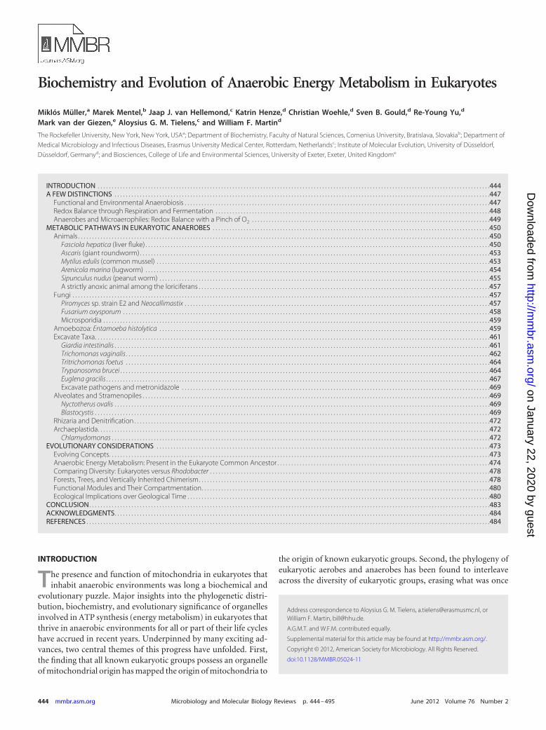
Even the spectrum of organelles specified by the term “mito-chondria” has changed in recent years. Traditionally, the termmitochondria refers to the classical double-membrane-bounded,oxygen-respiring, crista-bearing organelles from rat liver that har-bor the enzymes of the Krebs cycle (also known as the citric acid ortricarboxylic acid [TCA] cycle) and oxidative phosphorylationand that synthesize, and export to the cytosol, ATP with the help ofproton-pumping electron transport chain complexes, ATP syn-thases, and ADP/ATP carriers (AACs) (Fig. 1A). Such would havebeen the description of a mitochondrion in 1973, the year whenhydrogenosomes were reported for the parabasalid flagellateTritrichomonas foetus (279). Hydrogenosomes are double-mem-brane-bounded, oxygen-sensitive, and H2-producing organellesthat occur among several groups of eukaryotic anaerobes and thatsynthesize ATP exclusively via substrate-level phosphorylation(Fig. 1B). For the 20 years following their discovery, the biochem-ical links between parabasalid hydrogenosomes and rat liver mi-tochondria, as shown in Fig. 1, appeared sufficiently few and suf-ficiently scarce that a common ancestry with mitochondria longseemed unlikely.
The first proteins characteristic of hydrogenosomes provided
no links to mitochondria, for example, pyruvate:ferredoxin oxi-doreductase (PFO) (also abbreviated PFOR and sometimes calledpyruvate synthase) and iron-only hydrogenase ([Fe]-Hyd) (279).Although the investigation of hydrogenosomal [2Fe-2S]-ferre-doxin (Fd) could have provided links to mitochondria because ofthe similarity of amino acid sequences and biochemical propertiesto mitochondrial [2Fe-2S]-Fd (164, 228, 303), those clues werenot pursued at the time. Subsequent studies of hydrogenosomalsuccinyl coenzyme A (succinyl-CoA) synthetase (SCS) hintedmore distinctly at evolutionary links between mitochondria andhydrogenosomes (57, 259, 260). The situation changed markedly,however, when four parallel reports of chaperonins common tothe two organelles appeared, making it clear that hydrogenosomesare mitochondria in the evolutionary sense (63, 156, 201, 415).The discovery of more heavily reduced forms of mitochondriathat do not produce ATP—mitosomes—furthermore indicatedthat mitochondria might indeed be ubiquitous among eukaryotes(163, 300, 507, 508, 512, 558). In the meantime, abundant evi-dence supporting the common ancestry of all three organelles andthe ubiquity of mitochondria was amply reviewed (41, 127, 134–136, 178, 454, 520–522, 524).
Although hydrogenosomes typically lack DNA (351), some hy-drogenosomes were eventually found to have preserved a genomethat is homologous to mitochondrial DNA (mtDNA) (49), leav-ing no doubt that hydrogenosomes are anaerobic forms of mito-chondria. Besides the trichomonad lineage, hydrogenosomes havebeen characterized for ciliates (569), chytridiomycete fungi (570),and the heterolobosean amoeboflagellate Psalteriomonas lanterna(56). Newly characterized organelles from the human parasiteBlastocystis hominis share some properties with hydrogenosomes(270, 380, 468), even though evidence for H2 production, hithertodefining for hydrogenosomes, is so far lacking for the Blastocystisorganelles. Conversely, H2 production has been reported for Gi-ardia, a protist that lacks hydrogenosomes and synthesizes its ATPin the cytosol (287), and truncated iron-only hydrogenases arenow found to be ubiquitous among eukaryotes, where they are
FIG 1 Two organelles in comparison. (A) Generalized metabolic scheme of pyruvate oxidation and oxidative phosphorylation in a typical oxygen-respiringmitochondrion, for example, from rat liver. (B) Generalized metabolic scheme of fermentative pyruvate oxidation in trichomonad hydrogenosomes, as proposedin the early 1970s. The presence and absence of organellar genomes are indicated. End products are boxed. Abbreviations: CI to CIV, respiratory complexes I toIV; UQ, ubiquinone; C, cytochrome c; A, ATPase; Fd, ferredoxin; [1], pyruvate:ferredoxin oxidoreductase; [2], acetate:succinate CoA-transferase; [3], succinyl-CoA synthetase; [4], hydrogenase; [5], malic enzyme; [6], pyruvate dehydrogenase complex.
Anaerobic Energy Metabolism in Eukaryotes
June 2012 Volume 76 Number 2 mmbr.asm.org 445
on January 22, 2020 by guesthttp://m
mbr.asm
.org/D
ownloaded from
involved in FeS cluster biogenesis (162, 199). In addition, thechloroplasts of some eukaryotic algae have been found to producecopious amounts of H2 under certain conditions, a developmentof immense biotechnological interest (184, 322, 323). Thus, theproduction of H2 in eukaryotes is no longer synonymous withhydrogenosomes, while hydrogenosomes have become perma-nent members of the mitochondrial family of organelles.
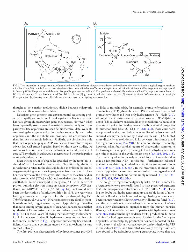
Those developments have witnessed hundreds of original newreports over the past years dealing with mitochondria, hydrog-enosomes, and mitosomes in protists. This was accompanied bythe appearance of the terms “mitochondrion-like organelles”(MLOs) or “mitochondrion-related organelles” (MROs) for or-ganelles that are mitochondria in the evolutionary sense, causingsome confusion as to “who is who” among organelles of mito-chondrial origin. A functional classification and a short descrip-tion of the various classes among the mitochondrial family oforganelles reported so far, along with a terse delineation of criteriafor that classification, are given in Fig. 2.
Mitochondria produce ATP via oxidative phosphorylation.They harbor a mitochondrial genome (167), as the protein com-plexes of proton-pumping electron transport chains contain es-sential subunits that are always mitochondrially encoded (10).The canonical, textbook-type mitochondrion uses oxygen as theterminal electron acceptor.
Anaerobic mitochondria are otherwise typical mitochondria,but they function anaerobically, using compounds other than ox-ygen as the final electron acceptor. Most organisms with anaerobicmitochondria use an endogenously produced electron acceptor,such as fumarate, generating succinate as a major excreted endproduct, but environmental acceptors, such as nitrate (410), canalso be used. Membrane-associated fumarate reduction is usuallyassociated with rhodoquinone (RQ) as an electron carrier, protonpumping, and chemiosmotic ATP synthesis (501).
Hydrogen-producing mitochondria possess, in addition totheir proton-pumping electron transport chain, an iron-only hy-drogenase that allows them to use protons as terminal electronacceptors, resulting in hydrogen production. They can harbor cy-tochromes. They possess a membrane-associated, proton-pump-ing electron transport chain but do not use oxygen as a terminalacceptor, and they produce H2, like hydrogenosomes. An exampleis found in the ciliate Nyctotherus ovalis (49).
Hydrogenosomes are organelles of mitochondrial origin thatgenerate ATP via hydrogen-producing fermentations (351). Theylack cytochromes, a membrane-associated electron transportchain, and a genome. They produce ATP exclusively via substrate-level phosphorylation. Hydrogenosomes have so far been foundamong the trichomonads, the ciliates, the chytridiomycete fungi,and excavate taxa such as Psalteriomonas (520).
FIG 2 Organelles of mitochondrial origin. (A) The mitochondrial family of organelles, divided along functional lines (classes 1 to 5). Class 1, the canonical, ratliver-type mitochondrion (as described in most textbooks), which uses oxygen as the terminal electron acceptor; class 2, an anaerobically functioning mito-chondrion, which uses an endogenously produced electron acceptor, such as fumarate, instead of oxygen; class 3, a hydrogen-producing mitochondrion, whichpossesses (besides a proton-pumping electron transport chain) a hydrogenase and hence can use protons as a terminal electron acceptor and is therefore qualitatequa also a hydrogenosome; class 4, hydrogenosomes, anaerobically functioning ATP-producing organelles of mitochondrial origin that can use protons as anelectron acceptor, which results in the formation of hydrogen; class 5, mitosomes, organelles of mitochondrial origin that are not involved in ATP production.Red indicates that oxygen is consumed in the production of ATP, blue indicates the production of ATP without the use of oxygen, and yellow indicates that theorganelle is not involved in the production of ATP. (B) Criteria for the functional classification of organelles of mitochondrial origin.
Müller et al.
446 mmbr.asm.org Microbiology and Molecular Biology Reviews
on January 22, 2020 by guesthttp://m
mbr.asm
.org/D
ownloaded from
Mitosomes are organelles of mitochondrial origin that do notproduce ATP. Mitosomes of some lineages have retained compo-nents of FeS cluster assembly (163, 508), and others have retainedcomponents of sulfate activation (332).
This classification of the organelles takes into account newerfindings and hence should replace the older type I and type IIdesignations for energy metabolic types (309, 349). To signifytheir common origin and to underscore that these organelles arenot mitochondrion-like organelles but are in fact mitochondria,in that they all evolved from the same ancestral endosymbiont asall other mitochondria, we use the term “organelle of mitochon-drial origin” to designate them collectively. This is an opportunespot to cover another nomenclatural point: about 20 years ago, one ofus introduced into the literature the term “amitochondriate” to des-ignate those eukaryotes that lack typical, cytochrome-possessingmitochondria (349, 350). Although seemingly a good idea at thetime, from today’s perspective, it has turned out that none of theprotists then designated by the term, nor any protists subse-quently discovered, are indeed “amitochondriate” in the sense ofancestrally lacking mitochondria (although many do indeed lackcytochromes). Although the term “amitochondriate” has been ex-tensively used and is still occasionally used in the literature, now isa good time to stop using it altogether.
The purpose of this review is to survey energy metabolism—with an emphasis on mitochondrial metabolism—in eukaryoteswith anaerobic life-styles for part or all of their life cycles, focusingon organisms, including metazoans, where enough biochemicaldata are available on the main enzyme activities and excreted endproducts to allow the presentation of more or less realistic meta-bolic maps. Another nomenclatural point deserves mention. Inthe metazoan literature, where differing degrees of hypoxia (oxy-gen levels below ambient atmospheric levels) are often distin-guished, it is common practice to designate environments as hy-poxic or anoxic, while the term anaerobic is traditionally used todescribe metabolism (169). Because we are focusing on mitochon-dria, here we use the term anaerobic more generally to designatelife-styles, organisms, or pathways in which oxygen is not requiredas the terminal electron acceptor, and we will encounter caseswhere oxygen can be used even though it is not required.
Numerous aspects of the mitochondria of anaerobes and par-asites have recently been reviewed elsewhere, including proteinimport (121), FeS cluster assembly (454, 520), bioenergetic as-pects (266, 267), and reductive evolutionary trends (193). Mono-graphs on the topic of hydrogenosomes and mitosomes have ap-peared (84, 310, 480), as have books on the evolutionarysignificance of mitochondria, including anaerobic forms (264,265). Recent progress from the study of protists needs to be inte-grated with existing knowledge of the anaerobically functioningmitochondria of metazoans (61, 498); hence, we will cover bothprotists and metazoans here, but we will not address metazoansthat can survive short-term hypoxia or anoxia through ethanol,lactate, or similar fermentations, aspects that have been reviewedelsewhere (194). Rather, we focus on metabolic pathways that aregermane to eukaryotic anaerobes and their mitochondria. By pre-senting current metabolic maps for these eukaryotes in one placefor comparison, and by presenting them within a current phylo-genetic framework, we aim to provide a comparative picture thatconveys the unity among oxygen-independent mitochondrial en-ergy metabolic pathways among diverse eukaryotic lineages whilenot blurring the distinctions. We also briefly consider mitochon-
drial energy metabolic pathways in a more general evolutionaryand earth history context.
Key reports of previous findings on this topic and related topicscan be found, including reports on early progress on the topic ofenergy metabolism in anaerobic protists (98), general overviews(142, 349, 352, 426, 516), hydrogenosomes (32, 33, 177, 136, 351),anaerobic mitochondria (501), Giardia intestinalis (3, 59, 221,222, 286, 444), Entamoeba histolytica (320, 406, 423), Trichomonasvaginalis and Tritrichomonas foetus (280, 381), and mitochondrialfunction in various medically relevant parasites (305).
A FEW DISTINCTIONS
Functional and Environmental Anaerobiosis
Particularly among animals, a distinction has to be made betweenenvironmental hypoxia (low oxygen availability) and functionalhypoxia, because energy metabolism without the use of oxygencan occur either by a reduction in the availability of oxygen in theenvironment or as a consequence of burst-type muscular activity(61, 169). Functional hypoxia related to burst-type activity occursusually in animals trying to escape from predators or in those inpursuit of their prey. The rate of the use of ATP by the musclesinvolved is too high to be met by their aerobic energy metabolismand oxygen supply. Therefore, the organism necessarily reverts tothe rapid anaerobic production of ATP in its rapidly contractingmuscles. In some species and tissues, phosphagens like creatinephosphate or phospho-L-arginine can be physiologically relevant“preformed” sources of ATP reserves for burst activity (169). En-vironmental hypoxia, on the other hand, occurs when organismsnaturally inhabit niches where oxygen is scarce, such as parasiticworms living in the intestinal tract of their host (249) or protiststhat inhabit anoxic sediments (38, 129, 142, 291, 473). Environ-mental changes can also result in a decreased availability of oxygenfor organisms unable to relocate to more oxic habitats.
As a consequence, environmental hypoxia is found mainly inorganisms inhabiting an environment where the scarcity of oxy-gen is compensated for by a surplus of fermentable substrates (forexample, parasites in the intestinal tract of their host) or in organ-isms with limited mobility that prevents them from actively escap-ing periods of environmental hypoxia. This occurs, for example,in intertidal marine organisms, such as the sea mussel Mytilusedulis, where daily interruptions of oxygen availability are forcedupon the organism by the tidal cycle. During low tide, the emergedmussel closes its valves to avoid dehydration and therefore has toadapt its metabolism to hypoxic conditions and thus switches toanaerobic energy metabolism (66, 169). Freshwater snails, on theother hand, usually live at the water surface but may occasionallyinhabit bottom layers low in oxygen, for instance, when they expelair from their lung cavities and sink as a response to danger.
Another strategy among various animal groups to survive pro-longed periods of hypoxia is a physiological response called met-abolic repression: energy metabolism is more or less turned off toreduce ATP consumption, thereby requiring little production(195). For example, marine snails that inhabit the intertidal zonecan respond to severe hypoxia or anoxia with a strongly repressedmetabolic rate until oxygen levels sufficient to support respirationreturn (181). Various marine invertebrates respond similarly, en-abling them to stretch their endogenous glycogen reserves (169).Metabolic repression can be seen as a kind of metabolic restingstage; it also occurs in vertebrates, especially among diving marine
Anaerobic Energy Metabolism in Eukaryotes
June 2012 Volume 76 Number 2 mmbr.asm.org 447
on January 22, 2020 by guesthttp://m
mbr.asm
.org/D
ownloaded from
animals such as turtles and seals (109, 174, 195). However, even ifthe organism responds to hypoxia with a reduced metabolic rate,maintenance amounts of ATP still have to be produced withoutoxygen. In most free-living marine invertebrates, succinate, ace-tate, and propionate along with alanine and opines are the mainend products of anaerobic energy metabolism (108) either duringrepression or during normal physiological activity. Among para-sitic metazoans, the main anaerobic end products are usually lac-tate, succinate, acetate, and propionate, whereby, notably, thesame spectrum of end products tends to be produced in the pres-ence and in the absence of oxygen (28).
Redox Balance through Respiration and Fermentation
Since we do not consider photosynthetic ATP synthesis in thispaper, when we refer to eukaryotes, we are referring to hetero-trophs or heterotrophic growth. Core ATP synthesis in eukaryotesentails the oxidation of reduced carbon compounds. Glycolysis—the oxidative breakdown of glucose to pyruvate via the Embden-Meyerhof pathway—is the backbone of ATP synthesis in eu-karyotes (354). Some exceptions to that rule in specializedenvironments entail, for example, the arginine dihydrolase path-way (155, 243, 338) in some anaerobes. The arginine dihydrolasepathway involves the conversion of arginine to ammonia and cit-rulline, with the phosphorolysis of the latter yielding ornithineand carbamoylphosphate, which is readily converted into NH3,CO2, and ATP (338). It has a function analogous to that of the ureacycle in that it is nonoxidative, and it removes nitrogen fromamino acids, but in contrast to the urea cycle, it generates 1 ATPper arginine. Under physiological growth conditions, it satisfies arelevant but small fraction of the total ATP requirements of theeukaryotic organisms that possess it (59, 571) and is therefore anauxiliary rather than a core metabolic pathway. Another exceptionis the methylotrophic pathway in some obligately aerobic fungi, inwhich methanol is oxidized to CO2, with the electrons entering themitochondrial respiratory chain (525), or outright energy parasit-ism, as found for some microsporidians that appear to siphon offnot only metabolites but also ATP (in exchange for ADP) fromtheir host cells (512). Further exceptions (although they have beenrare so far) are the mitochondria in gills of the lugworm Arenicolamarina, which can use H2S instead of glucose as a source of elec-trons for proton pumping and mitochondrial ATP synthesis viathe electron transport chain (115, 170).
The oxidation of glucose or other organic compounds generatesreduced cofactors (NADH and reduced flavin adenine dinucle-otide [FADH2]) that have to be reoxidized by the donation of thegained electrons to a terminal acceptor that, in its reduced form, isexcreted as a metabolic end product (waste). Reduced cofactorscan be reoxidized in two ways: respiration and fermentation.
Respiration is the use of a terminal electron acceptor that isobtained from the environment. Prokaryotic anaerobes display agreat diversity of environmentally available terminal acceptorsthat they can use during ATP synthesis. Amend and Shock (13)compiled data for over 140 different core metabolic reactions in-volving environmentally available terminal acceptors from just131 thermophilic anaerobic prokaryotic species alone. In eu-karyotes, the entire known spectrum of core energy metabolicreactions—aerobic or anaerobic—involves closer to a dozen dif-ferent main overall reactions, a paucity of biochemical diversitywhich, however, powers millions of known species. Perhaps themost common form of anaerobic respiration in eukaryotes, fuma-
rate respiration, is not truly respiration at all, because fumarate(the terminal electron acceptor) is generated endogenously dur-ing metabolism, for which reason it constitutes fermentation (butfermentation involving the generation of a proton gradient).Among eukaryotes, the use of environmentally available acceptorsother than oxygen is still considered to be rare, but it might bemore widespread than currently thought. Nitrate is used as theterminal acceptor by denitrifying foraminiferans, which can bevery widespread in marine environments (384, 410), several fun-gal species (248, 486, 513, 573, 574), and diatoms (232) and whichwas found in ciliates in one report (146). There was also one reportof elemental sulfur being used as the terminal acceptor to generateH2S as the metabolic end product (1). From today’s standpoint,however, that is about all in terms of anaerobic respirations ineukaryotes.
Far more common in eukaryotic anaerobes are fermentations,the donation of electrons onto terminal acceptors that are gener-ated by the organism during metabolism, such as pyruvate (toproduce lactate and/or opines), acetaldehyde (to produce etha-nol), fumarate (to produce succinate), protons (to produce hy-drogen), or acetyl-CoA (to produce fatty acids and their deriva-tives). Fermentations entail disproportionation reactions inwhich the organic substrate is converted into a more reduced anda more oxidized form, for example, the conversion of glucose intoethanol and CO2. Carbohydrates are thus suitable substrates forfermentation. Lipids, however, are too reduced to be fermented,as both oxidation and reduction of the substrate must occur. Fer-mentations in eukaryotes can occur entirely in the cytosol: exam-ples include protistan parasites such as Giardia and Entamoeba(354) or specialized cells such as mature human erythrocytes,which are devoid of mitochondria (51). Fermentations can alsooccur partly in hydrogenosomes, as in the case of Trichomonas(351). Among animals, fermentation often entails malate dismu-tation, involving parts of the mitochondrial electron transportchain, as in the case of the anaerobic mitochondria of many ma-rine invertebrates and parasitic worms (500, 501). Details of thesefermentations are presented in later sections.
It should also be stressed that many eukaryotes live in fully oxichabitats but without using O2 for oxidative phosphorylation.When O2 is present, it can be used as a terminal acceptor, or it canbe ignored. Oxygen is an opportunity, one that is surprisinglyoften declined, with yeast being the classical example. Given asufficient amount of a fermentable substrate in the presence ofoxygen, many yeasts “choose” to ferment rather than use O2 as theterminal acceptor in mitochondria (395, 526, 535). Furthermore,many yeast species show a distinct Crabtree effect: the occurrenceof alcoholic fermentation under aerobic conditions in the pres-ence of excess sugar (395, 527). Maintenance of the baker’s yeast,Saccharomyces cerevisiae, under aerobic cultivation conditionsdoes not result in completely respiratory sugar metabolism, aseven fully aerobic cultures exhibit a mixture of respiration andfermentation, unless they are grown with a limited sugar supply atlow specific growth rates (23, 393). In S. cerevisiae, the biosynthesisof functional mitochondria is furthermore controlled by environ-mental stimuli, including the availability of oxygen and the type ofcarbon source (171, 344). In the laboratory, yeast can, however, beforced to use its mitochondria via growth on nonfermentable sub-strates such as lactate or ethanol (446).
Trypanosomes are another example of organisms that some-times “just say no” to the use of O2 for oxidative phosphorylation.
Müller et al.
448 mmbr.asm.org Microbiology and Molecular Biology Reviews
on January 22, 2020 by guesthttp://m
mbr.asm
.org/D
ownloaded from
When they live in the mammalian bloodstream, where there isample oxygen and glucose, they synthesize all of their ATPthrough substrate-level phosphorylation in glycolysis, but theyexcrete an oxidized end product (pyruvate) instead of a reducedend product (lactate). They maintain redox balance with the helpof an alternative oxidase (AOX) in mitochondria, which uses O2 asthe terminal electron acceptor but without mitochondrial ATPsynthesis. Thus, bloodstream-form trypanosomes use their mito-chondria with O2 as the terminal acceptor but without oxidativephosphorylation. This is another example of what is sometimescalled aerobic fermentation (100). The bloodstream forms of themalaria parasite Plasmodium falciparum follow yet a differentstrategy. Their core energy metabolism is lactate fermentation(368a). Despite this, they have a fully functional electron transportchain in their mitochondria (505a), which is, however, usedmainly to support a ubiquinone (UQ)-dependent dihydroorotatedehydrogenase of the inner mitochondrial membrane (375a). Ac-cordingly, bloodstream malaria parasites use O2 in their mito-chondria but to reoxidize UQH2 (ubiquinol) for pyrimidine bio-synthesis and not for energy metabolism.
There is then the caveat that fermentation as a strategy to sur-vive anoxia is not restricted to microbes; goldfish (Carassius aura-tus) are a classic example. At 10°C, goldfish can survive completeanoxia for more than a week; the end products of metabolism areethanol and CO2 (519), with the ethanol coming from a very stan-dard yeast-type pyruvate decarboxylase (PDC) and alcohol dehy-drogenase (ADH) reaction, with much higher activities of the lat-ter, so that acetaldehyde (a cytotoxin) does not accumulate (536).The ethanol diffuses into the surrounding water. Carp (Carassiuscarassius) can also survive anaerobiosis via ethanol excretion butfor periods of 140 days at 2°C (536). These ethanol-producing fishfermentations, while remarkable, do not entail the participation ofmitochondria.
Anaerobes and Microaerophiles: Redox Balance with aPinch of O2
Several eukaryotes that lack complexes I to IV of membrane-associ-ated electron transport in the inner mitochondrial membrane as wellas the associated cytochromes and quinones can respire to a limitedextent by using O2 as the terminal acceptor, yielding H2O as an im-portant but very inconspicuous end product of energy metabolism.For example, oxygen-reducing NADH oxidases are present in thecytosol of Entamoeba histolytica (289), Giardia lamblia (278),Trichomonas vaginalis (281, 356, 489), and Tritrichomonas foetus (78,345). Again, we designate eukaryotes that have no requirement for O2
in their core energy metabolism “anaerobes,” using the term anaero-bic to indicate that the organisms in question do not require free O2
for survival and multiplication. Furthermore, and importantly, thegrowth of anaerobes is typically inhibited by atmospheric O2 concen-trations, which are 21% (vol/vol) in air, or roughly 250�M in water at25°C, for which reason yeast does not qualify as an anaerobe. Thebasis for growth inhibition by O2 in anaerobes is generally thought toinvolve the inhibition or inactivation of one or more oxygen-sensitiveFeS-cluster-containing enzymes involved in core energy metabolism,such as pyruvate:ferredoxin oxidoreductase (79).
Many anaerobic protists can readily multiply in the presence oflow O2 concentrations, in the 3 to 30 �M range, corresponding to1 to 10% of the present atmospheric levels. Some even growslightly faster under low O2 levels than in the complete absence ofO2 (285, 373). The basis for this slight increase is clear in some
cases. Levels of O2 in the 1 �M range elicit metabolic shifts towardthe excretion of more highly oxidized end products of energy me-tabolism, and this in turn results in increased levels of ATP gen-eration by the same substrate-level phosphorylation reactions thatfunction in the absence of O2 (286, 375). This circumstance is thebasis for suggestions to designate oxygen-shunning protists “mi-croaerophilic” instead of the more generic designation “anaero-bic” (40, 285). Many protists inhabit environments where virtu-ally no free O2 is available, such as anaerobic sediments (38, 142,473). In addition, numerous species, in particular among theciliates, harbor methanogenic archaebacteria that live as endo-symbionts within their hosts (132), whereby methanogens areamong the strictest anaerobes known.
Thus, the terms microaerophilic and anaerobic can be equallyapplicable for many protists. Few eukaryotes studied in detail sofar are truly strict anaerobes, in that most of them regularly en-counter a bit of O2 in their natural habitats. Accordingly, they havebiochemical means for dealing with O2 and can readily tolerate itin small amounts. For example, when a culture of T. vaginalis isgrown in the laboratory, the culture medium does not need to bepurged of O2 at all. Rather, one simply adds a large inoculum ofviable cells to the medium, and the T. vaginalis cells start bybreaking down glucose to pyruvate but without multiplying.Trichomonads possess a cytosolic NADH oxidase, also called di-aphorase, that transfers four electrons from glucose oxidation di-rectly to O2, yielding water (345): 2 NADH � 2 H� � O2 ¡2 NAD� � 2 H2O.
The free energy available in this highly exergonic reaction is notconserved by the NADH oxidase (neither as a proton gradient noras ATP), and the reaction proceeds until the medium is essentiallyfree of O2. Once the O2 is consumed, the cells commence normalgrowth, channeling carbon flux into O2-sensitive pathways, but inan environment that they have themselves made anaerobic in or-der for their full complement of enzymes to function.
Thus, O2 is both a toxin and a minor alternative acceptor forachieving redox balance in many anaerobic protists, and NADHoxidases of the type possessed by Trichomonas (402) are very wide-spread. The enzyme from Trichomonas (281, 489) and that fromGiardia (58, 364) have been characterized. Homologs of theNADH oxidase genes reported for Giardia and Entamoeba (364)are common among eukaryotic genomes. Trichomonas possessestwo diaphorases in the cytosol; the NADH-dependent enzymeyields H2O only, whereas the NADPH-dependent enzyme in ad-dition yields H2O2 (81). Giardia possesses a cytosolic NADHoxidase and a membrane-associated NADH peroxidase (59).Based on their biochemical properties, the eukaryotic enzymes aresimilar to the prokaryotic NADH oxidases and NADH peroxi-dases, which produce H2O2 instead of water (157).
In addition to NADH oxidases, eukaryote anaerobes can pos-sess flavodiiron proteins that function as O2 scavengers, as re-cently characterized for Trichomonas hydrogenosomes (463) andfor Giardia (112). These enzymes have close homologs encoded byseveral sequenced eukaryote genomes, including Entamoeba andseveral green algae. One might wonder why green algae, which aretypically O2 producers, should possess O2-scavenging enzymestypical of anaerobes. The answer is probably one of successfulgeneralist strategies. Some algae, such as Chlamydomonas rein-hardtii, can switch from O2 production to vigorous anaerobicgrowth in the dark within 30 min, producing large amounts of H2
using very O2 sensitive enzymes for fermentative ATP synthesis
Anaerobic Energy Metabolism in Eukaryotes
June 2012 Volume 76 Number 2 mmbr.asm.org 449
on January 22, 2020 by guesthttp://m
mbr.asm
.org/D
ownloaded from
(331, 357); accordingly, O2 detoxification is an issue for Chlamy-domonas and similar algae during anaerobic growth. Since Chla-mydomonas is a typical soil inhabitant (326), it can regularly en-counter anaerobic conditions.
METABOLIC PATHWAYS IN EUKARYOTIC ANAEROBES
In eukaryotic heterotrophs, glycolysis—the Embden-Meyerhofpathway—is the backbone of carbon and energy metabolism.During glycolysis, 1 mol glucose is oxidized to 2 mol pyruvate,with a net yield of 2 mol ATP and producing 2 mol NADH. Ineukaryotes that use O2 as the terminal electron acceptor in theirmitochondria, pyruvate is further oxidized in the mitochondriathrough the pyruvate dehydrogenase (PDH) complex, the Krebscycle, and O2 respiration to yield CO2 and water, with the synthe-sis of roughly an additional 25 mol ATP per mol glucose. Eu-karyotes that are specialized to aerobic environments, such as landplants and land vertebrates, typically possess in addition simplecytosolic fermentations to endure short-term anaerobic function-ing, resulting in end products such as lactate, via lactate dehydro-genase (LDH), or ethanol, via pyruvate decarboxylase (PDC) andalcohol dehydrogenase (ADH) (284, 387).
Another cytosolic fermentation variant is found in severaltrichomonad and yeast species, which increases their glycerol pro-duction upon anoxia (469, 517). In this case, the glycolytic inter-mediate dihydroxyacetone phosphate (DHAP) is converted toglycerol-3-phosphate by a glycerol-3-phosphate dehydrogenasethat is NADPH dependent in trichomonads (469) and NADHdependent in yeast (517) and is subsequently converted to glycerolby glycerol-3-phosphatase. Glycerol production from glucose re-sults in a net consumption of NAD(P)H and is essential in respi-ratory-incompetent yeast cells (517).
In eukaryotes that do not use O2 as the terminal acceptor, amodest diversity is known among energy metabolic pathways,which consist of components that overlap in one way or anotherand which are summarized in the following sections, with threemain caveats mentioned here. One is core pathways. In the textand the metabolic maps, we have focused on major metabolicpathways and major end products. In many cases, there are addi-tional minor end products, sometimes accumulated only underspecific conditions. In most cases, the minor end products are notmentioned here, but in some cases they are, with H2 production inGiardia being an example (287). Another caveat is strains. For thesame species, there are numerous strain-specific differences withregard to the spectrum of end products detected, underscored, forexample, by Trichomonas vaginalis (403), Euglena gracilis (514),and Chlamydomonas reinhardtii (331). A third caveat is stage-specific differences. Many of the organisms covered in this reviewhave a life cycle in which pronounced stage-specific differences inenergy metabolism exist, for example, juvenile versus adult formsof parasitic metazoans (498) or bloodstream versus insect stages ofTrypanosoma (54, 532).
We present these pathways following a phylogenetic approach,based upon current views of eukaryote relationships, as compris-ing roughly six supergroups, whose exact membership and rela-tionships to one another remain discussed (4, 180, 237). This phy-logenetic framework is shown in Fig. 3, along with the kinds ofmitochondria that have been found in some of the biochemicallycharacterized anaerobes.
Animals
Animals belong to the eukaryotic supergroup currently calledOpisthokonta, which includes the animals and the fungi (180).The metazoans harbor many forms that are adapted to life onland, above the soil line, where oxygen is always available, yetmany invertebrates can also survive without oxygen for at leastpart of their life cycle, and some vertebrates, for example, carp,survive complete anoxia for months via ethanol fermentation(536). More recently, some animals were reported to completetheir entire life cycle in sediments below deep-sea hypersaline ha-loclines (99), where there is no oxygen at all, and the mitochondriaof these species of the animal phylum Loricifera appear to lackcristae, but the function of these organelles is as yet unknown.Among freshwater, marine, and parasitic invertebrates, functionalhypoxia and environmental hypoxia are common. All adult para-sitic worms studied so far have a fermentative metabolism and donot use oxygen to completely oxidize glucose to carbon dioxide.The anaerobic metabolism of some of these animals has beenstudied in detail, and representative examples are discussed insubsequent sections.
A leitmotiv common to anaerobic energy metabolism in ani-mals surveyed here is that the anaerobically functioning animalmitochondria do not reveal any fundamental differences in theirenzymatic repertoires relative to those of aerobically specializedlineages. Hence, in the evolutionary sense, there are no differencesbetween aerobic and anaerobic animal lineages with respect togenes and enzymes for which to account. A caveat is that the pres-ence of rhodoquinone seems to be restricted to organisms that canfunction anaerobically. In contrast, among plants, fungi, and pro-tists, there are some notable differences among lineages with re-gard to the enzymes employed by anaerobes versus aerobes. How-ever, as we discuss in the corresponding sections, these differencesare minor, and so far, there are no enzymes involved in the majorpathways of anaerobic energy metabolism that are truly specific toany one eukaryotic lineage; that is, enzymes used for anaerobicenergy metabolism by one eukaryotic supergroup are also foundin at least one other, and they are virtually all present in the greenalga Chlamydomonas, indicating that—just as for oxidative phos-phorylation—the differences reflect a presence in the eukaryotecommon ancestor and differential loss. This observation indicatesthat the evolutionary process of specialization to an anaerobiclife-style in metazoans is distinct from the process in protists andleads to a less diverse spectrum of end products. In other words,among animals, the anaerobic species divert their core carbon fluxthrough enzymes that are also possessed by the aerobes but in sucha manner as to generate a different spectrum of end products,shunting and obviating the need for O2-dependent terminal oxi-dation reactions.
Fasciola hepatica (liver fluke). The adult liver fluke, Fasciolahepatica, is a parasitic flatworm that lives in the bile ducts of itshost, mainly cattle and sheep, but humans can also become in-fected. In the free-living and early larval stages of the animal’s lifecycle, mitochondria of F. hepatica use oxygen in a conventionalway, and these stages degrade glucose completely to CO2 via theKrebs cycle. Upon the penetration of the host and the develop-ment of the parasite in the liver and, subsequently, the bile ducts,this fully aerobic metabolism is gradually replaced by an anaerobicpartial oxidation of glucose, which nevertheless still involves mi-tochondria (502). During this metabolic development, first the
Müller et al.
450 mmbr.asm.org Microbiology and Molecular Biology Reviews
on January 22, 2020 by guesthttp://m
mbr.asm
.org/D
ownloaded from
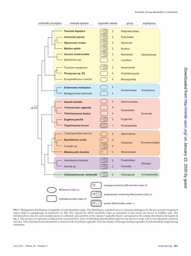
FIG 3 Phylogenetic distribution of organelles of mitochondrial origin. The distribution is plotted across a schematic phylogeny for the six currently recognizedmajor clades or supergroups of eukaryotes (4, 180, 237). Species for which metabolic maps are presented in this review are shown in boldface type. Themitochondrion class for each example species is indicated, and numbers in the column “organelle classes” correspond to the scheme described in the legend ofFig. 2. The presence of a genome is indicated by encircled DNA. Sizes of individual mitochondrial types are not drawn to scale. LECA, last eukaryotic commonancestor. Note that functional information is absent for the loriciferan organelle. Note the absence of lineages lacking organelles of mitochondrial origin amongeukaryotes.
Anaerobic Energy Metabolism in Eukaryotes
June 2012 Volume 76 Number 2 mmbr.asm.org 451
on January 22, 2020 by guesthttp://m
mbr.asm
.org/D
ownloaded from
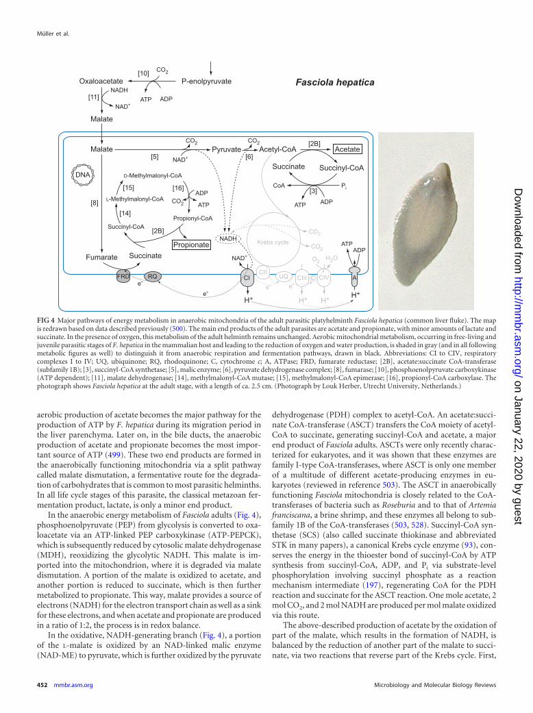
aerobic production of acetate becomes the major pathway for theproduction of ATP by F. hepatica during its migration period inthe liver parenchyma. Later on, in the bile ducts, the anaerobicproduction of acetate and propionate becomes the most impor-tant source of ATP (499). These two end products are formed inthe anaerobically functioning mitochondria via a split pathwaycalled malate dismutation, a fermentative route for the degrada-tion of carbohydrates that is common to most parasitic helminths.In all life cycle stages of this parasite, the classical metazoan fer-mentation product, lactate, is only a minor end product.
In the anaerobic energy metabolism of Fasciola adults (Fig. 4),phosphoenolpyruvate (PEP) from glycolysis is converted to oxa-loacetate via an ATP-linked PEP carboxykinase (ATP-PEPCK),which is subsequently reduced by cytosolic malate dehydrogenase(MDH), reoxidizing the glycolytic NADH. This malate is im-ported into the mitochondrion, where it is degraded via malatedismutation. A portion of the malate is oxidized to acetate, andanother portion is reduced to succinate, which is then furthermetabolized to propionate. This way, malate provides a source ofelectrons (NADH) for the electron transport chain as well as a sinkfor these electrons, and when acetate and propionate are producedin a ratio of 1:2, the process is in redox balance.
In the oxidative, NADH-generating branch (Fig. 4), a portionof the L-malate is oxidized by an NAD-linked malic enzyme(NAD-ME) to pyruvate, which is further oxidized by the pyruvate
dehydrogenase (PDH) complex to acetyl-CoA. An acetate:succi-nate CoA-transferase (ASCT) transfers the CoA moiety of acetyl-CoA to succinate, generating succinyl-CoA and acetate, a majorend product of Fasciola adults. ASCTs were only recently charac-terized for eukaryotes, and it was shown that these enzymes arefamily I-type CoA-transferases, where ASCT is only one memberof a multitude of different acetate-producing enzymes in eu-karyotes (reviewed in reference 503). The ASCT in anaerobicallyfunctioning Fasciola mitochondria is closely related to the CoA-transferases of bacteria such as Roseburia and to that of Artemiafranciscana, a brine shrimp, and these enzymes all belong to sub-family 1B of the CoA-transferases (503, 528). Succinyl-CoA syn-thetase (SCS) (also called succinate thiokinase and abbreviatedSTK in many papers), a canonical Krebs cycle enzyme (93), con-serves the energy in the thioester bond of succinyl-CoA by ATPsynthesis from succinyl-CoA, ADP, and Pi via substrate-levelphosphorylation involving succinyl phosphate as a reactionmechanism intermediate (197), regenerating CoA for the PDHreaction and succinate for the ASCT reaction. One mole acetate, 2mol CO2, and 2 mol NADH are produced per mol malate oxidizedvia this route.
The above-described production of acetate by the oxidation ofpart of the malate, which results in the formation of NADH, isbalanced by the reduction of another part of the malate to succi-nate, via two reactions that reverse part of the Krebs cycle. First,
FIG 4 Major pathways of energy metabolism in anaerobic mitochondria of the adult parasitic platyhelminth Fasciola hepatica (common liver fluke). The mapis redrawn based on data described previously (500). The main end products of the adult parasites are acetate and propionate, with minor amounts of lactate andsuccinate. In the presence of oxygen, this metabolism of the adult helminth remains unchanged. Aerobic mitochondrial metabolism, occurring in free-living andjuvenile parasitic stages of F. hepatica in the mammalian host and leading to the reduction of oxygen and water production, is shaded in gray (and in all followingmetabolic figures as well) to distinguish it from anaerobic respiration and fermentation pathways, drawn in black. Abbreviations: CI to CIV, respiratorycomplexes I to IV; UQ, ubiquinone; RQ, rhodoquinone; C, cytochrome c; A, ATPase; FRD, fumarate reductase; [2B], acetate:succinate CoA-transferase(subfamily 1B); [3], succinyl-CoA synthetase; [5], malic enzyme; [6], pyruvate dehydrogenase complex; [8], fumarase; [10], phosphoenolpyruvate carboxykinase(ATP dependent); [11], malate dehydrogenase; [14], methylmalonyl-CoA mutase; [15], methylmalonyl-CoA epimerase; [16], propionyl-CoA carboxylase. Thephotograph shows Fasciola hepatica at the adult stage, with a length of ca. 2.5 cm. (Photograph by Louk Herber, Utrecht University, Netherlands.)
Müller et al.
452 mmbr.asm.org Microbiology and Molecular Biology Reviews
on January 22, 2020 by guesthttp://m
mbr.asm
.org/D
ownloaded from
malate is converted to fumarate via the enzyme fumarase runningin reverse, and the fumarate produced then serves as the terminalelectron acceptor for the electrons from the oxidative branch ofmalate dismutation and is reduced to succinate. This fumaratereduction is coupled to an anaerobically functioning electrontransport chain in which electrons are transferred from NADH tofumarate via complex I, rhodoquinone (RQ), and a membrane-associated fumarate reductase (FRD) (Fig. 4). Complex I is similarto complex I in aerobic mitochondria, but RQ and FRD are spe-cific for anaerobic mitochondria. The use of RQ instead of ubiqui-none (UQ), the quinone used by aerobically functioning mito-chondria, is essential. RQ is important because it has a lowerstandard redox potential than UQ, which is the reason why re-duced RQ can be used by FRD to donate electrons to fumarate,producing succinate, a reaction where UQ would not readily work(530).
The membrane-associated FRD found in Fasciola resemblesstructurally, and is related to, complex II (succinate dehydroge-nase [SDH]) (501) but is distinct from the soluble NADH-depen-dent FRD found in several protists, including trichomonads andtrypanosomes, that catalyzes the NADH-dependent reduction offumarate to succinate (90, 354).
The succinate produced by FRD is a major excreted end productin many organisms that perform malate dismutation. In Fasciola,however, this succinate is decarboxylated to propionate, and one ad-ditional ATP is gained (383) (Fig. 4). The CoA moiety from propio-nyl-CoA is transferred to succinate by virtue of a dual substrate spec-ificity of ASCT (528), generating propionate as a metabolic endproduct and succinyl-CoA. A cycle regenerates propionyl-CoA, in-volving the vitamin B12-dependent enzyme methylmalonyl-CoAmutase, methylmalonyl-CoA epimerase, and propionyl-CoA car-boxylase. The propionyl-CoA carboxylase reaction generates propi-onyl-CoA and produces ATP via substrate-level phosphorylation(Fig. 4).
During this malate dismutation, protons are pumped by com-plex I, and the resulting proton gradient is harnessed by the mito-chondrial ATP synthase, i.e., oxidative phosphorylation viapumping at complex I alone and without O2 as the terminal ac-ceptor (67a, 248a, 435a). Overall, this anaerobic energy metabo-lism in Fasciola generates propionate and acetate (in addition toCO2) as major end products, in a ratio 2:1, respectively. The pro-cess yields roughly 5 ATPs per glucose: ATP is formed via sub-strate-level phosphorylation during glycolysis in the cytosol (2ATPs per glucose) and inside the mitochondria by the formationof acetate (1 ATP) as well as propionate (1 ATP), and next to that,ATP is formed via proton pumping at complex I and the mito-chondrial ATP synthase (�1 ATP per glucose) but in the absenceof oxygen.
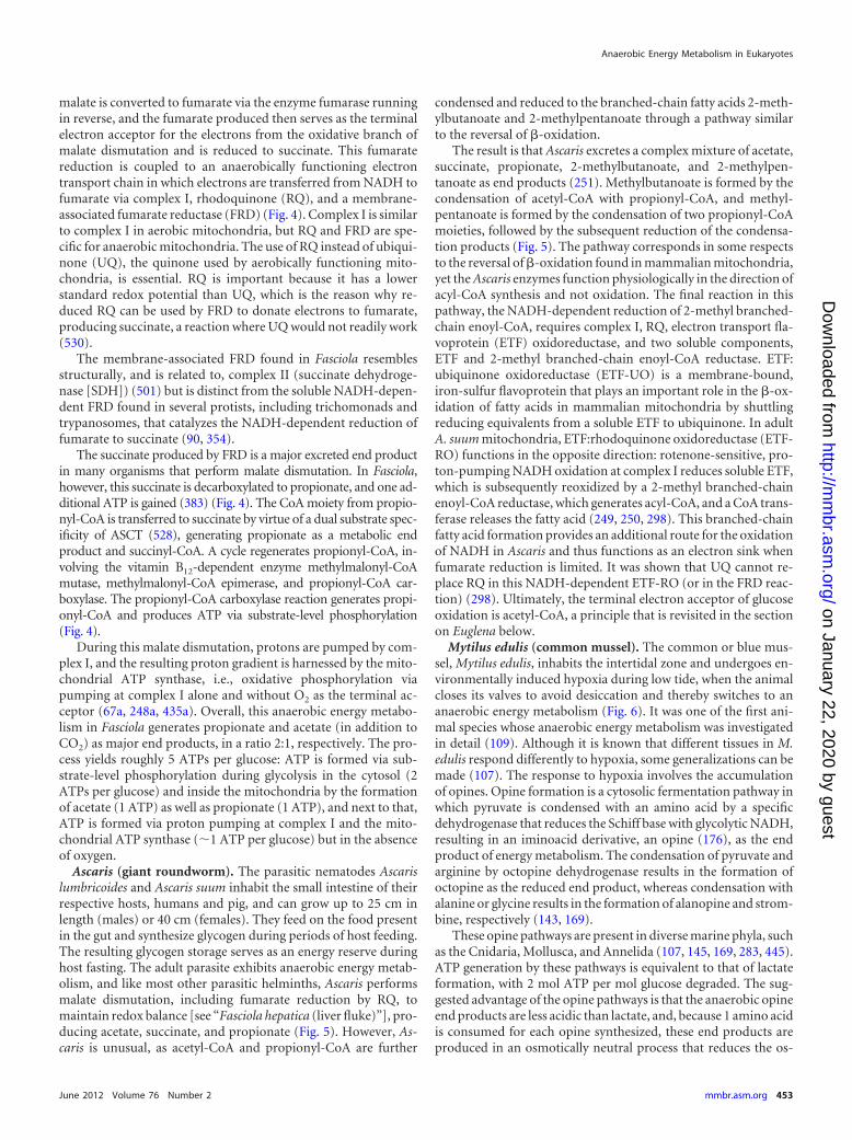
Ascaris (giant roundworm). The parasitic nematodes Ascarislumbricoides and Ascaris suum inhabit the small intestine of theirrespective hosts, humans and pig, and can grow up to 25 cm inlength (males) or 40 cm (females). They feed on the food presentin the gut and synthesize glycogen during periods of host feeding.The resulting glycogen storage serves as an energy reserve duringhost fasting. The adult parasite exhibits anaerobic energy metab-olism, and like most other parasitic helminths, Ascaris performsmalate dismutation, including fumarate reduction by RQ, tomaintain redox balance [see “Fasciola hepatica (liver fluke)”], pro-ducing acetate, succinate, and propionate (Fig. 5). However, As-caris is unusual, as acetyl-CoA and propionyl-CoA are further
condensed and reduced to the branched-chain fatty acids 2-meth-ylbutanoate and 2-methylpentanoate through a pathway similarto the reversal of �-oxidation.
The result is that Ascaris excretes a complex mixture of acetate,succinate, propionate, 2-methylbutanoate, and 2-methylpen-tanoate as end products (251). Methylbutanoate is formed by thecondensation of acetyl-CoA with propionyl-CoA, and methyl-pentanoate is formed by the condensation of two propionyl-CoAmoieties, followed by the subsequent reduction of the condensa-tion products (Fig. 5). The pathway corresponds in some respectsto the reversal of �-oxidation found in mammalian mitochondria,yet the Ascaris enzymes function physiologically in the direction ofacyl-CoA synthesis and not oxidation. The final reaction in thispathway, the NADH-dependent reduction of 2-methyl branched-chain enoyl-CoA, requires complex I, RQ, electron transport fla-voprotein (ETF) oxidoreductase, and two soluble components,ETF and 2-methyl branched-chain enoyl-CoA reductase. ETF:ubiquinone oxidoreductase (ETF-UO) is a membrane-bound,iron-sulfur flavoprotein that plays an important role in the �-ox-idation of fatty acids in mammalian mitochondria by shuttlingreducing equivalents from a soluble ETF to ubiquinone. In adultA. suum mitochondria, ETF:rhodoquinone oxidoreductase (ETF-RO) functions in the opposite direction: rotenone-sensitive, pro-ton-pumping NADH oxidation at complex I reduces soluble ETF,which is subsequently reoxidized by a 2-methyl branched-chainenoyl-CoA reductase, which generates acyl-CoA, and a CoA trans-ferase releases the fatty acid (249, 250, 298). This branched-chainfatty acid formation provides an additional route for the oxidationof NADH in Ascaris and thus functions as an electron sink whenfumarate reduction is limited. It was shown that UQ cannot re-place RQ in this NADH-dependent ETF-RO (or in the FRD reac-tion) (298). Ultimately, the terminal electron acceptor of glucoseoxidation is acetyl-CoA, a principle that is revisited in the sectionon Euglena below.
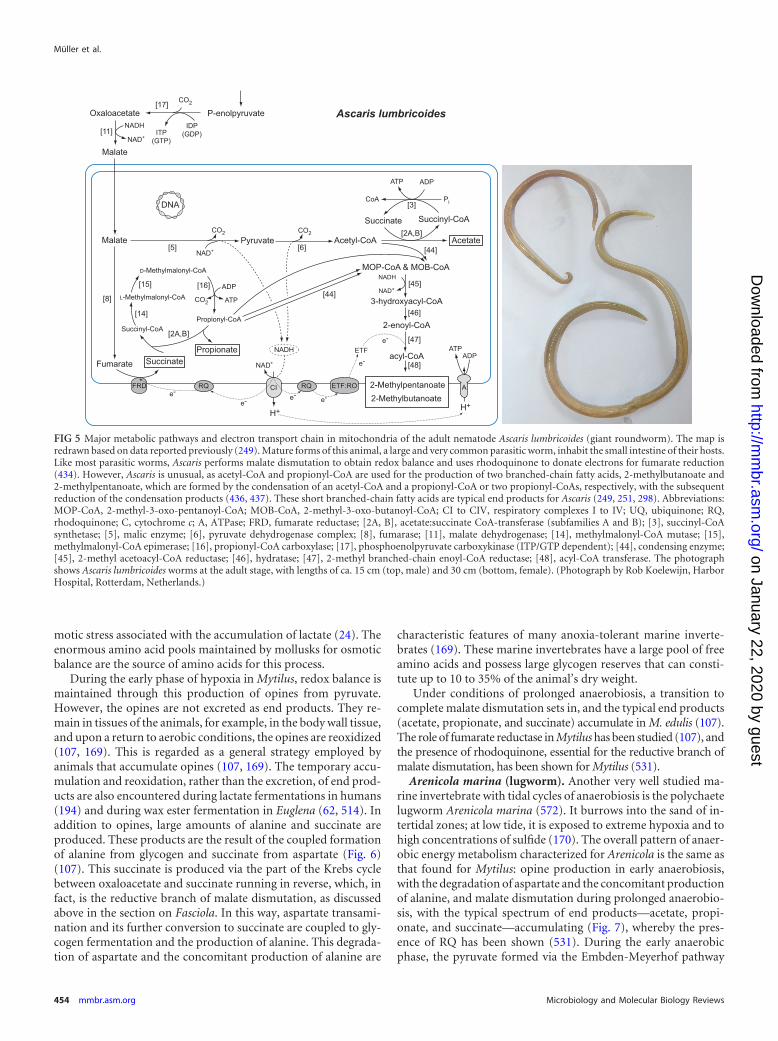
Mytilus edulis (common mussel). The common or blue mus-sel, Mytilus edulis, inhabits the intertidal zone and undergoes en-vironmentally induced hypoxia during low tide, when the animalcloses its valves to avoid desiccation and thereby switches to ananaerobic energy metabolism (Fig. 6). It was one of the first ani-mal species whose anaerobic energy metabolism was investigatedin detail (109). Although it is known that different tissues in M.edulis respond differently to hypoxia, some generalizations can bemade (107). The response to hypoxia involves the accumulationof opines. Opine formation is a cytosolic fermentation pathway inwhich pyruvate is condensed with an amino acid by a specificdehydrogenase that reduces the Schiff base with glycolytic NADH,resulting in an iminoacid derivative, an opine (176), as the endproduct of energy metabolism. The condensation of pyruvate andarginine by octopine dehydrogenase results in the formation ofoctopine as the reduced end product, whereas condensation withalanine or glycine results in the formation of alanopine and strom-bine, respectively (143, 169).
These opine pathways are present in diverse marine phyla, suchas the Cnidaria, Mollusca, and Annelida (107, 145, 169, 283, 445).ATP generation by these pathways is equivalent to that of lactateformation, with 2 mol ATP per mol glucose degraded. The sug-gested advantage of the opine pathways is that the anaerobic opineend products are less acidic than lactate, and, because 1 amino acidis consumed for each opine synthesized, these end products areproduced in an osmotically neutral process that reduces the os-
Anaerobic Energy Metabolism in Eukaryotes
June 2012 Volume 76 Number 2 mmbr.asm.org 453
on January 22, 2020 by guesthttp://m
mbr.asm
.org/D
ownloaded from
motic stress associated with the accumulation of lactate (24). Theenormous amino acid pools maintained by mollusks for osmoticbalance are the source of amino acids for this process.
During the early phase of hypoxia in Mytilus, redox balance ismaintained through this production of opines from pyruvate.However, the opines are not excreted as end products. They re-main in tissues of the animals, for example, in the body wall tissue,and upon a return to aerobic conditions, the opines are reoxidized(107, 169). This is regarded as a general strategy employed byanimals that accumulate opines (107, 169). The temporary accu-mulation and reoxidation, rather than the excretion, of end prod-ucts are also encountered during lactate fermentations in humans(194) and during wax ester fermentation in Euglena (62, 514). Inaddition to opines, large amounts of alanine and succinate areproduced. These products are the result of the coupled formationof alanine from glycogen and succinate from aspartate (Fig. 6)(107). This succinate is produced via the part of the Krebs cyclebetween oxaloacetate and succinate running in reverse, which, infact, is the reductive branch of malate dismutation, as discussedabove in the section on Fasciola. In this way, aspartate transami-nation and its further conversion to succinate are coupled to gly-cogen fermentation and the production of alanine. This degrada-tion of aspartate and the concomitant production of alanine are
characteristic features of many anoxia-tolerant marine inverte-brates (169). These marine invertebrates have a large pool of freeamino acids and possess large glycogen reserves that can consti-tute up to 10 to 35% of the animal’s dry weight.
Under conditions of prolonged anaerobiosis, a transition tocomplete malate dismutation sets in, and the typical end products(acetate, propionate, and succinate) accumulate in M. edulis (107).The role of fumarate reductase in Mytilus has been studied (107), andthe presence of rhodoquinone, essential for the reductive branch ofmalate dismutation, has been shown for Mytilus (531).
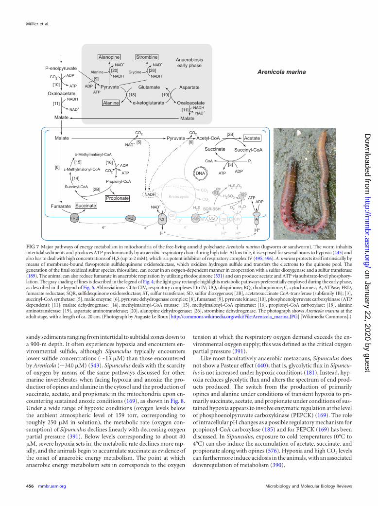
Arenicola marina (lugworm). Another very well studied ma-rine invertebrate with tidal cycles of anaerobiosis is the polychaetelugworm Arenicola marina (572). It burrows into the sand of in-tertidal zones; at low tide, it is exposed to extreme hypoxia and tohigh concentrations of sulfide (170). The overall pattern of anaer-obic energy metabolism characterized for Arenicola is the same asthat found for Mytilus: opine production in early anaerobiosis,with the degradation of aspartate and the concomitant productionof alanine, and malate dismutation during prolonged anaerobio-sis, with the typical spectrum of end products—acetate, propi-onate, and succinate—accumulating (Fig. 7), whereby the pres-ence of RQ has been shown (531). During the early anaerobicphase, the pyruvate formed via the Embden-Meyerhof pathway
FIG 5 Major metabolic pathways and electron transport chain in mitochondria of the adult nematode Ascaris lumbricoides (giant roundworm). The map isredrawn based on data reported previously (249). Mature forms of this animal, a large and very common parasitic worm, inhabit the small intestine of their hosts.Like most parasitic worms, Ascaris performs malate dismutation to obtain redox balance and uses rhodoquinone to donate electrons for fumarate reduction(434). However, Ascaris is unusual, as acetyl-CoA and propionyl-CoA are used for the production of two branched-chain fatty acids, 2-methylbutanoate and2-methylpentanoate, which are formed by the condensation of an acetyl-CoA and a propionyl-CoA or two propionyl-CoAs, respectively, with the subsequentreduction of the condensation products (436, 437). These short branched-chain fatty acids are typical end products for Ascaris (249, 251, 298). Abbreviations:MOP-CoA, 2-methyl-3-oxo-pentanoyl-CoA; MOB-CoA, 2-methyl-3-oxo-butanoyl-CoA; CI to CIV, respiratory complexes I to IV; UQ, ubiquinone; RQ,rhodoquinone; C, cytochrome c; A, ATPase; FRD, fumarate reductase; [2A, B], acetate:succinate CoA-transferase (subfamilies A and B); [3], succinyl-CoAsynthetase; [5], malic enzyme; [6], pyruvate dehydrogenase complex; [8], fumarase; [11], malate dehydrogenase; [14], methylmalonyl-CoA mutase; [15],methylmalonyl-CoA epimerase; [16], propionyl-CoA carboxylase; [17], phosphoenolpyruvate carboxykinase (ITP/GTP dependent); [44], condensing enzyme;[45], 2-methyl acetoacyl-CoA reductase; [46], hydratase; [47], 2-methyl branched-chain enoyl-CoA reductase; [48], acyl-CoA transferase. The photographshows Ascaris lumbricoides worms at the adult stage, with lengths of ca. 15 cm (top, male) and 30 cm (bottom, female). (Photograph by Rob Koelewijn, HarborHospital, Rotterdam, Netherlands.)
Müller et al.
454 mmbr.asm.org Microbiology and Molecular Biology Reviews
on January 22, 2020 by guesthttp://m
mbr.asm
.org/D
ownloaded from
can be converted to strombine and to alanine. In the case of Areni-cola, however, the L-alanine formed can be converted to D-alanineby alanine racemase (445).
In contrast to Mytilus, Arenicola burrows into the sulfidic sed-iment (solfatara) and is therefore regularly exposed to high sulfideconcentrations. Sulfide inhibits cytochrome c oxidase (170), forwhich reason sulfide is toxic during aerobic growth. Arenicoladeals with this sulfide with the help of sulfide:quinone oxi-doreductase (SQR), which oxidizes sulfide and donates the elec-trons to quinones in the mitochondrial electron transport chain(496). Mitochondrial SQR oxidizes sulfide to an enzyme-boundpersulfide (495). The final product of mitochondrial sulfide oxi-dation in Arenicola is thiosulfate, the production of which appearsto require oxygen via a sulfur dioxygenase and the sulfur-trans-ferase activity of mitochondrial rhodanese (189). The sulfur di-oxygenase homolog in humans was recently characterized and is
directly involved in mitochondrial sulfur metabolism, and nullmutants induce severe disease phenotypes (505). Long considereda toxin, sulfide has recently been recognized as a signaling mole-cule and a mediator of cardiovascular function in mammals(45, 231).
Although homologous SQR genes are very widespread in ani-mals and fungi (496), still comparatively little is known aboutmitochondrial sulfide metabolism. In the ribbed mussel Geuken-sia, sulfide oxidation can drive ATP synthesis (116). Recent workon another inhabitant of marine sediments, the annelid Urechisunicinctus, revealed that it possesses the same SQR enzyme andthat its mitochondria, especially mitochondria from the hindgut,can also synthesize ATP with the help of protons pumped by usingelectrons stemming from sulfide (299).
Sipunculus nudus (peanut worm). The free-living peanut ma-rine worm, Sipunculus nudus, is an annelid (358) that lives in
FIG 6 Major pathways of facultative anaerobic energy metabolism in mitochondria of the free-living mollusk Mytilus edulis (blue mussel). The map is redrawnbased on data reported previously (107). Living attached to hard substrates, for example, rocks in intertidal habitats, the bivalve has to face anaerobiosisperiodically. Oxygen-independent cytosolic energy metabolism produces ATP via substrate-level phosphorylation accompanied by the formation of various endproducts, including alanopine, strombine, and alanine (11). Under conditions of prolonged anaerobiosis, propionate is preferentially formed instead of succinatein mitochondria. Fumarate reduction is electron transfer chain coupled, and rhodoquinone serves as an electron donor to fumarate reductase, as in otheranaerobic mitochondria (107, 531). The light gray rectangle highlights metabolic pathways preferentially employed during the early phase of anaerobiosis. Thegray shading of lines is described in the legend of Fig. 4. Abbreviations: CI to CIV, respiratory complexes I to IV; UQ, ubiquinone; RQ, rhodoquinone; C,cytochrome c; A, ATPase; FRD, fumarate reductase; [2B], acetate:succinate CoA-transferase (subfamily 1B); [3], succinyl-CoA synthetase; [5], malic enzyme; [6],pyruvate dehydrogenase complex; [8], fumarase; [9], pyruvate kinase; [10], phosphoenolpyruvate carboxykinase (ATP dependent); [11], malate dehydrogenase;[14], methylmalonyl-CoA mutase; [15], methylmalonyl-CoA epimerase; [16], propionyl-CoA carboxylase; [18], alanine aminotransferase; [19], aspartateaminotransferase; [26], strombine dehydrogenase; [27], octopine dehydrogenase. The photograph shows Mytilus edulis at the adult stage, with a length of ca. 6cm. (Photograph by Louk Herber, Utrecht University, Netherlands.)
Anaerobic Energy Metabolism in Eukaryotes
June 2012 Volume 76 Number 2 mmbr.asm.org 455
on January 22, 2020 by guesthttp://m
mbr.asm
.org/D
ownloaded from
sandy sediments ranging from intertidal to subtidal zones down toa 900-m depth. It often experiences hypoxia and encounters en-vironmental sulfide, although Sipunculus typically encounterslower sulfide concentrations (�13 �M) than those encounteredby Arenicola (�340 �M) (543). Sipunculus deals with the scarcityof oxygen by means of the same pathways discussed for othermarine invertebrates when facing hypoxia and anoxia: the pro-duction of opines and alanine in the cytosol and the production ofsuccinate, acetate, and propionate in the mitochondria upon en-countering sustained anoxic conditions (169), as shown in Fig. 8.Under a wide range of hypoxic conditions (oxygen levels belowthe ambient atmospheric level of 159 torr, corresponding toroughly 250 �M in solution), the metabolic rate (oxygen con-sumption) of Sipunculus declines linearly with decreasing oxygenpartial pressure (391). Below levels corresponding to about 40�M, severe hypoxia sets in, the metabolic rate declines more rap-idly, and the animals begin to accumulate succinate as evidence ofthe onset of anaerobic energy metabolism. The point at whichanaerobic energy metabolism sets in corresponds to the oxygen
tension at which the respiratory oxygen demand exceeds the en-vironmental oxygen supply; this was defined as the critical oxygenpartial pressure (391).
Like most facultatively anaerobic metazoans, Sipunculus doesnot show a Pasteur effect (440); that is, glycolytic flux in Sipuncu-lus is not increased under hypoxic conditions (181). Instead, hyp-oxia reduces glycolytic flux and alters the spectrum of end prod-ucts produced. The switch from the production of primarilyopines and alanine under conditions of transient hypoxia to pri-marily succinate, acetate, and propionate under conditions of sus-tained hypoxia appears to involve enzymatic regulation at the levelof phosphoenolpyruvate carboxykinase (PEPCK) (169). The roleof intracellular pH changes as a possible regulatory mechanism forpropionyl-CoA carboxylase (185) and for PEPCK (169) has beendiscussed. In Sipunculus, exposure to cold temperatures (0°C to4°C) can also induce the accumulation of acetate, succinate, andpropionate along with opines (576). Hypoxia and high CO2 levelscan furthermore induce acidosis in the animals, with an associateddownregulation of metabolism (390).
FIG 7 Major pathways of energy metabolism in mitochondria of the free-living annelid polychaete Arenicola marina (lugworm or sandworm). The worm inhabitsintertidal sediments and produces ATP predominantly by an aerobic respiratory chain during high tide. At low tide, it is exposed for several hours to hypoxia (445) andalso has to deal with high concentrations of H2S (up to 2 mM), which is a potent inhibitor of respiratory complex IV (495, 496). A. marina protects itself intrinsically bymeans of membrane-bound flavoprotein sulfide:quinone oxidoreductase, which oxidizes hydrogen sulfide and transfers the electrons to the quinone pool. Thegeneration of the final oxidized sulfur species, thiosulfate, can occur in an oxygen-dependent manner in cooperation with a sulfur dioxygenase and a sulfur transferase(189). The animal can also reduce fumarate in anaerobic respiration by utilizing rhodoquinone (531) and can produce acetate and ATP via substrate-level phosphory-lation. The gray shading of lines is described in the legend of Fig. 4; the light gray rectangle highlights metabolic pathways preferentially employed during the early phase,as described in the legend of Fig. 6. Abbreviations: CI to CIV, respiratory complexes I to IV; UQ, ubiquinone; RQ, rhodoquinone; C, cytochrome c; A, ATPase; FRD,fumarate reductase; SQR, sulfide:quinone oxidoreductase; ST, sulfur transferase; SD, sulfur dioxygenase; [2B], acetate:succinate CoA-transferase (subfamily 1B); [3],succinyl-CoA synthetase; [5], malic enzyme; [6], pyruvate dehydrogenase complex; [8], fumarase; [9], pyruvate kinase; [10], phosphoenolpyruvate carboxykinase (ATPdependent); [11], malate dehydrogenase; [14], methylmalonyl-CoA mutase; [15], methylmalonyl-CoA epimerase; [16], propionyl-CoA carboxylase; [18], alanineaminotransferase; [19], aspartate aminotransferase; [20], alanopine dehydrogenase; [26], strombine dehydrogenase. The photograph shows Arenicola marina at theadult stage, with a length of ca. 20 cm. (Photograph by Auguste Le Roux [http://commons.wikimedia.org/wiki/File:Arenicola_marina.JPG] [Wikimedia Commons].)
Müller et al.
456 mmbr.asm.org Microbiology and Molecular Biology Reviews
on January 22, 2020 by guesthttp://m
mbr.asm
.org/D
ownloaded from
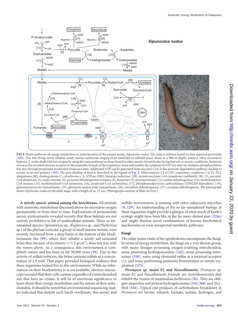
A strictly anoxic animal among the loriciferans. All animalswith anaerobic metabolism discussed above do encounter oxygenpermanently or from time to time. Explorations of permanentlyanoxic environments revealed recently that these habitats are notentirely prohibitive to life of multicellular animals. Three so-far-unnamed species (Spinoloricus sp., Rugiloricus sp., and Pliciloricussp.) of the phylum Loricata, a group of small marine worms, wererecently recovered from a deep basin at the bottom of the Medi-terranean Sea (99), where they inhabit a nearly salt-saturatedbrine that, because of its density (�1.2 g/cm3), does not mix withthe waters above. As a consequence, this environment is com-pletely anoxic and has been so for 50,000 years (99). Due to theactivity of sulfate reducers, the brine contains sulfide at a concen-tration of 2.9 mM. That paper provided biological evidence thatthese organisms indeed live in this environment. While no infor-mation on their biochemistry is as yet available, electron micros-copy revealed that their cells contain organelles of a mitochondrialsize that have no cristae. It will be of enormous significance tolearn about their energy metabolism and the nature of their mito-chondria. It should be noted that environmental sequencing stud-ies indicated that despite such harsh conditions, this anoxic and
sulfidic environment is teeming with other eukaryotic microbes(9, 129). An understanding of the so-far-unexplored biology ofthese organisms might provide a glimpse of what much of Earth’secology might have been like in the far more distant past (324a)and might disclose novel combinations of known biochemicalmechanisms or even unexpected metabolic pathways.
Fungi
The other major clade of the opisthokonts encompasses the fungi.In terms of energy metabolism, the fungi are a very diverse group,with many lineages possessing oxygen-respiring mitochondria,some possessing hydrogenosomes (142), some possessing mito-somes (558), some using elemental sulfur as a terminal acceptor(1), and some performing ammonia fermentation or nitrate res-piration (573).
Piromyces sp. strain E2 and Neocallimastix. Piromyces sp.strain E2 and Neocallimastix frontalis are chytridiomycetes thatinhabit the rumen of mammalian herbivores (30). They are obli-gate anaerobes and possess hydrogenosomes (343, 568) and [Fe]-Hyd (544). Typical end products of carbohydrate breakdown inPiromyces are lactate, ethanol, formate, acetate, hydrogen, and
FIG 8 Major pathways of energy metabolism in mitochondria of the peanut worm, Sipunculus nudus. The map is redrawn based on data reported previously(169). This free-living worm inhabits sandy marine sediments ranging from intertidal to subtidal zones down to a 900-m depth, where it often encountershypoxia. S. nudus deals with low oxygen by using the same pathways as those found in other marine invertebrates facing hypoxic or anoxic conditions. Fumarateserves as the terminal electron acceptor in the anaerobic branch of the respiratory chain and enables the synthesis of ATP not only via oxidative phosphorylationbut also through propionate production from succinate. Additional ATP can be generated from succinyl-CoA in the pyruvate degradation pathway, leading toacetate as an end product (169). The gray shading of lines is described in the legend of Fig. 4. Abbreviations: CI to CIV, respiratory complexes I to IV; UQ,ubiquinone; RQ, rhodoquinone; C, cytochrome c; A, ATPase; FRD, fumarate reductase; [2B], acetate:succinate CoA-transferase (subfamily 1B); [3], succinyl-CoA thiokinase; [5], malic enzyme; [6], pyruvate dehydrogenase complex; [8], fumarase; [9], pyruvate kinase; [11], malate dehydrogenase; [14], methylmalonyl-CoA mutase; [15], methylmalonyl-CoA epimerase; [16], propionyl-CoA carboxylase; [17], phosphoenolpyruvate carboxykinase (ITP/GTP dependent); [18],glutamate:pyruvate transaminase; [19], glutamate:oxaloacetate transaminase; [26], strombine dehydrogenase; [27], octopine dehydrogenase. The photographshows Sipunculus nudus at the adult stage, with a length of ca. 15 cm. (Photograph courtesy of Matt du Fort.)
Anaerobic Energy Metabolism in Eukaryotes
June 2012 Volume 76 Number 2 mmbr.asm.org 457
on January 22, 2020 by guesthttp://m
mbr.asm
.org/D
ownloaded from
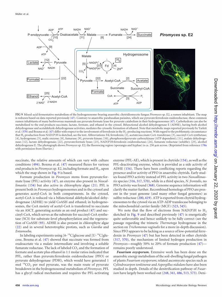
succinate, the relative amounts of which can vary with cultureconditions (494). Boxma et al. (47) measured fluxes for variousend products in Piromyces sp. E2, including formate and H2, uponwhich the map shown in Fig. 9 is based.
Formate production in Piromyces stems from pyruvate:for-mate lyase (PFL) activity (47), an enzyme also present in Neocal-limastix (154) but also active in chlorophyte algae (21). PFL ispresent both in Piromyces hydrogenosomes and in the cytosol andgenerates acetyl-CoA in both compartments. In the cytosol,acetyl-CoA is reduced via a bifunctional aldehyde/alcohol dehy-drogenase (ADHE) to yield CoASH and ethanol; in hydrogeno-somes, the CoA moiety of acetyl-CoA is transferred to succinatevia an ASCT, generating acetate as an end product (47) and suc-cinyl-CoA, which serves as the substrate for succinyl-CoA synthe-tase (SCS) for substrate-level phosphorylation and the regenera-tion of CoASH (93). ADHE is also present in chlorophyte algae(22) and in several heterotrophic protists, such as Giardia andEntamoeba.
In labeling experiments using [6-14C]glucose and [U-14C]glu-cose, Boxma et al. (47) showed that succinate was formed fromoxaloacetate via a malate intermediate and involving a solublefumarate reductase. The lack of labeled CO2 and the formation offormate and acetate plus ethanol in 1:1 molar ratios indicated thatPFL, rather than pyruvate:ferredoxin oxidoreductase (PFO) orpyruvate dehydrogenase (PDH), which would have generated 1mol 14CO2 per mol pyruvate, was the main route of pyruvatebreakdown in the hydrogenosomal metabolism of Piromyces. PFLhas a glycyl radical mechanism and requires the PFL-activating
enzyme (PFL-AE), which is present in chytrids (154), as well as thePFL-deactivating enzyme, which is provided as a side activity ofADHE (154). There have been conflicting reports regarding thepresence and/or activity of PFO in anaerobic chytrids. Early stud-ies found PFO activity instead of PFL activity in two Neocallimas-tix species (316, 317, 570), while in a third species, N. frontalis, noPFO activity was found (368). Genome sequence information willclarify the matter further. Recombined homologs of PFO are pres-ent in the yeast genome (and many other fungal genomes) assulfite reductase (200, 419). ATP is exported from chytrid hydrog-enosomes to the cytosol via an ATP-ADP translocase belonging tothe mitochondrial carrier family (MCF) (523, 545).
We note that the flow of electrons from NAD(P)H to H2
sketched in Fig. 9 and described previously (47) is energeticallyquite unfavorable and hence unlikely to be fully correct (see thepassage regarding the trimeric Thermotoga hydrogenase in thesection on Trichomonas vaginalis for a more in-depth discussion).Since PFO appears to be lacking as a source of low-potential ferre-doxin in Piromyces (47) but has been detected in other chytrids(317, 570), the mechanism of limited hydrogen production inPiromyces—roughly 50% to 20% of formate production (47)—remains poorly understood.
Fusarium oxysporum. Extensive work has been done on theanaerobic energy metabolism of the soil-dwelling fungal pathogenof plants Fusarium oxysporum; related ascomycete species such asCylindrocarpon tonkinense and Aspergillus nidulans have also beenstudied in depth. Details of the denitrification pathway of Fusar-ium have largely been worked out (248, 341, 486, 513, 573). Deni-
FIG 9 Mixed-acid fermentative metabolism of the hydrogenosome-bearing anaerobic chytridiomycete fungus Piromyces sp. E2, a rumen inhabitant. The mapis redrawn based on data reported previously (47). Contrary to anaerobic parabasalian parasites, which use pyruvate:ferredoxin oxidoreductase, these commonrumen inhabitants of many herbivorous mammals use pyruvate:formate lyase for pyruvate catabolism in their hydrogenosomes (47). Carbohydrate can also bemetabolized to the end products succinate, lactate, formate, and ethanol in the cytosol. Bifunctional alcohol dehydrogenase E (ADHE), having both alcoholdehydrogenase and acetaldehyde dehydrogenase activities, mediates the cytosolic formation of ethanol. Note that metabolic maps reported previously by Yarlettet al. (570) and Boxma et al. (47) differ with respect to the involvement of ferredoxin in the H2-producing reactions. With regard to the problematic circumstancethat H2 production from NAD(P)H is sketched, see the text. Abbreviations: Fd, ferredoxin; [2], acetate:succinate CoA-transferase; [3], succinyl-CoA synthetase;[4], hydrogenase; [5], malic enzyme; [8], fumarase; [9], pyruvate kinase; [10], phosphoenolpyruvate carboxykinase (ATP dependent); [11], malate dehydroge-nase; [12], lactate dehydrogenase; [22], pyruvate:formate lyase; [23], NAD(P)H:ferredoxin oxidoreductase; [24], fumarate reductase (soluble); [25], alcoholdehydrogenase E. The photograph shows Piromyces sp. E2; the fluorescing region (sporangia and hyphae) is ca. 250 �m across. (Reprinted from reference 178awith permission from Elsevier.)
Müller et al.
458 mmbr.asm.org Microbiology and Molecular Biology Reviews
on January 22, 2020 by guesthttp://m
mbr.asm
.org/D
ownloaded from
trification among various ascomycetes has been described (574).In Fusarium, denitrification occurs under low-oxygen conditions,involves mitochondria, and entails the oxidation of reduced car-bon sources, such as ethanol, to acetate with a deposition of theelectrons onto nitrate to generate N2O, which is excreted as an endproduct (487, 573). Dissimilatory nitrate reductase (dNar) anddissimilatory nitrite reductase (dNir) in the mitochondrion re-duce NO3
� to NO, which is further reduced by a soluble nitricoxide reductase, a cytochrome P450 enzyme called p450nor, toproduce N2O (150, 487, 573). Fusarium dNar has not been char-acterized at the gene level but has properties in common withbacterial NarGHI proteins (515, 573). The nitrite reductases,NirK, from Fusarium and Aspergillus are homologous to copper-containing proteobacterial nitrite reductases, have conservedhomologs in Acanthamoeba castellanii and Chlamydomonas rein-hardtii, and likely have a mitochondrial origin (241, 242, 359). TheFusarium NO reductase, NoR, is not homologous to prokaryotic ni-trate reductases but is a recruited P450 enzyme (488). Fusarium deni-trification takes place in mitochondria and is coupled to the mito-chondrial electron transport chain and ATP synthesis (487, 573).
Under more strictly anoxic conditions, the reduction of nitrateproceeds to the level of NH4
� as the excreted end product, a pro-cess called ammonia fermentation (485, 574). This involves a dif-ferent set of nitrate-metabolizing enzymes altogether, assimila-tory nitrate reductase (aNar or niaD) and assimilatory nitritereductase (aNir or niiA), which are localized in the cytosol (573).Also involved in the ammonia fermentation pathway of Fusariumis acetate kinase (ACK) (485), an enzyme that is also found inChlamydomonas (21, 357). Formate dehydrogenase was also re-ported to be involved in Fusarium denitrification (515), and therehas been one report of the participation of PFL activity in thegeneration of endogenous formate (258).
Abe et al. (1) showed that Fusarium will grow under anaerobicconditions on a variety of reduced carbon sources using elementalsulfur, S8, as the terminal electron acceptor, generating H2S as thereduced end product in a 2:1 molar ratio relative to acetate.Among eukaryotes studied so far, the use of elemental sulfur as aterminal electron acceptor is restricted to Fusarium. Hypoxic A.nidulans can also excrete large amounts of branched-chain aminoacids as an alternative pathway to achieve redox balance (455).
Denitrification has been reported for foraminifera that inhabitaquatic sediments (410) and for diatoms (232), but none of theenzymes in these systems have been characterized so far, in con-trast to Fusarium. Nitrite reduction coupled to ATP synthesis wasalso measured for mitochondria of higher-plant roots, where NOwas the end product (474), but the quantitative contribution ofthis route to the core ATP supply is as yet unknown.
Microsporidia. Microsporidia are obligate intracellular para-sites that can infect a broad spectrum of hosts (110). The groupharbors important human pathogens, including Encephalitozooncuniculi, which causes digestive and nervous system syndromes(234). The microsporidia are now recognized as highly derivedfungi (192, 555), although their exact placement within the fungiis not clear (220). Their metabolic organization is the result ofreductive evolution due to their specialized obligate parasitic life-style. The microsporidians Trachipleistophora hominis (558) andE. cuniculi (234, 512) are energy parasites; they have little in theway of energy metabolism at all. They can steal the ATP that theyneed from their host via ADP/ATP translocases localized in theirplasma membranes. This type of reduction can go to an extreme.
The microsporidian Enterocytozoon bieneusi, for example, has lostall but 2 genes for the 21 enzymes of glycolysis, all genes for thepentose phosphate pathway, and all genes for trehalose metabo-lism (238). This not only makes the parasite strictly dependent onthe host for energy but also poses a problem with regard to therecycling of NADH. A possibility might be a glycerol-3-phosphateshuttle that moves reducing equivalents across the mitochondrialmembrane, as glycerol-3-phosphate dehydrogenase has beenidentified on microsporidian genomes. The reducing equivalentscould subsequently feed into the microsporidian alternative oxi-dase (556). In the microsporidian Antonospora locustae, both glyc-erol-3-phosphate dehydrogenase and the alternative oxidase, inaddition to Hsp70, localize to the mitosome (122).
As another example, the energy parasite E. cuniculi has lost allof the mitochondrial carrier family (MCF) members, even includ-ing the ADP/ATP carrier (AAC); instead, it now uses the bacterialtype of nucleotide transporter, which is not related to the AAC butis found in plastids and the intracellular pathogenic bacteria Rick-ettsia and Chlamydia, to import ATP from the cytosol of theireukaryotic host cells (512). The E. cuniculi genome harbors fourgenes for this nucleotide transporter, and all four gene productsare expressed: three are localized to the E. cuniculi plasma mem-brane, while the fourth is localized to mitosomes, where it isthought to provide cytosolic ATP to the mitosome (512). Con-trary to E. cuniculi, the microsporidian A. locustae possesses theMCF-type AAC, which was suggested to provide energy forHsp70-dependent protein import into mitosomes and the exportof iron-sulfur clusters to the cytosol (557). Transport processes(including ADP/ATP transport) on the surface of microsporidiawere recently reviewed (554). Microsporidia appear to recruithost mitochondria around their surface, apparently in line withtheir life-style as ATP-stealing energy parasites (439, 554).
The mitochondrial-type protein import system in microspo-ridian mitosomes has been reviewed (549), as have protein importand tRNA import into organelles of mitochondrial origin in gen-eral (282). The characterization of mitochondrial-type iron-sulfurassembly proteins in microsporidia suggests that iron-sulfur pro-tein assembly is the key function of their mitosomes (163), whichdo not appear to play a direct role in energy metabolism.
Amoebozoa: Entamoeba histolytica
The eukaryotic group called Amoebozoa (4) includes membersthat lack ATP-producing organelles, such as Entamoeba histo-lytica, an intestinal pathogen of humans, and members that haveoxygen-respiring mitochondria, such as Dictyostelium discoideum,a free-living amoeba that inhabits soil and compost (25). E. histo-lytica is an anaerobe, and the main end products of its energymetabolism are alanine, CO2, ethanol, and acetate (337, 349),whereby the relative amounts of ethanol and acetate produceddepend upon trace oxygen levels. The enzymes of its energy me-tabolism are localized exclusively in the cytosol, as reviewed else-where (354), from which the map shown in Fig. 10 is adapted.
In Entamoeba, glycolytic phosphoenolpyruvate (PEP) is con-verted to pyruvate via high levels of activity of pyruvate:or-thophosphate dikinase (PPDK) (405) rather than the classical py-ruvate kinase (PYK). However, a homolog of PYK encoded by theEntamoeba genome (290) was subsequently found to be expressedat levels of activity approaching that of PPDK (422) and thus ap-pears to represent an alternative route to pyruvate under someconditions. Another alternative shunt pathway to pyruvate, by-
Anaerobic Energy Metabolism in Eukaryotes
June 2012 Volume 76 Number 2 mmbr.asm.org 459
on January 22, 2020 by guesthttp://m
mbr.asm
.org/D
ownloaded from
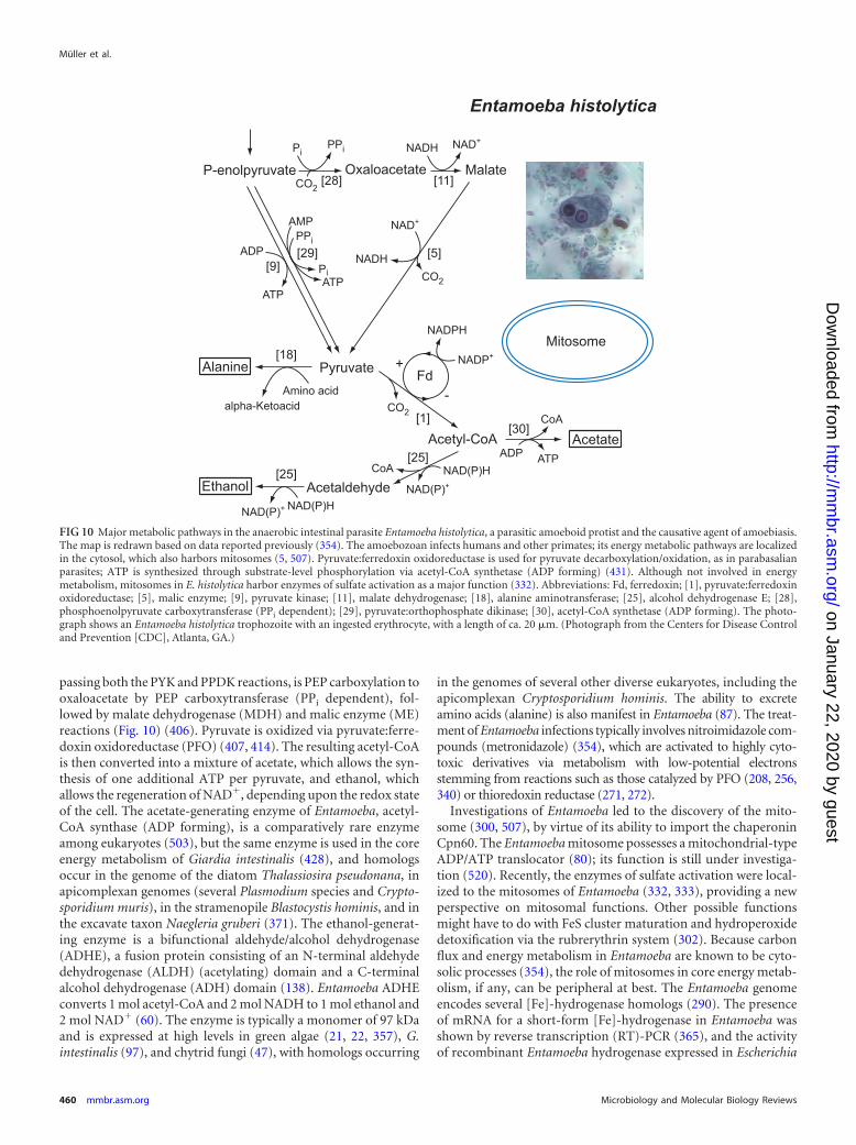
passing both the PYK and PPDK reactions, is PEP carboxylation tooxaloacetate by PEP carboxytransferase (PPi dependent), fol-lowed by malate dehydrogenase (MDH) and malic enzyme (ME)reactions (Fig. 10) (406). Pyruvate is oxidized via pyruvate:ferre-doxin oxidoreductase (PFO) (407, 414). The resulting acetyl-CoAis then converted into a mixture of acetate, which allows the syn-thesis of one additional ATP per pyruvate, and ethanol, whichallows the regeneration of NAD�, depending upon the redox stateof the cell. The acetate-generating enzyme of Entamoeba, acetyl-CoA synthase (ADP forming), is a comparatively rare enzymeamong eukaryotes (503), but the same enzyme is used in the coreenergy metabolism of Giardia intestinalis (428), and homologsoccur in the genome of the diatom Thalassiosira pseudonana, inapicomplexan genomes (several Plasmodium species and Crypto-sporidium muris), in the stramenopile Blastocystis hominis, and inthe excavate taxon Naegleria gruberi (371). The ethanol-generat-ing enzyme is a bifunctional aldehyde/alcohol dehydrogenase(ADHE), a fusion protein consisting of an N-terminal aldehydedehydrogenase (ALDH) (acetylating) domain and a C-terminalalcohol dehydrogenase (ADH) domain (138). Entamoeba ADHEconverts 1 mol acetyl-CoA and 2 mol NADH to 1 mol ethanol and2 mol NAD� (60). The enzyme is typically a monomer of 97 kDaand is expressed at high levels in green algae (21, 22, 357), G.intestinalis (97), and chytrid fungi (47), with homologs occurring
in the genomes of several other diverse eukaryotes, including theapicomplexan Cryptosporidium hominis. The ability to excreteamino acids (alanine) is also manifest in Entamoeba (87). The treat-ment of Entamoeba infections typically involves nitroimidazole com-pounds (metronidazole) (354), which are activated to highly cyto-toxic derivatives via metabolism with low-potential electronsstemming from reactions such as those catalyzed by PFO (208, 256,340) or thioredoxin reductase (271, 272).
Investigations of Entamoeba led to the discovery of the mito-some (300, 507), by virtue of its ability to import the chaperoninCpn60. The Entamoeba mitosome possesses a mitochondrial-typeADP/ATP translocator (80); its function is still under investiga-tion (520). Recently, the enzymes of sulfate activation were local-ized to the mitosomes of Entamoeba (332, 333), providing a newperspective on mitosomal functions. Other possible functionsmight have to do with FeS cluster maturation and hydroperoxidedetoxification via the rubrerythrin system (302). Because carbonflux and energy metabolism in Entamoeba are known to be cyto-solic processes (354), the role of mitosomes in core energy metab-olism, if any, can be peripheral at best. The Entamoeba genomeencodes several [Fe]-hydrogenase homologs (290). The presenceof mRNA for a short-form [Fe]-hydrogenase in Entamoeba wasshown by reverse transcription (RT)-PCR (365), and the activityof recombinant Entamoeba hydrogenase expressed in Escherichia
FIG 10 Major metabolic pathways in the anaerobic intestinal parasite Entamoeba histolytica, a parasitic amoeboid protist and the causative agent of amoebiasis.The map is redrawn based on data reported previously (354). The amoebozoan infects humans and other primates; its energy metabolic pathways are localizedin the cytosol, which also harbors mitosomes (5, 507). Pyruvate:ferredoxin oxidoreductase is used for pyruvate decarboxylation/oxidation, as in parabasalianparasites; ATP is synthesized through substrate-level phosphorylation via acetyl-CoA synthetase (ADP forming) (431). Although not involved in energymetabolism, mitosomes in E. histolytica harbor enzymes of sulfate activation as a major function (332). Abbreviations: Fd, ferredoxin; [1], pyruvate:ferredoxinoxidoreductase; [5], malic enzyme; [9], pyruvate kinase; [11], malate dehydrogenase; [18], alanine aminotransferase; [25], alcohol dehydrogenase E; [28],phosphoenolpyruvate carboxytransferase (PPi dependent); [29], pyruvate:orthophosphate dikinase; [30], acetyl-CoA synthetase (ADP forming). The photo-graph shows an Entamoeba histolytica trophozoite with an ingested erythrocyte, with a length of ca. 20 �m. (Photograph from the Centers for Disease Controland Prevention [CDC], Atlanta, GA.)
Müller et al.
460 mmbr.asm.org Microbiology and Molecular Biology Reviews
on January 22, 2020 by guesthttp://m
mbr.asm
.org/D
ownloaded from
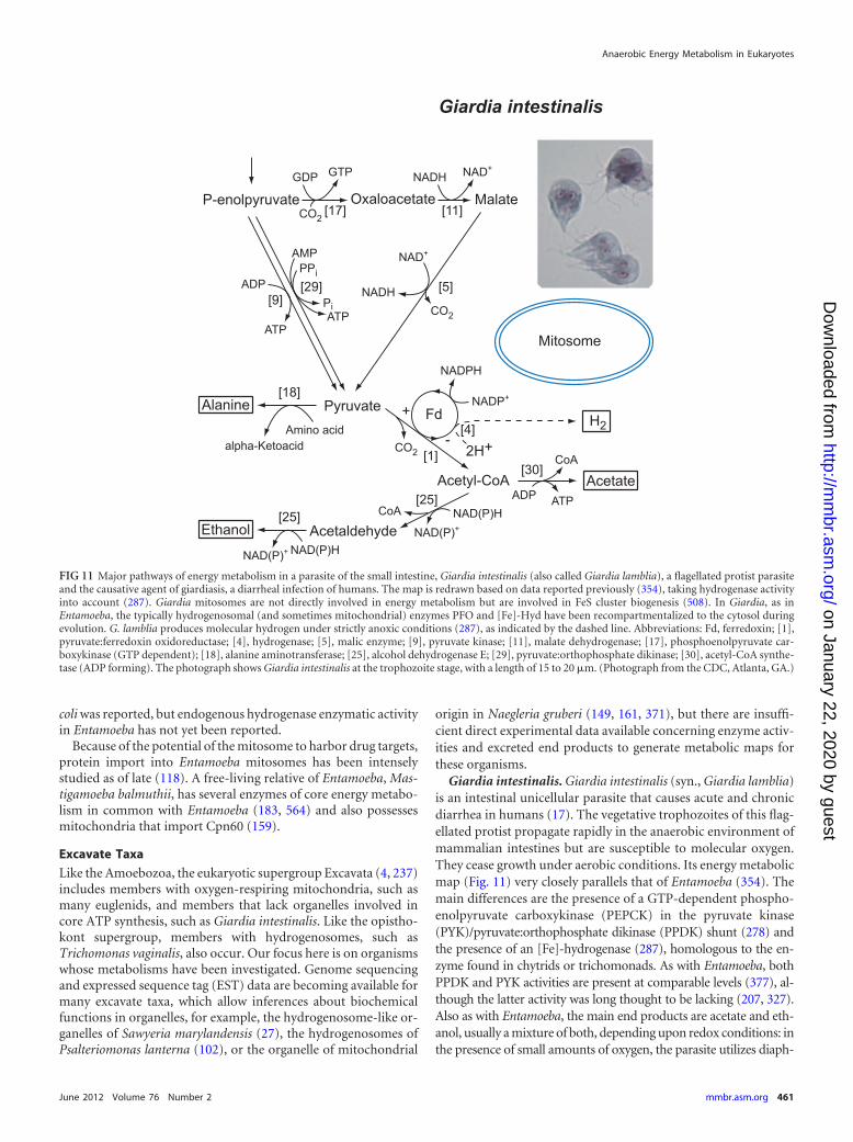
coli was reported, but endogenous hydrogenase enzymatic activityin Entamoeba has not yet been reported.
Because of the potential of the mitosome to harbor drug targets,protein import into Entamoeba mitosomes has been intenselystudied as of late (118). A free-living relative of Entamoeba, Mas-tigamoeba balmuthii, has several enzymes of core energy metabo-lism in common with Entamoeba (183, 564) and also possessesmitochondria that import Cpn60 (159).
Excavate Taxa
Like the Amoebozoa, the eukaryotic supergroup Excavata (4, 237)includes members with oxygen-respiring mitochondria, such asmany euglenids, and members that lack organelles involved incore ATP synthesis, such as Giardia intestinalis. Like the opistho-kont supergroup, members with hydrogenosomes, such asTrichomonas vaginalis, also occur. Our focus here is on organismswhose metabolisms have been investigated. Genome sequencingand expressed sequence tag (EST) data are becoming available formany excavate taxa, which allow inferences about biochemicalfunctions in organelles, for example, the hydrogenosome-like or-ganelles of Sawyeria marylandensis (27), the hydrogenosomes ofPsalteriomonas lanterna (102), or the organelle of mitochondrial
origin in Naegleria gruberi (149, 161, 371), but there are insuffi-cient direct experimental data available concerning enzyme activ-ities and excreted end products to generate metabolic maps forthese organisms.
Giardia intestinalis. Giardia intestinalis (syn., Giardia lamblia)is an intestinal unicellular parasite that causes acute and chronicdiarrhea in humans (17). The vegetative trophozoites of this flag-ellated protist propagate rapidly in the anaerobic environment ofmammalian intestines but are susceptible to molecular oxygen.They cease growth under aerobic conditions. Its energy metabolicmap (Fig. 11) very closely parallels that of Entamoeba (354). Themain differences are the presence of a GTP-dependent phospho-enolpyruvate carboxykinase (PEPCK) in the pyruvate kinase(PYK)/pyruvate:orthophosphate dikinase (PPDK) shunt (278) andthe presence of an [Fe]-hydrogenase (287), homologous to the en-zyme found in chytrids or trichomonads. As with Entamoeba, bothPPDK and PYK activities are present at comparable levels (377), al-though the latter activity was long thought to be lacking (207, 327).Also as with Entamoeba, the main end products are acetate and eth-anol, usually a mixture of both, depending upon redox conditions: inthe presence of small amounts of oxygen, the parasite utilizes diaph-
FIG 11 Major pathways of energy metabolism in a parasite of the small intestine, Giardia intestinalis (also called Giardia lamblia), a flagellated protist parasiteand the causative agent of giardiasis, a diarrheal infection of humans. The map is redrawn based on data reported previously (354), taking hydrogenase activityinto account (287). Giardia mitosomes are not directly involved in energy metabolism but are involved in FeS cluster biogenesis (508). In Giardia, as inEntamoeba, the typically hydrogenosomal (and sometimes mitochondrial) enzymes PFO and [Fe]-Hyd have been recompartmentalized to the cytosol duringevolution. G. lamblia produces molecular hydrogen under strictly anoxic conditions (287), as indicated by the dashed line. Abbreviations: Fd, ferredoxin; [1],pyruvate:ferredoxin oxidoreductase; [4], hydrogenase; [5], malic enzyme; [9], pyruvate kinase; [11], malate dehydrogenase; [17], phosphoenolpyruvate car-boxykinase (GTP dependent); [18], alanine aminotransferase; [25], alcohol dehydrogenase E; [29], pyruvate:orthophosphate dikinase; [30], acetyl-CoA synthe-tase (ADP forming). The photograph shows Giardia intestinalis at the trophozoite stage, with a length of 15 to 20 �m. (Photograph from the CDC, Atlanta, GA.)
Anaerobic Energy Metabolism in Eukaryotes
June 2012 Volume 76 Number 2 mmbr.asm.org 461
on January 22, 2020 by guesthttp://m
mbr.asm
.org/D
ownloaded from
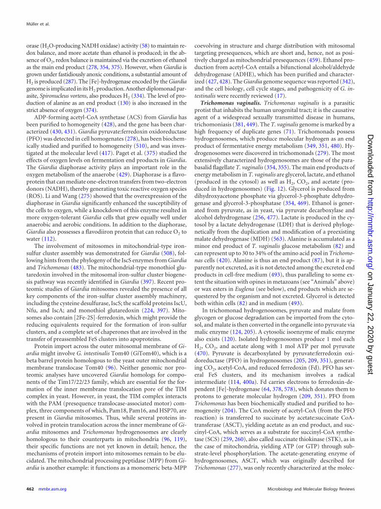
orase (H2O-producing NADH oxidase) activity (58) to maintain re-dox balance, and more acetate than ethanol is produced; in the ab-sence of O2, redox balance is maintained via the excretion of ethanolas the main end product (278, 354, 375). However, when Giardia isgrown under fastidiously anoxic conditions, a substantial amount ofH2 is produced (287). The [Fe]-hydrogenase encoded by the Giardiagenome is implicated in its H2 production. Another diplomonad par-asite, Spironucleus vortens, also produces H2 (334). The level of pro-duction of alanine as an end product (130) is also increased in thestrict absence of oxygen (374).
ADP-forming acetyl-CoA synthetase (ACS) from Giardia hasbeen purified to homogeneity (428), and the gene has been char-acterized (430, 431). Giardia pyruvate:ferredoxin oxidoreductase(PFO) was detected in cell homogenates (278), has been biochem-ically studied and purified to homogeneity (510), and was inves-tigated at the molecular level (417). Paget et al. (375) studied theeffects of oxygen levels on fermentation end products in Giardia.The Giardia diaphorase activity plays an important role in theoxygen metabolism of the anaerobe (429). Diaphorase is a flavo-protein that can mediate one-electron transfers from two-electrondonors (NADH), thereby generating toxic reactive oxygen species(ROS). Li and Wang (275) showed that the overexpression of thediaphorase in Giardia significantly enhanced the susceptibility ofthe cells to oxygen, while a knockdown of this enzyme resulted inmore oxygen-tolerant Giardia cells that grew equally well underanaerobic and aerobic conditions. In addition to the diaphorase,Giardia also possesses a flavodiiron protein that can reduce O2 towater (112).
The involvement of mitosomes in mitochondrial-type iron-sulfur cluster assembly was demonstrated for Giardia (508), fol-lowing hints from the phylogeny of the IscS enzymes from Giardiaand Trichomonas (483). The mitochondrial-type monothiol glu-taredoxin involved in the mitosomal iron-sulfur cluster biogene-sis pathway was recently identified in Giardia (397). Recent pro-teomic studies of Giardia mitosomes revealed the presence of allkey components of the iron-sulfur cluster assembly machinery,including the cysteine desulfurase, IscS; the scaffold proteins IscU,Nfu, and IscA; and monothiol glutaredoxin (224, 397). Mito-somes also contain [2Fe-2S]-ferredoxin, which might provide thereducing equivalents required for the formation of iron-sulfurclusters, and a complete set of chaperones that are involved in thetransfer of preassembled FeS clusters into apoproteins.
Protein import across the outer mitosomal membrane of Gi-ardia might involve G. intestinalis Tom40 (GiTom40), which is abeta barrel protein homologous to the yeast outer mitochondrialmembrane translocase Tom40 (96). Neither genomic nor pro-teomic analyses have uncovered Giardia homologs for compo-nents of the Tim17/22/23 family, which are essential for the for-mation of the inner membrane translocation pore of the TIMcomplex in yeast. However, in yeast, the TIM complex interactswith the PAM (presequence translocase-associated motor) com-plex, three components of which, Pam18, Pam16, and HSP70, arepresent in Giardia mitosomes. Thus, while several proteins in-volved in protein translocation across the inner membrane of Gi-ardia mitosomes and Trichomonas hydrogenosomes are clearlyhomologous to their counterparts in mitochondria (96, 119),their specific functions are not yet known in detail; hence, themechanisms of protein import into mitosomes remain to be elu-cidated. The mitochondrial processing peptidase (MPP) from Gi-ardia is another example: it functions as a monomeric beta-MPP
coevolving in structure and charge distribution with mitosomaltargeting presequences, which are short and, hence, not as posi-tively charged as mitochondrial presequences (459). Ethanol pro-duction from acetyl-CoA entails a bifunctional alcohol/aldehydedehydrogenase (ADHE), which has been purified and character-ized (427, 428). The Giardia genome sequence was reported (342),and the cell biology, cell cycle stages, and pathogenicity of G. in-testinalis were recently reviewed (17).
Trichomonas vaginalis. Trichomonas vaginalis is a parasiticprotist that inhabits the human urogenital tract; it is the causativeagent of a widespread sexually transmitted disease in humans,trichomoniasis (381, 449). The T. vaginalis genome is marked by ahigh frequency of duplicate genes (71). Trichomonads possesshydrogenosomes, which produce molecular hydrogen as an endproduct of fermentative energy metabolism (349, 351, 480). Hy-drogenosomes were discovered in trichomonads (279). The mostextensively characterized hydrogenosomes are those of the para-basalid flagellate T. vaginalis (354, 355). The main end products ofenergy metabolism in T. vaginalis are glycerol, lactate, and ethanol(produced in the cytosol) as well as H2, CO2, and acetate (pro-duced in hydrogenosomes) (Fig. 12). Glycerol is produced fromdihydroxyacetone phosphate via glycerol-3-phosphate dehydro-genase and glycerol-3-phosphatase (354, 469). Ethanol is gener-ated from pyruvate, as in yeast, via pyruvate decarboxylase andalcohol dehydrogenase (256, 477). Lactate is produced in the cy-tosol by a lactate dehydrogenase (LDH) that is derived phyloge-netically from the duplication and modification of a preexistingmalate dehydrogenase (MDH) (563). Alanine is accumulated as aminor end product of T. vaginalis glucose metabolism (82) andcan represent up to 30 to 34% of the amino acid pool in Trichomo-nas cells (420). Alanine is thus an end product (87), but it is ap-parently not excreted, as it is not detected among the excreted endproducts in cell-free medium (493), thus paralleling to some ex-tent the situation with opines in metazoans (see “Animals” above)or wax esters in Euglena (see below), end products which are se-questered by the organism and not excreted. Glycerol is detectedboth within cells (82) and in medium (493).
In trichomonad hydrogenosomes, pyruvate and malate fromglycogen or glucose degradation can be imported from the cyto-sol, and malate is then converted in the organelle into pyruvate viamalic enzyme (124, 205). A cytosolic isoenzyme of malic enzymealso exists (120). Isolated hydrogenosomes produce 1 mol eachH2, CO2, and acetate along with 1 mol ATP per mol pyruvate(470). Pyruvate is decarboxylated by pyruvate:ferredoxin oxi-doreductase (PFO) in hydrogenosomes (205, 209, 351), generat-ing CO2, acetyl-CoA, and reduced ferredoxin (Fd). PFO has sev-eral FeS clusters, and its mechanism involves a radicalintermediate (114, 400a). Fd carries electrons to ferredoxin-de-pendent [Fe]-hydrogenase (64, 378, 578), which donates them toprotons to generate molecular hydrogen (209, 351). PFO fromTrichomonas has been biochemically studied and purified to ho-mogeneity (204). The CoA moiety of acetyl-CoA (from the PFOreaction) is transferred to succinate by acetate:succinate CoA-transferase (ASCT), yielding acetate as an end product, and suc-cinyl-CoA, which serves as a substrate for succinyl-CoA synthe-tase (SCS) (259, 260), also called succinate thiokinase (STK), as inthe case of mitochondria, yielding ATP (or GTP) through sub-strate-level phosphorylation. The acetate-generating enzyme ofhydrogenosomes, ASCT, which was originally described forTrichomonas (277), was only recently characterized at the molec-
Müller et al.
462 mmbr.asm.org Microbiology and Molecular Biology Reviews
on January 22, 2020 by guesthttp://m
mbr.asm
.org/D
ownloaded from
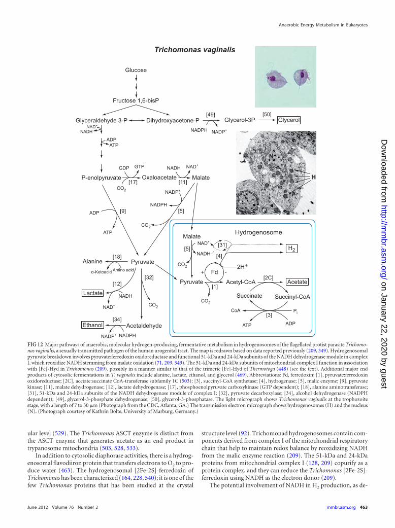
ular level (529). The Trichomonas ASCT enzyme is distinct fromthe ASCT enzyme that generates acetate as an end product intrypanosome mitochondria (503, 528, 533).
In addition to cytosolic diaphorase activities, there is a hydrog-enosomal flavodiiron protein that transfers electrons to O2 to pro-duce water (463). The hydrogenosomal [2Fe-2S]-ferredoxin ofTrichomonas has been characterized (164, 228, 540); it is one of thefew Trichomonas proteins that has been studied at the crystal
structure level (92). Trichomonad hydrogenosomes contain com-ponents derived from complex I of the mitochondrial respiratorychain that help to maintain redox balance by reoxidizing NADHfrom the malic enzyme reaction (209). The 51-kDa and 24-kDaproteins from mitochondrial complex I (128, 209) copurify as aprotein complex, and they can reduce the Trichomonas [2Fe-2S]-ferredoxin using NADH as the electron donor (209).
The potential involvement of NADH in H2 production, as de-
FIG 12 Major pathways of anaerobic, molecular hydrogen-producing, fermentative metabolism in hydrogenosomes of the flagellated protist parasite Trichomo-nas vaginalis, a sexually transmitted pathogen of the human urogenital tract. The map is redrawn based on data reported previously (209, 349). Hydrogenosomalpyruvate breakdown involves pyruvate:ferredoxin oxidoreductase and functional 51-kDa and 24-kDa subunits of the NADH dehydrogenase module in complexI, which reoxidize NADH stemming from malate oxidation (71, 209, 349). The 51-kDa and 24-kDa subunits of mitochondrial complex I function in associationwith [Fe]-Hyd in Trichomonas (209), possibly in a manner similar to that of the trimeric [Fe]-Hyd of Thermotoga (448) (see the text). Additional major endproducts of cytosolic fermentations in T. vaginalis include alanine, lactate, ethanol, and glycerol (469). Abbreviations: Fd, ferredoxin; [1], pyruvate:ferredoxinoxidoreductase; [2C], acetate:succinate CoA-transferase subfamily 1C (503); [3], succinyl-CoA synthetase; [4], hydrogenase; [5], malic enzyme; [9], pyruvatekinase; [11], malate dehydrogenase; [12], lactate dehydrogenase; [17], phosphoenolpyruvate carboxykinase (GTP dependent); [18], alanine aminotransferase;[31], 51-kDa and 24-kDa subunits of the NADH dehydrogenase module of complex I; [32], pyruvate decarboxylase; [34], alcohol dehydrogenase (NADPHdependent); [49], glycerol-3-phosphate dehydrogenase; [50], glycerol-3-phosphatase. The light micrograph shows Trichomonas vaginalis at the trophozoitestage, with a length of 7 to 30 �m (Photograph from the CDC, Atlanta, GA.) The transmission electron micrograph shows hydrogenosomes (H) and the nucleus(N). (Photograph courtesy of Kathrin Bolte, University of Marburg, Germany.)
Anaerobic Energy Metabolism in Eukaryotes
June 2012 Volume 76 Number 2 mmbr.asm.org 463
on January 22, 2020 by guesthttp://m
mbr.asm
.org/D
ownloaded from
scribed by Hrdy et al. (209) and as shown in Fig. 12, might appearto be problematic at first sight, because the midpoint potential ofNADH is not sufficiently negative to generate H2. Although notyet shown for eukaryotes, the likely solution to this situationcomes from a process recently discovered for prokaryotes calledelectron bifurcation (274): Schut and Adams (448) showed thatthe trimeric [Fe]-hydrogenase of Thermotoga maritima operatesin such a way as to accept 1 electron from NADH and 1 electronfrom the low-potential reduced Fd generated by PFO per mole-cule of H2 produced. Despite the involvement of NADH, the over-all reaction is energetically favorable because of the participationof the low-potential ferredoxin, which drives the reaction for-ward. The trimeric Thermotoga [Fe]-hydrogenase consists of threesubunits, �, �, and �. The �-subunit is homologous to the [Fe]-hydrogenase of Trichomonas, while the �- and �- subunits arehomologous to the 51- and 24-kDa subunits of complex I (209),respectively, as sequence comparisons have readily shown. Thisfinding suggests that eukaryote [Fe]-hydrogenase might be trim-eric and function similarly to the Thermotoga enzyme in somespecies, such as Trichomonas. Consistent with this view, in theanaerobic chytrids, the 51- and 24-kDa subunits of complex I arepresent, and in the ciliate Nyctotherus, they are C-terminally fusedto the [Fe]-hydrogenase catalytic subunit (48). However, not alleukaryotic hydrogenases will be trimeric, because the 51- and 24-kDa subunits of complex I are lacking in some eukaryotic anaer-obes (Giardia, for example), but they are present in others.
Protein import into Trichomonas hydrogenosomes can involvethe mitochondrial-type processing peptidase (459), but some pro-teins are imported into hydrogenosomes without processing andwithout the help of N-terminal transit peptides (66a, 325). Severalof the enzymes underpinning Trichomonas core carbon and en-ergy metabolism are abundant proteins possessing FeS clusters(518), and iron metabolism in Trichomonas and related parasiteshas been studied in depth (476, 481). Trichomonas hydrogeno-somes have a mitochondrial-type FeS cluster assembly machinery(478), and frataxin, a mitochondrial protein of iron homeostasis,from Trichomonas hydrogenosomes has been studied (117). [Fe]-hydrogenase of the type found in Trichomonas hydrogenosomesrequires specific maturases, HydEFG, for the assembly of the FeSclusters and the attachment of ligands to the active-site H-cluster(396). One of the [Fe]-hydrogenases in Trichomonas is a fusionprotein carrying at its C terminus the same NAD-, flavin mono-nucleotide (FMN)-, and flavin adenine dinucleotide (FAD)-bind-ing domain as that found at the C terminus of the pyruvate:NADP� oxidoreductase (PNO) of Euglena (419).
The maturation of eukaryotic Fe-Hyd was first studied for thegreen alga Chlamydomonas, which requires at least three addi-tional proteins, HydE, HydF, and HydG (392), that are also pres-ent in trichomonad hydrogenosomes (396) and in other eu-karyotes that express Fe-Hyd (211), but their presence in chytridshas not yet been established. The function of these hydrogenasematurases involves the synthesis and insertion of small ligands(319, 453), including CN� and CO, that are attached to Fe and Satoms of the H-cluster, the active site of the enzyme.
Several Trichomonas Fe-hydrogenases are found as fusion pro-teins possessing an FAD-NAD one-electron- to two-electron-converting module (209), which also occurs in Euglena, Crypto-sporidium, and Blastocystis PNOs (270, 419). However, thisfunctional module is distinct from the 51-kDa and 24-kDa com-plex I subunits that are associated with the trimeric Thermotoga
hydrogenase (the �- and �-subunits thereof) that are involved inlow-potential ferredoxin-dependent NADH oxidation (448) andthat are also present in Trichomonas hydrogenosomes (209).
The ATP generated in trichomonad hydrogenosomes isthought to reach the cytosol via a mitochondrial-type ADP/ATPcarrier homolog, Hmp31 (126, 545). The spectrum of mitochon-drial carrier family (MCF) proteins is remarkably small, in addi-tion to the ATP/ADP carrier. Rada et al. (398) identified five otherMCF members in Trichomonas. There are 44 distinct MCF pro-teins in humans, for comparison. In addition to the pathwaysshown in Fig. 12, there is also a contribution of the arginine dihy-drolase pathway to energy metabolism, but it is a quantitativelyminor contribution (571). The first enzyme of the arginine dihy-drolase pathway, arginine deiminase, is present in the hydrogeno-some, while other components are cytosolic (338).
Tritrichomonas foetus. Tritrichomonas foetus is an importantparasite of livestock that can infect the placenta, lung, and lymphnodes of fetal cattle, causing abortion (449). Hydrogenosomeswere first discovered in this anaerobic flagellate (279). Whengrown under conditions of iron starvation, T. foetus has reducedPFO levels (518).
The spectrum of end products of T. foetus (Fig. 13) parallelsthat of Trichomonas vaginalis (Fig. 12), with the exceptions thatlactate is missing (256) and that in T. foetus, up to 50% of thecarbon end product is excreted as succinate (421). In T. foetus,succinate is produced from phosphoenolpyruvate (PEP) via PEPcarboxykinase (PEPCK), entailing a cytosolic fumarase and a sol-uble NADH-dependent fumarate reductase (354), by a route par-alleling those also found for trypanosomes (39) and anaerobicfungi (47). This mode of succinate formation is thus distinctlydifferent from that found for anaerobic metazoans, where a mem-brane-bound fumarate reductase and rhodoquinone are involved[see “Fasciola hepatica (liver fluke)” for details].
Like T. vaginalis, T. foetus possesses a hydrogenosomal adenyl-ate kinase (113). The cytosolic malate dehydrogenase (202), thehydrogenosomal malic enzyme (ME) (203, 205), and the MEisoenzymes (206) have been characterized, as has the ATP-con-serving hydrogenosomal succinyl-CoA synthetase (SCS) (227).Succinate is produced via a soluble, cytosolic fumarate reductase(346). The enzymes of the T. foetus glycolytic route leading to PEPhave been studied (328, 329).
Differences between T. vaginalis and T. foetus metabolisms aremore pronounced in metronidazole-resistant lines. Both parasitesshunt hydrogenosomal metabolism during resistance, but met-ronidazole-resistant T. vaginalis cells excrete mainly lactate,whereas resistant T. foetus cells excrete mainly ethanol (256). Themode of action of metronidazole (321) involves the activation ofthe compound through low-potential electron donors derivedfrom metabolism (273, 340). The Tritrichomonas ferredoxin hasbeen characterized (303). ME, [Fe]-hydrogenase, SCS, and ade-nylate kinase were purified from T. foetus hydrogenosomes (280).
Trypanosoma brucei. Trypanosoma brucei is a parasitic protistof the order Kinetoplastida that causes African sleeping sickness inhumans and Nagana in livestock. Trypanosomatids exhibit sev-eral unusual cell biological features, such as (i) the presence ofonly a single mitochondrion containing a special structure, thekinetoplast, that comprises a giant network of thousands of cate-nated circular DNAs (106, 292); (ii) that most mitochondrialmRNAs of trypanosomatids are subject to elaborate and preciseposttranscriptional RNA processing that inserts hundreds and de-
Müller et al.
464 mmbr.asm.org Microbiology and Molecular Biology Reviews
on January 22, 2020 by guesthttp://m
mbr.asm
.org/D
ownloaded from
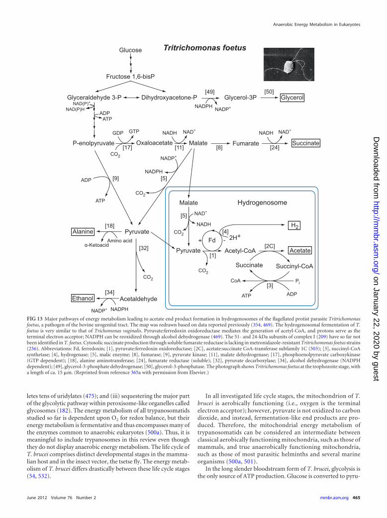
letes tens of uridylates (475); and (iii) sequestering the major partof the glycolytic pathway within peroxisome-like organelles calledglycosomes (182). The energy metabolism of all trypanosomatidsstudied so far is dependent upon O2 for redox balance, but theirenergy metabolism is fermentative and thus encompasses many ofthe enzymes common to anaerobic eukaryotes (500a). Thus, it ismeaningful to include trypanosomes in this review even thoughthey do not display anaerobic energy metabolism. The life cycle ofT. brucei comprises distinct developmental stages in the mamma-lian host and in the insect vector, the tsetse fly. The energy metab-olism of T. brucei differs drastically between these life cycle stages(54, 532).
In all investigated life cycle stages, the mitochondrion of T.brucei is aerobically functioning (i.e., oxygen is the terminalelectron acceptor); however, pyruvate is not oxidized to carbondioxide, and instead, fermentation-like end products are pro-duced. Therefore, the mitochondrial energy metabolism oftrypanosomatids can be considered an intermediate betweenclassical aerobically functioning mitochondria, such as those ofmammals, and true anaerobically functioning mitochondria,such as those of most parasitic helminths and several marineorganisms (500a, 501).
In the long slender bloodstream form of T. brucei, glycolysis isthe only source of ATP production. Glucose is converted to pyru-
FIG 13 Major pathways of energy metabolism leading to acetate end product formation in hydrogenosomes of the flagellated protist parasite Tritrichomonasfoetus, a pathogen of the bovine urogenital tract. The map was redrawn based on data reported previously (354, 469). The hydrogenosomal fermentation of T.foetus is very similar to that of Trichomonas vaginalis. Pyruvate:ferredoxin oxidoreductase mediates the generation of acetyl-CoA, and protons serve as theterminal electron acceptor; NADPH can be reoxidized through alcohol dehydrogenase (469). The 51- and 24-kDa subunits of complex I (209) have so far notbeen identified in T. foetus. Cytosolic succinate production through soluble fumarate reductase is lacking in metronidazole-resistant Tritrichomonas foetus strains(256). Abbreviations: Fd, ferredoxin; [1], pyruvate:ferredoxin oxidoreductase; [2C], acetate:succinate CoA-transferase subfamily 1C (503); [3], succinyl-CoAsynthetase; [4], hydrogenase; [5], malic enzyme; [8], fumarase; [9], pyruvate kinase; [11], malate dehydrogenase; [17], phosphoenolpyruvate carboxykinase(GTP dependent); [18], alanine aminotransferase; [24], fumarate reductase (soluble); [32], pyruvate decarboxylase; [34], alcohol dehydrogenase (NADPHdependent); [49], glycerol-3-phosphate dehydrogenase; [50], glycerol-3-phosphatase. The photograph shows Tritrichomonas foetus at the trophozoite stage, witha length of ca. 15 �m. (Reprinted from reference 365a with permission from Elsevier.)
Anaerobic Energy Metabolism in Eukaryotes
June 2012 Volume 76 Number 2 mmbr.asm.org 465
on January 22, 2020 by guesthttp://m
mbr.asm
.org/D
ownloaded from
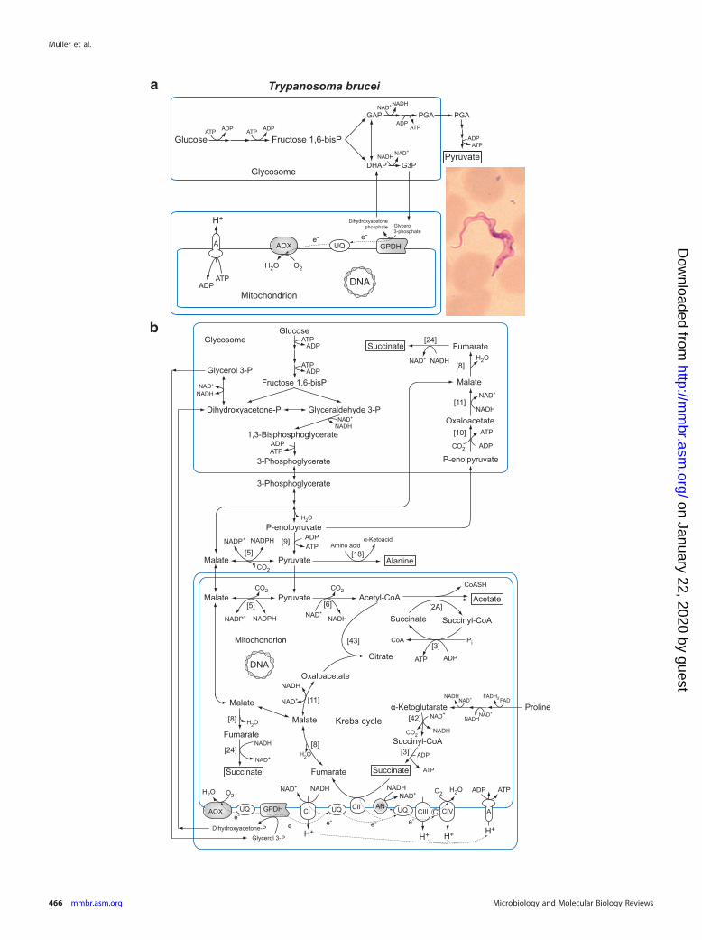
Müller et al.
466 mmbr.asm.org Microbiology and Molecular Biology Reviews
on January 22, 2020 by guesthttp://m
mbr.asm
.org/D
ownloaded from
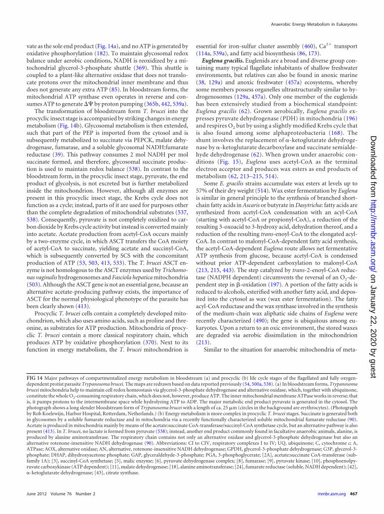
vate as the sole end product (Fig. 14a), and no ATP is generated byoxidative phosphorylation (182). To maintain glycosomal redoxbalance under aerobic conditions, NADH is reoxidized by a mi-tochondrial glycerol-3-phosphate shuttle (369). This shuttle iscoupled to a plant-like alternative oxidase that does not translo-cate protons over the mitochondrial inner membrane and thusdoes not generate any extra ATP (85). In bloodstream forms, themitochondrial ATP synthase even operates in reverse and con-sumes ATP to generate by proton pumping (365b, 442, 539a).
The transformation of bloodstream form T. brucei into theprocyclic insect stage is accompanied by striking changes in energymetabolism (Fig. 14b). Glycosomal metabolism is then extended,such that part of the PEP is imported from the cytosol and issubsequently metabolized to succinate via PEPCK, malate dehy-drogenase, fumarase, and a soluble glycosomal NADH:fumaratereductase (39). This pathway consumes 2 mol NADH per molsuccinate formed, and therefore, glycosomal succinate produc-tion is used to maintain redox balance (538). In contrast to thebloodstream form, in the procyclic insect stage, pyruvate, the endproduct of glycolysis, is not excreted but is further metabolizedinside the mitochondrion. However, although all enzymes arepresent in this procyclic insect stage, the Krebs cycle does notfunction as a cycle; instead, parts of it are used for purposes otherthan the complete degradation of mitochondrial substrates (537,538). Consequently, pyruvate is not completely oxidized to car-bon dioxide by Krebs cycle activity but instead is converted mainlyinto acetate. Acetate production from acetyl-CoA occurs mainlyby a two-enzyme cycle, in which ASCT transfers the CoA moietyof acetyl-CoA to succinate, yielding acetate and succinyl-CoA,which is subsequently converted by SCS with the concomitantproduction of ATP (53, 503, 413, 533). The T. brucei ASCT en-zyme is not homologous to the ASCT enzymes used by Trichomo-nas vaginalis hydrogenosomes and Fasciola hepatica mitochondria(503). Although the ASCT gene is not an essential gene, because analternative acetate-producing pathway exists, the importance ofASCT for the normal physiological phenotype of the parasite hasbeen clearly shown (413).
Procyclic T. brucei cells contain a completely developed mito-chondrion, which also uses amino acids, such as proline and thre-onine, as substrates for ATP production. Mitochondria of procy-clic T. brucei contain a more classical respiratory chain, whichproduces ATP by oxidative phosphorylation (370). Next to itsfunction in energy metabolism, the T. brucei mitochondrion is
essential for iron-sulfur cluster assembly (460), Ca2� transport(114a, 539a), and fatty acid biosynthesis (86, 173).
Euglena gracilis. Euglenids are a broad and diverse group con-taining many typical flagellate inhabitants of shallow freshwaterenvironments, but relatives can also be found in anoxic marine(38, 129a) and anoxic freshwater (457a) ecosystems, wherebysome members possess organelles ultrastructurally similar to hy-drogenosomes (129a, 457a). Only one member of the euglenidshas been extensively studied from a biochemical standpoint:Euglena gracilis (62). Grown aerobically, Euglena gracilis ex-presses pyruvate dehydrogenase (PDH) in mitochondria (196)and respires O2 but by using a slightly modified Krebs cycle thatis also found among some alphaproteobacteria (168). Theshunt involves the replacement of �-ketoglutarate dehydroge-nase by �-ketoglutarate decarboxylase and succinate semialde-hyde dehydrogenase (62). When grown under anaerobic con-ditions (Fig. 15), Euglena uses acetyl-CoA as the terminalelectron acceptor and produces wax esters as end products ofmetabolism (62, 213–215, 514).
Some E. gracilis strains accumulate wax esters at levels up to57% of their dry weight (514). Wax ester fermentation by Euglenais similar in general principle to the synthesis of branched short-chain fatty acids in Ascaris or butyrate in Dasytricha: fatty acids aresynthesized from acetyl-CoA condensation with an acyl-CoA(starting with acetyl-CoA or propionyl-CoA), a reduction of theresulting 3-oxoacid to 3-hydroxy acid, dehydration thereof, and areduction of the resulting trans-enoyl-CoA to the elongated acyl-CoA. In contrast to malonyl-CoA-dependent fatty acid synthesis,the acetyl-CoA-dependent Euglena route allows net fermentativeATP synthesis from glucose, because acetyl-CoA is condensedwithout prior ATP-dependent carboxylation to malonyl-CoA(213, 215, 443). The step catalyzed by trans-2-enoyl-CoA reduc-tase (NADPH dependent) circumvents the reversal of an O2-de-pendent step in �-oxidation (197). A portion of the fatty acids isreduced to alcohols, esterified with another fatty acid, and depos-ited into the cytosol as wax (wax ester fermentation). The fattyacyl-CoA reductase and the wax synthase involved in the synthesisof the medium-chain wax aliphatic side chains of Euglena wererecently characterized (490); the gene is ubiquitous among eu-karyotes. Upon a return to an oxic environment, the stored waxesare degraded via aerobic dissimilation in the mitochondrion(213).
Similar to the situation for anaerobic mitochondria of meta-
FIG 14 Major pathways of compartmentalized energy metabolism in bloodstream (a) and procyclic (b) life cycle stages of the flagellated and fully oxygen-dependent protist parasite Trypanosoma brucei. The maps are redrawn based on data reported previously (54, 500a, 538). (a) In bloodstream forms, Trypanosomabrucei mitochondria help to maintain cell redox homeostasis via glycerol-3-phosphate dehydrogenase and alternative oxidase, which, together with ubiquinone,constitute the whole O2-consuming respiratory chain, which does not, however, produce ATP. The inner mitochondrial membrane ATPase works in reverse; thatis, it pumps protons to the intermembrane space while hydrolyzing ATP to ADP. The major metabolic end product pyruvate is generated in the cytosol. Thephotograph shows a long slender bloodstream form of Trypanosoma brucei with a length of ca. 25 �m (circles in the background are erythrocytes). (Photographby Rob Koelewijn, Harbor Hospital, Rotterdam, Netherlands.) (b) Energy metabolism is more complex in procyclic T. brucei stages. Succinate is generated bothin glycosomes by a soluble fumarate reductase and in mitochondria via a recently functionally characterized soluble mitochondrial fumarate reductase (90).Acetate is produced in mitochondria mainly by means of the acetate:succinate CoA-transferase/succinyl-CoA synthetase cycle, but an alternative pathway is alsopresent (413). In T. brucei, no lactate is formed from pyruvate (538); instead, another end product commonly found in facultative anaerobic animals, alanine, isproduced by alanine aminotransferase. The respiratory chain contains not only an alternative oxidase and glycerol-3-phosphate dehydrogenase but also analternative rotenone-insensitive NADH dehydrogenase (90). Abbreviations: CI to CIV, respiratory complexes I to IV; UQ, ubiquinone; C, cytochrome c; A,ATPase; AOX, alternative oxidase; AN, alternative, rotenone-insensitive NADH dehydrogenase; GPDH, glycerol-3-phosphate dehydrogenase; G3P, glycerol-3-phosphate; DHAP, dihydroxyacetone phosphate; GAP, glyceraldehyde-3-phosphate; PGA, 3-phosphoglycerate; [2A], acetate:succinate CoA-transferase (sub-family 1A); [3], succinyl-CoA synthetase; [5], malic enzyme; [6], pyruvate dehydrogenase complex; [8], fumarase; [9], pyruvate kinase; [10], phosphoenolpy-ruvate carboxykinase (ATP dependent); [11], malate dehydrogenase; [18], alanine aminotransferase; [24], fumarate reductase (soluble, NADH dependent); [42],�-ketoglutarate dehydrogenase; [43], citrate synthase.
Anaerobic Energy Metabolism in Eukaryotes
June 2012 Volume 76 Number 2 mmbr.asm.org 467
on January 22, 2020 by guesthttp://m
mbr.asm
.org/D
ownloaded from

zoa, wax ester fermentation of Euglena involves mitochondrialfumarate reduction and thus utilizes rhodoquinone (RQ) (196)for the synthesis of propionyl-CoA, although reduced RQ can alsodonate electrons to other components of the Euglena mitochon-drial respiratory chain (73), including the cytochrome bc1 com-plex and the alternative oxidase (72). Propionyl-CoA is used as thestarter for the synthesis of fatty acids with odd-number chainlengths, which comprise about 50% (by weight) of hydrocarbonchains in accumulated wax esters under various conditions (235)and in different Euglena strains (514). Propionyl-CoA is producedvia the same short methylmalonyl-CoA route as that found foranimal mitochondria (443), and homologs of the underlying en-zymes, methylmalonyl-CoA mutase and propionyl-CoA carbox-ylase (which provides ATP via substrate-level phosphorylation),are abundantly expressed in euglenid EST data (6). Methylmalo-nyl-CoA mutase from Euglena was recently characterized (335);
the enzyme is also present in humans, where it is one of our onlytwo vitamin B12 (cobalamin)-dependent enzymes (418), wherebyEuglena was once the standard assay for serum B12 levels, becauseof its B12-dependent ribonucleotide reductase (506).
When grown under anaerobic conditions, Euglena expressesPNO (216–218), which performs the oxidative decarboxylation ofpyruvate and is a fusion protein, with an N-terminal PFO domainfused to a C-terminal flavoprotein domain with NAD-, FMN-,and FAD-binding modules (360, 419). The flavoprotein domain isfound in many other proteins, sometimes alone as NADPH:cyto-chrome P450 reductase and sometimes fused to other domains,such as in nitric oxide synthases of metazoans. The flavoproteindomain is best understood as a transducer of one-electron trans-port (from the FeS clusters of the PFO domain) to two-electrontransport (NADPH). PNO supplies acetyl-CoA and NADPH forthe production of wax esters. PNO was first described for Euglena
FIG 15 Wax ester fermentation in mitochondria of the facultatively anaerobic photosynthetic flagellate Euglena gracilis. The map is redrawn based on datareported previously (514). Under anaerobic conditions, this photosynthetic euglenid, which acquired its plastids through secondary endosymbiosis, usesacetyl-CoA produced by pyruvate:NADP� oxidoreductase (419) as the terminal electron acceptor, leading to the formation of an unusual end product amongeukaryotes: wax esters (213, 443, 514). Mitochondrial wax ester fermentation includes anaerobic fumarate respiration and the same propionyl-CoA formationpathway as the one found in mitochondria of facultative anaerobic animals excreting and/or accumulating propionate. Abbreviations: CI to CIV, respiratorycomplexes I to IV; UQ, ubiquinone; RQ, rhodoquinone; C, cytochrome c; A, ATPase; FRD, fumarate reductase; [3], succinyl-CoA synthetase; [5], malic enzyme;[6], pyruvate dehydrogenase complex; [7], pyruvate:NADP� oxidoreductase; [8], fumarase; [9], pyruvate kinase; [10], phosphoenolpyruvate carboxykinase(ATP dependent); [11], malate dehydrogenase; [14], methylmalonyl-CoA mutase; [15], methylmalonyl-CoA epimerase/racemase; [16], propionyl-CoA car-boxylase; [35], �-ketoacyl synthase; [36], �-ketoacyl reductase; [37], �-hydroxyacyl dehydrogenase; [38], trans-2-enoyl-CoA reductase. (Photograph of Euglenagracilis [American Type Culture Collection] by D. J. Patterson, L. Amaral-Zettler, M. Peglar, and T. Nerad, reprinted from the Encyclopedia of Life [http://eol.org/pages/918864/overview], which was published under a Creative Commons license.)
Müller et al.
468 mmbr.asm.org Microbiology and Molecular Biology Reviews
on January 22, 2020 by guesthttp://m
mbr.asm
.org/D
ownloaded from
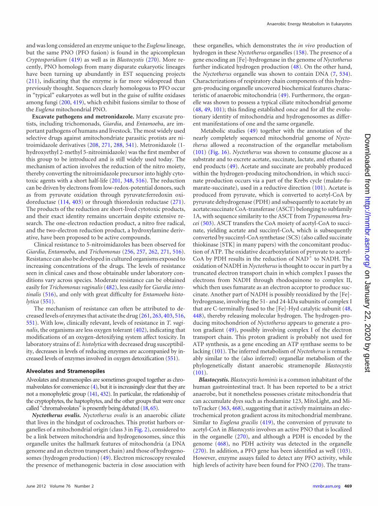
and was long considered an enzyme unique to the Euglena lineage,but the same PNO (PFO fusion) is found in the apicomplexanCryptosporidium (419) as well as in Blastocystis (270). More re-cently, PNO homologs from many disparate eukaryotic lineageshave been turning up abundantly in EST sequencing projects(211), indicating that the enzyme is far more widespread thanpreviously thought. Sequences clearly homologous to PFO occurin “typical” eukaryotes as well but in the guise of sulfite oxidasesamong fungi (200, 419), which exhibit fusions similar to those ofthe Euglena mitochondrial PNO.
Excavate pathogens and metronidazole. Many excavate pro-tists, including trichomonads, Giardia, and Entamoeba, are im-portant pathogens of humans and livestock. The most widely usedselective drugs against amitochondriate parasitic protists are ni-troimidazole derivatives (208, 271, 288, 541). Metronidazole (1-hydroxyethyl 2-methyl 5-nitroimidazole) was the first member ofthis group to be introduced and is still widely used today. Themechanism of action involves the reduction of the nitro moiety,thereby converting the nitroimidazole precursor into highly cyto-toxic agents with a short half-life (201, 348, 516). The reductioncan be driven by electrons from low-redox-potential donors, suchas from pyruvate oxidation through pyruvate:ferredoxin oxi-doreductase (114, 403) or through thioredoxin reductase (271).The products of the reduction are short-lived cytotoxic products,and their exact identity remains uncertain despite extensive re-search. The one-electron reduction product, a nitro free radical,and the two-electron reduction product, a hydroxylamine deriv-ative, have been proposed to be active compounds.
Clinical resistance to 5-nitroimidazoles has been observed forGiardia, Entamoeba, and Trichomonas (256, 257, 262, 271, 516).Resistance can also be developed in cultured organisms exposed toincreasing concentrations of the drugs. The levels of resistanceseen in clinical cases and those obtainable under laboratory con-ditions vary across species. Moderate resistance can be obtainedeasily for Trichomonas vaginalis (482), less easily for Giardia intes-tinalis (516), and only with great difficulty for Entamoeba histo-lytica (551).
The mechanism of resistance can often be attributed to de-creased levels of enzymes that activate the drug (261, 263, 403, 516,551). With low, clinically relevant, levels of resistance in T. vagi-nalis, the organisms are less oxygen tolerant (402), indicating thatmodifications of an oxygen-detoxifying system affect toxicity. Inlaboratory strains of E. histolytica with decreased drug susceptibil-ity, decreases in levels of reducing enzymes are accompanied by in-creased levels of enzymes involved in oxygen detoxification (551).
Alveolates and Stramenopiles
Alveolates and stramenopiles are sometimes grouped together as chro-malveolates for convenience (4), but it is increasingly clear that they arenot a monophyletic group (141, 432). In particular, the relationship ofthe cryptophytes, the haptophytes, and the other groups that were oncecalled “chromalveolates” is presently being debated (18, 65).
Nyctotherus ovalis. Nyctotherus ovalis is an anaerobic ciliatethat lives in the hindgut of cockroaches. This protist harbors or-ganelles of a mitochondrial origin (class 3 in Fig. 2), considered tobe a link between mitochondria and hydrogenosomes, since thisorganelle unites the hallmark features of mitochondria (a DNAgenome and an electron transport chain) and those of hydrogeno-somes (hydrogen production) (49). Electron microscopy revealedthe presence of methanogenic bacteria in close association with
these organelles, which demonstrates the in vivo production ofhydrogen in these Nyctotherus organelles (158). The presence of agene encoding an [Fe]-hydrogenase in the genome of Nyctotherusfurther indicated hydrogen production (48). On the other hand,the Nyctotherus organelle was shown to contain DNA (7, 534).Characterizations of respiratory chain components of this hydro-gen-producing organelle uncovered biochemical features charac-teristic of anaerobic mitochondria (49). Furthermore, the organ-elle was shown to possess a typical ciliate mitochondrial genome(48, 49, 101); this finding established once and for all the evolu-tionary identity of mitochondria and hydrogenosomes as differ-ent manifestations of one and the same organelle.
Metabolic studies (49) together with the annotation of thenearly completely sequenced mitochondrial genome of Nycto-therus allowed a reconstruction of the organellar metabolism(101) (Fig. 16). Nyctotherus was shown to consume glucose as asubstrate and to excrete acetate, succinate, lactate, and ethanol asend products (49). Acetate and succinate are probably producedwithin the hydrogen-producing mitochondrion, in which succi-nate production occurs via a part of the Krebs cycle (malate-fu-marate-succinate), used in a reductive direction (101). Acetate isproduced from pyruvate, which is converted to acetyl-CoA bypyruvate dehydrogenase (PDH) and subsequently to acetate by anacetate:succinate CoA-transferase (ASCT) belonging to subfamily1A, with sequence similarity to the ASCT from Trypanosoma bru-cei (503). ASCT transfers the CoA moiety of acetyl-CoA to succi-nate, yielding acetate and succinyl-CoA, which is subsequentlyconverted by succinyl-CoA synthetase (SCS) (also called succinatethiokinase [STK] in many papers) with the concomitant produc-tion of ATP. The oxidative decarboxylation of pyruvate to acetyl-CoA by PDH results in the reduction of NAD� to NADH. Theoxidation of NADH in Nyctotherus is thought to occur in part by atruncated electron transport chain in which complex I passes theelectrons from NADH through rhodoquinone to complex II,which then uses fumarate as an electron acceptor to produce suc-cinate. Another part of NADH is possibly reoxidized by the [Fe]-hydrogenase, involving the 51- and 24-kDa subunits of complex Ithat are C-terminally fused to the [Fe]-Hyd catalytic subunit (48,448), thereby releasing molecular hydrogen. The hydrogen-pro-ducing mitochondrion of Nyctotherus appears to generate a pro-ton gradient (49), possibly involving complex I of the electrontransport chain. This proton gradient is probably not used forATP synthesis, as a gene encoding an ATP synthase seems to belacking (101). The inferred metabolism of Nyctotherus is remark-ably similar to the (also inferred) organellar metabolism of thephylogenetically distant anaerobic stramenopile Blastocystis(101).
Blastocystis. Blastocystis hominis is a common inhabitant of thehuman gastrointestinal tract. It has been reported to be a strictanaerobe, but it nonetheless possesses cristate mitochondria thatcan accumulate dyes such as rhodamine 123, MitoLight, and Mi-toTracker (363, 468), suggesting that it actively maintains an elec-trochemical proton gradient across its mitochondrial membrane.Similar to Euglena gracilis (419), the conversion of pyruvate toacetyl-CoA in Blastocystis involves an active PNO that is localizedin the organelle (270), and although a PDH is encoded by thegenome (468), no PDH activity was detected in the organelle(270). In addition, a PFO gene has been identified as well (103).However, enzyme assays failed to detect any PFO activity, whilehigh levels of activity have been found for PNO (270). The trans-
Anaerobic Energy Metabolism in Eukaryotes
June 2012 Volume 76 Number 2 mmbr.asm.org 469
on January 22, 2020 by guesthttp://m
mbr.asm
.org/D
ownloaded from
formation of acetyl-CoA to acetate leads to the synthesis of ATPvia substrate-level phosphorylation using an acetate:succinateCoA-transferase, succinyl-CoA synthetase cycle (197, 468), as isfound for T. vaginalis, and high levels of activity of both enzymesin the organelle were measured (270). Blastocystis has an activecytosolic lactate dehydrogenase (270). From these biochemical
data, it can be reasonably predicted that acetate and lactate couldbe major metabolic end products, but no information on accumu-lated Blastocystis end products is currently available. The genomedata suggest the presence of many more mitochondrial enzymes(103, 468); accordingly, the map shown in Fig. 17, suggesting thenature of major metabolic pathways, could be incomplete.
FIG 16 Tentative map of major pathways of energy metabolism in hydrogen-producing mitochondria of the anaerobic ciliate Nyctotherus ovalis. The map is redrawnbased on data reported previously (49). The ciliate lives in the hindgut of cockroaches and harbors organelles that provided a link between mitochondria and hydrog-enosomes (49). The Krebs cycle is incomplete and is likely used in the reductive direction (177). A proton gradient is generated, probably by a functional respiratorycomplex I, which passes the electrons from the NADH pool through rhodoquinone to complex II, acting as fumarate reductase synthesizing succinate (101). Redoxbalance is also achieved with the help of hydrogenase, releasing molecular hydrogen, hence the term hydrogen-producing anaerobic mitochondria (49). ATP can besynthesized by substrate-level phosphorylation, producing acetate. Abbreviations: CI, respiratory complex I; RQ, rhodoquinone; CII, fumarate reductase/succinatedehydrogenase; [2A], acetate:succinate CoA-transferase subfamily 1A (503); [3], succinyl-CoA synthetase; [4], hydrogenase; [5], malic enzyme; [6], pyruvate dehydro-genase complex; [8], fumarase (predicted); [9], pyruvate kinase; [12], lactate dehydrogenase; [18], alanine aminotransferase; [32], pyruvate decarboxylase; [33], alcoholdehydrogenase (NADH dependent); [42], �-ketoglutarate dehydrogenase (predicted); [41], glutamate dehydrogenase. The photograph on the left shows Nyctotherusovalis, with a length of ca. 80 �m. The hydrogenosomes (H) are surrounded by endosymbiotic methane-producing archaebacteria (dark spots). N, macronucleus; n,micronucleus; V, vacuole. (Reprinted from reference 7 with permission of Macmillan Publishers Ltd.) The right photograph shows a closeup view of a Nyctotherushydrogenosome (H) and an associated methanogen (M). (Reprinted from reference 49 with permission of Macmillan Publishers Ltd.)
Müller et al.
470 mmbr.asm.org Microbiology and Molecular Biology Reviews
on January 22, 2020 by guesthttp://m
mbr.asm
.org/D
ownloaded from

The situation regarding the Krebs cycle in Blastocystis is con-fusing, as different studies have suggested different results. Alarge-scale EST study suggested that the enzymes for the secondhalf of the Krebs cycle from succinyl-CoA to oxaloacetate are pres-ent (468), while no EST encoding enzymes capable of convertingacetyl-CoA into succinyl-CoA was identified (i.e., no citrate syn-thase, aconitase, isocitrate dehydrogenase, or �-ketoglutarate de-hydrogenase). On the other hand, in cell fractionation studies, theBlastocystis organelle was shown to contain malic enzyme, aconi-tase, isocitrate dehydrogenase, �-ketoglutarate dehydrogenase,and succinyl-CoA synthetase activities (270). Mitochondrial tar-geting sequences were predicted to be present on a putative fuma-rase (103, 468) and malate dehydrogenase (103), although theformer seems more likely to represent an aspartate ammonia-lyasebased on sequence comparisons. Blastocystis might represent aspecies complex of up to nine subtypes (471). A genome project(103) and a biochemical study (270) were conducted on a subtype
7 species, while an EST study (468) was conducted with subtype 1;since these subtypes belong to two different clades, this mightexplain many of the differences among those three studies.
Similar to Nyctotherus ovalis, a partial complex I seems to be pres-ent, suggesting that the organelle might have some proton-pumpingpotential (103, 468), in agreement with the ability of the organelle toaccumulate dyes such as rhodamine 123 (363). Genes encoding com-plex I components were also identified in the Blastocystis mitochon-drial genome (380, 552). In addition, all four complex II subunitswere identified (103, 468), although whether this complex functionsas a succinate dehydrogenase (SDH) or a fumarate reductase (FRD) isunknown, and it is not known which quinones are present. No evi-dence for complexes III and IV or an ATP synthase was found, inagreement with earlier biochemical studies (577). Genomic studiesidentified an alternative oxidase (AOX) and an [Fe]-hydrogenase.The latter protein was localized to the organelle by using antibodies(468), but hydrogenase activity was not detected (270).
FIG 17 Putative major pathways of anaerobic energy metabolism in the organelle of mitochondrial origin in Blastocystis hominis, a protozoan parasite and acommon inhabitant of the human gastrointestinal tract. The map is redrawn based on data reported previously (270). The conversion of pyruvate to acetyl-CoAis carried out by pyruvate:NADP� oxidoreductase (PNO) (270), the same fusion protein found in Euglena gracilis. The transformation of acetyl-CoA to acetateleads to the synthesis of ATP via substrate-level phosphorylation. The alternative oxidase might help Blastocystis cope with oxygen stress conditions in the hostintestine and prevent the formation of reactive oxygen species. Hydrogen production has not yet been shown for Blastocystis (as indicated by a question mark),although [Fe]-Hyd localizes to the organelle (468). In this sense, the mitochondria of Blastocystis resemble the hydrogen-producing mitochondria found in theciliate Nyctotherus ovalis and represent another example demonstrating that mitochondria and hydrogenosomes utilize a common enzyme toolkit. The questionmark indicates another possible pathway (103, 270, 468). Abbreviations: CI, respiratory complex I; Q?, quinone (of an uncertain nature); AOX, alternativeoxidase; [2B, C], acetate:succinate CoA-transferase (members of subfamilies 1B and 1C [503] are present in the genome); [3], succinyl-CoA synthetase; [4],hydrogenase; [5], malic enzyme; [7], pyruvate:NADP� oxidoreductase; [9], pyruvate kinase; [11], malate dehydrogenase; [12], lactate dehydrogenase; [17],phosphoenolpyruvate carboxykinase (GTP dependent). The photograph shows Blastocystis sp. at the trophozoite stage, with a length of ca. 35 �m. (Photographcourtesy of C. G. Clark, London School of Hygiene and Tropical Medicine, London, United Kingdom.)
Anaerobic Energy Metabolism in Eukaryotes
June 2012 Volume 76 Number 2 mmbr.asm.org 471
on January 22, 2020 by guesthttp://m
mbr.asm
.org/D
ownloaded from
Rhizaria and Denitrification
The biochemical details of anaerobic energy metabolism have notyet been characterized for any members of the eukaryotic super-group Rhizaria. However, it has long been known that foramin-iferans, members of the Rhizaria (4), are prominently representedin the protist communities of anoxic and sulfidic marine sedi-ments (36–38). Hence, there can be little doubt that there existbroadly based anaerobic eukaryote communities in such environ-ments. Foraminiferans have attracted considerable interest as oflate with a report by Risgaard-Petersen et al. (410) that sediment-inhabiting foraminiferans are capable of performing completedenitrification, that is, the conversion of NO3
� to N2. In contrastto the situation described for the fungus Fusarium oxysporum,where the enzyme activities involved in nitrate respiration to thelevel of N2 or to NH4
� have been characterized (574), the enzymesbehind the foraminiferan pathway are not at all clear, although itwas documented by 4=,6-diamidino-2-phenylindole (DAPI)staining and microscopy that the foraminiferans themselves, notassociated prokaryotes, are performing the denitrification reac-tion (410). In a more recent report demonstrating the broaderoccurrence of nitrate respiration among many facultative anaero-bic members of the Rhizaria (384), it is less certain that the deni-trification is not due to prokaryotic partners, although the seques-tration of nitrate by the rhizarians is highly suggestive that theprotists themselves, and not associated symbionts, are performingnitrate respiration.
Archaeplastida
Higher plants produce oxygen but can encounter anaerobic con-ditions in roots or at the seedling stage due to submersion. Archae-plastida (4) is the name currently used to designate plants andalgae with plastids derived from primary endosymbiosis and sur-rounded by two membranes (18, 472). The typical higher-plantresponse to anaerobiosis is the cytosolic fermentation of carbohy-drate reserves, involving pyruvate decarboxylase and either alco-hol dehydrogenase (ADH), lactate dehydrogenase (LDH), or both(539). In anaerobic maize seedlings, about 20 glycolytic and fer-mentation enzymes are induced, accounting for 70% of the solu-ble protein (424). However, these anaerobic responses do not in-volve mitochondria, except perhaps in a signaling role (336, 424).Under anaerobic conditions, some higher-plant mitochondriacan produce small amounts of NO from nitrite, and this mightinvolve ATP synthesis, but the NO respiratory rate is only about0.1% of that measured for oxygen (175), such that the quantitativecontribution to energy metabolism is minor. For unicellular greenalgae, the situation is very different: under conditions of anaero-biosis, Chlamydomonas (see below) behaves like Trichomonas.
Chlamydomonas. Chlamydomonas reinhardtii is a green algathat possesses the generalist, facultative anaerobic physiology of atypical soil microbe (326). Interest in the anaerobic metabolism ofC. reinhardtii has risen dramatically in recent years because of itsability to produce H2 as a possible biofuel under certain growthconditions (187, 322, 399), with the biology of hydrogenases play-ing a central role in that development (198, 542). When grownaerobically, the alga respires oxygen with a normal manifestationof oxidative decarboxylation via pyruvate dehydrogenase (PDH)and oxidative phosphorylation in mitochondria (70). Whengrown anaerobically, it produces acetate, formate, ethanol, andhydrogen as major end products (357) (Fig. 18). Different C. rein-
hardtii strains differ substantially with respect to the relativeamounts of different end products accumulated, with strain-spe-cific variations in formate and glycerol production being particu-larly pronounced (331).
The enzymes involved in anaerobic energy metabolism inChlamydomonas are to a large extent the same as those of otheranaerobic protists: [Fe]-Hyd (184), bifunctional alcohol dehydro-genase E (ADHE) (22); pyruvate:formate lyase (PFL) (21), and,characterized only at the gene and transcript levels so far, pyru-vate:ferredoxin oxidoreductase (PFO) (21, 357). There are dupli-cate gene copies for several of the enzymes involved, and the lo-calization of the enzymes is not yet fully resolved. The map shownin Fig. 18 is based on data presented previously (331, 357, 491,492). One [Fe]-Hyd isozyme, HydA1, is localized in the plastid,where it can accept electrons from ferredoxin (PetF) of the pho-tosynthetic electron transfer chain (560). Acetate production inChlamydomonas appears to differ from that in most eukaryotesstudied so far (503), in that a phosphotransacetylase (PTA) andacetate kinase (ACK) system is used, as earlier biochemical studies(255) and more recent mitochondrial proteomic studies (21) aswell as transcriptomic studies (357) have revealed. Based on thecurrent genome sample, ACK has numerous well-conserved ho-mologs in other green algae, many fungi, and Entamoeba, whilethe occurrence of PTA is more restricted among green algae, theoomycete (stramenopile) Phytophthora, and the alveolate Perkin-sus. The ACK activity of Fusarium is involved in the fungal am-monia fermentation pathway (485, 486, 574).
The catalytic mechanism of PFL involves a glycyl radical. Thisradical is generated by the PFL-activating enzyme (PFL-AE),which is present in Chlamydomonas and other eukaryotes thatpossess PFL (154, 467). The deactivating enzyme for PFL isADHE, which is also present in Chlamydomonas and generally ineukaryotes that possess PFL (21).
Many eukaryotes that do not produce hydrogen, such as mam-mals, lack [Fe]-Hyd but nonetheless express homologs of [Fe]-Hyd, which are called either Nar1, Narf (29), or IOP (iron-onlyhydrogenase-like protein). In humans, IOP1 is involved in cyto-solic FeS cluster assembly (465). In Chlamydomonas, the IOP ho-molog is named Hyd3 and also appears to be involved in cytosolicFeS cluster biogenesis (162). Several maturase proteins are re-quired for FeS cluster formation specific for [Fe]-Hyd; these pro-teins are called HydG and HydEF, the eukaryotic versions ofwhich were first characterized for Chlamydomonas (392).HydEFG are radical SAM (S-adenosyl methionine) proteins andare conserved via a common ancestry in Trichomonas vaginalis(396), which expresses the homologous [Fe]-Hyd.
The prospect of the use of green algae as a source of H2 inbiofuel production has sparked interest in the anaerobic metabo-lism of chlorophytes in general, and this is turning up some newinsights. For example, a Chlorella vulgaris strain isolated from an-aerobic sediment not only produces H2 in anaerobic fermentativegrowth but also accumulates butyrate in medium (14). The find-ing that the land plants (derived chlorophytes) apparently havelost many of their anaerobic capabilities, or have specialized themto ethanolic fermentations involving pyruvate decarboxylase andADH, is not surprising: like mammals, they are specialized to lifeabove the soil line, and today’s atmosphere is not prone to goinganoxic. The anaerobic capabilities of Chlamydomonas and of thechlorophytes in general strikingly underscore the conserved na-
Müller et al.
472 mmbr.asm.org Microbiology and Molecular Biology Reviews
on January 22, 2020 by guesthttp://m
mbr.asm
.org/D
ownloaded from
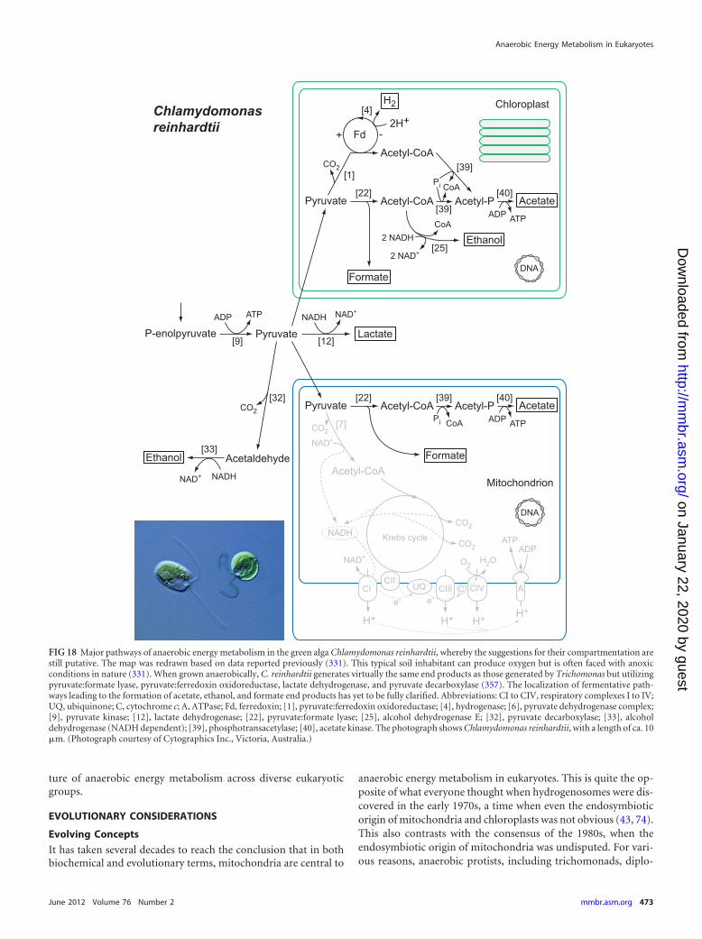
ture of anaerobic energy metabolism across diverse eukaryoticgroups.
EVOLUTIONARY CONSIDERATIONS
Evolving Concepts
It has taken several decades to reach the conclusion that in bothbiochemical and evolutionary terms, mitochondria are central to
anaerobic energy metabolism in eukaryotes. This is quite the op-posite of what everyone thought when hydrogenosomes were dis-covered in the early 1970s, a time when even the endosymbioticorigin of mitochondria and chloroplasts was not obvious (43, 74).This also contrasts with the consensus of the 1980s, when theendosymbiotic origin of mitochondria was undisputed. For vari-ous reasons, anaerobic protists, including trichomonads, diplo-
FIG 18 Major pathways of anaerobic energy metabolism in the green alga Chlamydomonas reinhardtii, whereby the suggestions for their compartmentation arestill putative. The map was redrawn based on data reported previously (331). This typical soil inhabitant can produce oxygen but is often faced with anoxicconditions in nature (331). When grown anaerobically, C. reinhardtii generates virtually the same end products as those generated by Trichomonas but utilizingpyruvate:formate lyase, pyruvate:ferredoxin oxidoreductase, lactate dehydrogenase, and pyruvate decarboxylase (357). The localization of fermentative path-ways leading to the formation of acetate, ethanol, and formate end products has yet to be fully clarified. Abbreviations: CI to CIV, respiratory complexes I to IV;UQ, ubiquinone; C, cytochrome c; A, ATPase; Fd, ferredoxin; [1], pyruvate:ferredoxin oxidoreductase; [4], hydrogenase; [6], pyruvate dehydrogenase complex;[9], pyruvate kinase; [12], lactate dehydrogenase; [22], pyruvate:formate lyase; [25], alcohol dehydrogenase E; [32], pyruvate decarboxylase; [33], alcoholdehydrogenase (NADH dependent); [39], phosphotransacetylase; [40], acetate kinase. The photograph shows Chlamydomonas reinhardtii, with a length of ca. 10�m. (Photograph courtesy of Cytographics Inc., Victoria, Australia.)
Anaerobic Energy Metabolism in Eukaryotes
June 2012 Volume 76 Number 2 mmbr.asm.org 473
on January 22, 2020 by guesthttp://m
mbr.asm
.org/D
ownloaded from
monads, and microsporidians, came to be viewed as primitivelineages of eukaryote evolution that branched off before the originof mitochondria (75, 349, 464, 546, 562).
There was also a window of time when the notion was enter-tained that hydrogenosomes were descended from a Clostridium-like endosymbiont that was distinct from the mitochondrion(347, 553), but in the 1990s, it became evident that the “early-branching” status of anaerobic protists was an artifact of molecu-lar phylogenetics and that the hydrogenosomes of various lineagesare mitochondria in the evolutionary sense (133, 382). The subse-quent discovery of mitosomes and their characterizations in sev-eral disparate lineages made a strong case for the ubiquity of mi-tochondria (300, 507, 508, 558). In the past 10 years, it becameclear that all eukaryotes possess an organelle of mitochondrialorigin (Fig. 2) (134, 454, 458). New order came to the eukaryotictree of life, in which anaerobic protists such as trichomonads,diplomonads, and microsporidians found homes next to mito-chondrion-bearing lineages within the six or so eukaryotic super-groups currently recognized (4, 25, 180, 192, 237, 457) (Fig. 3).Mitochondria are thus ubiquitous among eukaryotes, and they arealso germane to energy metabolism in eukaryotic anaerobes. Inenergetic terms, mitochondria themselves, whether aerobic or an-aerobic (not using oxygen in the role of the terminal electronacceptor), now appear causal to the origin of eukaryote complex-ity in that they removed the tight constraints that prokaryotic cellorganization places on the prokaryotic genome and cell complex-ity (267); that is, the foundation of the eukaryote cell complexityrests on the origin of �1,000 novel gene families that arose in theeukaryote common ancestor, those supporting the eukaryotic cellcycle, endomembrane traffic, flagella, and the like. That phase ofgene invention required amounts of energy per gene (or per ge-nome) that only the internalized configuration of bioenergeticmembranes afforded by mitochondria can supply (267).
Anaerobic Energy Metabolism: Present in the EukaryoteCommon Ancestor
Throughout those developments, the central evolutionary ques-tion has been, What is the origin of anaerobic energy metabolismin eukaryotes? From the previous sections of this review, it hashopefully become evident that anaerobic protists possess a fewenzymes, about 10, that mammals do not. In contrast, anaerobicmetazoans survive by using the same enzymes as humans do intheir mitochondria albeit with parts of the Krebs cycle runningbackwards—including a modified succinate dehydrogenase(SDH) functioning as a fumarate reductase (FRD) (416, 433,500)—and with complex I reducing rhodoquinone (RQ) insteadof ubiquinone (UQ). Thus, for the origin of anaerobic energymetabolism in metazoans, there is no differential gene contentwhatsoever to account for, only the origin of the cofactor RQ,whose biosynthesis is so far unresolved, whereby we recall that RQalso occurs in metazoans such as Caenorhabditis (484) and in pro-tists such as Euglena (196). For the origin of anaerobic energymetabolism in protists, the question is, Were the genes present inthe single eukaryote common ancestor, or do they clearly reflectmultiple origins, and if the former is true, does their single origincoincide with the origin of mitochondria? (131, 309, 501). Thishas in turn given rise to two main competing alternative hypoth-eses for the origin of anaerobic energy metabolism in protists: (i)the enzymes were present in the eukaryote ancestor and were in-
herited vertically by modern groups, or (ii) they were lacking inthe eukaryote ancestor (which would then implicitly have been astrict aerobe) and were acquired in different eukaryotes groupsindependently via lateral gene transfers (LGTs). Those views gen-erated very different predictions with regard to the evolutionarypatterns of the underlying genes.
If the enzymes of anaerobic energy metabolism were present inthe unicellular eukaryote common ancestor, different eukaryoticgroups should tend to have different subsets of the same ancestralcollection of genes, allowing for differential loss, and some eu-karyotes should have preserved nearly the entire set.
However, if anaerobic energy metabolism was lacking in theeukaryote common ancestor and had been acquired indepen-dently by different eukaryotic lineages, eukaryotic aerobes shouldlack such genes altogether, and the anaerobes of different eukary-otic lineages should tend to possess fundamentally different col-lections of genes reflecting acquisitions from the multitude ofways in which prokaryotic anaerobes can conserve energy in an-aerobic habitats.
The distribution of the enzymes for the pathways underlyinganaerobic energy metabolism presented here reveals that there isno fundamental difference between the major eukaryotic groupssurveyed with respect to either an aerobic or anaerobic life-style orthe enzymes involved. All major groups of eukaryotes currentlyrecognized harbor aerobes and anaerobes (Fig. 3), and if we com-pare the anaerobes, the following is observed.
For the metazoans, the core anaerobic pathway typified by Fas-ciola consists of (i) malate dismutation involving malic enzyme(ME), pyruvate dehydrogenase (PDH), and conventional Krebscycle enzymes and (ii) the propionyl-CoA cycle, involving fourenzymes that are also expressed in humans, methylmalonyl-CoAmutase, methylmalonyl-CoA epimerase, propionyl-CoA carbox-ylase, and propionate:succinate CoA-transferase. The metazoanFRD enzymes are currently indistinguishable from SDH at thelevel of the primary structure (501), acetate:succinate CoA-trans-ferase (ASCT) homologs function in aerobes (503), and complex Iaccepts both UQ and RQ as substrates, such that the only truly“special” component of the metazoan anaerobic mitochondrioncore pathway is RQ.
For the protists, the enzymes of Trichomonas hydrogenosomes,pyruvate:ferredoxin oxidoreductase (PFO), [Fe]-Hyd, ASCT, andsuccinyl-CoA synthetase (SCS) (a Krebs cycle enzyme), occur incombinations with various other enzymes which themselves arenot lineage specific (as genome database searching quickly re-veals). Examples include soluble FRD (present in Tritrichomonasand in Trypanosoma), pyruvate decarboxylase (PDC) and alcoholdehydrogenase (ADH) (Tritrichomonas and plants), a bifunc-tional aldehyde/alcohol dehydrogenase (ADHE) and acetyl-CoAsynthetase (ACS) (ADP forming) (Giardia and Entamoeba), thepropionyl-CoA cycle (Blastocystis), Krebs cycle enzymes (Nycto-therus and Euglena), pyruvate:formate lyase (PFL) (Piromyces andChlamydomonas), acetate kinase (ACK) and phosphotransacety-lase (PTA) (also called phosphate acetyltransferase) (Chlamy-domonas; both are also present in the oomycete Phytophthora, inthe apicomplexan relative Perkinsus, and in the moss Physcomi-trella), or PFO fused to an NADP�-reducing domain as pyruvate:NADP� oxidoreductase (PNO) (Euglena, Blastocystis, Perkinsus,and the apicomplexan Cryptosporidium) or fragmented acrossproteins as sulfite reductase in fungi (200, 419).
The differences in anaerobic energy metabolism observed
Müller et al.
474 mmbr.asm.org Microbiology and Molecular Biology Reviews
on January 22, 2020 by guesthttp://m
mbr.asm
.org/D
ownloaded from
among the protist lineages are minor and clearly reflect, in ourview, differential loss from a facultatively anaerobic ancestral stateof eukaryotic energy metabolism. The different lineages possessmerely different subsets of the same ancestral collection of genes.Furthermore, almost the entire collection is preserved withinChlamydomonas reinhardtii (21). We consider this to be verystrong evidence in favor of the enzymes being present in the eu-karyote common ancestor. Ginger et al. (161) surveyed the phy-logenetic distributions of some of the same enzymes and foundevidence for the presence of genes for anaerobic energy metabo-lism in the eukaryote common ancestor, followed by differentialloss.
Had the origin of the anaerobic life-style in the different eukary-otic lineages surveyed here entailed independent lateral gene ac-quisitions, as has been suggested (16, 211), one would expect dif-ferent eukaryotic lineages to have acquired different collections ofgenes as a result of their independent solutions to the anaerobicsurvival problem. However, this has not been observed. For ex-ample, there are no known eukaryotic anaerobes that performsulfate reduction, anaerobic ammonium oxidation, anaerobicmethane oxidation, uranium oxide reduction, or dozens of otheranaerobic energy metabolic pathways (13), even though eu-karyotes inhabit environments where such prokaryote physiolo-gies are abundant and successful strategies (291, 473).
Instead, eukaryotic anaerobes of different lineages possess differentsubsets of a common stock of only about 50 genes and enzymes (Ta-ble 1). This is a very modest number of basic tools in the anaerobictoolbox, similar to the number of genes required for subunits of mi-tochondrial complex I. These 50 enzymes are used in various combi-nations in different lineages—minor variations on a theme, as themaps presented in this paper show. If we examine the distribu-tion of the underlying genes across different eukaryotic groupsusing a standard database search approach (Fig. 19), it is clearthat there are no patterns to suggest lineage-specific acquisi-tions. Rather, the genes in question were present in the eu-karyote common ancestor and have undergone differential lossin various lineages.
It follows that the ancestral eukaryote stem was able to respireoxygen when present but in the absence of oxygen was also able toperform anaerobic respiration and fermentations of the typefound for the green alga C. reinhardtii. This free-living soil inhab-itant can gladly respire oxygen like humans, but after 30 min ofanaerobic growth in the dark, it uses the same enzymes as thoseused by Trichomonas or Piromyces to produce the same spectrumof metabolic end products as those heterotrophs (331, 357).Clearly, Chlamydomonas has preserved the largest subset of ances-tral eukaryote gene diversity for an anaerobic life-style (21) char-acterized so far (Fig. 19), while some highly reduced lineages, suchas the microsporidians, have lost almost all energy metabolic func-tions (Fig. 19), in accordance with their life-style as energy para-sites (238, 512, 554, 558).
Further evidence for the presence of anaerobic energy metabo-lism in the eukaryote common ancestor comes from the presenceof derived versions of [Fe]-Hyd and PFO in the genomes of eu-karyotic aerobes. Narf, also called Nar1 or Hyd3, is a eukaryote-specific derivative of [Fe]-Hyd that has conserved all but one (theH-cluster) of the FeS clusters of [Fe]-Hyd (199) and is present inall eukaryotes sampled so far. In Chlamydomonas, the Hyd3 pro-tein, like its homologs in yeast and humans, is involved in FeScluster biogenesis (162). This indicates that [Fe]-Hyd was present
in the eukaryote common ancestor, where it duplicated to give riseto Narf (Hyd3), which then acquired an essential role in FeS bio-genesis that has remained conserved across eukaryotes, even afterthe loss of the ancestral [Fe]-hydrogenase gene in many oxygen-adapted lineages. Another such example is the sulfite oxidase ofmany oxygen-adapted fungi, an enzyme consisting of two cata-lytic subunits corresponding to the yeast Met5 and Met10 genes(88), which are derived from eukaryotic PNO via domain rear-rangement (200, 419). The presence of functionally specializedforms of PNO [a fusion of PFO and a one-electron-accepting,NAD(P)�-reducing domain] and [Fe]-Hyd in diverse eukaryoticaerobes suggests that the genes were present in a facultatively an-aerobic eukaryote common ancestor.
A notable aspect of Fig. 19 is that virtually all of the enzymes ofeukaryotic energy metabolism are more similar to proteobacterialhomologs than to archaebacterial homologs, with enolase beingthe possible exception (183). This pattern of similarity is not at allsurprising for proteins germane to mitochondrial energy conser-vation, because of the eubacterial origin of mitochondria. Theeubacterial nature of cytosolic proteins involved in eukaryote en-ergy metabolism was initially more puzzling, however. It becameevident through studies of individual genes (183, 204, 304, 311,313) and stood in contrast to the findings, emerging at the sametime, that eukaryotic transcription (269) and translation (219,563) proteins are of an archaebacterial nature. This functional andphylogenetic dichotomy of eukaryotic genes into operational (forbiochemical functions, mainly eubacterial) and informational(for gene expression, mainly archaebacterial) classes (412) is nowwell documented (89, 139) and most easily attributed to endo-symbiosis and gene transfer at the origin of eukaryotes, in thesimplest scenario, gene transfer from the common ancestor ofmitochondria and hydrogenosomes to an archaebacterial host(91, 309). It could be argued that increased evolutionary rates inarchaebacterial proteins might be able to explain patterns such asthose shown in Fig. 19 (76), but specific investigations of ancientduplicate gene pairs showed that the evolutionary rate of archae-bacterial genomes is not increased at all; if anything, it is lowerthan that of eubacteria (95).
Hence, the similarity between enzymes of eukaryotic andeubacterial energy metabolisms, in particular the glycolyticpathway (309), reflects a eubacterial origin of the eukaryoticpathway. Indeed, the glycolytic (Embden-Meyerhof) pathway,as it is manifest in eukaryotes (148), is generally lacking inheterotrophic archaebacteria altogether; they use modifica-tions thereof or modifications of the Entner-Doudoroff path-way instead (34, 456). Moreover, archaebacterial enzymes en-tailing glucose oxidation to pyruvate are often altogetherunrelated to their eukaryotic and eubacterial counterparts.Many examples of archaebacterial enzymes with “glycolytic”activities that are unrelated to eukaryotic counterparts weresummarized by Siebers and Schönheit (456); these include glu-cokinase (ribokinase family), phosphoglucoisomerase (cupinfamily), the ADP-dependent phosphofructokinases (riboki-nase family), the archaeal type I aldolase, the phosphorylatingglyceraldehyde-3-phosphate dehydrogenase (GAPDH), andthe ferredoxin-dependent glyceraldehyde-3-phosphate oxi-doreductase. Underscoring how different the glucose-to-pyru-vate conversions in archaebacteria and eukaryotes are, Say andFuchs (435) recently discovered the archaebacterial fructose1,6-bisphosphate aldolase/phosphatase, a bifunctional enzyme
Anaerobic Energy Metabolism in Eukaryotes
June 2012 Volume 76 Number 2 mmbr.asm.org 475
on January 22, 2020 by guesthttp://m
mbr.asm
.org/D
ownloaded from
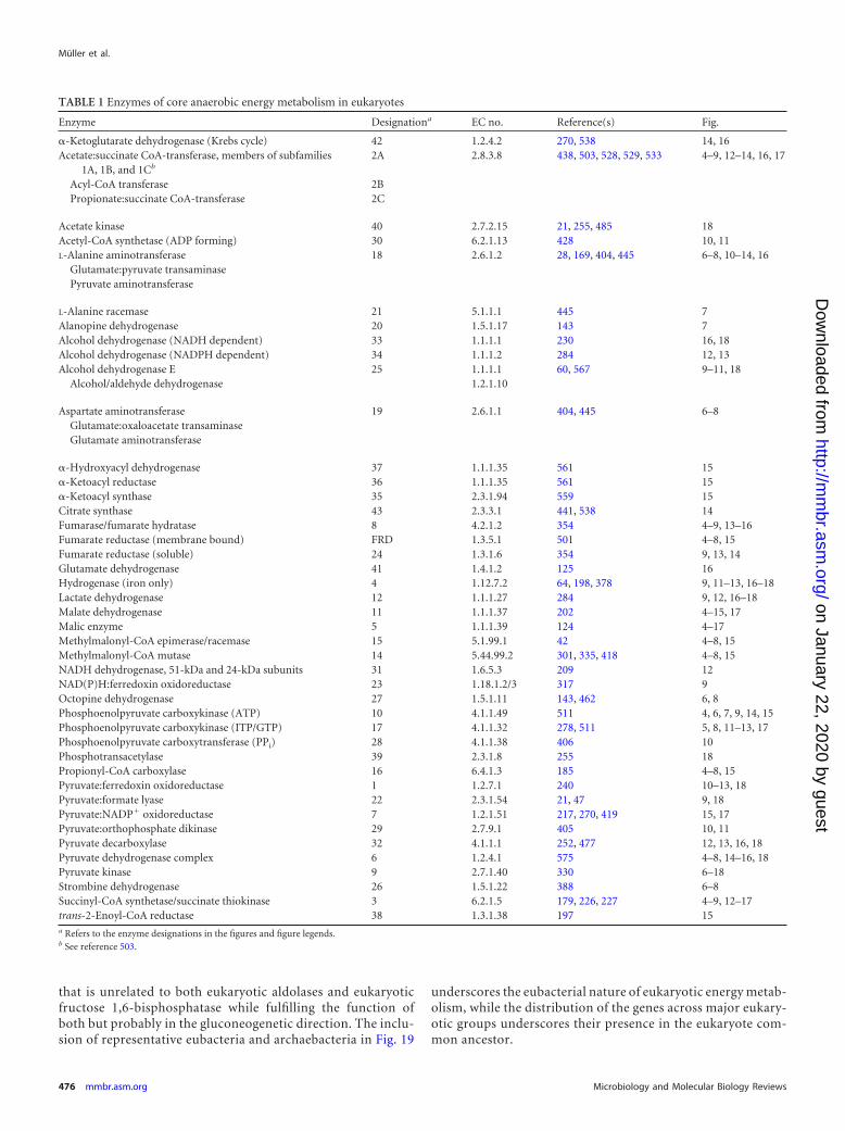
that is unrelated to both eukaryotic aldolases and eukaryoticfructose 1,6-bisphosphatase while fulfilling the function ofboth but probably in the gluconeogenetic direction. The inclu-sion of representative eubacteria and archaebacteria in Fig. 19
underscores the eubacterial nature of eukaryotic energy metab-olism, while the distribution of the genes across major eukary-otic groups underscores their presence in the eukaryote com-mon ancestor.
TABLE 1 Enzymes of core anaerobic energy metabolism in eukaryotes
Enzyme Designationa EC no. Reference(s) Fig.
�-Ketoglutarate dehydrogenase (Krebs cycle) 42 1.2.4.2 270, 538 14, 16Acetate:succinate CoA-transferase, members of subfamilies
1A, 1B, and 1Cb
2A 2.8.3.8 438, 503, 528, 529, 533 4–9, 12–14, 16, 17
Acyl-CoA transferase 2BPropionate:succinate CoA-transferase 2C
Acetate kinase 40 2.7.2.15 21, 255, 485 18Acetyl-CoA synthetase (ADP forming) 30 6.2.1.13 428 10, 11L-Alanine aminotransferase 18 2.6.1.2 28, 169, 404, 445 6–8, 10–14, 16
Glutamate:pyruvate transaminasePyruvate aminotransferase
L-Alanine racemase 21 5.1.1.1 445 7Alanopine dehydrogenase 20 1.5.1.17 143 7Alcohol dehydrogenase (NADH dependent) 33 1.1.1.1 230 16, 18Alcohol dehydrogenase (NADPH dependent) 34 1.1.1.2 284 12, 13Alcohol dehydrogenase E 25 1.1.1.1 60, 567 9–11, 18
Alcohol/aldehyde dehydrogenase 1.2.1.10
Aspartate aminotransferase 19 2.6.1.1 404, 445 6–8Glutamate:oxaloacetate transaminaseGlutamate aminotransferase
�-Hydroxyacyl dehydrogenase 37 1.1.1.35 561 15�-Ketoacyl reductase 36 1.1.1.35 561 15�-Ketoacyl synthase 35 2.3.1.94 559 15Citrate synthase 43 2.3.3.1 441, 538 14Fumarase/fumarate hydratase 8 4.2.1.2 354 4–9, 13–16Fumarate reductase (membrane bound) FRD 1.3.5.1 501 4–8, 15Fumarate reductase (soluble) 24 1.3.1.6 354 9, 13, 14Glutamate dehydrogenase 41 1.4.1.2 125 16Hydrogenase (iron only) 4 1.12.7.2 64, 198, 378 9, 11–13, 16–18Lactate dehydrogenase 12 1.1.1.27 284 9, 12, 16–18Malate dehydrogenase 11 1.1.1.37 202 4–15, 17Malic enzyme 5 1.1.1.39 124 4–17Methylmalonyl-CoA epimerase/racemase 15 5.1.99.1 42 4–8, 15Methylmalonyl-CoA mutase 14 5.44.99.2 301, 335, 418 4–8, 15NADH dehydrogenase, 51-kDa and 24-kDa subunits 31 1.6.5.3 209 12NAD(P)H:ferredoxin oxidoreductase 23 1.18.1.2/3 317 9Octopine dehydrogenase 27 1.5.1.11 143, 462 6, 8Phosphoenolpyruvate carboxykinase (ATP) 10 4.1.1.49 511 4, 6, 7, 9, 14, 15Phosphoenolpyruvate carboxykinase (ITP/GTP) 17 4.1.1.32 278, 511 5, 8, 11–13, 17Phosphoenolpyruvate carboxytransferase (PPi) 28 4.1.1.38 406 10Phosphotransacetylase 39 2.3.1.8 255 18Propionyl-CoA carboxylase 16 6.4.1.3 185 4–8, 15Pyruvate:ferredoxin oxidoreductase 1 1.2.7.1 240 10–13, 18Pyruvate:formate lyase 22 2.3.1.54 21, 47 9, 18Pyruvate:NADP� oxidoreductase 7 1.2.1.51 217, 270, 419 15, 17Pyruvate:orthophosphate dikinase 29 2.7.9.1 405 10, 11Pyruvate decarboxylase 32 4.1.1.1 252, 477 12, 13, 16, 18Pyruvate dehydrogenase complex 6 1.2.4.1 575 4–8, 14–16, 18Pyruvate kinase 9 2.7.1.40 330 6–18Strombine dehydrogenase 26 1.5.1.22 388 6–8Succinyl-CoA synthetase/succinate thiokinase 3 6.2.1.5 179, 226, 227 4–9, 12–17trans-2-Enoyl-CoA reductase 38 1.3.1.38 197 15a Refers to the enzyme designations in the figures and figure legends.b See reference 503.
Müller et al.
476 mmbr.asm.org Microbiology and Molecular Biology Reviews
on January 22, 2020 by guesthttp://m
mbr.asm
.org/D
ownloaded from
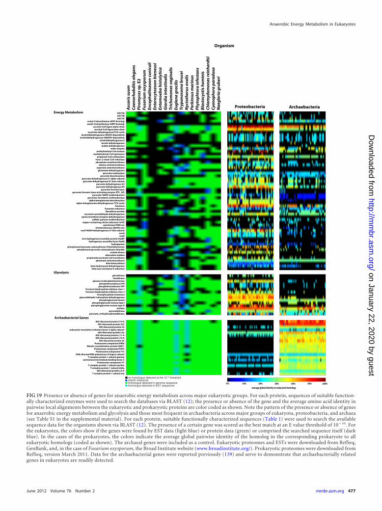
FIG 19 Presence or absence of genes for anaerobic energy metabolism across major eukaryotic groups. For each protein, sequences of suitable function-ally characterized enzymes were used to search the databases via BLAST (12); the presence or absence of the gene and the average amino acid identity inpairwise local alignments between the eukaryotic and prokaryotic proteins are color coded as shown. Note the pattern of the presence or absence of genesfor anaerobic energy metabolism and glycolysis and those most frequent in archaebacteria across major groups of eukaryota, proteobacteria, and archaea(see Table S1 in the supplemental material). For each protein, suitable functionally characterized sequences (Table 1) were used to search the availablesequence data for the organisms shown via BLAST (12). The presence of a certain gene was scored as the best match at an E value threshold of 10�10. Forthe eukaryotes, the colors show if the genes were found by EST data (light blue) or protein data (green) or comprised the searched sequence itself (darkblue). In the cases of the prokaryotes, the colors indicate the average global pairwise identity of the homolog in the corresponding prokaryote to alleukaryotic homologs (coded as shown). The archaeal genes were included as a control. Eukaryotic proteomes and ESTs were downloaded from RefSeq,GenBank, and, in the case of Fusarium oxysporum, the Broad Institute website (www.broadinstitute.org/). Prokaryotic proteomes were downloaded fromRefSeq, version March 2011. Data for the archaebacterial genes were reported previously (139) and serve to demonstrate that archaebacterially relatedgenes in eukaryotes are readily detected.
Anaerobic Energy Metabolism in Eukaryotes
June 2012 Volume 76 Number 2 mmbr.asm.org 477
on January 22, 2020 by guesthttp://m
mbr.asm
.org/D
ownloaded from
Comparing Diversity: Eukaryotes versus Rhodobacter
The gene presence or absence criterion is a robust source of evi-dence for the presence of anaerobic energy metabolism in theeukaryote common ancestor. Similar evidence has been found forthe distribution and utilization of rhodoquinone (RQ). RQ is acomparatively rare quinone among prokaryotes (366), reportedso far only for facultatively anaerobic Rhodobacter-related alpha-proteobacteria (191) and two betaproteobacteria (50). Among eu-karyotes, it has been reported for organisms that use membrane-bound fumarate reductase in anaerobic redox balance, such asparasitic helminths (500), the free-living nematode Caenorhabdi-tis elegans (484), and Euglena (196). It is required for quinol-basedfumarate reduction instead of ubiquinone (UQ) because of itslower redox potential (501). RQ provides a robust biochemicallink between the anaerobic energy metabolism found in mito-chondria and that found in facultatively anaerobic alphaproteo-bacteria.
This brings us to a notable observation: the whole spectrum ofknown energy metabolic pathways in eukaryotes encompasses lessdiversity than can be found in one single individual typical gener-alist purple nonsulfur bacterium (alphaproteobacterium). This isillustrated by the example of species such as Rhodospirillumrubrum or Rhodobacter capsulatus, where aerobic respiration (8,31, 166) is found, where the same spectrum of fermentation endproducts is found as among eukaryotes during anaerobic hetero-trophy (447) and where FRD and RQ are found (190). However,on top of that, Rs. rubrum and Rd. capsulatus can also performanaerobic photolithoautotrophy (479, 550), anaerobic photohet-erotrophy (35), nitrate respiration (451), dimethyl sulfoxide res-piration (425), or growth on carbon monoxide (239), to name afew of the possibilities, in addition to being able to fix nitrogen inthe light or in the dark (425), if needed.
Seen from this perspective, anaerobic energy metabolism in eu-karyotes carries the rather unmistakable imprint of a single acqui-sition via endosymbiosis in that it was acquired once, it representsa very narrow sample of preexisting prokaryotic metabolic diver-sity, and, at least with regard to an important quinone, RQ, it istraced physiologically to a particular group (facultatively anaero-bic alphaproteobacteria). This situation is similar to the cyano-bacterial origin of plastids, which also entailed the single acquisi-tion of a small sample of preexisting photosynthetic diversity via aplastid endosymbiont (165), followed by specialization and differ-ential loss (of phycobilisomes in some lineages, for example). Be-yond the mitochondrion, there is no evidence for the participationof any other endosymbionts in the origin of the primarily hetero-trophic eukaryotic groups, involving microbodies (152), flagella(225), or otherwise (314).
Forests, Trees, and Vertically Inherited Chimerism
The origin of anaerobic energy metabolism in eukaryotes has beenstudied by using phylogenetic analyses. Investigations have beenof two basic types: (i) studies addressing the relationship of mito-chondria to free-living prokaryotes, generating inferences aboutthe physiology of the free-living ancestor of mitochondria, and (ii)single-gene phylogenies inferring the phylogenies of individualenzymes involved in anaerobic energy metabolism. There havebeen more reports of both approaches than we can cover in anydepth; hence, we focus on a summary of recent trends.
The origin of mitochondria has been extensively studied from
the standpoint of phylogenetic analyses of gene products encodedby mitochondrial DNA (167). Conflicting results have been ob-tained with mitochondrial genes, which are usually strung to-gether (concatenated) into one big alignment for such studies,with different analyses pointing to three basic kinds of results: anorigin of mitochondria from Rickettsia-like ancestors (16), an or-igin from Rhodobacter-like facultatively anaerobic alphaproteo-bacteria (2, 565), or an origin from free-living oceanic alphapro-teobacteria of the SAR clade (52, 497). Phylogenetic analyses ofmitochondrial genes together with homologs from free-livingprokaryotes are technically challenging because of the AT richnessand rapid evolutionary rate of mitochondrial DNA. This can leadto artifacts and conflicting phylogenetic results, depending onhow these effects are computationally taken into account, espe-cially because the alphaproteobacteria harbor several AT-rich andrapidly evolving lineages (497).
Mitochondrial genes are not, however, the only source of phy-logenetic information to investigate the evolutionary affinities ofthe mitochondrial ancestor, whereby analyses of nuclear-encodedproteins of mitochondrial origin have also produced conflictingresults. Trees for pyruvate dehydrogenase subunits pointed toRickettsia-like ancestors (16). Trees for Krebs cycle and glyoxylatecycle enzymes (441) as well as trees for �200 nuclear-encodedmitochondrial proteins from Chlamydomonas point more fre-quently to origins from generalist, facultatively anaerobic alpha-proteobacteria (20) than to origins from Rickettsia-like ancestors,whereby many proteins indicated a eubacterial but not a specifi-cally alphaproteobacterial ancestry. A recent analysis of 86 yeastnuclear-encoded mitochondrial proteins produced a similar re-sult: some pointed to Rickettsia-like ancestors, and some pointedto facultatively anaerobic Rhodobacter-like ancestors (2). Clearly,nuclear genes for anaerobic energy metabolism cannot implicateRickettsia-like ancestors, because rickettsias are strict aerobes thatharbor no genes of anaerobic energy metabolism for comparison.Using an automated pipeline for phylogenetic trees, Gabaldonand Huynen (153) identified 630 nuclear-encoded protein fami-lies that trace to the ancestor of mitochondria, whereby the alpha-proteobacteria seldom formed a monophyletic group. Those re-searchers also found that a large fraction of nuclear-encoded geneproducts that were acquired from the mitochondrial ancestor nowfunction in the cytosol or other compartments (153), the sameresult that earlier studies of gene acquisitions from cyanobacteriain plants had uncovered (312). This makes sense, because genesthat were transferred from the mitochondrion to the host beforethe mitochondrial protein import machinery had evolved wereexpressed in the host’s cytosol (309). In a genomewide supertreeanalysis, nuclear genomes of nonphotosynthetic eukaryotes evenbranched within the alphaproteobacteria (385), a result most eas-ily explained by the circumstance that eukaryotes acquired manyof their nuclear genes from the ancestor of mitochondria (139,253, 254, 267, 308, 504).
However, supertrees and concatenated alignments blend oftenconflicting phylogenetic results from many genes into a singleresult. If sequence alignments are analyzed individually, ratherthan as a concatenated alignment, both mitochondrial genes andnuclear genes of a mitochondrial origin trace not to a single ap-parent donor but to many different apparent donors. The mito-chondrial genome of the jacobid Reclinomonas americana (268) isstill the most gene-rich mtDNA known and, hence, figures prom-inently in such studies. It contains 63 protein-coding genes, only
Müller et al.
478 mmbr.asm.org Microbiology and Molecular Biology Reviews
on January 22, 2020 by guesthttp://m
mbr.asm
.org/D
ownloaded from
about 55 of which are well conserved enough to permit the con-struction of alignments and trees. Trees for those 55 proteins en-coded by Reclinomonas mitochondrial DNA give conflicting re-sults, some proteins point to Rickettsia-like ancestors, some pointto Rhodobacter-like ancestors, and some point to Rhizobium-likeancestors, while some Reclinomonas mitochondrially encodedproteins do not reflect an alphaproteobacterial ancestry at all but,more generally, a eubacterial ancestry (139). A more recent re-analysis of the Reclinomonas mitochondrially encoded protein setproduced a similar result: some pointed to Rickettsia-like ances-tors, and some pointed to Rhodobacter-like ancestors (2).
The fact that different genes of mitochondrial origin or differentgenes of mitochondrial DNA can be traced to many different pro-karyotic genomes, rather than one single contemporary lineage, isnot usually taken to indicate that mitochondria had multiple ori-gins. Indeed, we can be sure that mitochondria had only one singleorigin (121, 134, 167, 521). Gene trees indicating origins of mito-chondria from different lineages have at least two main causes:phylogenetic error and lateral gene transfer (LGT) among pro-karyotes. Many branching discrepancies can be readily explainedby the errors inherent to phylogenetic inference, such as long-branch attraction, shifts in functional constraints, lineage-specificbiases, rate differences, AT richness, and the like (212, 497). As aparticularly prominent example of the influence that phylogeneticmethods can have on the issue of inferences of mitochondrial andhydrogenosomal evolution, Dyall et al. (128) analyzed the 51-kDasubunit of complex I from Trichomonas hydrogenosomes and re-ported evidence for a separate origin of mitochondria and hydro-genosomes, while Hrdy et al. (209) analyzed the same data usingdifferent phylogenetic methods that can recover more ancientevolutionary information from the data and found evidence for acommon ancestry for mitochondria and hydrogenosomes.
The second reason why different genes of a mitochondrial ori-gin point to different mitochondrial ancestors is that the role ofLGT among free-living prokaryotes complicates the issue in amanner that was long overlooked. The proteobacterium that gaverise to mitochondria lived �1.5 billion years (Ga) ago. It had aspecific (as-yet-unknown) collection of genes in its genome, andsince the time of mitochondrial origin, there has been ample op-portunity for its free-living sisters (the immediate relatives of mi-tochondria that did not become endosymbionts) to undergo genetransfer with other prokaryotes in the wild (2, 140, 409, 494a). Theendosymbiont itself remained isolated from further gene ex-change, as we see for modern endosymbiotic bacteria (339). Theresult of this effect as it applies to gene phylogenies involvingmodern genomes is that different genes in mtDNA and differentnuclear genes of mitochondrial origin appear to stem from differ-ent and diverse bacterial lineages, even though they were broughtin by one and the same mitochondrial endosymbiont (2, 306,409). A gene acquired via the mitochondrion should thus tend toshow a single origin for eukaryotes (131, 134), but it might notshow a specifically alphaproteobacterial ancestry, because likeother proteobacteria, alphaproteobacteria have been undergoingLGT over evolutionary time (244).
Hence, very many genes of eukaryotes reflect a presence in theeukaryote common ancestor and a eubacterial ancestry but notspecifically an alphaproteobacterial ancestry (89, 411, 412, 494a).A recent example for a single origin is pyruvate:formate lyase(PFL) (467), where one can interpret the data as evidence for (i)lateral gene transfer from clostridia to a eukaryote, followed by
eukaryote-to-eukaryote gene transfers of an unidentified nature,mimicking a single origin of the eukaryotic gene (467), or (ii) asingle acquisition from a mitochondrial endosymbiont in the eu-karyote ancestor, followed by the transfer of the gene amongprokaryotes (494a). The latter interpretation acknowledges thatprokaryotes, especially proteobacteria (244), do indeed acquireand donate genes avidly during evolution (123), via known mech-anisms (94, 389). There is no reason to assume that the free-livingrelatives of mitochondria were immune to LGT.
It is becoming increasingly apparent that the genome of theancestor of mitochondria was itself a mosaic of genes from differ-ent sources (2, 306, 409), just like modern alphaproteobacterialgenomes are today (140, 244). Thus, the collection of genes thateukaryote nuclear genomes acquired from the mitochondrial en-dosymbiont represents a kind of vertically inherited chimerism.
Without forgetting those many trees, there is also a more forest-like observation to be made regarding eukaryotic anaerobes: aslineage and genome samplings for eukaryotic anaerobes havegradually improved over 4 decades since the discovery of hydro-genosomes, the same few genes for anaerobic energy metabolismhave been found again and again in the various new eukaryoticlineages sampled (161, 349, 353, 355), pointing clearly to the pres-ence of those genes in the eukaryote common ancestor. During thesame time, exactly the opposite trend has been observed for pro-karyotic anaerobes: as prokaryotic lineage sampling has increasedover the last 4 decades, the diversity of known metabolic types,core energy metabolic reactions, and the genes underlying bothhave skyrocketed (13). Bearing witness to the burgeoning meta-bolic diversity of prokaryotes is the circumstance that fundamen-tally new core metabolic enzymes and pathways are still beingdiscovered among prokaryotes (34, 188, 233, 274, 372, 400, 408,435). In contrast, the Chlamydomonas nuclear genome comesclose to containing the complete collection of genes involved inanaerobic energy metabolism across all eukaryotes (21). No pro-karyote genome comes anywhere close to containing the full spec-trum of genes involved in anaerobic energy metabolism across allprokaryotes.
There is clearly a fundamental difference between prokaryotesand eukaryotes with respect to how they generate lineage-specificgenetic novelty: prokaryotes use LGT, allowing different lineagesto assemble virtually limitless new combinations of genes, whileeukaryotes use genome duplications and sex (26), allowing themplenty of room to generate new morphological types but littleroom to improvise on energy metabolism. LGT among pro-karyotes operates with known mechanisms of conjugation, trans-duction, transformation (367, 389), and gene transfer agents(318). Similar mechanisms do not exist in eukaryotes, which havemeiosis instead. Accordingly, two individuals of a eukaryotic spe-cies will tend to have the same numbers of genes and loci, but thisis not so for prokaryotes. E. coli individuals typically harbor 4,500genes, but this is only about 25% of the pangenome of the species,which, at current count, contains some 18,000 genes (293). Exceptfor gene transfers from organelles to the nucleus, which are wellcharacterized at the mechanistic level (186, 210, 461, 504), mech-anisms of LGT from prokaryotes to eukaryotes remain unknownand uncharacterized at the molecular level.
None of this is to say that there have been no lateral gene trans-fers whatsoever in eukaryotic evolution. For example, a carefulphylogenetic analysis of the Entamoeba and Trichomonas genomesuncovered evidence for LGT from prokaryotes, possibly affecting
Anaerobic Energy Metabolism in Eukaryotes
June 2012 Volume 76 Number 2 mmbr.asm.org 479
on January 22, 2020 by guesthttp://m
mbr.asm
.org/D
ownloaded from
about 1% to 2% of protein-coding genes (71, 290), a far lower LGTfrequency for eukaryotes than other surveys might imply (15,236). All things being equal, one might then expect that LGT couldhave affected 1% to 2% of the genes involved in anaerobic energymetabolism in eukaryotes, but with more samplings of eukaryoticlineages, those numbers should decline. From studies of eukary-otic anaerobes that unfolded after the discovery of hydrogeno-somes in trichomonads (279) and chytrids (570), one very clearcentral trend has emerged: as lineage sampling increases, we arenot discovering fundamentally new energy metabolic pathways;rather, we are finding the same genes over and over again, andmore and more genes trace to the eukaryote common ancestor.Lineage-specific acquisitions in core anaerobic energy metabo-lism—if there are any at all—are clearly the exception rather thanthe rule. Eukaryote nuclear genomes acquired a colorful collectionof genes from the ancestor of mitochondria, but they acquired itonly once and then inherited that chimerism vertically.
Functional Modules and Their Compartmentation
The function of core energy metabolism is to supply ATP and toafford redox balance. In anaerobic energy metabolism in eukaryotes,this entails a small number of discrete functional modules that aredifferentially distributed across the cytosol and in mitochondria indifferent lineages, as summarized in Table 2. Taking an updated lookat the eight main carbon flux modules recognized previously amonganaerobic protists (353), here we distinguish electron-donating pro-cesses, electron-accepting processes, and ATP-generating processesas well as their occurrences in the cytosol and the organelle. In anaer-obes, the most widespread oxidative process in the cytosol is, ofcourse, the reaction catalyzed by glyceraldehyde-3-phosphate dehy-drogenase (GAPDH) (77, 304). The oxidation of malate to pyruvateusually occurs in mitochondria but has been transferred to the cytosolin trichomonads, with a parallel route existing in its hydrogeno-somes. In Giardia and Entamoeba, the mitochondrion has undergonereduction to the level of a mitosome and has lost its function in corecarbon and energy metabolism; as a consequence, pyruvate:ferre-doxin oxidoreductase (PFO) in those lineages has been relocated tothe cytosol, where it catalyzes pyruvate oxidation to acetyl-CoA.
Electron-accepting processes in the cytosol lead to the forma-tion of the five major end products of cytosolic fermentationscovered here: lactate, opines, ethanol, succinate, glycerol, and, as aminor cytosolic product only for Giardia so far but suspected forEntamoeba, H2. The list of major cytosolic ATP-generating pro-cesses is devoid of lineage-specific variants (Table 2). The list oforganellar processes contains many processes that also occur inthe cytosol, and in several cases, the same enzymes are involved;exceptions are the acetyl-CoA-to-acetate pathways, which entailacetyl-CoA synthase (ADP forming) in the cytosol but phospho-transacetylase and acetate kinase (PTA/ACK) in the Chlamydomo-nas mitochondrion. Also of note for Chlamydomonas is that the[Fe]-Hyd is localized to the chloroplast in that species, at leastunder some conditions (560), and that an additional set of PTA/ACK isoenzymes appears to operate in the chloroplast (331). In asimilar vein, the ethanol-producing enzyme in Chlamydomonas,ADHE, is chloroplast localized but is mitochondrial in a very closealgal relative, Polytomella (22).
The examples of cytosolic PFO localization in Giardia and En-tamoeba versus the hydrogenosomal localization in trichomonadsand Piromyces versus the chloroplast localization in Chlamydomo-nas, in addition to the example of the ADHE localization shift for
Polytomella (mitochondrial) versus Chlamydomonas (chloro-plast), point to another important evolutionary aspect: compart-mentation. During evolution, enzymes and pathways can readilyundergo recompartmentation between mitochondrial, cytosolic,and, in the case of H2-producing algae, plastidic compartments.This has been evident since studies of chloroplast-cytosol isoen-zymes (315), comparative studies of the anaerobic protists (353),studies of the differential localization of isoprenoid biosynthesisto plastids and the cytosol of algae (276), studies of starch metab-olism in algae (104), studies of the localization of fatty acid me-tabolism in peroxisomes of some species and tissues (466), studiesof peroxisomal pathways in general (151), and—the classic exam-ple—studies of the glycosome in trypanosome evolution (172).
Each of these examples underscores the power of natural selec-tion to bring forth different and useful variations of enzymaticcompartmentation and sometimes variation in metabolic func-tion as well, starting from the one basic set of enzymatic tools. Thevast majority of mitochondrial and plastid proteins, and all hy-drogenosomal proteins, are nuclear encoded, and many of thosenuclear-encoded gene products are acquisitions from the endo-symbiotic ancestors of organelles (504). When the gene is trans-ferred from the organelle to the chromosomes of the host duringevolution, there is no guarantee or homing device to ensure thatits product will be targeted back to the donor organelle; naturalvariation and natural selection determine the outcome of the en-zyme compartmentation process. Moreover, during the earlyphases of organelle origins, before the evolution of an organelle-specific protein import machinery, any genes that were trans-ferred to the host’s chromosomes, became expressed, and ulti-mately became fixed gave rise to cytosolic products (315). Thisprocess of endosymbiotic gene transfer is how whole pathwayscould easily be transferred from DNA-containing organelles to thecytosol (309). Slightly more puzzling was the problem of how totransfer whole pathways (or major segments thereof) consisting ofseveral enzymes from the cytosol to a new organelle, for example,in the case of glycolysis moving to the glycosome (172). This isbecause a single enzyme in a new compartment is of no use and,hence, hardly selectable in evolution; the whole functional unithas to move. One solution to this problem involves the concept ofminor mistargeting (307). If protein targeting in eukaryotes is not100% specific (and many examples of dual targeting of enzymesare known), then minor amounts of whole pathways could con-ceivably end up in the wrong compartment. Such minor mistar-geting is all the more likely for highly expressed proteins, such asthose involved in core energy metabolism, and if a small amountof a whole pathway is present, it could readily become a unit offunction and, hence, a unit upon which natural selection could actto generate more or less of that specific compartmentation vari-ant. Clearly, pathways and enzymes are often recompartmental-ized during evolution, and the minor mistargeting mechanism(307) could, in principle, explain how whole pathways can attainnew states of subcellular localization (160).
Ecological Implications over Geological Time
The uniform nature and broadly shared conservation of genes foranaerobic energy metabolism across the major eukaryotic groupsindicate their presence in the common ancestor of eukaryotes,which can be viewed from the physiological standpoint as a “gar-den-variety” facultatively anaerobic heterotroph. When available,oxygen could have readily served as a terminal electron acceptor,
Müller et al.
480 mmbr.asm.org Microbiology and Molecular Biology Reviews
on January 22, 2020 by guesthttp://m
mbr.asm
.org/D
ownloaded from

TA
BLE
2Fu
nction
almodu
lesan
dth
eircom
partmen
tationin
anaerobic
energy
metabolism
ineu
karyotesa
Com
partmen
t,process,and
enzym
e
Presen
ceofen
zyme
Opisth
okonta
Am
oebozoaE
xcavataC
hrom
alveolatesA
rchaeplastida
FasciolaA
renicolaSipunculus
Mytilus
Ascaris
Pirom
ycesE
ntamoeba
Giardia
Trichom
onasT
ritrichomonas
Euglena
Nyctotherus
Blastocystis
Chlam
ydomonas
CytosolicE
lectrondon
orsG
lyceraldehyde-3-ph
osphate¡
1,3-BP
G●
●●
●●
●●
●●
●●
●●
●P
yruvate¡
AcC
oA●
●
Electron
acceptorsP
yruvate¡
lactate●
●●
●●
Pyru
vate¡opin
es●
●●
Pyru
vate¡eth
anol
●●
●●
AcC
oA¡
ethan
ol●
●●
Oxaloacetate¡
malate
●●
●●
●●
●●
●●
●Fu
marate¡
succin
ate(N
AD
H)
●●
Pyru
vate¡m
alate●
●●
H�
¡H
2●
DH
AP¡
glycerol●
●
AT
Pprodu
ctionP
hosph
oglyceratekin
ase●
●●
●●
●●
●●
●●
●●
●P
yruvate
kinase
●●
●●
●●
●●
●●
●●
PE
Pcarboxykin
ase●
●●
●●
●●
●●
●●
●A
cCoA
¡acetate
(AC
S[A
DP
])●
●
Organ
ellarE
lectrondon
orsM
alate¡pyru
vate●
●●
●●
●●
●●
●P
yruvate¡
AcC
oA(P
DH
)●
●●
●●
●●
Pyru
vate¡A
cCoA
(PFO
)●
●●
b
Pyru
vate¡A
cCoA
(PN
O)
●●
Pro/G
lu¡�
KG
¡su
ccinate
●
Electron
acceptorsFu
marate¡
succin
ate(R
Q)
●●
●●
●●
●H
�¡
H2
●●
●●
●?
●b
AcC
oA¡
ethan
ol●
b
Acyl-C
oA¡
lipids●
●N
itrate¡n
itrite*Su
lfur
compou
nds*
AT
Pprodu
ctionA
SCT
/SCS
cycle●
●●
●●
●●
●●
●A
cCoA
¡acetate
(PT
A/A
CK
)●
Propion
atecycle
●●
●●
●●
Ox
Ph
os(an
aerobic)●
●●
●●
●
aA
nasterisk
forth
epath
ways
involvin
gn
itratean
dsu
lfur
asterm
inalacceptors
refersto
denitrifi
cationin
foramin
iferans
(384,410)an
dFusarium
(241,242,359)an
dsu
lfur
reduction
inFusarium
(1).Aqu
estionm
arkin
dicatesth
ath
ydrogenprodu
ctionin
Blastocystis
(270)h
asn
otyet
beendirectly
demon
strated.BP
G,bisph
osphoglycerate;A
cCoA
,acetyl-CoA
;�K
G,�
-ketoglutarate;O
xP
hos,oxidative
phosph
orylation.
bH
2produ
ctionan
deth
anolprodu
ctionvia
AD
HE
inC
hlamydom
onasappear
tobe
plastidic,but
not
mitoch
ondrial,processes.
Anaerobic Energy Metabolism in Eukaryotes
June 2012 Volume 76 Number 2 mmbr.asm.org 481
on January 22, 2020 by guesthttp://m
mbr.asm
.org/D
ownloaded from
either with energy conservation in mitochondria via the respira-tory chain or in the cytosol using diaphorases that support redoxbalance without conserving energy (see “Anaerobes and Microae-rophiles: Redox Balance with a Pinch of O2”) or in the mitochon-drion without conserving energy, for example, via the alternativeoxidase (AOX). This leads to three broader questions. (i) How oldare eukaryotes as a group? (ii) How much oxygen is required foroxygen-dependent pathways? (iii) How available was oxygenwhen the eukaryote common ancestor lived and the earliest, fac-ultatively anaerobic eukaryotic lineages were diversifying?
Let us first look at eukaryote age. Statements that eukaryotes areabout 2.5 billion years (Ga) old are common (294) but are not wellsupported by data. Fossil microorganisms with a size and mor-phological complexity clearly representing eukaryotic microbesappear in rocks 1.45 Ga of age (223, 246), with the first fossils thatclearly represent eukaryotic algae appearing slightly later, in rocks1.2 Ga of age (67). Thus, by at least 1.2 Ga ago, eukaryotes hademerged and had commenced— or completed— diversificationinto the six major lineages recognized today. Molecular dates forthe major events in eukaryote evolution (83) are consistent withthat view. In terms of the geological time scale (548), eukaryotesarose and diversified during the Proterozoic eon, 2,500 to 542million years (Ma) ago. Current studies integrating molecular andfossil data indicate that eukaryotes started diversifying about 1.8Ga ago, at a time when the Earth’s chemistry, especially with re-gard to oxygen, was much different from today’s (376). Earlierreports, based on sterol biomarkers (geologically preserved or-ganic compounds), that eukaryotes can be assumed to be 2.7 Gaold (55) turned out to be doubly incorrect because (i) the sterolbiomarkers were younger than the rocks in which they were found(147, 401) and (ii) several groups of prokaryotes, including pro-teobacteria, synthesize sterols (379); sterols are synthesized by eu-karyotes but are in no way specific to eukaryotes.
Sterols bring us to the second question, regarding how muchoxygen is required for oxygen-dependent pathways. Sterols figureinto issues both on eukaryote age and on the relationship of eu-karyotes to oxygen. This is because oxygen is required as a cofactorin known eukaryotic sterol biosynthesis pathways: a squalene ep-oxidation step, three demethylation reactions, and other modifi-cations consume about 12 molecules of O2 per sterol backbone(547). Accordingly, sterols are widely used as geochemical proxiesfor the presence of oxygen in the ancient Earth environment, butthe crucial question is, How much oxygen does biological sterolsynthesis indicate? Waldbauer et al. (547) recently addressed thisquestion by using yeast and found that yeast requires only traceamounts of O2 for sterol biosynthesis. Unimpaired sterol accumu-lation was observed at 7 nM, the lowest dissolved oxygen concen-tration tested (547). In contrast, current atmospheric oxygen lev-els correspond to about 250 �M dissolved O2, and the Pasteurpoint (the concentration at which facultative anaerobic eu-karyotes start to respire O2) is about 2 �M dissolved O2. Thisindicates two things. First, sterol biomarkers indicate the presenceof oxygen at levels corresponding to a trace gas in the atmosphere(547) and, hence, make no statement about the oxygen require-ments of energy metabolism. Second, the ubiquity of sterol syn-thesis across diverse eukaryotic lineages (105) does not indicatethat eukaryotes arose in oxic environments; it merely suggests thatthe eukaryote common ancestor arose subsequent to the origin ofoxygenic photosynthesis (547).
What ecological setting confronted the emerging, pioneer eu-
karyotic lineages? The geosciences are currently delivering a new,detailed, and differentiated view of global ocean chemistry andecology during the Proterozoic era, one that meshes well with ourpresent understanding of anaerobic energy metabolism in eu-karyotes. This view, based on abundant findings, as reviewed else-where (111, 296) and supported by additional newer findings(229, 273, 362), is outlined in Fig. 20. As Lyons et al. (296) formu-lated it, the available data “point to global oxygen deficiency in thedeep ocean and perhaps euxinia throughout most, if not all, of theProterozoic, and likely extending into the Paleozoic.”
On the bottom line, the current view of Proterozoic oceanchemistry has it that prior to the appearance of oxygen in theatmosphere 2.4 Ga ago (295), the oceans were (of course) anoxicbut, in marked contrast to older views, remained anoxic for al-most 2 billion years after the onset of atmosphere oxygenation.More specifically, during the time from 2.4 to 1.8 Ga ago, theoceans below the photic zone were anoxic (19, 297) and ferrugi-nous (containing much dissolved iron). In the time from 1.8 Ga to�750 Ma ago, the oceans below the photic zone were euxinic(anoxic and sulfidic) (394), whereby Planavsky et al. (386) re-cently made the case for mixed ferruginous and sulfidic subsurfacewaters throughout the Proterozoic era. At �750 Ma ago, ferrugi-nous conditions persisted with continued anoxia (68), and atabout 580 Ma ago, the oceans finally became oxygenated belowthe photic zone (14, 68, 450). The geological and biological pro-cesses that generated these three apparent phases of Proterozoicmarine anoxia are far from pinpointed (386), and current discus-sions involve the interplay between a plethora of factors, includingthe oxygen-dependent weathering of continental sulfide depositscausing an influx of sulfate to the oceans, the growth of marinesulfate-reducing bacteria that produce H2S, marine anoxygenicphotosynthesizers that consume H2S, diversity among marine pri-mary producers, global glaciations, carbon burial rates, stratifiedocean chemistries (229, 273, 296), and various elemental cycles.With regard to eukaryote ecology, and given the age of eukaryotes,this suggests that early eukaryotic evolution was influenced byanoxic and sulfidic water masses in the oceans of their day (324,376). Today, the oxygen-poor sulfidic waters of hypersaline Med-iterranean basins (129) or the Black Sea (565a) abound with eu-karyotic microbes from the six supergroups, providing windowsinto protist ecology from the ancient past; the cultivation of theseorganisms and studies of their pathways provide exciting chal-lenges for the future.
The late oxygenation of the oceans at about 580 Ma ago is gen-erally brought into connection with the sudden appearance oflarge, diversified animal lineages in Ediacaran (630 to 542 Ma ago)fauna (69, 273, 452, 566); that is, the evolutionary lineages werealready in existence, but with the advent of oxygenated oceans, theanimals just increased in size (245, 247) and were furthermoreable to form more rigid bodies with the help of oxygen-dependentcollagen synthesis (509), consistent with the appearance of colla-gen in the earliest metazoan lineages (46). By inference, thesmaller metazoan ancestors from which the Ediacaran fauna arose(361) must have been well suited to life with low or no oxygen, inline with the biochemical findings presented here. The mitochon-drial sulfide-oxidizing metabolism of animals such as the annelidArenicola (496) or Urechis (299) that still inhabit sulfidic environ-ments can be seen as a relic from early metazoan evolution.
The recognition that anaerobic energy metabolism in moderneukaryotes is an ancestral character calls for a general rethinking of
Müller et al.
482 mmbr.asm.org Microbiology and Molecular Biology Reviews
on January 22, 2020 by guesthttp://m
mbr.asm
.org/D
ownloaded from
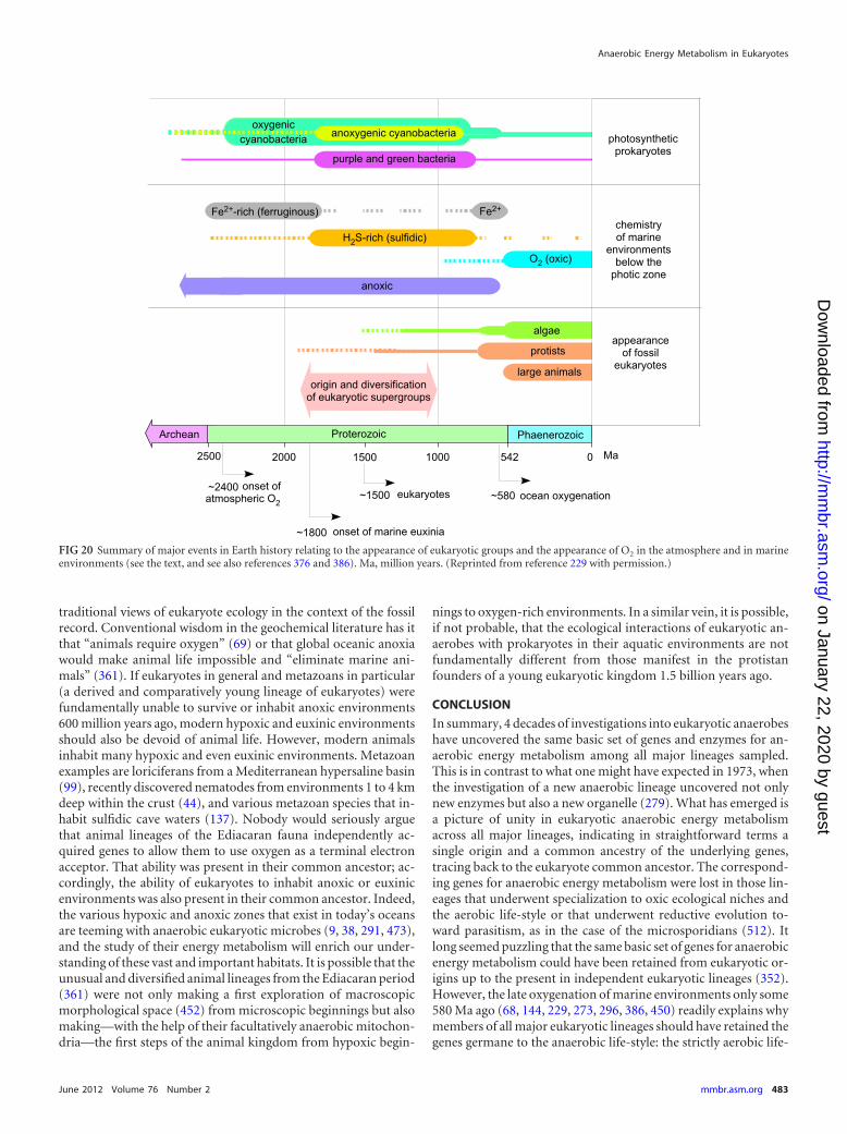
traditional views of eukaryote ecology in the context of the fossilrecord. Conventional wisdom in the geochemical literature has itthat “animals require oxygen” (69) or that global oceanic anoxiawould make animal life impossible and “eliminate marine ani-mals” (361). If eukaryotes in general and metazoans in particular(a derived and comparatively young lineage of eukaryotes) werefundamentally unable to survive or inhabit anoxic environments600 million years ago, modern hypoxic and euxinic environmentsshould also be devoid of animal life. However, modern animalsinhabit many hypoxic and even euxinic environments. Metazoanexamples are loriciferans from a Mediterranean hypersaline basin(99), recently discovered nematodes from environments 1 to 4 kmdeep within the crust (44), and various metazoan species that in-habit sulfidic cave waters (137). Nobody would seriously arguethat animal lineages of the Ediacaran fauna independently ac-quired genes to allow them to use oxygen as a terminal electronacceptor. That ability was present in their common ancestor; ac-cordingly, the ability of eukaryotes to inhabit anoxic or euxinicenvironments was also present in their common ancestor. Indeed,the various hypoxic and anoxic zones that exist in today’s oceansare teeming with anaerobic eukaryotic microbes (9, 38, 291, 473),and the study of their energy metabolism will enrich our under-standing of these vast and important habitats. It is possible that theunusual and diversified animal lineages from the Ediacaran period(361) were not only making a first exploration of macroscopicmorphological space (452) from microscopic beginnings but alsomaking—with the help of their facultatively anaerobic mitochon-dria—the first steps of the animal kingdom from hypoxic begin-
nings to oxygen-rich environments. In a similar vein, it is possible,if not probable, that the ecological interactions of eukaryotic an-aerobes with prokaryotes in their aquatic environments are notfundamentally different from those manifest in the protistanfounders of a young eukaryotic kingdom 1.5 billion years ago.
CONCLUSION
In summary, 4 decades of investigations into eukaryotic anaerobeshave uncovered the same basic set of genes and enzymes for an-aerobic energy metabolism among all major lineages sampled.This is in contrast to what one might have expected in 1973, whenthe investigation of a new anaerobic lineage uncovered not onlynew enzymes but also a new organelle (279). What has emerged isa picture of unity in eukaryotic anaerobic energy metabolismacross all major lineages, indicating in straightforward terms asingle origin and a common ancestry of the underlying genes,tracing back to the eukaryote common ancestor. The correspond-ing genes for anaerobic energy metabolism were lost in those lin-eages that underwent specialization to oxic ecological niches andthe aerobic life-style or that underwent reductive evolution to-ward parasitism, as in the case of the microsporidians (512). Itlong seemed puzzling that the same basic set of genes for anaerobicenergy metabolism could have been retained from eukaryotic or-igins up to the present in independent eukaryotic lineages (352).However, the late oxygenation of marine environments only some580 Ma ago (68, 144, 229, 273, 296, 386, 450) readily explains whymembers of all major eukaryotic lineages should have retained thegenes germane to the anaerobic life-style: the strictly aerobic life-
FIG 20 Summary of major events in Earth history relating to the appearance of eukaryotic groups and the appearance of O2 in the atmosphere and in marineenvironments (see the text, and see also references 376 and 386). Ma, million years. (Reprinted from reference 229 with permission.)
Anaerobic Energy Metabolism in Eukaryotes
June 2012 Volume 76 Number 2 mmbr.asm.org 483
on January 22, 2020 by guesthttp://m
mbr.asm
.org/D
ownloaded from
style (ours) is the one that arrived late on the scene of eukaryoticecological specialization.
ACKNOWLEDGMENTS
We thank Martin Embley, Manfred Grieshaber, Andy Knoll, Paul Mi-chels, Fred Opperdoes, and Jan Tachezy for numerous helpful commentsand suggestions on the manuscript.
We thank the NIH (grant no. AI 11942 to Miklós Müller), the DFG(Marek Mentel, Katrin Henze, and William F. Martin), the NWO (Jaap J.van Hellemond and Aloysius G. M. Tielens), the SFF fund of the HHUD(Sven B. Gould), the Slovak grant agency VEGA (grant no. 1/0870/11 toMarek Mentel), and the European Research Council (grant no. 232975 toWilliam F. Martin) for funding.
REFERENCES1. Abe T, Hoshino T, Nakamura A, Takaya N. 2007. Anaerobic elemental
sulfur reduction by fungus Fusarium oxysporum. Biosci. Biotechnol.Biochem. 71:2402–2407.
2. Abhishek A, Bavishi A, Choudhary M. 2011. Bacterial genome chima-erism and the origin of mitochondria. Can. J. Microbiol. 57:49 – 61.
3. Adam RD. 2001. Biology of Giardia lamblia. Clin. Microbiol. Rev. 14:447– 475.
4. Adl SM, et al. 2005. The new higher level classification of eukaryoteswith emphasis on the taxonomy of protists. J. Eukaryot. Microbiol. 52:399 – 451.
5. Aguilera P, Barry T, Tovar J. 2008. Entamoeba histolytica mitosomes:organelles in search of a function. Exp. Parasitol. 118:10 –16.
6. Ahmadinejad N, Dagan T, Martin W. 2007. Genome history in thesymbiotic hybrid Euglena gracilis. Gene 402:35–39.
7. Akhmanova A, et al. 1998. A hydrogenosome with a genome. Nature396:527–528.
8. Aklujkar M, Prince RC, Beatty JT. 2005. The puhE gene of Rhodobactercapsulatus is needed for optimal transition from aerobic to photosyn-thetic growth and encodes a putative negative modulator of bacterio-chlorophyll production. Arch. Biochem. Biophys. 437:186 –198.
9. Alexander E, et al. 2009. Microbial eukaryotes in the hypersaline anoxicL’Atalante deep-sea basin. Environ. Microbiol. 11:360 –381.
10. Allen JF. 2003. The function of genomes in bioenergetic organelles.Philos. Trans. R. Soc. Lond. B 358:19 –37.
11. Al-Subiai SN, Jha AN, Moody AJ. 2009. Contamination of bivalvehaemolymph samples by adductor muscle components: implications forbiomarker studies. Ecotoxicology 18:334 –342.
12. Altschul, et al. 1997. Gapped BLAST and PSI-BLAST: a new generationof protein database search programs. Nucleic Acids Res. 25:3389 –3402.
13. Amend JP, Shock EL. 2001. Energetics of overall metabolic reactions ofthermophilic and hyperthermophilic Archaea and Bacteria. FEMS Mi-crobiol. Rev. 25:175–243.
14. Amutha KB, Murugesan AG. 2011. Biological hydrogen production bythe algal biomass Chlorella vulgaris MSU 01 strain isolated from pondsediment. Bioresour. Technol. 102:194 –199.
15. Andersson JO, et al. 2007. A genomic survey of the fish parasite Spiro-nucleus salmonicida indicates genomic plasticity among diplomonadsand significant lateral gene transfer in eukaryote genome evolution.BMC Genomics 8:51. doi:10.1186/1471-2164-8-51.
16. Andersson SGE, Kurland CG. 1999. Origins of mitochondria and hy-drogenosomes. Curr. Opin. Microbiol. 2:535–541.
17. Ankarklev J, Jerlström-Hultqvist J, Ringqvist E, Troell K, Svärd SG.2010. Behind the smile: cell biology and disease mechanisms of Giardiaspecies. Nat. Rev. Microbiol. 8:413– 422.
18. Archibald JM. 2009. The puzzle of plastid evolution. Curr. Biol. 19:R81–R88.
19. Arnold GL, Anbar AD, Barling J, Lyons TW. 2004. Molybdenumisotope evidence for widespread anoxia in mid-Proterozoic oceans. Sci-ence 304:87–90.
20. Atteia A, et al. 2009. A proteomic survey of Chlamydomonas reinhardtiimitochondria sheds new light on the metabolic plasticity of the organelleand on the nature of the alpha-proteobacterial mitochondrial ancestor.Mol. Biol. Evol. 26:1533–1548.
21. Atteia A, et al. 2006. Pyruvate:formate lyase and a novel route of eukary-otic ATP-synthesis in anaerobic Chlamydomonas mitochondria. J. Biol.Chem. 281:9909 –9918.
22. Atteia A, et al. 2003. Bifunctional aldehyde/alcohol dehydrogenase(ADHE) in chlorophyte algal mitochondria. Plant Mol. Biol. 53:175–188.
23. Bakker BM, et al. 2001. Stoichiometry and compartmentation of NADHmetabolism in Saccharomyces cerevisiae. FEMS Microbiol. Rev. 25:15–37.
24. Ballantyne JS. 2004. Mitochondria: aerobic and anaerobic design—lessons from molluscs and fishes. Comp. Biochem. Physiol. B Biochem.Mol. Biol. 139:461– 467.
25. Bapteste E, et al. 2002. The analysis of 100 genes supports the groupingof three highly divergent amoebae: Dictyostelium, Entamoeba, and Mas-tigamoeba. Proc. Natl. Acad. Sci. U. S. A. 99:1414 –1419.
26. Bapteste E, et al. 2009. Prokaryotic evolution and the tree of life are twodifferent things. Biol. Direct 4:34. doi:10.1186/1745-6150-4-34.
27. Barbera MJ, et al. 2010. Sawyeria marylandensis (Heterolobosea) has ahydrogenosome with novel metabolic properties. Eukaryot. Cell 9:1913–1924.
28. Barrett J. 1991. Parasitic helminths, p 146 –164. In Bryant C (ed), Meta-zoan life without oxygen. Chapman and Hall, London, United Kingdom.
29. Barton RM, Worman HJ. 1999. Prenylated prelamin A interacts withNarf, a novel nuclear protein. J. Biol. Chem. 274:30008 –30018.
30. Bauchop T. 1989. Biology of gut anaerobic fungi. Biosystems 23:53– 64.31. Bauer CE, Bird TH. 1996. Regulatory circuits controlling photosynthe-
sis gene expression. Cell 85:5– 8.32. Benchimol M. 1999. The hydrogenosome. Acta Microsc. 8:1–22.33. Benchimol M. 2009. Hydrogenosomes under microscopy. Tissue Cell
41:151–168.34. Berg IA, et al. 2010. Autotrophic carbon fixation in archaea. Nat. Rev.
Microbiol. 8:447– 460.35. Berg IA, Krasilnikova EN, Ivanovsky RN. 2000. Investigation of the
dark metabolism of acetate in photoheterotrophically grown cells ofRhodospirillum rubrum. Microbiology 69:7–12.
36. Bernhard JM. 1993. Experimental and field evidence of Antarctic fora-miniferal tolerance to anoxia and hydrogen sulfide. Mar. Micropalaeon-tol. 20:203–213.
37. Bernhard JM, Habura A, Bowser SS. 2006. An endobiont-bearingallogromiid from the Santa Barbara Basin. Implications for the earlydiversification of foraminifera. J. Geophys. Res. 111:G03002. doi:10.1029/2005JG000158.
38. Bernhard JM, Buck KR, Farmer MA, Bowser SS. 2000. The SantaBarbara Basin is a symbiosis oasis. Nature 403:77– 80.
39. Besteiro S, et al. 2002. Succinate secreted by Trypanosoma brucei isproduced by a novel and unique glycosomal enzyme, NADH-dependentfumarate reductase. J. Biol. Chem. 277:38001–38012.
40. Biagini GA, Bernard C. 2000. Primitive anaerobic protozoa: a falseconcept? Microbiology 1148:1019 –1020.
41. Biagini GA, Finlay BJ, Lloyd D. 1997. Evolution of the hydrogenosome.FEMS Microbiol. Lett. 155:133–140.
42. Bobik TA, Rasche ME. 2003. HPLC assay for methylmalonyl-CoA epi-merase. Anal. Bioanal. Chem. 375:344 –349.
43. Bogorad L. 1975. Evolution of organelles and eukaryotic genomes. Sci-ence 188:891– 898.
44. Borgonie G, et al. 2011. Nematoda from the terrestrial deep subsurfaceof South Africa. Nature 474:79 – 82.
45. Bouillaud F, Blachier F. 2011. Mitochondria and sulfide: a very old storyof poisoning, feeding, and signaling? Antioxid. Redox Signal. 15:379 –391.
46. Boute N, et al. 1996. Type IV collagen in sponges, the missing link inbasement membrane ubiquity. Biol. Cell 88:37– 44.
47. Boxma B, et al. 2004. The anaerobic chytridiomycete fungus Piromycessp E2 produces ethanol via pyruvate:formate lyase and an alcohol dehy-drogenase E. Mol. Microbiol. 51:1389 –1399.
48. Boxma B, et al. 2007. The [FeFe] hydrogenase of Nyctotherus ovalis hasa chimeric origin. BMC Evol. Biol. 7:230. doi:10.1186/1471-2148-7-230.
49. Boxma B, et al. 2005. An anaerobic mitochondrion that produces hy-drogen. Nature 434:74 –79.
50. Brajcich BC, et al. 2010. Evidence that ubiquinone is a required inter-mediate for rhodoquinone biosynthesis in Rhodospirillum rubrum. J.Bacteriol. 192:436 – 445.
51. Bratosin D, et al. 2001. Programmed cell death in mature erythrocytes:a model for investigating death effector pathways operating in the ab-sence of mitochondria. Cell Death Differ. 8:1143–1156.
52. Brindefalk B, Ettema TJ, Viklund J, Thollesson M, Andersson SG.2011. A phylometagenomic exploration of oceanic alphaproteobacteria
Müller et al.
484 mmbr.asm.org Microbiology and Molecular Biology Reviews
on January 22, 2020 by guesthttp://m
mbr.asm
.org/D
ownloaded from
reveals mitochondrial relatives unrelated to the SAR11 clade. PLoS One6:e24457. doi:10.1371/journal.pone.0024457.
53. Bringaud F, Ebikeme C, Boshart M. 2010. Acetate and succinate pro-duction in amoebae, helminths, diplomonads, trichomonads andtrypanosomatids: common and diverse metabolic strategies used by par-asitic lower eukaryotes. Parasitology 137:1315–1331.
54. Bringaud F, Rivière L, Coustou V. 2006. Energy metabolism oftrypanosomatids: adaptation to available carbon sources. Mol. Biochem.Parasitol. 149:1–9.
55. Brocks JJ, Logan GA, Buick R, Summons RE. 1999. Archean molecularfossils and the early rise of eukaryotes. Science 285:1033–1036.
56. Broers CA, Stumm CK, Vogels GD, Brugerolle G. 1990. Psalteriomonaslanterna gen. nov., sp. nov., a free-living amoeboflagellate isolated fromfreshwater anaerobic sediments. Eur. J. Protistol. 25:369 –380.
57. Brondijk THC, et al. 1996. scsB, a cDNA encoding the hydrogenosomalbeta subunit of succinyl-CoA synthetase from the anaerobic fungus Neo-callimastix frontalis. Mol. Gen. Genet. 253:315–323.
58. Brown DM, Upcroft JA, Upcroft P. 1996. A H2O producing NADHoxidase from the protozoan parasite Giardia duodenalis. Eur. J. Biochem.241:155–161.
59. Brown DM, Upcroft JA, Edwards MR, Upcroft P. 1998. Anaerobicbacterial metabolism in the ancient eukaryote Giardia duodenalis. Int. J.Parasitol. 28:149 –164.
60. Bruchhaus I, Tannich E. 1994. Purification and molecular characteriza-tion of the NAD�-dependent acetaldehyde/alcohol dehydrogenase fromEntamoeba histolytica. Biochem. J. 303:743–748.
61. Bryant C (ed). 1991. Metazoan life without oxygen. Chapman and Hall,London, United Kingdom.
62. Buetow DE. 1989. The mitochondrion, p 247–314. In Buetow DE (ed),The biology of Euglena, vol 4. Academic Press, San Diego, CA.
63. Bui ETN, Bradley PJ, Johnson PJ. 1996. A common evolutionary originfor mitochondria and hydrogenosomes. Proc. Natl. Acad. Sci. U. S. A.93:9651–9656.
64. Bui ET, Johnson PJ. 1996. Identification and characterization of [Fe]-hydrogenases in the hydrogenosome of Trichomonas vaginalis. Mol.Biochem. Parasitol. 76:305–310.
65. Burki F, Shalchian-Tabrizi K, Pawlowski J. 2008. Phylogenomics re-veals a new ‘megagroup’ including most photosynthetic eukaryotes. Biol.Lett. 4:366 –369.
66. Burnett LE. 1997. The challenges of living in hypoxic and hypercapnicaquatic environments. Am. Zool. 37:633– 640.
66a.Burstein D, et al. 2012. A machine-learning approach to identify hy-drogenosomal proteins in Trichomonas vaginalis. Eukaryot. Cell 11:217–228.
67. Butterfield NJ. 2000. Bangiomorpha pubescens n. gen., n. sp.: implica-tions for the evolution of sex, multicellularity, and the Mesoproterozoic/Neoproterozoic radiation of eukaryotes. Paleobiology 26:386 – 404.
67a.Campbell T, Rubin N, Komuniecki R. 1989. Succinate-dependent en-ergy generation in Ascaris suum mitochondria. Mol. Biochem. Parasitol.33:1–12.
68. Canfield DE, et al. 2008. Ferruginous conditions dominated later neo-proterozoic deep-water chemistry. Science 321:949 –952.
69. Canfield DE, Poulton SW, Narbonne GM. 2007. Late-Neoproterozoicdeep-ocean oxygenation and the rise of animal life. Science 315:92–95.
70. Cardol P, et al. 2005. The mitochondrial oxidative phosphorylationproteome of Chlamydomonas reinhardtii deduced from the genome se-quencing project. Plant Physiol. 137:447– 459.
71. Carlton JM, et al. 2007. Draft genome sequence of the sexually trans-mitted pathogen Trichomonas vaginalis. Science 315:207–212.
72. Castro-Guerrero NA, Krab K, Moreno-Sanchez R. 2004. The alterna-tive respiratory pathway of Euglena mitochondria. J. Bioenerg.Biomembr. 36:459 – 469.
73. Castro-Guerrero NA, Jasso-Chavez R, Moreno-Sanchez R. 2005. Phys-iological role of rhodoquinone in Euglena gracilis mitochondria.Biochim. Biophys. Acta 1710:113–121.
74. Cavalier-Smith T. 1975. The origin of nuclei and of eukaryotic cells.Nature 256:463– 468.
75. Cavalier-Smith T. 1987. Eukaryotes with no mitochondria. Nature 326:332–333.
76. Cavalier-Smith T. 2010. Origin of the cell nucleus, mitosis and sex: rolesof intracellular coevolution. Biol. Direct 5:7. doi:10.1186/1745-6150-5-7.
77. Cerff R, Chambers S. 1979. Subunit structure of higher plant glyceral-
dehyde-3-phosphate dehydrogenases (EC 1.2.1.12 and 1.2.1.13). J. Biol.Chem. 254:6094 – 6098.
78. Cerkasovova A, Cerkasov J. 1974. Location of NADH-oxidase activityin fractions of Tritrichomonas foetus homogenate. Folia Parasitol. 21:193–203.
79. Chabriere E, et al. 1999. Crystal structures of the key anaerobic enzymepyruvate:ferredoxin oxidoreductase, free and in complex with pyruvate.Nat. Struct. Biol. 6:182–190.
80. Chan KW, et al. 2005. A novel ADP/ATP transporter in the mitosome ofthe microaerophilic human parasite Entamoeba histolytica. Curr. Biol.15:737–742.
81. Chapman A, Linstead DJ, Lloyd D. 1999. Hydrogen peroxide is aproduct of oxygen consumption by Trichomonas vaginalis. J. Biosci. 24:339 –344.
82. Chapman A, Linstead DJ, Lloyd D, Williams J. 1985. 13C-NMR revealsglycerol as an unexpected major metabolite of the protozoan parasiteTrichomonas vaginalis. FEBS Lett. 191:287–292.
83. Chernikova D, Motamedi S, Csuros M, Koonin EV, Rogozin IB.2011. A late origin of the extant eukaryotic diversity: divergence timeestimates using rare genomic changes. Biol. Direct 6:26. doi:10.1186/1745-6150-6-26.
84. Clark CG, Johnson PJ, Adam RD (ed). 2010. Anaerobic parasiticprotozoa: genomics and molecular biology. Caister Academic Press,Norfolk, United Kingdom.
85. Clarkson AB, Jr, Bienen EJ, Pollakis G, Grady RW. 1989. Respirationof bloodstream forms of the parasite Trypanosoma brucei brucei is depen-dent on a plant-like alternative oxidase. J. Biol. Chem. 264:17770 –17776.
86. Clayton AM, et al. 2011. Depletion of mitochondrial acyl carrier proteinin bloodstream-form Trypanosoma brucei causes a kinetoplast segrega-tion defect. Eukaryot. Cell 10:286 –292.
87. Coombs GH, Müller M. 1995. Energy metabolism in anaerobic proto-zoa, p 33– 47. In Marr JJ, M̈uller M (ed), Biochemistry and molecularbiology of parasites. Academic Press, London, United Kingdom.
88. Cordente AG, Heinrich A, Pretorius IS, Swiegers JH. 2009. Isolation ofsulfite reductase variants of a commercial wine yeast with significantlyreduced hydrogen sulfide production. FEMS Yeast Res. 9:446 – 459.
89. Cotton JA, McInerney JO. 2010. Eukaryotic genes of archaebacterialorigin are more important than the more numerous eubacterial genes,irrespective of function. Proc. Natl. Acad. Sci. U. S. A. 107:17252–17255.
90. Coustou V, et al. 2005. A mitochondrial NADH-dependent fumaratereductase involved in the production of succinate excreted by procyclicTrypanosoma brucei. J. Biol. Chem. 280:16559 –16570.
91. Cox CJ, Foster PG, Hirt RP, Harris SR, Embley TM. 2008. Thearchaebacterial origin of eukaryotes. Proc. Natl. Acad. Sci. U. S. A. 105:20356 –20361.
92. Crossnoe CR, Germanas JP, LeMagueres P, Mustata G, Krause KL.2002. The crystal structure of Trichomonas vaginalis ferredoxin providesinsight into metronidazole activation. J. Mol. Biol. 318:503–518.
93. Dacks JB, Dyal PL, Embley TM, van der Giezen M. 2006. Hydrogeno-somal succinyl-CoA synthetase from the rumen-dwelling fungus Neocal-limastix patriciarum; an energy-producing enzyme of mitochondrial or-igin. Gene 373:75– 82.
94. Dagan T. 2011. Phylogenomic networks. Trends Microbiol. 19:483–491.
95. Dagan T, Roettger M, Bryant D, Martin W. 2010. Genome networksroot the tree of life between prokaryotic domains. Genome Biol. Evol.2:379 –392.
96. Dagley MJ, et al. 2009. The protein import channel in the outer mito-somal membrane of Giardia intestinalis. Mol. Biol. Evol. 26:1941–1947.
97. Dan M, Wang CC. 2000. Role of alcohol dehydrogenase E (ADHE) inthe energy metabolism of Giardia lamblia. Mol. Biochem. Parasitol. 109:25–36.
98. Danforth WF. 1967. Respiratory metabolism, p 201–306. In Chen T-T(ed), Research in protozoology, vol 1. Pergamon Press, Oxford, UnitedKingdom.
99. Danovaro R, et al. 2010. The first metazoa living in permanently anoxicconditions. BMC Biol. 8:30. doi:10.1186/1741-7007-8-30.
100. De Deken RH. 1966. The Crabtree effect: a regulatory system in yeast. J.Gen. Microbiol. 44:149 –156.
101. de Graaf RM, et al. 2011. The organellar genome and metabolic poten-tial of the hydrogen-producing mitochondrion of Nyctotherus ovalis.Mol. Biol. Evol. 28:2379 –2391.
Anaerobic Energy Metabolism in Eukaryotes
June 2012 Volume 76 Number 2 mmbr.asm.org 485
on January 22, 2020 by guesthttp://m
mbr.asm
.org/D
ownloaded from
102. de Graaf RM, et al. 2009. The hydrogenosomes of Psalteriomonas lan-terna. BMC Evol. Biol. 9:287. doi:10.1186/1471-2148-9-287.
103. Denoeud F, et al. 2011. Genome sequence of the stramenopile Blasto-cystis, a human anaerobic parasite. Genome Biol. 12:R29. doi:10.1186/gb-2011-12-3-r29.
104. Deschamps P, et al. 2008. Metabolic symbiosis and the birth of the plantkingdom. Mol. Biol. Evol. 25:536 –548.
105. Desmond E, Gribaldo S. 2009. Phylogenomics of sterol synthesis: in-sights into the origin, evolution, and diversity of a key eukaryotic feature.Genome Biol. Evol. 1:364 –381.
106. de Souza W, Attias M, Rodrigues JC. 2009. Particularities of mitochon-drial structure in parasitic protists (Apicomplexa and Kinetoplastida).Int. J. Biochem. Cell Biol. 41:2069 –2080.
107. de Zwaan A. 1991. Molluscs, p 186 –217. In Bryant C (ed), Metazoan lifewithout oxygen. Chapman and Hall, London, United Kingdom.
108. de Zwaan A, Eertman RHM. 1996. Anoxic or aerial survival of bivalvesand other euryoxic invertebrates as a useful response to environmentalstress—a comprehensive review. Comp. Biochem. Physiol. C 113:299 –312.
109. de Zwaan A, Wijsman TCM. 1976. Anaerobic metabolism in Bivalvia(Mollusca)— characteristics of anaerobic metabolism. Comp. Biochem.Physiol. B 54:313–323.
110. Didier ES, Weiss LM. 2006. Microsporidiosis: current status. Curr.Opin. Infect. Dis. 19:485– 492.
111. Dietrich LEP, Tice MM, Newman DK. 2006. The co-evolution of lifeand Earth. Curr. Biol. 16:R395–R400.
112. Di Matteo A, et al. 2008. The O2-scavenging flavodiiron protein in thehuman parasite Giardia intestinalis. J. Biol. Chem. 283:4061– 4068.
113. Dinbergs ID, Lindmark DG. 1990. Tritrichomonas foetus: purificationand characterization of hydrogenosomal ATP:AMP phosphotransferase(adenylate kinase). Exp. Parasitol. 69:150 –156.
114. Docampo R, Moreno SN, Mason RP. 1987. Free radical intermediatesin the reaction of pyruvate:ferredoxin oxidoreductase in Tritrichomonasfoetus hydrogenosomes. J. Biol. Chem. 262:12417–12420.
114a.Docampo R, Lukes J. 2012. Trypanosomes and the solution to a 50-yearmitochondrial calcium mystery. Trends Parasitol. 28:31–37.
115. Doeller JE, Gaschen BK, Parrino V, Kraus DW. 1999. Chemolithohet-erotrophy in a metazoan tissue: sulfide supports cellular work in ciliatedmussel gills. J. Exp. Biol. 202:1953–1961.
116. Doeller JE, Grieshaber MK, Kraus DW. 2001. Chemolithoheterotrophyin a metazoan tissue: thiosulfate production matches ATP demand inciliated mussel gills. J. Exp. Biol. 204:3755–3764.
117. Dolezal P, et al. 2007. Frataxin, a conserved mitochondrial protein, inthe hydrogenosome of Trichomonas vaginalis. Eukaryot. Cell 6:1431–1438.
118. Dolezal P, et al. 2010. The essentials of protein import in the degeneratemitochondrion of Entamoeba histolytica. PLoS Pathog. 6:e1000812. doi:10.1371/journal.ppat.1000812.
119. Dolezal P, et al. 2005. Giardia mitosomes and trichomonad hydrogeno-somes share a common mode of protein targeting. Proc. Natl. Acad. Sci.U. S. A. 102:10924 –10929.
120. Dolezal P, Vanacova S, Tachezy J, Hrdy I. 2004. Malic enzymes ofTrichomonas vaginalis: two enzyme families, two distinct origins. Gene329:81–92.
121. Dolezal P, Likic V, Tachezy J, Lithgow T. 2006. Evolution of themolecular machines for protein import into mitochondria. Science 313:314 –318.
122. Dolgikh VV, Senderskiy IV, Pavlova OA, Naumov AM, BeznoussenkoGV. 2011. Immunolocalization of an alternative respiratory chain inAntonospora (Paranosema) locustae spores: mitosomes retain their role inmicrosporidial energy metabolism. Eukaryot. Cell 10:588 –593.
123. Doolittle WF. 1999. Phylogenetic classification and the universal tree.Science 284:2124 –2128.
124. Drmota T, et al. 1996. Iron-ascorbate cleavable malic enzyme fromhydrogenosomes of Trichomonas vaginalis: purification and character-ization. Mol. Biochem. Parasitol. 83:221–234.
125. Duschak VG, Cazzulo JJ. 1991. Subcellular localization of glutamatedehydrogenases and alanine aminotransferase in epimastigotes ofTrypanosoma cruzi. FEMS Microbiol. Lett. 83:131–135.
126. Dyall SD, et al. 2000. Presence of a member of the mitochondrial carrierfamily in hydrogenosomes: conservation of membrane-targeting path-ways between hydrogenosomes and mitochondria. Mol. Cell. Biol. 20:2488 –2497.
127. Dyall SD, Brown MT, Johnson PJ. 2004. Ancient invasions: from en-dosymbionts to organelles. Science 304:253–257.
128. Dyall SD, et al. 2004. Non-mitochondrial complex I proteins in a hy-drogenosomal oxidoreductase complex. Nature 431:1103–1107.
129. Edgcomb VP, et al. 2009. Protistan community patterns within the brineand halocline of deep hypersaline anoxic basins in the eastern Mediter-ranean Sea. Extremophiles 13:151–167.
129a.Edgcomb VP, et al. 2011. Identity of epibiotic bacteria on symbiontideuglenozoans in O2-depleted marine sediments: evidence for symbiontand host co-evolution. ISME J. 5:231–243.
130. Edwards MR, Gilroy FV, Jimenez BM, O’Sullivan WJ. 1989. Alanine isa major end product of metabolism by Giardia lamblia: a proton nuclearmagnetic resonance study. Mol. Biochem. Parasitol. 37:19 –26.
131. Embley TM, Martin W. 1998. A hydrogen-producing mitochondrion.Nature 396:517–519.
132. Embley TM, Finlay BJ. 1994. The use of small subunit rRNA sequencesto unravel the relationships between anaerobic ciliates and their meth-anogen endosymbionts. Microbiology 140:225–235.
133. Embley TM, Hirt RP. 1998. Early branching eukaryotes? Curr. Opin.Genet. Dev. 8:655– 661.
134. Embley TM, Martin W. 2006. Eukaryotic evolution, changes and chal-lenges. Nature 440:623– 630.
135. Embley TM, Horner DA, Hirt RP. 1997. Anaerobic eukaryote evolu-tion: hydrogenosomes as biochemically modified mitochondria? TrendsEcol. Evol. 12:437– 441.
136. Embley TM, van der Giezen M, Horner DS, Dyal PL, Foster P. 2003.Hydrogenosomes and mitochondria: phenotypic variants of the samefundamental organelle. Philos. Trans. R. Soc. Lond. B Biol. Sci. 358:191–203.
137. Engel AS. 2007. Observations on the biodiversity of sulfidic karst habi-tats. J. Cave Karst Stud. 69:187–206.
138. Espinosa A, et al. 2001. The bifunctional Entamoeba histolytica alcoholdehydrogenase 2 (EhADH2) protein is necessary for amoebic growth andsurvival and requires an intact C-terminal domain for both alcohol de-hydrogenase and acetaldehyde dehydrogenase activity. J. Biol. Chem.276:20136 –20143.
139. Esser C, et al. 2004. A genome phylogeny for mitochondria among�-proteobacteria and a predominantly eubacterial ancestry of yeast nu-clear genes. Mol. Biol. Evol. 21:1643–1660.
140. Esser C, Martin W, Dagan T. 2007. The origin of mitochondria in lightof a fluid prokaryotic chromosome model. Biol. Lett. 3:180 –184.
141. Felsner G, et al. 2011. ERAD components in organisms with complexred plastids suggest recruitment of a preexisting protein transport path-way for the periplastid membrane. Genome Biol. Evol. 3:140 –150.
142. Fenchel T, Finlay BJ. 1995. Ecology and evolution in anoxic worlds.Oxford University Press, Oxford, United Kingdom.
143. Fields JHA, Eng AK, Ramsden WD, Hochachka PW, Weinstein B.1980. Alanopine and strombine are novel imino acids produced by adehydrogenase found in the adductor muscle of the oyster, Crassostreagigas. Arch. Biochem. Biophys. 201:110 –114.
144. Fike DA, Grotzinger JP, Pratt LM, Summons RE. 2006. Oxidation ofthe Ediacaran ocean. Nature 444:744 –747.
145. Finke E, Portner HO, Lee PG, Webber DM. 1996. Squid (Lolligunculabrevis) life in shallow waters: oxygen limitation of metabolism and swim-ming performance. J. Exp. Biol. 199:911–921.
146. Finlay BJ, Span ASW, Harman JMP. 1983. Nitrate respiration in prim-itive eukaryotes. Nature 303:333–336.
147. Fischer WW. 2008. Biogeochemistry—life before the rise of oxygen.Nature 455:1051–1052.
148. Fothergill-Gilmore LA, Michels PAM. 1993. Evolution of glycolysis.Prog. Biophys. Mol. Biol. 59:105–235.
149. Fritz-Laylin LK, et al. 2010. The genome of Naegleria gruberi illuminatesearly eukaryotic versatility. Cell 140:631– 642.
150. Fuji T, Takaya N. 2008. Denitrification by the fungus Fusarium oxyspo-rum involves NADH-nitrate reductase. Biosci. Biotechnol. Biochem. 72:412– 420.
151. Gabaldon T. 2010. Peroxisome diversity and evolution. Philos. Trans. R.Soc. Lond. B Biol. Sci. 365:765–773.
152. Gabaldon T, et al. 2006. Origin and evolution of the peroxisomal pro-teome. Biol. Direct 1:8. doi:10.1186/1745-6150-1-8.
153. Gabaldon T, Huynen MA. 2003. Reconstruction of the proto-mitochondrial metabolism. Science 301:609.
154. Gelius-Dietrich G, Henze K. 2004. Pyruvate formate lyase (PFL) and
Müller et al.
486 mmbr.asm.org Microbiology and Molecular Biology Reviews
on January 22, 2020 by guesthttp://m
mbr.asm
.org/D
ownloaded from
PFL activating enzyme in the chytrid fungus Neocallimastix frontalis: afree-radical enzyme system conserved across divergent eukaryotic lin-eages. J. Eukaryot. Microbiol. 51:456 – 463.
155. Gelius-Dietrich G, Ter Braak M, Henze K. 2007. Mitochondrialsteps of arginine biosynthesis are conserved in the hydrogenosomes ofthe chytridiomycete Neocallimastix frontalis. J. Eukaryot. Microbiol.54:42– 44.
156. Germot A, Philippe H, Le Guyader H. 1996. Presence of a mitochon-drial-type 70-kDa heat shock protein in Trichomonas vaginalis suggests avery early mitochondrial endosymbiosis in eukaryotes. Proc. Natl. Acad.Sci. U. S. A. 93:14614 –14617.
157. Gibson CM, Mallett TC, Claiborne A, Caparon MG. 2000. Contribu-tion of NADH oxidases to aerobic metabolism of Streptococcus pyogenes.J. Bacteriol. 182:448 – 455.
158. Gijzen HJ, Broers CA, Barughare M, Stumm CK. 1991. Methanogenicbacteria as endosymbionts of the ciliate Nyctotherus ovalis in the cock-roach hindgut. Appl. Environ. Microbiol. 57:1630 –1634.
159. Gill EE, et al. 2007. Novel mitochondrion-related organelles in theanaerobic amoeba Mastigamoeba balamuthi. Mol. Microbiol. 66:1306 –1320.
160. Ginger ML, McFadden GI, Michels PAM. 2010. Rewiring and regula-tion of cross-compartmentalized metabolism in protists. Philos. Trans.R. Soc. Lond. B Biol. Sci. 365:831– 845.
161. Ginger ML, Fritz-Laylin LK, Fulton C, Cande WZ, Dawson SC. 2010.Intermediary metabolism in protists: a sequence-based view of faculta-tive anaerobic metabolism in evolutionarily diverse eukaryotes. Protist161:642– 671.
162. Godman JE, Molnar A, Baulcombe DC, Balk J. 2010. RNA silencing ofhydrogenase (-like) genes and investigation of their physiological roles inthe green alga Chlamydomonas reinhardtii. Biochem. J. 431:345–351.
163. Goldberg AV, et al. 2008. Localization and functionality of microspo-ridian iron-sulphur cluster assembly proteins. Nature 452:624 – 628.
164. Gorrell TE, Yarlett N, Müller M. 1984. Isolation and characterization ofTrichomonas vaginalis ferredoxin. Carlsberg Res. Comm. 49:259 –268.
165. Gould SB, Waller RR, McFadden GI. 2008. Plastid evolution. Annu.Rev. Plant Biol. 59:491–517.
166. Grammel H, Gilles ED, Ghosh R. 2003. Microaerophilic cooperation ofreductive and oxidative pathways allows maximal photosynthetic mem-brane biosynthesis in Rhodospirillum rubrum. Appl. Environ. Microbiol.69:6577– 6586.
167. Gray MW, Burger G, Lang BF. 1999. Mitochondrial evolution. Science283:1476 –1481.
168. Green LS, Li Y, Emerich DW, Bergersen FJ, Day DA. 2000. Catabolismof �-ketoglutarate by a sucA mutant of Bradyrhizobium japonicum: evi-dence for an alternative tricarboxylic acid cycle. J. Bacteriol. 182:2838 –2844.
169. Grieshaber MK, Hardewig I, Kreutzer U, Pörtner HO. 1994. Physio-logical and metabolic responses to hypoxia in invertebrates. Rev. Physiol.Biochem. Pharmacol. 125:43–147.
170. Grieshaber M, Völkel S. 1998. Animal adaptations for tolerance andexploitation of poisonous sulfide. Annu. Rev. Physiol. 60:33–53.
171. Grivell LA. 1989. Nucleo-mitochondrial interactions in yeast mitochon-drial biogenesis. Eur. J. Biochem. 182:477– 493.
172. Gualdrón-López M, et al. 2011. When, how and why became glycolysiscompartmentalized in the Kinetoplastea? A new look at an ancient or-ganelle. Int. J. Parasitol. 41:1–20.
173. Guler JL, Kriegova E, Smith TK, Lukes J, Englund PT. 2008. Mito-chondrial fatty acid synthesis is required for normal mitochondrial mor-phology and function in Trypanosoma brucei. Mol. Microbiol. 67:1125–1142.
174. Guppy M. 2004. The biochemistry of metabolic depression: a history ofperceptions. Comp. Biochem. Physiol. B Biochem. Mol. Biol. 139:435–442.
175. Gupta KJ, Igamberdiev AU. 2011. The anoxic plant mitochondrion as anitrite:NO reductase. Mitochondrion 11:537–543.
176. Guyon P, Chilton MD, Petit A, Tempé J. 1980. Agropine in “null-type”crown gall tumors: evidence for generality of the opine concept. Proc.Natl. Acad. Sci. U. S. A. 77:2693–2697.
177. Hackstein JHP, Akhmanova A, Boxma B, Harhangi HR, Voncken GJ.1999. Hydrogenosomes: eukaryotic adaptations to anaerobic environ-ments. Trends Microbiol. 7:441– 447.
178. Hackstein JH, Tjaden J, Huynen M. 2006. Mitochondria, hydrogeno-
somes and mitosomes: products of evolutionary tinkering. Curr. Genet.50:225–245.
178a.Hackstein JHP, et al. 2001. Hydrogenosomes: convergent adaptations ofmitochondria to anaerobic environments. Zoology 104:290 –302.
179. Hamblin K, et al. 2008. Localisation and nucleotide specificity of Blas-tocystis succinyl-CoA synthetase. Mol. Microbiol. 68:1395–1405.
180. Hampl V, et al. 2009. Phylogenomic analyses support the monophyly ofExcavata and resolve relationships among eukaryotic “supergroups.”Proc. Natl. Acad. Sci. U. S. A. 106:3859 –3864.
181. Hand SC, Hardewig I. 1996. Downregulation of cellular metabolismduring environmental stress. Annu. Rev. Physiol. 58:539 –563.
182. Hannaert V, Bringaud F, Opperdoes FR, Michels PA. 2003. Evolutionof energy metabolism and its compartmentation in Kinetoplastida. Kin-etoplastid Biol. Dis. 2:11.
183. Hannaert V, et al. 2000. Enolase from Trypanosoma brucei, from theamitochondriate protist Mastigamoeba balamuthi, and from the chloro-plast and cytosol of Euglena gracilis: pieces in the evolutionary puzzle ofthe eukaryotic glycolytic pathway. Mol. Biol. Evol. 17:989 –1000.
184. Happe T, Naber JD. 1993. Isolation, characterization and N-terminalamino-acid-sequence of hydrogenase from the green-alga Chlamydomo-nas reinhardtii. Eur. J. Biochem. 214:475– 481.
185. Hardewig I, Pörtner HO, Grieshaber MK. 1994. Interactions of anaer-obic propionate formation and acid-base status in Arenicola marina: ananalysis of propionyl-CoA carboxylase. Physiol. Zool. 67:892–909.
186. Hazkani-Covo E, Zeller RM, Martin W. 2010. Molecular poltergeists:mitochondrial DNA copies (numts) in sequenced nuclear genomes.PLoS Genet. 6:e1000834. doi:10.1371/journal.pgen.1000834.
187. Hemschemeier A, Melis A, Happe T. 2009. Analytical approaches tophotobiological hydrogen production in unicellular green algae. Photo-synth. Res. 102:523–540.
188. Herrmann G, Elamparithi J, Galina M, Buckel W. 2008. Energy con-servation via electron-transferring flavoprotein in anaerobic bacteria. J.Bacteriol. 190:784 –791.
189. Hildebrandt TM, Grieshaber MK. 2008. Three enzymatic activities cat-alyze the oxidation of sulfide to thiosulfate in mammalian and inverte-brate mitochondria. FEBS J. 275:3352–3361.
190. Hiraishi A. 1988. Fumarate reduction systems in members of the familyRhodospirillaceae with different quinone types. Arch. Microbiol. 150:56 – 60.
191. Hirashi A, Hoshino Y. 1984. Distribution of rhodoquinone in Rho-dospirillaceae and its taxonomic implications. J. Gen. Appl. Microbiol.30:435– 448.
192. Hirt RP, et al. 1999. Microsporidia are related to fungi: evidence fromthe largest subunit of RNA polymerase II and other proteins. Proc. Natl.Acad. Sci. U. S. A. 96:580 –585.
193. Hjort K, Goldberg AV, Tsaousis AD, Hirt RP, Embley TM. 2010.Diversity and reductive evolution of mitochondria among microbial eu-karyotes. Philos. Trans. R. Soc. Lond. B Biol. Sci. 365:713–727.
194. Hochachka PW, Somero GN. 2002. Biochemical adaptation. Mecha-nism and process in physiological evolution. Oxford University Press,Oxford, United Kingdom.
195. Hochachka PW, Buck LT, Doll CJ, Land SC. 1996. Unifying theory ofhypoxia tolerance: molecular metabolic defense and rescue mechanismsfor surviving oxygen lack. Proc. Natl. Acad. Sci. U. S. A. 93:9493–9498.
196. Hoffmeister M, et al. 2004. Euglena gracilis rhodoquinone:ubiquinoneratio and mitochondrial proteome differ under aerobic and anaerobicconditions. J. Biol. Chem. 279:22422–22429.
197. Hoffmeister M, Piotrowski M, Nowitzki U, Martin W. 2005. Mito-chondrial trans-2-enoyl-CoA reductase of wax ester fermentation fromEuglena gracilis defines a new family of enzymes involved in lipid synthe-sis. J. Biol. Chem. 280:4329 – 4338.
198. Horner DS, Heil B, Happe T, Embley TM. 2002. Iron hydrogenases—ancient enzymes in modern eukaryotes. Trends Biochem. Sci. 27:148 –153.
199. Horner DS, Foster PG, Embley TM. 2000. Iron hydrogenases and theevolution of anaerobic eukaryotes Mol. Biol. Evol. 17:1695–1709.
200. Horner DS, Hirt RP, Embley TM. 1999. A single eubacterial origin ofeukaryotic pyruvate:ferredoxin oxidoreductase genes: implications forthe evolution of anaerobic eukaryotes. Mol. Biol. Evol. 16:1280 –1291.
201. Horner DS, Hirt RP, Kilvington S, Lloyd D, Embley TM. 1996.Molecular data suggest an early acquisition of the mitochondrion endo-symbiont. Proc. Biol. Sci. 263:1053–1059.
202. Hrdy I. 1993. Purification and partial characterization of cytosolic
Anaerobic Energy Metabolism in Eukaryotes
June 2012 Volume 76 Number 2 mmbr.asm.org 487
on January 22, 2020 by guesthttp://m
mbr.asm
.org/D
ownloaded from
malate dehydrogenase from Tritrichomonas foetus. Folia Parasitol. 40:181–185.
203. Hrdy I, Mertens E. 1993. Purification and partial characterization ofmalate dehydrogenase (decarboxylating) from Tritrichomonas foetus hy-drogenosomes. Parasitology 107:379 –385.
204. Hrdy I, Müller M. 1995. Primary structure and eubacterial relationshipsof the pyruvate:ferredoxin oxidoreductase of the amitochondriate eu-karyote, Trichomonas vaginalis. J. Mol. Evol. 41:388 –396.
205. Hrdy I, Müller M. 1995. Primary structure of the hydrogenosomal malicenzyme of Trichomonas vaginalis and its relationship to homologousenzymes. J. Eukaryot. Microbiol. 42:593– 603.
206. Hrdy I, Mertens E, van Schaftingen E. 1993. Idenfication, purificationand separation of different isozymes of NADP-specific malic enzymefrom Tritrichomonas foetus. Mol. Biochem. Parasitol. 57:253–260.
207. Hrdy I, Mertens E, Nohynkova E. 1993. Giardia intestinalis: detectionand characterization of a pyruvate phosphate dikinase. Exp. Parasitol.76:438 – 441.
208. Hrdy I, Cammack R, Stopka P, Kulda J, Tachezy J. 2005. Alternativepathway of metronidazole activation in Trichomonas vaginalis hydrog-enosomes. Antimicrob. Agents Chemother. 49:5033–5036.
209. Hrdy I, et al. 2004. Trichomonas hydrogenosomes contain the NADHdehydrogenase module of mitochondrial complex I. Nature 432:618 –622.
210. Huang CY, Ayliffe MA, Timmis JN. 2003. Direct measurement of thetransfer rate of chloroplast DNA into the nucleus. Nature 422:72–76.
211. Hug LA, Stechmann A, Roger AJ. 2010. Phylogenetic distributions andhistories of proteins involved in anaerobic pyruvate metabolism in eu-karyotes. Mol. Biol. Evol. 27:311–324.
212. Inagaki Y, Susko E, Fast NM, Roger AJ. 2004. Covarion shifts cause along-branch attraction artifact that unites microsporidia and archaebac-teria in EF-1 alpha phylogenies. Mol. Biol. Evol. 21:1340 –1349.
213. Inui H, Miyatake K, Nakano Y, Kitaoka S. 1982. Wax ester fermenta-tion in Euglena gracilis. FEBS Lett. 150:89 –93.
214. Inui H, Miyatake K, Nakano Y, Kitaoka S. 1983. Production andcomposition of wax esters by fermentation of Euglena gracilis. Agric. Biol.Chem. 47:2669 –2671.
215. Inui H, Miyatake K, Nakano Y, Kitaoka S. 1984. Fatty acid synthesis inmitochondria of Euglena gracilis. Eur. J. Biochem. 142:121–126.
216. Inui H, Miyatake K, Nakano Y, Kitaoka S. 1985. The physiological roleof oxygen-sensitive pyruvate dehydrogenase in mitochondrial fatty acidsynthesis in Euglena gracilis. Arch. Biochem. Biophys. 237:423– 429.
217. Inui H, Ono K, Miyatake K, Nakano Y, Kitaoka S. 1987. Purificationand characterization of pyruvate:NADP oxidoreductase in Euglena gra-cilis. J. Biol. Chem. 262:9130 –9135.
218. Inui H, et al. 1991. Pyruvate:NADP oxidoreductase from Euglena gra-cilis: limited proteolysis of the enzyme with trypsin. Arch. Biochem. Bio-phys. 286:270 –276.
219. Iwabe N, Kuma K, Hasegawa M, Osawa S, Miyata T. 1989. Evolution-ary relationship of archaebacteria, eubacteria, and eukaryotes inferredfrom phylogenetic trees of duplicated genes. Proc. Natl. Acad. Sci.U. S. A. 86:9355–9359.
220. James TY, et al. 2006. Reconstructing the early evolution of Fungi usinga six-gene phylogeny. Nature 443:818 – 822.
221. Jarroll EL, Paget TA. 1995. Carbohydrate and amino acid metabolism inGiardia: a review. Folia Parasitol. 42:81– 89.
222. Jarroll EL, Manning P, Berrada A, Hare D, Lindmark DG. 1989.Biochemistry and metabolism of Giardia lamblia. J. Protozool. 36:190 –197.
223. Javaux EJ, Knoll AH, Walter MR. 2001. Morphological and ecologicalcomplexity in early eukaryotic ecosystems. Nature 412:66 – 69.
224. Jedelsky PL, et al. 2011. The minimal proteome in the reduced mito-chondrion of the parasitic protist Giardia intestinalis. PLoS One6:e17285. doi:10.1371/journal.pone.0017285.
225. Jekely G, Arendt D. 2006. Evolution of intraflagellar transport fromcoated vesicles and autogenous origin of the eukaryotic cilium. Bioessays28:191–198.
226. Jenkins TM, Weitzman PDJ. 1988. Metabolic roles of succinate thioki-nase. Biochem. Soc. Trans. 16:795–796.
227. Jenkins TM, Gorrell TE, Müller M, Weitzman PD. 1991. Hydrogeno-somal succinate thiokinase in Tritrichomonas foetus and Trichomonasvaginalis. Biochem. Biophys. Res. Commun. 179:892– 896.
228. Johnson PJ, D’Oliveira CE, Gorrell TE, Müller M. 1990. Molecular
analysis of the hydrogenosomal ferredoxin of the anaerobic protistTrichomonas vaginalis. Proc. Natl. Acad. Sci. U. S. A. 87:6097– 6101.
229. Johnston DT, Wolfe-Simon F, Pearson A, Knoll AH. 2009. Anoxygenicphotosynthesis modulated Proterozoic oxygen and sustained Earth’smiddle age. Proc. Natl. Acad. Sci. U. S. A. 106:16925–16929.
230. Jörnvall H, Persson B, Jeffery J. 1987. Characteristics of alcohol/polyoldehydrogenases. Eur. J. Biochem. 167:195–201.
231. Kabil O, Banerjee R. 2010. Redox biochemistry of hydrogen sulfide. J.Biol. Chem. 285:21903–21907.
232. Kamp A, de Beer D, Nitsch JL, Lavik G, Stief P. 2011. Diatoms respirenitrate to survive dark and anoxic conditions. Proc. Natl. Acad. Sci.U. S. A. 108:5649 –5654.
233. Kaster A-K, Moll J, Parey K, Thauer RK. 2011. Coupling of ferredoxinand heterodisulfide reduction via electron bifurcation in hydrog-enotrophic methanogenic archaea. Proc. Natl. Acad. Sci. U. S. A. 108:2981–2986.
234. Katinka MD, et al. 2001. Genome sequence and gene compaction of theeukaryote parasite Encephalitozoon cuniculi. Nature 414:450 – 453.
235. Kawabata A, Kaneyama M. 1989. The effect of growth temperature onwax ester content and composition of Euglena gracilis. J. Gen. Microbiol.135:1461–1467.
236. Keeling PJ, Palmer PD. 2008. Horizontal gene transfer in eukaryoticevolution. Nat. Rev. Genet. 9:605– 618.
237. Keeling PJ, et al. 2005. The tree of eukaryotes. Trends Ecol. Evol. 20:670 – 676.
238. Keeling PJ, et al. 2010. The reduced genome of the parasitic microspo-ridian Enterocytozoon bieneusi lacks genes for core carbon metabolism.Genome Biol. Evol. 2:304 –309.
239. Kerby RL, Ludden PW, Roberts GP. 1995. Carbon monoxide-dependent growth of Rhodospirillum rubrum. J. Bacteriol. 177:2241–2244.
240. Kerscher L, Oesterhelt D. 1982. Pyruvate ferredoxin oxidoreductasenew findings on an ancient enzyme. Trends Biochem. Sci. 7:371–374.
241. Kim SW, Fushinobu S, Zhou SM, Wakagi T, Shoun H. 2009. Eukary-otic nirK genes encoding copper-containing nitrite reductase: originat-ing from the protomitochondrion? Appl. Environ. Microbiol. 75:2652–2658.
242. Kim SW, Fushinobu S, Zhou M, Wakagi T, Shoun H. 2010. Thepossible involvement of copper-containing nitrite reductase (NirK) andflavohemoglobin in denitrification by the fungus Cylindrocarpon tonki-nense. Biosci. Biotechnol. Biochem. 74:1403–1407.
243. Kleydman Y, Yarlett N, Gorrell TE. 2004. Production of ammonia byTritrichomonas foetus and Trichomonas vaginalis. Microbiology 150:1139 –1145.
244. Kloesges T, Martin W, Dagan T. 2011. Networks of gene sharing among329 proteobacterial genomes reveal differences in lateral gene transferfrequency at different phylogenetic depths. Mol. Biol. Evol. 28:1057–1074.
245. Knoll AH. 1996. Breathing room for early animals. Nature 382:111–112.246. Knoll AH, Javaux EJ, Hewitt D, Cohen P. 2006. Eukaryotic organisms
in Proterozoic oceans. Philos. Trans. R. Soc. Lond. B Biol. Sci. 361:1023–1038.
247. Knoll AH, Carroll SB. 1999. Early animal evolution: emerging viewsfrom comparative biology and geology. Science 284:2129 –2137.
248. Kobayashi M, et al. 1996. Denitrification, a novel type of respiratorymetabolism in fungal mitochondrion. J. Biol. Chem. 271:16263–16267.
248a.Köhler P, Bachmann RR. 1980. Mechanism of respiration and phos-phorylation in Ascaris muscle mitochondria. Mol. Biochem. Parasitol.1:75–90.
249. Komuniecki R, Tielens AGM. 2003. Carbohydrate and energy metabo-lism in helminths, p 339 –358. In Marr JJ, Nilsen TW, Komuniecki RW(ed), Molecular medical parasitology. Academic Press, London, UnitedKingdom.
250. Komuniecki R, Harris BG. 1995. Carbohydrate and energy metabolismin helminths, p 49 – 66. In Marr JJ, Müller M (ed), Biochemistry andmolecular biology of parasites. Academic Press, London, United King-dom.
251. Komuniecki R, McCrury J, Thissen J, Rubin N. 1989. Electron-transferflavoprotein from anaerobic Ascaris suum mitochondria and its role inNADH-dependent 2-methyl branched-chain enoyl-CoA reduction.Biochim. Biophys. Acta 975:127–131.
252. König S. 1998. Subunit structure, function and organisation of pyruvate
Müller et al.
488 mmbr.asm.org Microbiology and Molecular Biology Reviews
on January 22, 2020 by guesthttp://m
mbr.asm
.org/D
ownloaded from
decarboxylases from various organisms. Biochim. Biophys. Acta 1385:271–286.
253. Koonin EV. 2010. The origin and early evolution of eukaryotes in thelight of phylogenomics. Genome Biol. 11:209. doi:10.1186/gb-2010-11-5-209.
254. Koonin EV. 2012. The logic of chance: the nature and origin of biologicalevolution. FT Press, Upper Saddle River, NJ.
255. Kreuzberg K, Klöch G, Grobheiser D. 1987. Subcellular distribution ofpyruvate-degrading enzymes in Chlamydomonas reinhardtii studied byan improved protoplast fractionation procedure. Physiol. Plant. 69:481–488.
256. Kulda J. 1999. Trichomonads, hydrogenosomes and drug resistance. Int.J. Parasitol. 29:199 –212.
257. Kulda J, Tachezy J, Cerkasovová A. 1993. In vitro induced anaerobic-resistance to metronidazole in Trichomonas vaginalis. J. Eukaryot. Mi-crobiol. 40:262–269.
258. Kuwazaki S, Takaya N, Nakamura A, Shoun H. 2003. Formate-forming fungal catabolic pathway to supply electrons to nitrate respira-tion. Biosci. Biotechnol. Biochem. 67:937–939.
259. Lahti CJ, d’Oliveira CE, Johnson PJ. 1992. Beta-succinyl-coenzyme-Asynthetase from Trichomonas vaginalis is a soluble hydrogenosomal pro-tein with an amino-terminal sequence that resembles mitochondrial pre-sequences. J. Bacteriol. 174:6822– 6830.
260. Lahti CJ, Bradley PJ, Johnson PJ. 1994. Molecular characterization ofthe alpha-subunit of Trichomonas vaginalis hydrogenosomal succinylCoA synthetase. Mol. Biochem. Parasitol. 66:309 –318.
261. Land KM, Johnson PJ. 1997. Molecular mechanisms underlying met-ronidazole resistance in trichomonads. Exp. Parasitol. 87:305–308.
262. Land KM, Clemens DL, Johnson PJ. 2001. Loss of multiple hydrogeno-somal proteins associated with organelle metabolism and high-level drugresistance in trichomonads. Exp. Parasitol. 97:102–110.
263. Land KM, et al. 2004. Targeted gene replacement of a ferredoxin gene inTrichomonas vaginalis does not lead to metronidazole resistance. Mol.Microbiol. 51:115–122.
264. Lane N. 2005. Sex, power, suicide: mitochondria and the meaning of life.Oxford University Press, Oxford, United Kingdom.
265. Lane N. 2009. Life ascending: the ten greatest inventions of evolution.WW Norton, New York, NY.
266. Lane N. 2011. Genetics and bioenergetics across the prokaryote-eukaryote divide. Biol. Direct 6:35. doi:10.1186/1745-6150-6-35.
267. Lane N, Martin W. 2010. The energetics of genome complexity. Nature467:929 –934.
268. Lang BF, et al. 1997. An ancestral mitochondrial DNA resembling aeubacterial genome in miniature. Nature 387:493– 497.
269. Langer D, Hain J, Thuriaux P, Zillig W. 1995. Transcription in Ar-chaea: similarity to that in Eukarya. Proc. Natl. Acad. Sci. U. S. A. 92:5768 –5772.
270. Lantsman Y, Tan KSW, Morada M, Yarlett N. 2008. Biochemicalcharacterization of a mitochondrial-like organelle from Blastocystis sp.subtype 7. Microbiology 154:2757–2766.
271. Leitsch D, Kolarich D, Duchêne M. 2010. The flavin inhibitor di-phenyleneiodonium renders Trichomonas vaginalis resistant to met-ronidazole, inhibits thioredoxin reductase and flavin reductase, andshuts off hydrogenosomal enzymatic pathways. Mol. Biochem. Para-sitol. 171:17–24.
272. Leitsch D, et al. 2009. Trichomonas vaginalis: metronidazole and othernitroimidazole drugs are reduced by the flavin enzyme thioredoxin re-ductase and disrupt the cellular redox system. Implications for nitroim-idazole toxicity and resistance. Mol. Microbiol. 72:518 –536.
273. Li C, et al. 2010. A stratified redox model for the ediacaran ocean.Science 328:80 – 83.
274. Li F, et al. 2008. Coupled ferredoxin and crotonyl coenzyme A (CoA)reduction with NADH catalyzed by the butyryl-CoA dehydrogenase/Etfcomplex from Clostridium kluyveri. J. Bacteriol. 190:843– 850.
275. Li L, Wang CC. 2006. A likely molecular basis of the susceptibility ofGiardia lamblia towards oxygen. Mol. Microbiol. 59:202–211.
276. Lichtenthaler HK. 1999. The 1-deoxy-d-xylulose-5-phosphate pathwayof isoprenoid biosynthesis in plants. Annu. Rev. Plant Physiol. Plant Mol.Biol. 50:47– 65.
277. Lindmark DG. 1976. Acetate production by Tritrichomonas foetus, p15–21. In Van den Bossche H (ed), Biochemistry of parasites and host-parasite relationships. Elsevier, Amsterdam, Netherlands.
278. Lindmark DG. 1980. Energy metabolism of the anaerobic protozoonGiardia lamblia. Mol. Biochem. Parasitol. 1:1–12.
279. Lindmark DG, Müller M. 1973. Hydrogenosome, a cytoplasmic organ-elle of the anaerobic flagellate, Tritrichomonas foetus and its role in pyru-vate metabolism. J. Biol. Chem. 248:7724 –7728.
280. Lindmark DG, Eckenrode BL, Halberg LA, Dinbergs ID. 1989. Car-bohydrate, energy and hydrogenosomal metabolism of Tritrichomonasfoetus and Trichomonas vaginalis. J. Protozool. 36:214 –216.
281. Linstead DJ, Bradley S. 1988. The purification and properties of twosoluble reduced nicotinamide:acceptor oxidoreductases from Trichomo-nas vaginalis. Mol. Biochem. Parasitol. 27:125–134.
282. Lithgow T, Schneider A. 2010. Evolution of macromolecular importpathways in mitochondria, hydrogenosomes and mitosomes. Philos.Trans. R. Soc. Lond. B Biol. Sci. 365:799 – 817.
283. Livingstone DR. 1983. Invertebrate and vertebrate pathways of an-aerobic metabolism: evolutionary considerations. J. Geol. Soc. Lond.140:27–37.
284. Livingstone DR. 1991. Origins and evolution of pathways of anaerobicmetabolism in the animal kingdom. Am. Zool. 31:522–534.
285. Lloyd D. 1996. Obligate anaerobe or not? Nature 381:121–122.286. Lloyd D, Harris JC. 2002. Giardia: highly evolved parasite or early
branching eukaryote. Trends Microbiol. 10:122–127.287. Lloyd D, Ralphs JR, Harris JC. 2002. Giardia intestinalis, a eukaryote
without hydrogenosomes, produces hydrogen. Microbiology 148:727–733.
288. Lloyd D, Yarlett N, Yarlett NC. 1986. Inhibition of hydrogen produc-tion in drug-resistant and susceptible Trichomonas vaginalis strains by arange of nitroimidazole derivatives. Biochem. Pharmacol. 35:61– 64.
289. Lo H-S, Chang C-J. 1982. Purification and properties of NADP-linkedalcohol dehydrogenase from Entamoeba histolytica. J. Parasitol. 68:372–377.
290. Loftus B, et al. 2005. The genome of the protist parasite Entamoebahistolytica. Nature 433:865– 868.
291. Lopez-Garcia P, Philippe H, Gail F, Moreira D. 2003. Autochthonouseukaryotic diversity in hydrothermal sediment and experimental micro-colonizers at the Mid-Atlantic Ridge. Proc. Natl. Acad. Sci. U. S. A. 100:697–702.
292. Lukes J, et al. 2002. Kinetoplast DNA network: evolution of an improb-able structure. Eukaryot. Cell 1:495–502.
293. Lukjancenko O, Wassenaar TM, Ussery DW. 2010. Comparison of 61sequenced Escherichia coli genomes. Microb. Ecol. 60:708 –720.
294. Lynch M, Bobay LM, Catania F, Gout JF, Rho M. 2011. The repat-terning of eukaryotic genomes by random genetic drift. Annu. Rev.Genomics Hum. Genet. 12:347–366.
295. Lyons TW. 2007. Oxygen’s rise reduced. Nature 448:1005–1006.296. Lyons TW, Anbar AD, Severmann S, Scott C, Gill BC. 2009. Tracking
euxinia in the ancient ocean: a multiproxy perspective and Proterozoiccase study. Annu. Rev. Earth Planet. Sci. 37:507–534.
297. Lyons TW, Reinhard CT. 2009. An early productive ocean unfit foraerobics. Proc. Natl. Acad. Sci. U. S. A. 106:18045–18046.
298. Ma YC, Funk M, Dunham WR, Komuniecki R. 1993. Purification andcharacterization of electron-transfer flavoprotein:rhodoquinone oxi-doreductase from anaerobic mitochondria of the adult parasitic nema-tode Ascaris suum. J. Biol. Chem. 268:20360 –20365.
299. Ma ZJ, Bao ZM, Wang SF, Zhang ZF. 2010. Sulfide-based ATP pro-duction in Urechis unicinctus. Chin. J. Oceanol. Limnol. 28:521–526.
300. Mai ZM, et al. 1999. Hsp60 is targeted to a cryptic mitochondrion-derived organelle (“crypton”) in the microaerophilic protozoan parasiteEntamoeba histolytica. Mol. Cell. Biol. 19:2198 –2205.
301. Mancia F, et al. 1996. How coenzyme B12 radicals are generated: thecrystal structure of methylmalonyl-coenzyme A mutase at 2Å resolution.Structure 4:339 –350.
302. Maralikova B, et al. 2010. Bacterial-type oxygen detoxification andiron-sulfur cluster assembly in amoebal relict mitochondria. Cell. Micro-biol. 12:331–342.
303. Marczak R, Gorrell TE, Müller M. 1983. Hydrogenosomal ferredoxin ofthe anaerobic protozoon Tritrichomonas foetus. J. Biol. Chem. 258:12427–12433.
304. Markos A, Miretsky A, Müller M. 1993. A glyceraldehyde-3-phosphatedehydrogenase with eubacterial features in the amitochondriate eu-karyote Trichomonas vaginalis. J. Mol. Evol. 37:631– 643.
305. Marr JJ, Nielsen RTW, Komuniecki R (ed). 2003. Molecular medicalparasitology. Academic Press, London, United Kingdom.
Anaerobic Energy Metabolism in Eukaryotes
June 2012 Volume 76 Number 2 mmbr.asm.org 489
on January 22, 2020 by guesthttp://m
mbr.asm
.org/D
ownloaded from
306. Martin W. 1999. Mosaic bacterial chromosomes—a challenge en routeto a tree of genomes. Bioessays 21:99 –104.
307. Martin W. 2010. Evolutionary origins of metabolic compartmentationin eukaryotes. Philos. Trans. R. Soc. Lond. B Biol. Sci. 365:847– 855.
308. Martin W, Koonin EV. 2006. Introns and the origin of nucleus-cytosolcompartmentalization. Nature 440:41– 45.
309. Martin W, Müller M. 1998. The hydrogen hypothesis for the first eu-karyote. Nature 392:37– 41.
310. Martin W, Müller M (ed). 2007. Origin of mitochondria and hydrog-enosomes. Springer-Verlag, Berlin, Germany.
311. Martin W, Cerff R. 1986. Prokaryotic features of a nucleus encodedenzyme: cDNA sequences for chloroplast and cytosolyic glyceraldehyde-3-phosphate dehydrogenases from mustard (Sinapis alba). Eur. J.Biochem. 159:323–331.
312. Martin W, et al. 2002. Evolutionary analysis of Arabidopsis, cyanobac-terial, and chloroplast genomes reveals plastid phylogeny and thousandsof cyanobacterial genes in the nucleus. Proc. Natl. Acad. Sci. U. S. A.99:12246 –12251.
313. Martin W, Brinkmann H, Savona C, Cerff R. 1993. Evidence for achimaeric nature of nuclear genomes: eubacterial origin of eukaryoticglyceraldehyde-3-phosphate dehydrogenase genes. Proc. Natl. Acad. Sci.U. S. A. 90:8692– 8696.
314. Martin W, Hoffmeister M, Rotte C, Henze K. 2001. An overview ofendosymbiotic models for the origins of eukaryotes, their ATP-producing organelles (mitochondria and hydrogenosomes), and theirheterotrophic lifestyle. Biol. Chem. 382:1521–1539.
315. Martin W, Schnarrenberger C. 1997. The evolution of the Calvin cyclefrom prokaryotic to eukaryotic chromosomes: a case study of functionalredundancy in ancient pathways through endosymbiosis. Curr. Genet.32:1–18.
316. Marvin-Sikkema FD, Dreissen AJM, Gottschal JC, Prins RA. 1994.Metabolic energy generation in hydrogenosomes of the anaerobic fungusNeocallimastix— evidence for a functional-relationship with mitochon-dria. Mycol. Res. 98:205–212.
317. Marvin-Sikkema FD, Gomes TMP, Grivet JP, Gottschal JC, Prins RA.1993. Characterization of hydrogenosomes and their role in glucose me-tabolism of Neocallimastix sp. L2. Arch. Microbiol. 160:388 –396.
318. McDaniel LD, et al. 2010. High frequency of horizontal gene transfer inthe oceans. Science 330:50.
319. McGlynn SE, et al. 2007. In vitro activation of [FeFe] hydrogenase: newinsights into hydrogenase maturation J. Biol. Inorg. Chem. 12:443– 447.
320. McLaughlin J, Aley S. 1985. The biochemistry and functional morphol-ogy of Entamoeba. J. Protozool. 32:221–240.
321. Meingassner JG, Mieth H, Czok R, Lindmark DG, Müller M. 1978.Assay conditions and the demonstration of nitroimidazole resistance inTritrichomonas foetus. Antimicrob. Agents Chemother. 13:1–3.
322. Melis A, Happe T. 2001. Hydrogen production. Green algae as a sourceof energy. Plant Physiol. 127:740 –748.
323. Melis A, Zhang LP, Forestier M, Ghirardi ML, Seibert M. 2000.Sustained photobiological hydrogen gas production upon reversible in-activation of oxygen evolution in the green alga Chlamydomonas rein-hardtii. Plant Physiol. 122:127–135.
324. Mentel M, Martin W. 2008. Energy metabolism among eukaryotic an-aerobes in light of Proterozoic ocean chemistry. Philos. Trans. R. Soc.Lond. B Biol. Sci. 363:2717–2729.
324a.Mentel M, Martin W. 2010. Anaerobic animals from an ancient, anoxicecological niche. BMC Biol. 8:32. doi:10.1186/1741-7007-8-32.
325. Mentel M, Zimorski V, Haferkamp P, Martin W, Henze K. 2008.Protein import into hydrogenosomes of Trichomonas vaginalis involvesboth N-terminal and internal targeting signals—a case study of thiore-doxin reductases. Eukaryot. Cell 7:1750 –1757.
326. Merchant SS, et al. 2006. Between a rock and a hard place: trace elementnutrition in Chlamydomonas. Biochim. Biophys. Acta 1763:578 –594.
327. Mertens E. 1993. ATP versus pyrophosphate: glycolysis revisited in par-asitic protists. Parasitol. Today 9:122–126.
328. Mertens E, Müller M. 1990. Glucokinase and fructokinase of Trichomo-nas vaginalis and Tritrichomonas foetus. J. Protozool. 37:384 –388.
329. Mertens E, van Schaftingen E, Müller M. 1989. Presence of a fructose-2,6-bisphosphate-insensitive pyrophosphate:fructose-6-phosphatephosphotransferase in the anaerobic protozoa Tritrichomonas foetus,Trichomonas vaginalis and Isotricha prostoma. Mol. Biochem. Parasitol.37:183–190.
330. Mertens E, van Schaftingen E, Müller M. 1992. Pyruvate kinase from
Trichomonas vaginalis, an allosteric enzyme stimulated by ribose 5-phos-phate and glycerate 3-phosphate. Mol. Biochem. Parasitol. 54:13–20.
331. Meuser JE, et al. 2009. Phenotypic diversity of hydrogen production inchlorophycean algae reflects distinct anaerobic metabolisms. J. Biotech-nol. 142:21–30.
332. Mi-ichi, F, Yousuf MA, Nakada-Tsukui K, Nozaki T. 2009. Mitosomesin Entamoeba histolytica contain a sulfate activation pathway. Proc. Natl.Acad. Sci. U. S. A. 106:21731–21736.
333. Mi-ichi, F, Makiuchi T, Furukawa A, Sato D, Nozaki T. 2011. Sulfateactivation in mitosomes plays an important role in the proliferation ofEntamoeba histolytica. PLoS Negl. Trop. Dis. 5:e1263. doi:10.1371/journal.pntd.0001263.
334. Millet COM, Cable J, Lloyd D. 2010. The diplomonad fish parasiteSpironucleus vortens produces hydrogen. J. Eukaryot. Microbiol. 57:400 –404.
335. Miyamoto E, et al. 2010. Characterization of methylmalonyl-CoA mu-tase involved in the propionate photoassimilation of Euglena gracilis Z.Arch. Microbiol. 192:437– 446.
336. Moldovan D, et al. 2010. Hypoxia-responsive microRNAs and trans-acting small interfering RNAs in Arabidopsis. J. Exp. Bot. 61:165–177.
337. Montalvo FE, Reeves RE, Warren LG. 1971. Aerobic and anaerobicmetabolism in Entamoeba histolytica. Exp. Parasitol. 30:249 –256.
338. Morada M, et al. 2011. Hydrogenosome-localization of arginine deimi-nase in Trichomonas vaginalis. Mol. Biochem. Parasitol. 176:51–54.
339. Moran NA. 2007. Symbiosis as an adaptive process and source of phe-notypic complexity. Proc. Natl. Acad. Sci. U. S. A. 104:8627– 8633.
340. Moreno SN, Mason RP, Docampo R. 1984. Distinct reduction of ni-trofurans and metronidazole to free radical metabolites by Tritrichomo-nas foetus hydrogenosomal and cytosolic enzymes. J. Biol. Chem. 259:8252– 8259.
341. Morozkina EV, Kurakov AV. 2007. Dissimilatory nitrate reduction infungi under conditions of hypoxia and anoxia: a review. Appl. Biochem.Microbiol. 43:544 –549.
342. Morrison HG, et al. 2007. Genomic minimalism in the early divergingintestinal parasite Giardia lamblia. Science 317:1921–1926.
343. Mountfort DO, Orpin CG (ed). 1994. Anaerobic fungi: biology, ecologyand function. Marcel Dekker Inc, New York, NY.
344. Mulder W, Scholten IH, Grivell LA. 1995. Distinct transcriptionalregulation of a gene coding for a mitochondrial protein in the yeastsSaccharomyces cerevisiae and Kluyveromyces lactis despite similar pro-moter structures. Mol. Microbiol. 17:813– 824.
345. Müller M. 1973. Biochemical cytology of trichomonad flagellates. I.Subcellular localization of hydrolases, dehydrogenases, and catalase inTritrichomonas foetus. J. Cell Biol. 57:453– 474.
346. Müller M. 1976. Carbohydrate and energy metabolism of Tritrichomo-nas foetus, p 3–14. In Van den Bossche H (ed), Biochemistry of parasitesand host-parasite relationships. North Holland, Amsterdam, Nether-lands.
347. Müller M. 1980. The hydrogenosome. Symp. Soc. Gen. Microbiol. 30:127–142.
348. Müller M. 1986. Reductive activation of nitroimidazoles in anaerobicmicroorganisms. Biochem. Pharmacol. 35:37– 41.
349. Müller M. 1988. Energy metabolism of protozoa without mitochondria.Annu. Rev. Microbiol. 42:465– 488.
350. Müller M. 1992. Energy-metabolism of ancestral eukaryotes—a hypoth-esis based on the biochemistry of amitochondriate parasitic protists. Bio-systems 28:33– 40.
351. Müller M. 1993. The hydrogenosome. J. Gen. Microbiol. 139:2879 –2889.
352. Müller M. 1996. Energy metabolism of amitochondriate protists, anevolutionary puzzle, p 63–76. In Schlegel M, Hausmann K (ed), ChristianGottfried Ehrenberg-Festschrift anläßlich der 14. WissenschaftlichenJahrestagung der Deutschen Gesellschaft für Protozoologie, 9-11 Marz1995 in Delitzsch (Sachsen). Leipziger Universitätsverlag, Leipzig, Ger-many.
353. Müller M. 1998. Enzymes and compartmentation of core energy metab-olism of anaerobic protists—a special case in eukaryotic evolution?, p109 –131. In Coombs GH, Vickermann K, Sleigh MA, Warren A (ed),Evolutionary relationships among protozoa. Kluwer, Dordrecht, Neth-erlands.
354. Müller M. 2003. Energy metabolism. Part I. Anaerobic protozoa, p 125–139. In Marr JJ, Nilsen TW, Komuniecki RW (ed), Molecular medicalparasitology. Academic Press, London, United Kingdom.
Müller et al.
490 mmbr.asm.org Microbiology and Molecular Biology Reviews
on January 22, 2020 by guesthttp://m
mbr.asm
.org/D
ownloaded from
355. Müller M. 2007. The road to hydrogenosomes, p 1–12. In Martin WF,Müller M (ed), Origin of mitochondria and hydrogenosomes. Springer-Verlag, Heidelberg, Germany.
356. Müller M, Gorrell TE. 1983. Metabolism and metronidazole uptake inTrichomonas vaginalis isolates with different metronidazole susceptibili-ties. Antimicrob. Agents Chemother. 24:667– 673.
357. Mus F, Dubini A, Seibert M, Posewitz MC, Grossman AR. 2007.Anaerobic acclimation in Chlamydomonas reinhardtii anoxic gene ex-pression, hydrogenase induction, and metabolic pathways. J. Biol. Chem.282:25475–25486.
358. Mwinyi A, et al. 2009. Mitochondrial genome sequence and gene orderof Sipunculus nudus give additional support for an inclusion of Sipunculainto Annelida. BMC Genomics 10:27. doi:10.1186/1471-2164-10-27.
359. Nakanishi Y, et al. 2010. A eukaryotic copper-containing nitrite reduc-tase derived from a NirK homolog gene of Aspergillus oryzae. Biosci.Biotechnol. Biochem. 74:984 –991.
360. Nakazawa M, et al. 2003. Pyruvate:NADP� oxidoreductase is stabilizedby its cofactor, thiamin pyrophosphate, in mitochondria of Euglena gra-cilis. Arch. Biochem. Biophys. 411:183–188.
361. Narbonne GM. 2005. The Ediacara biota: Neoproterozoic origin of an-imals and their ecosystems. Annu. Rev. Earth Planet. Sci. 33:421– 442.
362. Narbonne GM. 2010. Ocean chemistry and early animals. Science 328:53–54.
363. Nasirudeen AM, Tan KS. 2004. Isolation and characterization of themitochondrion-like organelle from Blastocystis hominis. J. Microbiol.Methods 58:101–109.
364. Nixon JE, et al. 2002. Evidence for lateral transfer of genes encodingferredoxins, nitroreductases, NADH oxidase, and alcohol dehydroge-nase 3 from anaerobic prokaryotes to Giardia lamblia and Entamoebahistolytica. Eukaryot. Cell 1:181–190.
365. Nixon JE, et al. 2003. Iron-dependent hydrogenases of Entamoeba his-tolytica and Giardia lamblia: activity of the recombinant entamoebic en-zyme and evidence for lateral gene transfer. Biol. Bull. 204:1–9.
365a.Noakes DE, Parkinson TJ, England GCW, Arthur GH (ed). 2001.Arthur’s veterinary reproduction and obstetrics, 8th ed. WB Saunders,London, United Kingdom.
365b.Nolan DP, Voorheis HP. 1992. The mitochondrion in bloodstreamforms of Trypanosoma brucei is energized by the electrogenic pumping ofprotons catalysed by the F1F0-ATPase. Eur. J. Biochem. 209:207–216.
366. Nowicka B, Kruk J. 2010. Occurrence, biosynthesis and function ofisoprenoid quinones. Biochim. Biophys. Acta 1797:1587–1605.
367. Ochman H, Lawrence JG, Groisman EA. 2000. Lateral gene transfer andthe nature of bacterial innovation. Nature 405:299 –304.
368. O’Fallon JV, Jr, Wright RW, Calza RE. 1991. Glucose metabolic path-ways in the anaerobic rumen fungus Neocallimastix frontalis. Biochem. J.274:595–599.
368a.Olszewski KL, Llinas M. 2011. Central carbon metabolism of Plasmo-dium parasites. Mol. Biochem. Parasitol. 175:95–103.
369. Opperdoes FR, Borst P, Bakker S, Leene W. 1977. Localization ofglycerol-3-phosphate oxidase in the mitochondrion and particulateNAD�-linked glycerol-3-phosphate dehydrogenase in the microbod-ies of the bloodstream form to Trypanosoma brucei. Eur. J. Biochem.76:29 –39.
370. Opperdoes FR, Michels PAM. 2008. Complex I of Trypanosomatidae:does it exist? Trends Parasitol. 24:310 –317.
371. Opperdoes FR, De Jonckheere JF, Tielens AGM. 2011. Naegleria gru-beri metabolism. Int. J. Parasitol. 41:915–924.
372. Orcutt BN, Sylvan JB, Knab NJ, Edwards KJ. 2011. Microbial ecologyof the dark ocean above, at, and below the seafloor. Microbiol. Mol. Biol.Rev. 75:361– 422.
373. Paget TA, Lloyd D. 1990. Trichomonas vaginalis requires traces of oxy-gen and high concentrations of carbon dioxide for optimal growth. Mol.Biochem. Parasitol. 41:65–72.
374. Paget TA, Raynor MH, Shipp DW, Lloyd D. 1990. Giardia lambliaproduces alanine anaerobically but not in the presence of oxygen. Mol.Biochem. Parasitol. 42:63– 67.
375. Paget TA, Kelly ML, Jarroll EL, Lindmark DG, Lloyd D. 1993. Theeffects of oxygen on fermentation in Giardia lamblia. Mol. Biochem.Parasitol. 57:65–71.
375a.Painter HJ, Morrisey JM, Mather MW, Vaidya AB. 2007. Specific roleof mitochondrial electron transport in blood-stage Plasmodium falcipa-rum. Nature 446:88 –91.
376. Parfrey LW, Lahr DJG, Knoll AH, Katz LA. 2011. Estimating the timing
of early eukaryotic diversification with multigene molecular clocks. Proc.Natl. Acad. Sci. U. S. A. 108:13624 –13629.
377. Park JH, Schofield PJ, Edwards MR. 1997. Pyruvate kinase is present inGiardia intestinalis. Exp. Parasitol. 87:153–156.
378. Payne MJ, Chapman A, Cammack R. 1993. Evidence for an [Fe]-typehydrogenase in the parasitic protozoan Trichomonas vaginalis. FEBS Lett.317:101–104.
379. Pearson A, Budin M, Brocks JJ. 2003. Phylogenetic and biochemicalevidence for sterol synthesis in the bacterium Gemmata obscuriglobus.Proc. Natl. Acad. Sci. U. S. A. 100:15352–15357.
380. Perez-Brocal V, Clark CG. 2008. Analysis of two genomes from themitochondrion-like organelle of the intestinal parasite Blastocystis: com-plete sequences, gene content, and genome organization. Mol. Biol. Evol.25:2475–2482.
381. Petrin D, Delgaty K, Bhatt R, Garber GE. 1998. Clinical and microbi-ological aspects of Trichomonas vaginalis. Clin. Microbiol. Rev. 11:300 –317.
382. Philippe H, Germot A, Moreira D. 2000. The new phylogeny of eu-karyotes. Curr. Opin. Genet. Dev. 10:596 – 601.
383. Pietrzak SM, Saz HJ. 1981. Succinate decarboxylation to propionate andthe associated phosphorylation in Fasciola hepatica and Spirometra man-sonoides. Mol. Biochem. Parasitol. 3:61–70.
384. Pina-Ochoa E, et al. 2010. Widespread occurrence of nitrate storage anddenitrification among Foraminifera and Gromiida. Proc. Natl. Acad. Sci.U. S. A. 107:1148 –1153.
385. Pisani D, Cotton JA, McInerney JO. 2007. Supertrees disentangle thechimerical origin of eukaryotic genomes. Mol. Biol. Evol. 24:1752–1760.
386. Planavsky NJ, et al. 2011. Widespread iron-rich conditions in the mid-Proterozoic ocean. Nature 477:448 – 451.
387. Plaxton WC. 1996. The organization and regulation of plant glycolysis.Annu. Rev. Plant Physiol. Plant Mol. Biol. 47:185–214.
388. Plese B, et al. 2009. Strombine dehydrogenase in the demosponge Su-berites domuncula: characterization and kinetic properties of the enzymecrucial for anaerobic metabolism. Comp. Biochem. Physiol. B Biochem.Mol. Biol. 154:102–107.
389. Popa O, Dagan T. 2011. Trends and barriers to lateral gene transfer inprokaryotes. Curr. Opin. Microbiol. 14:615– 623.
390. Pörtner HO, Reipschlager A, Heisler N. 1998. Acid-base regulation,metabolism and energetics in Sipunculus nudus as a function of ambientcarbon dioxide level. J. Exp. Biol. 201:43–55.
391. Pörtner HO, Heisler N, Grieshaber MK. 1985. Oxygen-consumptionand mode of energy-production in the intertidal worm Sipunculus nudusL.: definition and characterization of the critical PO2 for an oxycon-former. Respir. Physiol. 59:361–377.
392. Posewitz MC, et al. 2004. Discovery of two novel radical S-adeno-sylmethionine proteins required for the assembly of an active [Fe] hy-drogenase. J. Biol. Chem. 279:25711–25720.
393. Postma E, Verduyn C, Scheffers WA, Van Dijken JP. 1989. Enzymicanalysis of the crabtree effect in glucose-limited chemostat cultures ofSaccharomyces cerevisiae. Appl. Environ. Microbiol. 55:468 – 477.
394. Poulton SW, Fralick PW, Canfield DE. 2004. The transition to a sul-phidic ocean, 1.84 billion years ago. Nature 431:173–177.
395. Pronk JT, Steensma HY, vanDijken JP. 1996. Pyruvate metabolism inSaccharomyces cerevisiae. Yeast 12:1607–1633.
396. Pütz S, et al. 2006. Fe-hydrogenase maturases in the hydrogenosomes ofTrichomonas vaginalis. Eukaryot. Cell 5:579 –586.
397. Rada P, et al. 2009. The monothiol single-domain glutaredoxin is con-served in the highly reduced mitochondria of Giardia intestinalis. Eu-karyot. Cell 8:1584 –1591.
398. Rada P, et al. 2011. The core components of organelle biogenesis andmembrane transport in the hydrogenosomes of Trichomonas vaginalis.PLoS One 6:e24428. doi:10.1371/journal.pone.0024428.
399. Radakovits R, Jinkerson RE, Darzins A, Posewitz MC. 2010. Geneticengineering of algae for enhanced biofuel production. Eukaryot. Cell9:486 –501.
400. Raghoebarsing AA, et al. 2006. A microbial consortium couples anaer-obic methane oxidation to denitrification. Nature 440:918 –921.
400a.Ragsdale SW. 2003. Pyruvate ferredoxin oxidoreductase and its radicalintermediate. Chem. Rev. 103:2333–2346.
401. Rasmussen B, Fletcher IR, Brocks JJ, Kilburn MR. 2008. Reassessingthe first appearance of eukaryotes and cyanobacteria. Nature 455:1101–1104.
402. Rasoloson D, Tomkova E, Cammack R, Kulda J, Tachezy J. 2001.
Anaerobic Energy Metabolism in Eukaryotes
June 2012 Volume 76 Number 2 mmbr.asm.org 491
on January 22, 2020 by guesthttp://m
mbr.asm
.org/D
ownloaded from
Metronidazole-resistant strains of Trichomonas vaginalis display in-creased susceptibility to oxygen. Parasitology 123:45–56.
403. Rasoloson D, et al. 2002. Mechanisms of in vitro development of resis-tance to metronidazole in Trichomonas vaginalis. Microbiology 148:2467–2477.
404. Read KRH. 1962. Transamination in certain tissue homogenates of thebivalved molluscs Mytilus edulis L. and Modiolus modiolus L. Comp.Biochem. Physiol. 7:15–22.
405. Reeves RE. 1968. A new enzyme with the glycolytic function of pyruvatekinase. J. Biol. Chem. 243:3202–3204.
406. Reeves RE. 1984. Metabolism of Entamoeba histolytica Schaudinn, 1903.Adv. Parasitol. 23:105–142.
407. Reeves RE, Warren LG, Susskind B, Lo HS. 1977. An energy-conservingpyruvate-to-acetate pathway in Entamoeba histolytica. Pyruvate synthaseand a new acetate thiokinase. J. Biol. Chem. 252:726–731.
408. Reher M, Fuhrer T, Bott M, Schönheit P. 2010. The nonphosphoryla-tive Entner-Doudoroff pathway in the thermoacidophilic euryarchaeonPicrophilus torridus involves a novel 2-keto-3-deoxygluconate-specificaldolase. J. Bacteriol. 192:964 –974.
409. Richards TA, Archibald JM. 2011. Gene transfer agents and the origin ofmitochondria. Curr. Biol. 21:R112–R114.
410. Risgaard-Petersen N, et al. 2006. Evidence for complete denitrificationin a benthic foraminifer. Nature 443:93–96.
411. Rivera MC, Lake JA. 2004. The ring of life provides evidence for agenome fusion origin of eukaryotes. Nature 431:152–155.
412. Rivera MC, Jain R, Moore JE, Lake JA. 1998. Genomic evidence for twofunctionally distinct gene classes. Proc. Natl. Acad. Sci. U. S. A. 95:6239 –6244.
413. Rivière L, et al. 2004. Acetyl:succinate CoA-transferase in procyclicTrypanosoma brucei. Gene identification and role in carbohydrate me-tabolism. J. Biol. Chem. 279:45337– 45346.
414. Rodríguez MA, Báez-Camargo M, Delgadillo DM, Orozco E. 1996.Cloning and expression of an Entamoeba histolytica NAD(P)�-dependent alcohol dehydrogenase gene. Biochim. Biophys. Acta 1306:23–26.
415. Roger AJ, Clark CG, Doolittle WF. 1996. A possible mitochondrial genein the early-branching amitochondriate protist Trichomonas vaginalis.Proc. Natl. Acad. Sci. U. S. A. 93:14618 –14622.
416. Roos MH, Tielens AGM. 1994. Differential expression of 2 succinate-dehydrogenase subunit-� genes and a transition in energy-metabolismduring the development of the parasitic nematode Haemonchus contor-tus. Mol. Biochem. Parasitol. 66:273–281.
417. Rosenthal B, et al. 1997. Evidence for the bacterial origin of genesencoding fermentation enzymes of the amitochondriate protozoan par-asite Entamoeba histolytica. J. Bacteriol. 179:3736 –3745.
418. Roth JR, Lawrence JG, Bobik TA. 1996. Cobalamin (coenzyme B-12):synthesis and biological significance. Annu. Rev. Microbiol. 50:137–181.
419. Rotte C, Stejskal F, Zhu G, Keithly JS, Martin W. 2001. Pyruvate:NADP� oxidoreductase from the mitochondrion of Euglena gracilis andfrom the apicomplexan Cryptosporidium parvum: a biochemical reliclinking pyruvate metabolism in mitochondriate and amitochondriateprotists. Mol. Biol. Evol. 18:710 –720.
420. Rowe AF, Lowe PN. 1986. Modulation of amino acid and 2-oxo acidpools in Trichomonas vaginalis by aspartate aminotransferase inhibitors.Mol. Biochem. Parasitol. 21:17–24.
421. Ryley JF. 1955. Studies on the metabolism of the protozoa. 5. Metabo-lism of the parasitic flagellate Trichomonas foetus. Biochem. J. 59:361–369.
422. Saavedra E, Olivos A, Encalada R, Moreno-Sánchez R. 2004. Entam-oeba histolytica: kinetic and molecular evidence of a previously uniden-tified pyruvate kinase. Exp. Parasitol. 106:11–21.
423. Saavedra-Lira E, Perez-Montfort R. 1996. Energy production in Enta-moeba histolytica: new perspectives in rational drug design. Arch. Med.Res. 27:257–264.
424. Sachs MM, Subbaiah CC, Saab IN. 1996. Anaerobic gene expressionand flooding tolerance in maize. J. Exp. Bot. 47:1–15.
425. Saeki K, Kumagai H. 1998. The rnf gene products in Rhodobacter cap-sulatus play an essential role in nitrogen fixation during anaerobicDMSO-dependent growth in the dark. Arch. Microbiol. 169:464 – 467.
426. Samuelson J. 1999. Why metronidazole is active against bacteria andparasites. Antimicrob. Agents Chemother. 43:1533–1541.
427. Sánchez LB. 1998. Aldehyde dehydrogenase (CoA-acetylating) and the
mechanism of ethanol formation in the amitochondriate protist, Giardialamblia. Arch. Biochem. Biophys. 354:57– 64.
428. Sánchez LB, Müller M. 1996. Purification and characterization of theacetate forming enzyme, acetyl-CoA synthetase (ADP-forming) from theamitochondriate prostist, Giardia lamblia. FEBS Lett. 378:240 –244.
429. Sánchez LB, Elmendorf H, Nash TE, Müller M. 2001. NAD(P)H:menadione oxidoreductase of the amitochondriate eukaryote Giardialamblia: a simpler homologue of the vertebrate enzyme. Microbiology147:561–570.
430. Sánchez LB, Morrison HG, Sogin ML, Müller M. 1999. Cloning andsequencing of an acetyl-CoA synthetase (ADP-forming) gene from theamitochondriate protist, Giardia lamblia. Gene 233:225–231.
431. Sánchez LB, Galperin MY, Müller M. 2000. Acetyl-CoA synthetasefrom the amitochondriate eukaryote Giardia lamblia belongs to thenewly recognized superfamily of acyl-CoA synthetases (nucleosidediphosphate-forming). J. Biol. Chem. 275:5794 –5803.
432. Sanchez-Puerta MV, Delwiche CF. 2008. A hypothesis for plastid evo-lution in chromalveolates. J. Phycol. 44:1097–1107.
433. Saruta F, et al. 1995. Stage-specific isoforms of complex-II (succinate-ubiquinone oxidoreductase) in mitochondria from the parasitic nema-tode, Ascaris suum. J. Biol. Chem. 270:928 –932.
434. Sato M, Ozawa H. 1969. Occurrence of ubiquinone and rhodoquinonein parasitic nematodes Metastrongylus elongatus and Ascaris lumbricoidesvar. suis. J. Biochem. 65:861– 867.
435. Say RF, Fuchs G. 2010. Fructose 1,6-bisphosphate aldolase/phosphatasemay be an ancestral gluconeogenic enzyme. Nature 464:1077–1081.
435a.Saz HJ. 1971. Anaerobic phosphorylation in Ascaris mitochondria andeffects of anthelmintics. Comp. Biochem. Physiol. B 39:627– 637.
436. Saz HJ, Weil A. 1962. Pathway of formation of alpha-methylvalerate byAscaris lumbricoides. J. Biol. Chem. 237:2053–2056.
437. Saz HJ, Weil A. 1960. Mechanism of the formation of alpha-methylbutyrate from carbohydrate by Ascaris lumbricoides muscle. J.Biol. Chem. 235:914 –918.
438. Saz HJ, deBruyn B, de Mata Z. 1996. Acyl-CoA transferase activities inhomogenates of Fasciola hepatica adults. J. Parasitol. 82:694 – 696.
439. Scanlon M, Leitch GJ, Visvesvara GS, Shaw AP. 2004. Relationshipbetween the host cell mitochondria and the parasitophorous vacuole incells infected with Encephalitozoon microsporidia. J. Eukaryot. Micro-biol. 51:81– 87.
440. Schmidt H, Kamp G. 1996. The Pasteur effect in facultative anaerobicmetazoa. Experentia 52:440 – 448.
441. Schnarrenberger C, Martin W. 2002. Evolution of the enzymes of thecitric acid cycle and the glyoxylate cycle of higher plants: a case study ofendosymbiotic gene transfer. Eur. J. Biochem. 269:1–15.
442. Schnaufer A, Clark-Walker GD, Steinberg AG, Stuart K. 2005. TheF1-ATP synthase complex in bloodstream stage trypanosomes has anunusual and essential function. EMBO J. 24:4029 – 4040.
443. Schneider T, Betz A. 1985. Wax ester fermentation in Euglena gracilis T.Factors favouring the synthesis of odd-numbered fatty acids and alco-hols. Planta 166:67–73.
444. Schofield PJ, Edwards MR, Krantz P. 1991. Glucose metabolism inGiardia lamblia. Mol. Biochem. Parasitol. 45:39 – 47.
445. Schöttler U, Bennet EM. 1991. Annelids, p 165–185. In Bryant C (ed),Metazoan life without oxygen. Chapman and Hall, London, UnitedKingdom.
446. Schüller HJ. 2003. Transcriptional control of nonfermentative metabo-lism in the yeast Saccharomyces cerevisiae. Curr. Genet. 43:139 –160.
447. Schultz JE, Weaver PF. 1982. Fermentation and anaerobic respirationby Rhodospirillum rubrum and Rhodopseudomonas capsulata. J. Bacteriol.149:181–190.
448. Schut GJ, Adams MWW. 2009. The iron-hydrogenase of Thermotogamaritima utilizes ferredoxin and NADH synergistically: a new perspec-tive on anaerobic hydrogen production. J. Bacteriol. 191:4451– 44657.
449. Schwebke JR, Burgess D. 2004. Trichomoniasis. Clinical Microbiol.Rev. 17:794 – 803.
450. Scott C, et al. 2008. Tracing the stepwise oxygenation of the Proterozoicocean. Nature 452:456 – 459.
451. Shapleigh JP. 2011. Oxygen control of nitrogen oxide respiration, focus-ing on alpha-proteobacteria. Biochem. Soc. Trans. 39:179 –183.
452. Shen B, Dong L, Xiao SH, Kowalewski M. 2008. The Avalon explosion:evolution of Ediacara morphospace. Science 319:81– 84.
453. Shepard EM, et al. 2010. Synthesis of the 2Fe subcluster of the [FeFe]-
Müller et al.
492 mmbr.asm.org Microbiology and Molecular Biology Reviews
on January 22, 2020 by guesthttp://m
mbr.asm
.org/D
ownloaded from
hydrogenase H cluster on the HydF scaffold. Proc. Natl. Acad. Sci.U. S. A. 107:10448 –10453.
454. Shiflett AM, Johnson PJ. 2010. Mitochondrion-related organelles ineukaryotic protists. Annu. Rev. Microbiol. 64:409 – 429.
455. Shimizu M, Fujii T, Masuo S, Takaya N. 2010. Mechanism of de novobranched-chain amino acid synthesis as an alternative electron sink inhypoxic Aspergillus nidulans cells. Appl. Environ. Microbiol. 76:1507–1515.
456. Siebers B, Schönheit S. 2005. Unusual pathways and enzymes of centralcarbohydrate metabolism in Archaea. Curr. Opin. Microbiol. 8:695–705.
457. Simpson AGB, Roger AJ. 2004. The real ‘kingdoms’ of eukaryotes. Curr.Biol. 14:R693–R696.
457a.Simpson AGB, van den Hoff J, Bernard C, Burton HR, Patterson DJ.1997. The ultrastructure and systematic position of the EuglenozoonPostgaardi mariagerensis, Fenchel et al. Arch. Protistenkd. 147:213–225.
458. Simpson AGB, Inagaki Y, Roger AJ. 2006. Comprehensive multigenephylogenies of excavate protists reveal the evolutionary positions of“primitive” eukaryotes. Mol. Biol. Evol. 23:615– 625.
459. Smid O, et al. 2008. Reductive evolution of the mitochondrial process-ing peptidases of the unicellular parasites Trichomonas vaginalis andGiardia intestinalis . PLoS Pathog. 4 :e1000243. doi:10.1371/journal.ppat.1000243.
460. Smíd O, et al. 2006. Knock-downs of iron-sulfur cluster assembly pro-teins IscS and IscU down-regulate the active mitochondrion of procyclicTrypanosoma brucei. J. Biol. Chem. 281:28679 –28686.
461. Smith DR, Crosby K, Lee RW. 2011. Correlation between nuclearplastid DNA abundance and plastid number supports the limited trans-fer window hypothesis. Genome Biol. Evol. 3:365–371.
462. Smits SHJ, Müller A, Schmitt L, Grieshaber MK. 2008. A structuralbasis for substrate selectivity and stereoselectivity in octopine dehydro-genase from Pecten maximus. J. Mol. Biol. 381:200 –211.
463. Smutná T, et al. 2009. Flavodiiron protein from Trichomonas vaginalishydrogenosomes: the terminal oxygen reductase. Eukaryot. Cell 8:47–55.
464. Sogin M, Gunderson J, Elwood H, Alonso R, Peattie D. 1989. Phylo-genetic meaning of the kingdom concept: an unusal ribosomal RNAfrom Giardia lamblia. Science 243:75–77.
465. Song D, Lee F. 2008. A role for IOP1 in mammalian cytosolic iron-sulfurprotein biogenesis. J. Biol. Chem. 283:9231–9238.
466. Speijer D. 2011. Oxygen radicals shaping evolution: why fatty acid ca-tabolism leads to peroxisomes while neurons do without it. Bioessays33:88 –94.
467. Stairs CW, Roger AJ, Hampl V. 2011. Eukaryotic pyruvate formate lyaseand its activating enzyme were acquired laterally from a firmicute. Mol.Biol. Evol. 28:2087–2099.
468. Stechmann A, et al. 2008. Organelles in Blastocystis that blur the distinc-tion between mitochondria and hydrogenosomes. Curr. Biol. 18:580 –585.
469. Steinbüchel A, Müller M. 1986. Glycerol, a metabolic end product ofTrichomonas vaginalis and Tritrichomonas foetus. Mol. Biochem. Parasi-tol. 20:45–55.
470. Steinbüchel A, Müller M. 1986. Anaerobic pyruvate metabolism ofTritrichomonas foetus and Trichomonas vaginalis hydrogenosomes. Mol.Biochem. Parasitol. 20:57– 65.
471. Stensvold CR, et al. 2007. Terminology for Blastocystis subtypes—aconsensus. Trends Parasitol. 23:93–96.
472. Stoebe B, Maier UG. 2002. One, two, three: nature’s tool box for build-ing plastids. Protoplasma 219:123–130.
473. Stoeck T, et al. 2009. Massively parallel tag sequencing reveals the com-plexity of anaerobic marine protistan communities. BMC Biol. 7:72. doi:10.1186/1741-7007-7-72.
474. Stoimenova M, Igamberdiev AU, Gupta KJ, Hill RD. 2007. Nitrite-driven anaerobic ATP synthesis in barley and rice root mitochondria.Planta 226:465– 474.
475. Stuart KD, Schnaufer A, Ernst NL, Panigrahi AK. 2005. Complexmanagement: RNA editing in trypanosomes. Trends Biochem. Sci. 30:97–105.
476. Sutak R, Lesuisse E, Tachezy J, Richardson DR. 2008. Crusade for iron:iron uptake in unicellular eukaryotes and its significance for virulence.Trends Microbiol. 6:261–268.
477. Sutak R, Tachezy J, Kulda J, Hrdy I. 2004. Pyruvate decarboxylase, thetarget for omeprazole in metronidazole-resistant and iron-restrictedTritrichomonas foetus. Antimicrob. Agents Chemother. 48:2185–2189.
478. Sutak R, et al. 2004. Mitochondrial-type assembly of FeS centers in the
hydrogenosomes of the amitochondriate eukaryote Trichomonas vagina-lis. Proc. Natl. Acad. Sci. U. S. A. 101:10368 –10373.
479. Tabita FR, et al. 2007. Function, structure, and evolution of theRubisCO-like proteins and their RubisCO homologs. Microbiol. Mol.Biol. Rev. 71:576 –599.
480. Tachezy J (ed). 2008. Hydrogenosomes and mitosomes: mitochondriaof anaerobic eukaryotes. Springer-Verlag, Heidelberg, Germany.
481. Tachezy J. 1999. More on iron acquisition by parasitic protozoa. Para-sitol. Today 15:207.
482. Tachezy J, Kulda J, Tomková E. 1993. Aerobic resistance of Trichomo-nas vaginalis to metronidazole induced in vitro. Parasitology 106:31–37.
483. Tachezy J, Sánchez LB, Müller M. 2001. Mitochondrial type iron-sulfurcluster assembly in the amitochondriate eukaryotes Trichomonas vagina-lis and Giardia intestinalis, as indicated by the phylogeny of IscS. Mol.Biol. Evol. 18:1919 –1928.
484. Takamiya S, et al. 1999. Free-living nematodes Caenorhabditis eleganspossess in their mitochondria an additional rhodoquinone, an essentialcomponent of the eukaryotic fumarate reductase system. Arch. Biochem.Biophys. 371:284 –289.
485. Takasaki K, et al. 2004. Fungal ammonia fermentation, a novel meta-bolic mechanism that couples the dissimilatory and assimilatory path-ways of both nitrate and ethanol—role of acetyl CoA synthetase in an-aerobic ATP synthesis. J. Biol. Chem. 279:12414 –12420.
486. Takaya N. 2009. Response to hypoxia, reduction of electron acceptors,and subsequent survival by filamentous fungi. Biosci. Biotechnol.Biochem. 73:1– 8.
487. Takaya N, et al. 2003. Hybrid respiration in the denitrifying mitochon-dria of Fusarium oxysporum. J. Biochem. 133:461– 465.
488. Takaya N, et al. 1999. Cytochrome P450nor, a novel class of mitochon-drial cytochrome P450 involved in nitrate respiration in the fungus Fus-arium oxysporum. Arch. Biochem. Biophys. 372:340 –346.
489. Tanabe M. 1979. Trichomonas vaginalis:NADH oxidase activity. Exp.Parasitol. 48:135–143.
490. Teerawanichpan P, Qiu X. 2010. Fatty acyl-CoA reductase and waxsynthase from Euglena gracilis in the biosynthesis of medium-chain waxesters. Lipids 45:263–273.
491. Terashima M, Specht M, Hippler M. 2011. The chloroplast proteome:a survey from the Chlamydomonas reinhardtii perspective with a focus ondistinctive features. Curr. Genet. 57:151–168.
492. Terashima M, Specht M, Naumann B, Hippler M. 2010. Characterizingthe anaerobic response of Chlamydomonas reinhardtii by quantitativeproteomics. Mol. Cell. Biol. 7:1514 –1532.
493. Ter Kuile BH. 1996. Metabolic adaptation of Trichomonas vaginalis togrowth rate and glucose availability. Microbiology 142:3337–3345.
494. Teunissen MJ, Op den Camp HJM, Orpin CG, Huis in’t Veld JHJ,Vogels GD. 1991. Comparison of growth characteristics of anaerobicfungi isolated from ruminant and non-ruminant herbivores during cul-tivation in a defined medium. J. Gen. Microbiol. 137:1401–1408.
494a.Thiergart T, Landan G, Schenk M, Dagan T, Martin WF. 21 February2012, posting date. An evolutionary network of genes present in theeukaryote common ancestor polls genomes on eukaryotic and mito-chondrial origin. Genome Biol. Evol. [Epub ahead of print.] doi:10.1093/gbe/evs018.
495. Theissen U, Martin W. 2008. Sulfide:quinone oxidoreductase (SQR)from the lugworm Arenicola marina shows cyanide- and thioredoxin-dependent activity. FEBS J. 257:1131–1139.
496. Theissen U, Hoffmeister M, Grieshaber M, Martin W. 2003. Singleeubacterial origin of eukaryotic sulfide:quinone oxidoreductase, a mito-chondrial enzyme conserved from the early evolution of eukaryotes dur-ing anoxic and sulfidic times. Mol. Biol. Evol. 20:1564 –1574.
497. Thrash JC, et al. 2011. Phylogenomic evidence for a common ancestorof mitochondria and the SAR11 clade. Sci. Rep. 1:e13. doi:10.1038/srep00013.
498. Tielens AGM. 1994. Energy generation in parasitic helminths. Parasitol.Today 10:346 –352.
499. Tielens AGM. 2000. The carbohydrate metabolism of Fasciola hepatica,an example of biochemical adaptations in parasitic helminths. Acta Para-sitol. 45:59 – 66.
500. Tielens AGM, van Hellemond JJ. 1998. The electron transport chainin anaerobically functioning eukaryotes. Biochim. Biophys. Acta1365:71–78.
500a.Tielens AGM, van Hellemond JJ. 2009. Surprising variety in energymetabolism within Trypanosomatidae. Trends Parasitol. 25:482– 490.
Anaerobic Energy Metabolism in Eukaryotes
June 2012 Volume 76 Number 2 mmbr.asm.org 493
on January 22, 2020 by guesthttp://m
mbr.asm
.org/D
ownloaded from

501. Tielens AGM, Rotte C, van Hellemond JJ, Martin W. 2002. Mitochon-dria as we don’t know them. Trends Biochem. Sci. 27:564 –572.
502. Tielens AGM, van den Heuvel JM, van den Bergh SG. 1984. The energymetabolism of Fasciola hepatica during its development in the final host.Mol. Biochem. Parasitol. 13:301–307.
503. Tielens AGM, van Grinsven K, Henze K, van Hellemond JJ, Martin W.2010. Acetate formation in the energy metabolism of parasitic helminthsand protists. Int. J. Parasitol. 40:387–397.
504. Timmis JN, Ayliffe MA, Huang CY, Martin W. 2004. Endosymbioticgene transfer: organelle genomes forge eukaryotic chromosomes. Nat.Rev. Genet. 5:123–135.
505. Tiranti V, et al. 2009. Loss of ETHE1, a mitochondrial dioxygenase,causes fatal sulfide toxicity in ethylmalonic encephalopathy. Nat. Med.15:200 –205.
505a.Torrentino-Madamet M, Desplans J, Travaille C, Jammes Y, Parzy D.2010. Microaerophilic respiratory metabolism of Plasmodium falciparummitochondrion as a drug target. Curr. Mol. Med. 10:29 – 46.
506. Torrents E, et al. 2006. Euglena gracilis ribonucleotide reductase: theeukaryotic class II enzyme and the antiquity of eukaryotic B12-dependence. J. Biol. Chem. 281:5604 –5611.
507. Tovar J, Fischer A, Clark CG. 1999. The mitosome, a novel organellerelated to mitochondria in the amitochondrial parasite Entamoeba histo-lytica. Mol. Microbiol. 32:1013–1021.
508. Tovar J, et al. 2003. Mitochondrial remnant organelles of Giardia func-tion in iron-sulphur protein maturation. Nature 426:172–176.
509. Towe KM. 1970. Oxygen collagen priority and early metazoan fossilrecord. Proc. Natl. Acad. Sci. U. S. A. 65:781–788.
510. Townson SM, Upcroft JA, Upcroft P. 1996. Characterization and pu-rification of pyruvate:ferredoxin oxidoreductase from Giardia duodena-lis. Mol. Biochem. Parasitol. 79:183–193.
511. Trapani S, et al. 2001. Crystal structure of the dimeric phosphoenolpy-ruvate carboxykinase (PEPCK) from Trypanosoma cruzi at 2 Å resolu-tion. J. Mol. Biol. 313:1059 –1072.
512. Tsaousis AD, et al. 2008. A novel route for ATP acquisition by theremnant mitochondria of Encephalitozoon cuniculi. Nature 453:553–556.
513. Tsuruta S, et al. 1998. Denitrification by yeasts and occurrence of cyto-chrome P450nor in Trichosporon cutaneum. FEMS Microbiol. Lett. 168:105–110.
514. Tucci S, Vacula R, Krajcovic J, Proksch P, Martin W. 2010. Variabilityof wax ester fermentation in natural and bleached Euglena gracilis strainsin response to oxygen and the elongase inhibitor flufenacet. J. Eukaryot.Microbiol. 57:63– 69.
515. Uchimura H, et al. 2002. Nitrate reductase-formate dehydrogenasecouple involved in the fungal denitrification by Fusarium oxysporum. J.Biochem. 131:579 –586.
516. Upcroft P, Upcroft JA. 2001. Drug targets and mechanisms of resitancein the anaerobic protozoa. Clin. Microbiol. Rev. 114:150 –164.
517. Valadi A, Granath K, Gustafsson L, Adler L. 2004. Distinct intracellularlocalization of Gpd1p and Gpd2p, the two yeast isoforms of NAD�-dependent glycerol-3-phosphate dehydrogenase, explains their differentcontributions to redox-driven glycerol production. J. Biol. Chem. 279:39677–39685.
518. Vanacova S, et al. 2001. Iron-induced changes in pyruvate metabolismof Tritrichomonas foetus and involvement of iron in expression of hy-drogenosomal proteins. Microbiology 147:53– 62.
519. van den Thillart G, van Berge-Henegouwen M, Kesbeke F. 1983.Anaerobic metabolism of goldfish, Carassius auratus (L.): ethanol andCO2 excretion rates and anoxia tolerance at 20, 10 and 5°C. Comp.Biochem. Physiol. A 76:295–300.
520. van der Giezen M. 2009. Hydrogenosomes and mitosomes: conserva-tion and evolution of functions J. Eukaryot. Microbiol. 56:221–231.
521. van der Giezen M. 2011. Mitochondria and the rise of eukaryotes.Bioscience 61:594 – 601.
522. van der Giezen M, Tovar J. 2005. Degenerate mitochondria. EMBORep. 6:525–530.
523. van der Giezen M, et al. 2002. Conserved properties of hydrogenosomaland mitochondrial ADP/ATP carriers: a common origin for both organ-elles. EMBO J. 21:572–579.
524. van der Giezen M, Tovar J, Clark CG. 2005. Mitochondrion-derivedorganelles in protists and fungi. Int. Rev. Cytol. 244:177–227.
525. van der Klei IJ, Harder W, Veenhuis M. 1991. Methanol metabolism ina peroxisome-deficient mutant of Hansenula polymorpha—a physiolog-ical study. Arch. Microbiol. 156:15–23.
526. van Dijken JP, Scheffers WA. 1986. Redox balances in the metabolismof sugars by yeasts. FEMS Microbiol. Rev. 32:199 –224.
527. van Dijken JP, Weusthuis RA, Pronk JT. 1993. Kinetics of growth andsugar consumption in yeasts. Antonie Van Leeuwenhoek 63:343–352.
528. van Grinsven KWA, van Hellemond JJ, Tielens AGM. 2009. Acetate:succinate CoA-transferase in the anaerobic mitochondria of Fasciola he-patica. Mol. Biochem. Parasitol. 164:74 –79.
529. van Grinsven KWA, et al. 2008. Acetate:succinate CoA-transferase inthe hydrogenosomes of Trichomonas vaginalis: identification and char-acterization. J. Biol. Chem. 283:1411–1418.
530. van Hellemond JJ, Tielens AGM. 1994. Expression and functional prop-erties of fumarate reductase. Biochem. J. 304:321–333.
531. van Hellemond JJ, Klockiewicz M, Gaasenbeek CPH, Roos MH, Tiel-ens AGM. 1995. Rhodoquinone and complex II of the electron transportchain in anaerobically functioning eukaryotes. J. Biol. Chem. 270:31065–31070.
532. van Hellemond JJ, Bakker BM, Tielens AGM. 2005. Energy metabolismand its compartmentation in Trypanosoma brucei. Adv. Microb. Physiol.50:199 –226.
533. van Hellemond JJ, Opperdoes FR, Tielens AGM. 1998. Trypanosoma-tidae produce acetate via a mitochondrial acetate:succinate CoA-transferase. Proc. Natl. Acad. Sci. U. S. A. 95:3036 –3041.
534. van Hoek AH, Akhmanova AS, Huynen MA, Hackstein JH. 2000. Amitochondrial ancestry of the hydrogenosomes of Nyctotherus ovalis.Mol. Biol. Evol. 17:202–206.
535. Van Urk H, Postma E, Scheffers WA, van Dijken JP. 1989. Glucosetransport in Crabtree-positive and Crabtree-negative yeasts. J. Gen. Mi-crobiol. 135:2399 –2406.
536. van Waarde A, Van den Thillart G, Verhagen M. 1993. Ethanolformation and pH regulation in fish, p 157–170. In Hochachka PW, et al(ed), Surviving hypoxia. CRC Press, Boca Raton, FL.
537. van Weelden SWH, et al. 2003. Procyclic Trypanosoma brucei do not useKrebs cycle activity for energy generation. J. Biol. Chem. 278:12854 –12863.
538. van Weelden SWH, van Hellemond JJ, Opperdoes FR, Tielens AGM.2005. New functions for parts of the Krebs cycle in procyclic Trypano-soma brucei, a cycle not operating as a cycle. J. Biol. Chem. 280:12451–12460.
539. Vartapetian BB, Jackson MB. 1997. Plant adaptations to anaerobicstress. Ann. Bot. 79:3–20.
539a.Vercesi, et al. 1992. Energization-dependent Ca2� accumulation inTrypanosoma brucei blood-stream and procyclic Trypomastigotes mito-chondria. Mol. Biochem. Parasitol. 56:251–257.
540. Vidakovic MS, Fraczkiewicz G, Germanas JP. 1996. Expression andspectroscopic characterization of the hydrogenosomal [2Fe-2S] ferre-doxin from the protozoan Trichomonas vaginalis. J. Biol. Chem. 271:14734 –14739.
541. Vidakovic M, et al. 2003. Reactivity of reduced [2Fe-2S] ferredoxinsparallels host susceptibility to nitroimidazoles. Antimicrob. Agents Che-mother. 47:302–308.
542. Vignais PM, Billoud B. 2007. Occurrence, classification, and biologicalfunction of hydrogenases: an overview. Chem. Rev. 107:4206 – 4272.
543. Völkel S, Grieshaber MK. 1992. Mechanisms of sulphide tolerance inthe peanut worm, Sipunculus nudus (Sipunculidae) and in the lugworm,Arenicola marina (Polychaeta). J. Comp. Physiol. B 162:469 – 477.
544. Voncken FGJ, et al. 2002. A hydrogenosomal [Fe]-hydrogenase fromthe anaerobic chytrid Neocallimastix sp. L2. Gene 284:103–112.
545. Voncken F, et al. 2002. Multiple origins of hydrogenosomes: functionaland phylogenetic evidence from the ADP/ATP carrier of the anaerobicchytrid Neocallimastix sp. Mol. Microbiol. 44:1441–1454.
546. Vossbrinck CR, Maddox JV, Friedman S, Debrunner-Vossbrinck BA,Woese CR. 1987. Ribosomal RNA sequence suggests microsporidia areextremely ancient eukaryotes. Nature 326:411– 414.
547. Waldbauer JR, Newman DK, Summons RE. 2011. Microaerobic steroidbiosynthesis and the molecular fossil record of Archean life. Proc. Natl.Acad. Sci. U. S. A. 108:13409 –13414.
548. Walker JD, Geissman JW. 2009. Geologic time scale. Geological Societyof America, Boulder, CO. doi:10.1130/2009.CTS004R2C.
549. Waller RF, et al. 2009. Evidence of a reduced and modified mitochon-drial protein import apparatus in microsporidian mitosomes. Eukaryot.Cell 8:19 –26.
550. Wang X, Modak HV, Tabita FR. 1993. Photolithoautotrophic growthand control of CO2 fixation in Rhodobacter sphaeroides and Rhodospiril-
Müller et al.
494 mmbr.asm.org Microbiology and Molecular Biology Reviews
on January 22, 2020 by guesthttp://m
mbr.asm
.org/D
ownloaded from
lum rubrum in the absence of ribulose bisphosphate carboxylase-oxygenase. J. Bacteriol. 175:7109 –7114.
551. Wassmann C, Hellberg A, Tannich E, Bruchhaus I. 1999. Metronida-zole resistance in the protozoan parasite Entamoeba histolytica is associ-ated with increased expression of iron-containing suproxide dismutaseand peroxiredoxin and decreased expression of ferredoxin and flavinreductase. J. Biol. Chem. 274:26051–26056.
552. Wawrzyniak I, et al. 2008. Complete circular DNA in the mitochondria-like organelles of Blastocystis hominis. Int. J. Parasitol. 38:1377–1382.
553. Whatley JM, John P, Whatley FR. 1979. From extracellular to intracel-lular: the establishment of mitochondria and chloroplasts. Proc. R. Soc.Lond. B 204:165–187.
554. Williams BA. 2009. Unique physiology of host-parasite interactions inmicrosporidia infections. Cell. Microbiol. 11:1551–1560.
555. Williams BA, Cali A, Takvorian PM, Keeling PJ. 2008. Distinctlocalization patterns of two putative mitochondrial proteins in themicrosporidian Encephalitozoon cuniculi. J. Eukaryot. Microbiol. 55:131–133.
556. Williams BA, et al. 2010. A broad distribution of the alternative oxidasein microsporidian parasites. PLoS Pathog. 6:e1000761. doi:10.1371/journal.ppat.1000761.
557. Williams BA, Haferkamp I, Keeling PJ. 2008. An ADP/ATP-specificmitochondrial carrier protein in the microsporidian Antonospora locus-tae. J. Mol. Biol. 375:1249 –1257.
558. Williams BA, Hirt RP, Lucocq JM, Embley TM. 2002. A mitochondrialremnant in the microsporidian Trachipleistophora hominis. Nature 418:865– 869.
559. Williams SG, Worsham LM, Ernst-Fonberg ML. 1991. Purification andpartial characterization of acyl carrier protein from Euglena gracilis vari-ety bacillaris. Protein Expr. Purif. 2:199 –204.
560. Winkler M, Kuhlgert S, Hippler M, Happe T. 2009. Characterization ofthe key step for light-driven hydrogen evolution in green algae. J. Biol.Chem. 284:36620 –36627.
561. Winkler U, Säftel W, Stabenau H. 2003. A new type of a multifunctionalbeta-oxidation enzyme in Euglena. Plant Physiol. 131:753–762.
562. Woese C, Kandler O, Wheelis ML. 1990. Towards a natural system oforganisms: proposal for the domains Archaea, Bacteria and Eukarya.Proc. Natl. Acad. Sci. U. S. A. 87:4576 – 4579.
563. Wu G, Fiser A, ter Kuile B, Sali A, Müller M. 1999. Convergent evolutionof Trichomonas vaginalis lactate dehydrogenase from malate dehydrogenase.Proc. Natl. Acad. Sci. U. S. A. 96:6285–6290.
564. Wu G, Müller M. 2003. Glycogen phosphorylase sequences from theamitochondriate protists, Trichomonas vaginalis, Mastigamoeba bal-amuthi, Entamoeba histolytica and Giardia intestinalis. J. Eukaryot. Mi-crobiol. 50:366 –372.
565. Wu M, et al. 2004. Phylogenomics of the reproductive parasite Wolba-chia pipientis wMel: a streamlined genome overrun by mobile geneticelements. PLoS Biol. 2:E69. doi:10.1371/journal.pbio.0020069.
565a.Wylezich C, Jürgens K. 2011. Protist diversity in suboxic and sulfidicwaters of the Black Sea. Environ. Microbiol. 13:2939 –2956.
566. Yang Q, Ma JY, Sun XY, Cong PY. 2007. Phylochronology of earlymetazoans: combined evidence from molecular and fossil data. Geol. J.42:281–295.
567. Yang WG, Li E, Kairong T, Stanley SL. 1994. Entamoeba histolyticahas an alcohol-dehydrogenase homologous to the multifunctionaladhE gene-product of Escherichia coli. Mol. Biochem. Parasitol. 64:253–260.
568. Yarlett N. 1994. Fermentation product formation, p 129 –146. InMountfort DO, Orpin CG (ed), Anaerobic fungi: biology, ecology andfunction. Marcel Dekker Inc, New York, NY.
569. Yarlett N, Hann AC, Lloyd D, Williams A. 1981. Hydrogenosomes inthe rumen protozoon Dasytricha ruminantium Schuberg. Biochem. J.200:365–372.
570. Yarlett N, Orpin CG, Munn EA, Yarlett NC, Greenwood CA. 1986.Hydrogenosomes in the rumen fungus Neocallimastix patriciarum.Biochem. J. 236:729 –739.
571. Yarlett N, Martinez MP, Moharrami MA, Tachezy J. 1996. The con-tribution of the arginine dihydrolase pathway to energy metabolism byTrichomonas vaginalis. Mol. Biochem. Parasitol. 78:117–125.
572. Zebe E. 1976. In vivo-Untersuchungen über den Glucoseabbau beiArenicola marina. J. Comp. Physiol. 112:263–272.
573. Zhou ZM, Takaya N, Shoun H. 2010. Multi-energy metabolic mecha-nisms of the fungus Fusarium oxysporum in low oxygen environments.Biosci. Biotechnol. Biochem. 74:2431–2437.
574. Zhou Z, et al. 2002. Ammonia fermentation, a novel anoxic metabolismof nitrate by fungi. J. Biol. Chem. 277:1892–1896.
575. Zhou ZH, McCarthy DB, O’Connor CM, Reed LJ, Stoops JK. 2001.The remarkable structural and functional organization of the eukaryoticpyruvate dehydrogenase complexes. Proc. Natl. Acad. Sci. U. S. A. 98:14802–14807.
576. Zielinski S, Pörtner HO. 1996. Energy metabolism and ATP free-energychange of the intertidal worm Sipunculus nudus below a critical temper-ature. J. Comp. Physiol. B 166:492–500.
577. Zierdt CH. 1986. Cytochrome-free mitochondria of an anaerobic pro-tozoan—Blastocystis hominis. J. Protozool. 33:67– 69.
578. Zwart KB, et al. 1988. Cytochemical-localization of hydrogenaseactivity in the anaerobic protozoa Trichomonas vaginalis, Plagio-pyla nasuta and Trimyema compressum. J. Gen. Microbiol. 134:2165–2170.
Anaerobic Energy Metabolism in Eukaryotes
June 2012 Volume 76 Number 2 mmbr.asm.org 495
on January 22, 2020 by guesthttp://m
mbr.asm
.org/D
ownloaded from