biochemical properties and crystal structure of a -phenylalanine
TRANSCRIPT

Biochemical Properties and Crystal Structure of a �-PhenylalanineAminotransferase from Variovorax paradoxus
Ciprian G. Crismaru,a Gjalt G. Wybenga,b Wiktor Szymanski,c Hein J. Wijma,a Bian Wu,a Sebastian Bartsch,a Stefaan de Wildeman,d
Gerrit J. Poelarends,e Ben L. Feringa,c Bauke W. Dijkstra,b Dick B. Janssena
Department of Biochemistry, Groningen Biomolecular Sciences and Biotechnology Institute, University of Groningen, Groningen, The Netherlandsa; Laboratory ofBiophysical Chemistry, Groningen Biomolecular Sciences and Biotechnology Institute, University of Groningen, Groningen, The Netherlandsb; Center for SystemsChemistry, Stratingh Institute for Chemistry, University of Groningen, Groningen, The Netherlandsc; DSM Pharmaceutical Products, Geleen, The Netherlandsd; Departmentof Pharmaceutical Biology, Groningen Research Institute of Pharmacy, University of Groningen, Groningen, The Netherlandse
By selective enrichment, we isolated a bacterium that can use �-phenylalanine as a sole nitrogen source. It was identified by 16SrRNA gene sequencing as a strain of Variovorax paradoxus. Enzyme assays revealed an aminotransferase activity. Partial ge-nome sequencing and screening of a cosmid DNA library resulted in the identification of a 1,302-bp aminotransferase gene,which encodes a 46,416-Da protein. The gene was cloned and overexpressed in Escherichia coli. The recombinant enzyme waspurified and showed a specific activity of 17.5 U mg�1 for (S)-�-phenylalanine at 30°C and 33 U mg�1 at the optimum tempera-ture of 55°C. The �-specific aminotransferase exhibits a broad substrate range, accepting ortho-, meta-, and para-substituted�-phenylalanine derivatives as amino donors and 2-oxoglutarate and pyruvate as amino acceptors. The enzyme is highly enantio-selective toward (S)-�-phenylalanine (enantioselectivity [E], >100) and derivatives thereof with different substituents on thephenyl ring, allowing the kinetic resolution of various racemic �-amino acids to yield (R)-�-amino acids with >95% enantio-meric excess (ee). The crystal structures of the holoenzyme and of the enzyme in complex with the inhibitor 2-aminooxyacetaterevealed structural similarity to the �-phenylalanine aminotransferase from Mesorhizobium sp. strain LUK. The crystal struc-ture was used to rationalize the stereo- and regioselectivity of V. paradoxus aminotransferase and to define a sequence motifwith which new aromatic �-amino acid-converting aminotransferases may be identified.
Various nonproteinogenic �-amino acids occur naturally asfree metabolites and as components of biosynthetic products
(1). The simplest �-amino acid, �-alanine, occurs in carnosine,coenzyme A, and pantothenic acid. Other �-amino acids are pres-ent in bioactive peptides, such as the protease inhibitor bestatin,which contains a (2S,3R)-3-amino-2-hydroxy-4-phenylbutanoylgroup, and microcystin, a cyclic nonribosomal peptide that acts asa phosphatase inhibitor and contains both an aliphatic and anaromatic �-amino acid moiety (2–4). Further examples are cryp-tophycins, which are antitumor agents containing an �-methyl-�-alanine group (5), and paclitaxel, an antitumor agent fromTaxus brevifolia that contains a (2R,3S)-N-benzoyl-3-phenyliso-serine group that is derived from (R)-�-phenylalanine (6). Other�-amino acids occur as building blocks in �-lactam antibiotics (4,7) and in antifungal compounds, such as jasplakinolide (8). Inview of the growing importance of pharmaceutical compoundscontaining �-amino acid groups, there is demand for new toolsfor their production in enantiopure form. Chemical and bio-chemical reactions yielding enantiopure �-amino acids have re-cently been reviewed (9). However, the possibilities for biocata-lytic processes are scarce, and most existing options rely on kineticresolution of racemates instead of the more attractive asymmetricconversions (10, 11).
Microorganisms that can synthesize or degrade specific or-ganic compounds are a rich source of enzymes for application inbiocatalytic processes. However, the microbial metabolism of�-amino acids has been poorly investigated. Some information isavailable about the formation of aliphatic �-amino acids, such as�-lysine, �-leucine, or �-glutamate, which can be formed from�-amino acids by catabolic bacterial aminomutases and which aresubject to deamination by aminotransferases (ATs) or ammonia
lyases (12–14). The formation of �-alanine in Escherichia coli pro-ceeds by decarboxylation of aspartate (15). Aromatic �-aminoacids that occur in secondary-metabolite biosynthesis can beformed from proteinogenic �-amino acids by 4-methylideneimi-dazole-5-one (MIO)-dependent aminomutases that have a bio-synthetic function. The uncommon MIO cofactor also plays a keyrole in catabolic ammonia lyases that act upon �-histidine, �-phe-nylalanine, and �-tyrosine (16).
Attractive enzymes for asymmetric synthesis are ATs. These arepyridoxal 5=-phosphate (PLP)-dependent enzymes that transferamino groups between different metabolites and are ubiquitouslypresent in prokaryotic and eukaryotic cells (17, 18). There is evi-dence for a role of aminotransferases (also known as transaminases)in the biodegradation of �-amino acids by microorganisms. For ex-ample, a study on a �-phenylalanine-utilizing strain of Mesorhizo-bium led to the discovery of a transaminase that converts �-phenyl-alanine into 3-oxo-3-phenylpropionic acid (19–21).
Based on the latest update of the B6 database, which compilesinformation on PLP-dependent enzymes, seven fold types can bedistinguished (22). The ATs occur in fold types I and IV. Typical
Received 15 August 2012 Accepted 12 October 2012
Published ahead of print 19 October 2012
Address correspondence to Dick B. Janssen, [email protected].
C.G.C. and G.G.W. contributed equally to this study.
Supplemental material for this article may be found at http://dx.doi.org/10.1128/AEM.02525-12.
Copyright © 2013, American Society for Microbiology. All Rights Reserved.
doi:10.1128/AEM.02525-12
January 2013 Volume 79 Number 1 Applied and Environmental Microbiology p. 185–195 aem.asm.org 185
on March 10, 2018 by guest
http://aem.asm
.org/D
ownloaded from
examples of fold type I ATs are aspartate AT, aromatic AT, and�-ATs (23). According to a broader classification introduced inthe 1980s, based on the reaction that is catalyzed, ATs are dividedinto two subgroups: I, �-ATs, which catalyze transamination ofamino groups at the �-carbon, and II, �-ATs, which performtransamination at a �-amino group, a �-amino group, or another,more distal amino group of the substrate (24). According to thisolder, nonphylogenetic classification, all ATs that convert�-amino acids are confusingly considered to be �-ATs. Enzymesfrom subgroup II include �-alanine AT, 4-aminobutyrate AT,ornithine AT, acetylornithine AT, and 7,8-diaminopelargonicacid AT (25, 26), as well as a �-transaminase from Mesorhizobiumsp. strain LUK (MesAT) (27).
Application of ATs in biocatalysis has mainly been investigatedfor the production of proteinogenic amino acids, unnaturalamino acids, and various other amines and amino alcohols (28–34). The catalytic activities can be quite high (apparent kcat valuesup to 50 s�1) (35), and apart from PLP, which is sometimes added,there is no requirement for an external cofactor. Aminotrans-ferases that catalyze synthesis or conversion of �-amino acidscould be attractive for biocatalysis if they are enantioselective (36)and stable and exhibit a wide substrate scope. Here, we describethe gene cloning, the biochemical properties, and the three-di-mensional (3D) structure of VpAT, an aromatic �-amino acidaminotransferase discovered in a strain of Variovorax paradoxusisolated from soil. Based on the crystal structure, we offer a ratio-nale for the regio- and stereoselectivity of �-transaminases andidentify a signature sequence motif that allows the discovery ofnew aromatic �-amino acid-converting aminotransferases.
MATERIALS AND METHODSChemicals. PLP and rac-3-amino-3-phenylpropionic acid (�-phenylala-nine) were purchased from Acros Organics, and 2-oxoglutarate (�-keto-glutarate) disodium salt and Brij 35 were purchased from Fluka. Ortho-phthalaldehyde (OPA), dimethyl sulfoxide (DMSO), trans-cinnamicacid, sodium pyruvate, rac-�-leucine, and 2-aminooxyacetic acid (AOA)were purchased from Sigma-Aldrich. (S)-�-Phenylalanine and (R)-�-phenylalanine were purchased from PepTech Corp. Racemic and enan-tiomerically pure ortho-, meta-, and para-substituted �-phenylalaninesand �-phenylalanines were either purchased from PepTech Corp. or syn-thesized according to published procedures (37). Other chemicals werepurchased as follows: (R)-3-amino-butyric acid (Chemcube), (R)-3-amino-5-methyl-hexanoic acid (Fluorochem), �-asparagine (Bachem),and rac-3-amino-3-(4-hydroxyphenyl)-propionic acid (�-tyrosine) (In-nochemie GmbH).
Enrichment of a �-phenylalanine-degrading microorganism. Sam-ples of grassland soil (1 to 2 g) were used as a source of microorganisms.Minimal medium, pH 7.0, contained (per liter) 5.3 g Na2HPO4·12H2O,1.4 g KH2PO4, 0.2 g MgSO4·7H2O, 1.0 g (NH4)2SO4, 1 ml vitamin solu-tion (38), and 5 ml trace element solution (39). In nitrogen-free minimalmedium, (NH4)2SO4 was replaced by Na2SO4 and Ca(NO3)2·4H2O wasomitted from the trace element solution. Stock solutions of a carbonsource (cinnamic acid or glucose) and a nitrogen source (rac-�-phenyl-alanine) were prepared in 50 mM sodium phosphate buffer (pH 7.5).Inoculated flasks containing 50 ml minimal medium supplemented with 5mM cinnamic acid and 1 mM rac-�-phenylalanine were incubated in thedark at 20°C without shaking. After 2 or 3 transfers, pure cultures wereisolated on minimal medium agar plates supplemented with �-phenylal-anine as a sole nitrogen source and cinnamic acid as a carbon source. Afast-growing strain, named CBF3, was chosen for further study.
Bacterial strains and plasmids. E. coli strains VCS257, DH5�,MC1061, and C41(DE3) were used as hosts for the construction of acosmid library and a sublibrary, for proliferation of cloned genes, and for
overexpression of protein, respectively. Plasmids pLAFR3, pZErO-2 (In-vitrogen), and pET28b� (Novagen) were used for DNA libraries, sub-cloning, and overexpression, respectively.
Preparation of cell extracts. To obtain a high yield of cells expressingaminotransferase, strain CBF3 was grown on glucose (10 mM) with rac-�-Phe (2 mM) as a nitrogen source. From a 1-liter culture, about 1 g of wetcells was obtained. The cells were washed with 50 mM Tris-SO4 buffer(pH 8.0) and then suspended in 3 ml of this buffer containing 0.01%(vol/vol) �-mercaptoethanol. Sonication was performed with a SonicWibra cell, followed by centrifugation at 15,000 rpm and 4°C for 1 h. Thesupernatants were used as cell-free extracts (CFEs).
Enzyme assays and amino acid analysis. To test the enzyme activity,an AT assay cocktail was prepared, consisting of 10 mM rac-�-phenylal-anine, 5 mM 2-oxoglutarate (or pyruvate), and 50 �M PLP in 50 mMMOPS (morpholinepropanesulfonic acid), pH 7.6. Reactions were startedby addition of purified enzyme or CFE and incubated at 30°C. The con-version was monitored by taking samples at different times. To a 50-�lsample, 50 �l 2 M HCl was added to quench the reaction, and the mixturewas kept on ice for 5 min. Then, 45 �l 2 M NaOH was added to neutralizethe pH and 50 �l of water was added for dilution. Immediately prior toinjection, 1 �l of sample was mixed with 2 �l OPA solution and 5 �l of 0.4M NaBO3, pH 10.4, in a high-performance liquid chromatography(HPLC) autosampler (40). The OPA solution was prepared by first dis-solving 15 mg OPA in 50 �l absolute ethanol, which then was added to amixture of 4.42 ml of 0.4 M NaBO3, pH 10.4, 15 �l of 30% (wt/vol) Brij 35,and 11 �l of �-mercaptoethanol.
Quantification of glutamate, alanine, and �-phenylalanine was per-formed with a C18 OPA Adsorbosphere column connected to a JascoHPLC system after prederivatization with OPA. Elution was done with 20mM sodium acetate, pH 5.5, containing 5% (vol/vol) tetrahydrofuran(THF) as eluent A and acetonitrile as eluent B, with a flow rate of 1 ml/min. Eluent A and eluent B were used with a gradient program as follows:0 to 5 min, 100:0; 5 to 12 min, from 100:0 to 80:20; 12 to 16 min, 80:20; 16to 24 min, from 80:20 to 40:60; 24 to 28 min, 40:60; from 28 to 30 min,40:60 to 100:0; from 30 to 35 min, reequilibration at 100:0. Detection wasdone with a fluorescence detector, using excitation at 350 nm and mea-suring emission at 450 nm. Retention times for derivatized L-�-glutamate,L-�-alanine, and �-phenylalanine were 2.3 min, 7.7 min, and 23.2 min,respectively. One unit is defined as the amount of enzyme that catalyzesthe formation of 1 �mol of L-�-glutamate min�1 at concentrations of 10mM rac-�-phenylalanine and 5 mM 2-oxoglutarate. Protein concentra-tions were determined with Coomassie brilliant blue.
To determine the pH optimum of VpAT, Britton-Robinson buffer wasused at a pH range from pH 2 to pH 12. The buffer consists of a mixture of0.04 M H3BO3, 0.04 M H3PO4, and 0.04 M CH3COOH titrated to thedesired pH with 0.2 M NaOH. A sufficient amount of enzyme was added,and its activity was assayed using rac-�-phenylalanine (10 mM) as theamino donor and 2-oxoglutarate (5 mM) as the amino acceptor. Theinitial reaction rates were plotted against pH.
The optimum temperature was determined by measuring the specificactivity of VpAT in MOPS buffer (50 mM, pH 7.6) at temperatures be-tween 20°C and 65°C. Enzyme was added, and activity was assayed withrac-�-phenylalanine (10 mM) as the amino donor and 2-oxoglutarate (5mM) as the amino acceptor.
To follow the kinetic resolution of rac-�-phenylalanines with VpAT,separation of enantiomers was performed using a Crownpak CR(�)HPLC column connected to a UV detector (210 nm), as described previ-ously (37). Because of the low solubility of the racemates, they were testedin 50 mM MOPS (pH 7.6) at a concentration of 3 mM, using 5 mM2-oxoglutarate and 50 �M PLP at 30°C.
Cloning and sequence analysis. All chemicals used in DNA manipu-lation procedures were purchased from Roche Diagnostics (Mannheim,Germany) and Qiagen NV (Venlo, The Netherlands) and used as recom-mended by the manufacturer.
The 16S rRNA gene of strain CBF3 was sequenced after PCR amplifi-
Crismaru et al.
186 aem.asm.org Applied and Environmental Microbiology
on March 10, 2018 by guest
http://aem.asm
.org/D
ownloaded from

cation. For amplification, two universal primers of the 16S rRNA genewere used, namely, 27F as the forward primer (5=-AGAGTTTGATCMTGGCTCAG-3=) and 1492R as the reverse primer (5=-GGYTACCTTGTTACGACTT-3=), with genomic DNA as the template (41). The PCR prod-uct was sequenced by GATC Biotech, Konstanz, Germany.
Genomic DNA was isolated from bacterial cells as described previ-ously (42) and subjected to paired-end sequencing by Baseclear BV (Le-iden, The Netherlands), using an Illumina GAIIx platform to obtain about50-bp reads as raw data. The DNA reads were assembled into contigsusing CLC Genomics Workbench software (CLC Bio).
For library construction, chromosomal DNA was partially digestedwith Sau3A, resulting in DNA fragments of 15 kb to 40 kb, which werecloned in BamHI-digested and dephosphorylated cosmid vector pLAFR3(43, 44). Ligated DNA was packaged in vitro and transfected to E. coliVCS257 according to the recommendations supplied with the kit (Strat-agene). Recombinant clones were stored as glycerol stocks at �20°C.Transformants were grown in 96-well microtiter plates (MTPs) contain-ing 1 ml of LB medium and tetracycline (25 �g/ml) at 30°C and 900 rpm.After 24 h, rac-�-phenylalanine was added to a final concentration of 7.5to 10 mM to each well of the MTP. Screening for AT activity was per-formed by testing for the formation of acetophenone, which is formed byspontaneous decarboxylation of the expected transaminase product3-oxo-3-phenylpropionic acid. The assay is based on the reaction of ace-tophenone with 2,4-dinitrophenylhydrazine (DNPH), forming a hydra-zone that appears as an orange-red precipitate (45). For screening, MTPswere covered with a paper filter impregnated with a DNPH solution andincubated for 2 days at 30°C and 900 rpm.
For subcloning, the vector pZErO-2 and the pLAFR3-positive clonewere digested with EcoRI, and fragments were ligated. DNA was trans-formed to E. coli DH5� ElectroMax cells (Invitrogen), and screening wasperformed as described for the pLAFR3 cosmid library. A 5-kb insertcontaining the CBF3 VpAT-encoding gene was isolated and sequenced byprimer walking (GATC Biotech). Sequence comparisons were performedwith Clustal � (46) and Geneious Pro software version 5.5 (47).
For amplification of the entire VpAT gene, two primers were designed,a forward primer (5=-GCGCGCATATGACCCATGCCGCCATAG-3=)(the NdeI site is underlined; the start codon is in boldface) and a reverseprimer (5=-CGCGCGCTCGAGTTAGTTCGCGCGGGGCAGC-3=) (theXhoI site is underlined; the stop codon is in boldface). The 1.3-kb PCRproduct was cloned using the NdeI and XhoI sites of the pET28b� plas-mid. The MesAT I56V/A312S/M414F triple mutant was expressed andpurified as previously reported for the MesAT wild type (WT) (48).
The VpAT R41A mutant was prepared by site-directed mutagenesis(QuikChange; Stratagene). The R41A forward primer (5=-GGAGCCAACAGCGCCTCCGTGCTGTTC-3=) and R41A reverse primer (5=-GAACAGCACGGAGGCGCTGTTGGCTCC-3=) (mutated codons are in bold-face) were used according to the manufacturer’s recommendations. Allconstructs were confirmed by sequencing (GATC Biotech AG, Konstantz,Germany). The MesAT I56V/A312S/M414F triple mutant was preparedby site-directed mutagenesis (QuikChange; Stratagene) in three rounds.
Overexpression and purification of VpAT in E. coli. The pET28b�construct containing the gene for VpAT was used to produce the enzymewith an N-terminal His6 tag (MGSSHHHHHH) followed by a 10-amino-acid linker (SSGLVPRGSH) in E. coli C41(DE3). Cells were grown at 37°Cin LB medium with 50 �g/ml kanamycin. Expression of VpAT was in-duced by adding 0.8 mM isopropyl-�-D-thiogalactopyranoside (IPTG) tothe growing cells when the optical density at 600 nm (OD600) reached 0.6.Cultivation was continued for 16 h at 28°C and 170 rpm. Cells were ob-tained by centrifugation and disrupted by sonication at 4°C, followed bycentrifugation for 45 min at 15,000 rpm to obtain CFE. The enzyme waspurified in two steps using immobilized metal affinity chromatography(IMAC) (HisTrap HP column; 5 ml; GE Healthcare) and ion-exchangechromatography (IEXC) (Q-Sepharose HP column; 5 ml; GE Health-care). In the case of IMAC, VpAT was eluted at a flow rate of 1 ml/min with15 column volumes of a linear gradient of 0 to 0.5 M imidazole in a buffer
containing 20 mM Tris-HCl, pH 8.0, 0.5 M NaCl, and 0.01% (vol/vol)�-mercaptoethanol, whereas for IEXC, elution was performed with 15column volumes of a gradient of 0 to 1 M NaCl in a buffer containing 20mM Tris-HCl, pH 8.0, and 0.01% (vol/vol) �-mercaptoethanol. For usein crystallization experiments, the fractions containing active enzymewere pooled, concentrated (Ultracel 30K MWCO; Amicon), and appliedto a Superdex 200 10/300 GL size exclusion chromatography column (GEHealthcare) equilibrated in 20 mM Tris-HCl, pH 7.5, containing 200 mMNaCl. After elution, the fractions corresponding to the protein peak werepooled, concentrated (Amicon), and dialyzed overnight against a buffercontaining 20 mM Tris-HCl, pH 7.5, and 10 �M PLP and concentrated to20 mg/ml.
Protein crystallization. Crystallization experiments were set up at20°C, and a single crystal was obtained under 0.02 M sodium/potassiumphosphate, 0.1 M 1,3-bis[tris(hydroxyl-methyl)-methylamino]-propane,pH 6.5, and 20% (wt/vol) polyethylene glycol (PEG) 3350K. Crystals ofVpAT grew within a week, after which they were transferred to a cryopro-tection solution consisting of the mother liquor with 20% (vol/vol) glyc-erol. This was achieved in 4 steps of 5 min each, starting with a solutioncontaining 2% (vol/vol) glycerol, followed by solutions with 5, 10, andfinally 20% glycerol. Crystals from the last solution were cooled inliquid nitrogen. For the AOA binding study, the same steps were fol-lowed, but with the cryoprotection solutions supplemented with 2, 5,10, and 20 mM AOA.
Diffraction data collection and processing. Diffraction data were col-lected at beamline ID14-4 of the European Synchrotron Radiation Facility(ESRF) (Grenoble, France). Indexing and integration of reflections wasdone using XDS (49), and scaling and merging of the data were achievedusing SCALA (50) from the CCP4 software suite (51). For molecularreplacement, the Phaser program (52) was used with MesAT (ProteinData Bank [PDB] code 2YKU [48]) as the input model. The resultingmodel structure was subjected to successive rounds of automatic modelbuilding using ARP/wARP (53) at 1.7-Å resolution, followed by manualmodel building and manipulation in Coot (54). Refmac5 was used forrefinement of the atomic coordinates and atomic B factors (55). Afterrefinement, the model quality was validated with MolProbity (56). Het-erocompound coordinate files were obtained from the HIC-Up server(57), while the PRODRG2 server (58) was used to generate the stereo-chemical restraints. Structural homologues of VpAT were obtained withthe Dali server (59). PISA from the CCP4 software suite was used forprotein interface analysis (60), while PyMOL (http://www.pymol.org/)was used to make images of the protein structure. Data collection andrefinement statistics are given in Table S2 in the supplemental material.
Substrate docking. Docking of the PLP–(S)-�-phenylalanine inter-mediate was carried out with Rosetta software, which allows both sidechain and backbone flexibility during docking (61, 62). Rosetta’s redesignspecificity application (63) was used (without permitting mutations); theapplication allowed optimization of the surrounding protein structure tobind the (S)-�-phenylalanine intermediate while preserving the knownbinding orientation of the PLP (MesAT; PDB code 2YKY). To model theflexibility of the protein, Monte Carlo optimizations of side chain rotam-ers (collections of thermodynamically accessible conformations) werecarried out three times. Each of these optimization rounds was followedby an energy minimization, which also allowed backbone atoms to move.Residues up to 8 Å from the intermediate were allowed to change confor-mation. Rotamers for the PLP–(S)-�-phenylalanine intermediate wereprepared with Yasara (http://www.yasara.org) (64). AM1-BM3 charges ofthe PLP intermediate were assigned by OEchem (65). A total of 1,040docking runs were carried out, among which the lowest-energy solutionwas selected.
Accession numbers. The 16S rRNA gene sequence of V. paradoxusCBF3 has been deposited at GenBank under accession number JN990697.The sequences of the DNA contig containing the VpAT-encoding geneand of the VpAT enzyme were deposited at EMBL under accession num-bers HE608883 and CCE46017, respectively. Atomic coordinates and
�-Phenylalanine Aminotransferase
January 2013 Volume 79 Number 1 aem.asm.org 187
on March 10, 2018 by guest
http://aem.asm
.org/D
ownloaded from
structure factors have been deposited in the Protein Data Bank (http://www.pdb.org) under accession codes 4AO9 for the VpAT holoenzymeand 4AOA for VpAT complexed with 2-aminooxyacetic acid.
RESULTSIsolation of a �-phenylalanine-degrading bacterium. The isola-tion of a bacterial strain possessing �-phenylalanine transaminaseactivity was carried out by an enrichment procedure using rac-�-phenylalanine as the sole nitrogen source. After growth was ob-served, a pure culture was obtained by repeated transfer to freshmedium and streaking onto minimal medium plates supple-mented with �-phenylalanine and trans-cinnamic acid. The mostrapidly growing strain (named CBF3) was selected for further in-vestigation. The 16S rRNA gene (1,517 bp) of strain CBF3, iden-tified by PCR and paired-end genome sequencing, has 99% se-quence identity to the 16S rRNA genes of V. paradoxus S110(CP001635.1) and V. paradoxus EPS (CP002417.1) (66). This af-filiates strain CBF3 with the species V. paradoxus.
Activity assays with HPLC analysis showed that strain CBF3possesses a �-phenylalanine AT activity that converts �-phenylal-anine and 2-oxoglutarate (or pyruvate) to 3-oxo-3-phenylpropi-onic acid and L-�-glutamate (or L-�-alanine). Attempts to purifyVpAT using wild-type CBF3 as the source of enzyme failed due tolow protein recovery after partial purification.
Isolation of the VpAT gene. A DNA library, consisting ofabout 4,000 clones, was constructed in cosmid pLAFR3. The insert
size of the DNA fragments was between 15 and 25 kb. The genomesize of V. paradoxus is about 6.7 Mb (67); thus, the number ofclones obtained was sufficient for 9-fold coverage of the wholegenome. Screening individual clones for AT activity yielded twopositive clones, one of which was investigated further. The insertwas subcloned into vector pZErO-2, and rescreening for aceto-phenone formation yielded six positive hits. Restriction analysisidentified a shared 5-kb EcoRI fragment that likely contains theAT-encoding gene.
DNA sequence analysis showed the presence of a 1,302-bp geneencoding an aminotransferase, which was subsequently transferredto the pET28b� expression vector. The encoded 434-amino-acid protein has a theoretical pI of 6.06 and a calculated molecularmass of 46.42 kDa (http://web.expasy.org/compute_pi/). The se-quence of a 20.6-kb contig found by paired-end genome sequenc-ing indicated that around the VpAT gene there were no regulatoryregions or open reading frames related to other enzymes of aminoacid metabolism.
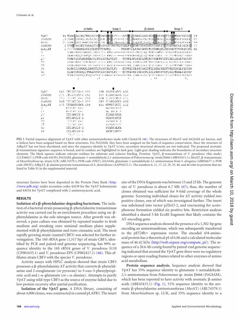
Protein sequence analysis. Sequence analysis showed thatVpAT has 55% sequence identity to glutamate-1-semialdehyde-2,1-aminomutase from Polaromonas sp. strain JS666 (PoGSAM),which has been reported to have activity with aromatic �-aminoacids (ABE43415.1) (Fig. 1), 51% sequence identity to the aro-matic �-phenylalanine aminotransferase (MesAT) (ABL74379.1)from Mesorhizobium sp. LUK, and 35% sequence identity to a
FIG 1 Partial sequence alignment of VpAT with other aminotransferases made with Clustal � (46). The structures of MesAT and SeGSAM are known, and�-helices have been assigned based on these structures. For PoGSAM, they have been assigned on the basis of sequence conservation. Since the structure ofAdbpAT has not been elucidated, and since the sequence identity to VpAT is low, secondary-structural elements are not indicated. The proposed aromatic�-transaminase signature sequence is boxed, and its residues are highlighted in dark gray. Light gray shading indicates the boundaries of secondary structureelements. The black squares indicate relevant residues for substrate and cofactor binding. Proteins: VpAT, �-transaminase of V. paradoxus (this study)(CCE46017.1; PDB code 4AO9); PoGSAM, glutamate-1-semialdehyde 2,1-aminomutase of Polaromonas sp. strain JS666 (ABE43415.1); MesAT, �-transaminaseof Mesorhizobium sp. strain LUK (ABL74379.1; PDB code 2YKY); SeGSAM, glutamate-1-semialdehyde 2,1-aminomutase from S. elongatus (ABB56677.1; PDBcode 2HOZ); AdbpAT, �-alanine:pyruvate transaminase of A. denitrificans (AAP92672.1). The numbers 6, 11, 17, 23, 29, 35, 40, and 46 refer to proteins that arelisted in Table S3 in the supplemental material.
Crismaru et al.
188 aem.asm.org Applied and Environmental Microbiology
on March 10, 2018 by guest
http://aem.asm
.org/D
ownloaded from
glutamate-1-semialdehyde-2,1-aminomutase from Synechococcuselongatus (SeGSAM) (ABB56677.1). These data suggest thatVpAT, just like MesAT and PoGSAM, is a fold type I aminotrans-ferase, and furthermore, that VpAT belongs to subgroup IItransaminases, which is based on an enzyme’s substrate specificity(27, 48, 68). VpAT has 19% sequence identity to a transaminasefrom Alcaligenes denitrificans that has been reported to have activ-ity toward aliphatic �-amino acids (AdbpAT) (AAP92672.1) (21).A sequence comparison between VpAT, MesAT, PoGSAM, andAdbpAT shows that the amino acid residues that are involved incofactor binding are conserved, in addition to several residuesthat, based on the structures of VpAT and MesAT, are involved insubstrate and cofactor binding (Fig. 1).
Purification of VpAT expressed in E. coli. The recombinantprotein was overproduced with an N-terminal His6 tag in E. colistrain C41(DE3). VpAT was mainly present as a soluble protein.The enzyme was purified by three chromatography steps. Size ex-clusion chromatography indicated a molecular mass of approxi-mately 100 kDa, suggesting that VpAT exists as a dimer in solu-tion. The purified protein showed a single band of about 48 kDa inan SDS-PAGE gel (see Fig. S1 in the supplemental material). Theoverall yield from 1 liter of culture was 40 to 50 mg (see Table S1 inthe supplemental material). The specific activity of the purifiedenzyme was 17.5 U mg�1 at 30°C, corresponding to a kcat of 11.8s�1 per monomer. The VpAT R41A mutant was overexpressedand purified under the same conditions as the wild-type enzyme,resulting in similar amounts of purified protein.
Catalytic properties. The pH activity profile of VpAT showedthat the enzyme has high activity over a broad pH range (4 to 11.2)at 30°C. The optimum temperature of the enzyme was tested bymeasuring the specific activity at temperatures between 20°C and65°C. VpAT exhibits a maximum specific activity of 33 U mg�1 at55°C, which is about 2-fold higher than the specific activity at 30°C(see Fig. S2 in the supplemental material). These data show thatVpAT is more active toward (S)-�-phenylalanine than other�-transaminases reported previously (21, 27, 68). The activity ofVpAT with pyruvate is 85% of that with �-ketoglutarate as theamino acceptor.
The relationship between the reaction rate and the substrateconcentration displayed Michaelis-Menten kinetics with sub-strate inhibition (69). The apparent Km and kcat values (per mono-mer) for (S)-�-phenylalanine in the presence of 10 mM �-keto-glutarate were 1.5 mM and 11.8 s�1, respectively, with an apparentsubstrate inhibition constant (Ki) of 40.3 mM. When using 10mM (S)-�-phenylalanine, the apparent Km and kcat for �-ketoglu-tarate were 0.3 mM and 10.6 s�1, respectively, with an apparent Ki
of 82.4 mM.Substrate scope and enantioselectivity (E) of VpAT. To inves-
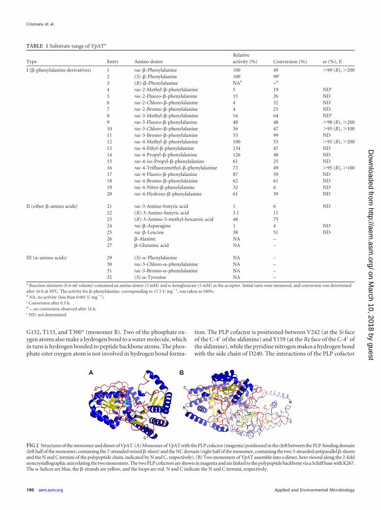
tigate the substrate scope of the enzyme, a number of �-aminoacids and �-amino acids were tested (Table 1). The results showthat VpAT acts exclusively on �-amino acids and that the enzymeprefers aromatic �-amino acids (Table 1, entries 1 to 3 and 8 to 20)over aliphatic �-amino acids (Table 1, entries 21 to 27). The mostfavorable substituent positions on the phenyl ring of �-phenylal-anine for activity were para- and meta-substituted analogues,while ortho-substituted analogues were poorly converted. The�-phenylalanine derivatives bearing linear alkyl substituents atthe para position, i.e., ethyl or propyl (Table 1, entries 13 to 14),were converted more efficiently than other substituted substrates(Table 1, entries 12 and 16 to 20). However, introduction of a
branched alkyl substituent, i.e., iso-propyl, at the para positionresults in a decrease of activity (Table 1, entry 15). The enzymeshowed no activity for �-phenylalanine or its meta-chloro-, meta-bromo-, and para-hydroxy-substituted (�-tyrosine) derivatives,which contrasts with MesAT, for which activity was reported with�-phenylalanine and other �-amino acids (27). However, wecould not confirm the activity of MesAT toward �-phenylalanine.
VpAT further preferentially converts the (S)-enantiomers ofaromatic �-amino acids (Table 1, entries 1 to 3, 9, 10, 12, and 16)and the (R)-enantiomers of aliphatic �-amino acids (which have asimilar configuration of functional groups around the chiral car-bon atom but a change in Cahn-Ingold-Prelog priority). How-ever, the activity of VpAT toward several aliphatic �-amino acidswas quite low (Table 1, entries 22 to 23). We also investigated theregio- and enantioselectivity of VpAT with several racemic sub-strates (Table 1, entries 1, 9, 10, 12, and 16). The enzyme appearedhighly enantioselective toward the (S)-enantiomers of �-phenyl-alanine and its meta- and para-ring-substituted derivatives,thereby producing highly pure (R)-enantiomer preparationswith high enantiomeric excess (ee; 95%).
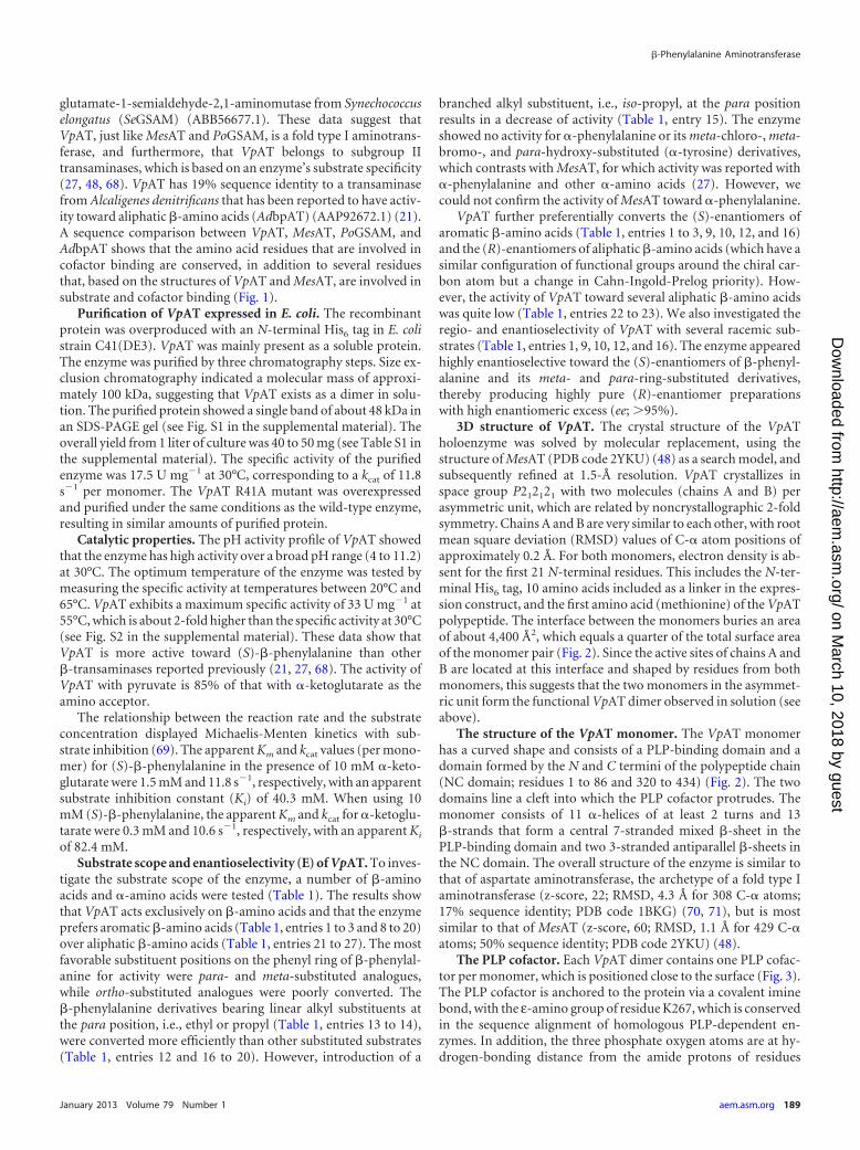
3D structure of VpAT. The crystal structure of the VpATholoenzyme was solved by molecular replacement, using thestructure of MesAT (PDB code 2YKU) (48) as a search model, andsubsequently refined at 1.5-Å resolution. VpAT crystallizes inspace group P212121 with two molecules (chains A and B) perasymmetric unit, which are related by noncrystallographic 2-foldsymmetry. Chains A and B are very similar to each other, with rootmean square deviation (RMSD) values of C-� atom positions ofapproximately 0.2 Å. For both monomers, electron density is ab-sent for the first 21 N-terminal residues. This includes the N-ter-minal His6 tag, 10 amino acids included as a linker in the expres-sion construct, and the first amino acid (methionine) of the VpATpolypeptide. The interface between the monomers buries an areaof about 4,400 Å2, which equals a quarter of the total surface areaof the monomer pair (Fig. 2). Since the active sites of chains A andB are located at this interface and shaped by residues from bothmonomers, this suggests that the two monomers in the asymmet-ric unit form the functional VpAT dimer observed in solution (seeabove).
The structure of the VpAT monomer. The VpAT monomerhas a curved shape and consists of a PLP-binding domain and adomain formed by the N and C termini of the polypeptide chain(NC domain; residues 1 to 86 and 320 to 434) (Fig. 2). The twodomains line a cleft into which the PLP cofactor protrudes. Themonomer consists of 11 �-helices of at least 2 turns and 13�-strands that form a central 7-stranded mixed �-sheet in thePLP-binding domain and two 3-stranded antiparallel �-sheets inthe NC domain. The overall structure of the enzyme is similar tothat of aspartate aminotransferase, the archetype of a fold type Iaminotransferase (z-score, 22; RMSD, 4.3 Å for 308 C-� atoms;17% sequence identity; PDB code 1BKG) (70, 71), but is mostsimilar to that of MesAT (z-score, 60; RMSD, 1.1 Å for 429 C-�atoms; 50% sequence identity; PDB code 2YKU) (48).
The PLP cofactor. Each VpAT dimer contains one PLP cofac-tor per monomer, which is positioned close to the surface (Fig. 3).The PLP cofactor is anchored to the protein via a covalent iminebond, with the ε-amino group of residue K267, which is conservedin the sequence alignment of homologous PLP-dependent en-zymes. In addition, the three phosphate oxygen atoms are at hy-drogen-bonding distance from the amide protons of residues
�-Phenylalanine Aminotransferase
January 2013 Volume 79 Number 1 aem.asm.org 189
on March 10, 2018 by guest
http://aem.asm
.org/D
ownloaded from
G132, T133, and T300* (monomer B). Two of the phosphate ox-ygen atoms also make a hydrogen bond to a water molecule, whichin turn is hydrogen bonded to peptide backbone atoms. The phos-phate ester oxygen atom is not involved in hydrogen bond forma-
tion. The PLP cofactor is positioned between V242 (at the Si faceof the C-4= of the aldimine) and Y159 (at the Re face of the C-4= ofthe aldimine), while the pyridine nitrogen makes a hydrogen bondwith the side chain of D240. The interactions of the PLP cofactor
TABLE 1 Substrate range of VpATa
Type Entry Amino donorRelativeactivity (%) Conversion (%) ee (%), E
I (�-phenylalanine derivatives) 1 rac-�-Phenylalanine 100 49 99 (R), 2002 (S)-�-Phenylalanine 100 99c
3 (R)-�-Phenylalanine NAb –d
4 rac-2-Methyl-�-phenylalanine 5 19 NDe
5 rac-2-Fluoro-�-phenylalanine 15 26 ND6 rac-2-Chloro-�-phenylalanine 4 32 ND7 rac-2-Bromo-�-phenylalanine 4 25 ND8 rac-3-Methyl-�-phenylalanine 54 64 NDe
9 rac-3-Fluoro-�-phenylalanine 48 48 98 (R), 20010 rac-3-Chloro-�-phenylalanine 56 47 95 (R), 10011 rac-3-Bromo-�-phenylalanine 53 99 ND12 rac-4-Methyl-�-phenylalanine 100 53 95 (R), 20013 rac-4-Ethyl-�-phenylalanine 134 47 ND14 rac-4-Propyl-�-phenylalanine 126 48 ND15 rac-4-iso-Propyl-�-phenylalanine 61 25 ND16 rac-4-Trifluoromethyl-�-phenylalanine 73 49 95 (R), 10017 rac-4-Fluoro-�-phenylalanine 87 59 ND18 rac-4-Bromo-�-phenylalanine 62 61 ND19 rac-4-Nitro-�-phenylalanine 32 6 ND20 rac-4-Hydroxy-�-phenylalanine 61 39 ND
II (other �-amino acids) 21 rac-3-Amino-butyric acid 1 6 ND22 (R)-3-Amino-butyric acid 3.1 1123 (R)-3-Amino-5-methyl-hexanoic acid 48 7524 rac-�-Asparagine 1 4 ND25 rac-�-Leucine 38 51 ND26 �-Alanine NA –27 �-Glutamic acid NA –
III (�-amino acids) 29 (S)-�-Phenylalanine NA –30 rac-3-Chloro-�-phenylalanine NA –31 rac-3-Bromo-�-phenylalanine NA –32 (S)-�-Tyrosine NA –
a Reaction mixtures (0.4-ml volume) contained an amino donor (3 mM) and �-ketoglutarate (5 mM) as the acceptor. Initial rates were measured, and conversion was determinedafter 16 h at 30°C. The activity for �-phenylalanine, corresponding to 17.5 U mg�1, was taken as 100%.b NA, no activity (less than 0.001 U mg�1).c Conversion after 0.5 h.d –, no conversion observed after 16 h.e ND, not determined.
FIG 2 Structures of the monomer and dimer of VpAT. (A) Monomer of VpAT with the PLP cofactor (magenta) positioned in the cleft between the PLP-binding domain(left half of the monomer, containing the 7-stranded mixed �-sheet) and the NC domain (right half of the monomer, containing the two 3-stranded antiparallel �-sheetsand the N and C termini of the polypeptide chain, indicated by N and C, respectively). (B) Two monomers of VpAT assemble into a dimer, here viewed along the 2-foldnoncrystallographic axis relating the two monomers. The two PLP cofactors are shown in magenta and are linked to the polypeptide backbone via a Schiff base with K267.The �-helices are blue, the �-strands are yellow, and the loops are red. N and C indicate the N and C termini, respectively.
Crismaru et al.
190 aem.asm.org Applied and Environmental Microbiology
on March 10, 2018 by guest
http://aem.asm
.org/D
ownloaded from
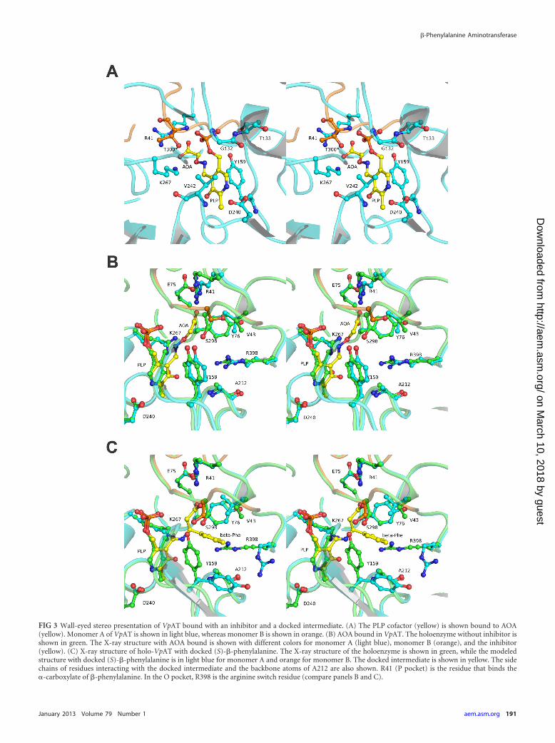
FIG 3 Wall-eyed stereo presentation of VpAT bound with an inhibitor and a docked intermediate. (A) The PLP cofactor (yellow) is shown bound to AOA(yellow). Monomer A of VpAT is shown in light blue, whereas monomer B is shown in orange. (B) AOA bound in VpAT. The holoenzyme without inhibitor isshown in green. The X-ray structure with AOA bound is shown with different colors for monomer A (light blue), monomer B (orange), and the inhibitor(yellow). (C) X-ray structure of holo-VpAT with docked (S)-�-phenylalanine. The X-ray structure of the holoenzyme is shown in green, while the modeledstructure with docked (S)-�-phenylalanine is in light blue for monomer A and orange for monomer B. The docked intermediate is shown in yellow. The sidechains of residues interacting with the docked intermediate and the backbone atoms of A212 are also shown. R41 (P pocket) is the residue that binds the�-carboxylate of �-phenylalanine. In the O pocket, R398 is the arginine switch residue (compare panels B and C).
�-Phenylalanine Aminotransferase
January 2013 Volume 79 Number 1 aem.asm.org 191
on March 10, 2018 by guest
http://aem.asm
.org/D
ownloaded from
with residues K267, V242, Y159, and D240 keep the PLP cofactorfixed in the active site (Fig. 3A).
The binding of 2-aminooxyacetic acid. AOA, a mimic of �-al-anine, is a known inhibitor of aminotransferases (72) and alsoinhibits VpAT. To investigate how AOA binds in VpAT, enzymecrystals were soaked with AOA. The structure of VpAT with AOA(Fig. 3A and B) shows that the amino group of the inhibitor cova-lently binds to the C-4A atom of the PLP cofactor, as also de-scribed for aspartate aminotransferase (73) and recently forMesAT (48). The ether-oxygen atom (O1) of AOA is positionedclose (3.0 Å) to the ε-amino group of residue K267. The carboxy-late group binds via a salt bridge to the Nε- and N�2-atoms ofR41. The binding of AOA in the active site of VpAT is similar to theway in which AOA binds in the active site of MesAT (PDB code2YKV). AOA inhibits aminotransferase activity because theamine-ether oxygen bond of AOA cannot be weakened by K267,and hence, hydrolysis and formation of the pyridoxamine 5=-phosphate (PMP) intermediate does not occur (Fig. 3B).
Docking of PLP–(S)-�-phenylalanine in the active site ofVpAT. To investigate how an aromatic �-amino acid, such as(S)-�-phenylalanine, binds in the active site of VpAT, initially, astructure was determined after a VpAT crystal had been soakedwith (S)-�-phenylalanine. However, this structure only revealedthe presence of a PMP intermediate in the active site of the en-zyme. Since trapping of (S)-�-phenylalanine in the active site ofVpAT apparently had not occurred, the external (S)-�-phenylal-anine aldimine intermediate was modeled into the active site ofVpAT using Rosetta, which allows docking with flexible backboneand side chains. In the docked structure, the aromatic ring of thesubstrate is bound between V43, Y76, Y159, and A212 (Fig. 3C),similar to the way (S)-�-phenylalanine is bound to MesAT (48).We previously termed this region the O pocket, because it is posi-tioned on the 3-hydroxyl side of the PLP cofactor (48) (see below).Similar to the conformational changes observed in MesAT uponformation of the external aldimine intermediate with (S)-�-phe-nylalanine (48), in VpAT the arginine switch residue R398 in the Opocket is also displaced during docking, freeing space for the phe-nyl ring of the substrate. Instead, the R398 side chain N�2 atomforms a hydrogen bond to the carbonyl oxygen of A212. Also, V43and Y159 have reoriented to better accommodate the docked ex-ternal aldimine intermediate. In the absence of (S)-�-phenylala-nine, the docking procedure resulted in minor shifts (less than 0.5Å) of the backbone atoms around the active site. While the orthohydrogen atoms of the phenyl ring of the bound intermediate areburied, the para and meta hydrogen atoms of the phenyl ring aresolvent exposed in the docked structure. The carboxylate of thedocked (S)-�-phenylalanine is buried in a region we term the Ppocket, which is at the phosphate group side of the PLP cofactor.Here, residue R41 is directly involved in binding of (S)-�-phenyl-alanine by making a salt bridge to the carboxylate group (Fig. 3C).The importance of R41 for the binding of (S)-�-phenylalanine isunderscored by the observation that the R41A mutant was inac-tive when tested with (S)-�-phenylalanine as amino donor and�-ketoglutarate as amino acceptor.
Identification of aromatic �-amino acid aminotransferases.The VpAT structure with bound AOA, a �-alanine mimic, and thedocking results with �-phenylalanine show that R41 is importantfor binding the �-carboxylate of �-amino acids, as is the corre-sponding residue R54 in MesAT (48). The side chain of R41 inter-acts with E75, which may assist the function of R41 by orienting its
side chain and shaping the carboxylate-binding P pocket. Wetherefore hypothesized that the presence of an R41-E75 pair in anaminotransferase protein sequence indicates enzymatic activitywith �-amino acids. A comparison of the crystal structures ofVpAT and MesAT showed that the R41-E75 salt bridge is con-served in MesAT (R54-E88). In addition, a sequence alignmentbetween VpAT and a glutamate-1-semialdehyde-2,1-aminomu-tase from Polaromonas sp. JS666 (PoGSAM), for which no struc-ture is available but which has activity toward aromatic�-amino acids (68), also showed conservation of R41 and E75(as R51 and E85, respectively) (Fig. 1). In contrast, the crystalstructure of S. elongatus glutamate-1-semialdehyde-2,1-ami-nomutase (SeGSAM) (PDB code 2HOZ) (74), which is struc-turally very similar to that of VpAT (z-score, 47; RMSD, 1.8 Åfor 404 C-� atoms; 35% sequence identity), shows that no Argand Glu residues equivalent to R41 and E75 of VpAT are pres-ent, in agreement with the enzyme’s lack of activity on �-aminoacids.
We subsequently tested whether R41 and E75 could be used todefine a sequence motif with which aromatic �-amino acid ami-notransferases can be distinguished from aminotransferases witha similar fold but with a different substrate specificity. For this,BLAST searches were performed using the VpAT, MesAT, andPoGSAM (ABE43415.1) protein sequences. For each query se-quence, 2,000 homologous sequences were retrieved with �18%sequence identity. After omitting duplicates, the resulting 2,222sequences were included in a multiple-sequence alignment andinspected for residues that are important for substrate binding.
We used the P-pocket R41-E75 pair as the starting point toidentify a motif with which aminotransferases with activity to-ward aromatic �-amino acids can be distinguished from amino-transferases with a similar fold but a different activity. The R41-E75 starting pair was subsequently broadened by including theO-pocket residues V43 and Y76, since the equivalent residues inMesAT (I56 and Y89) were shown to be involved in substratebinding. At position 43, Val, Ile, and Ala were allowed, but hydro-phobic residues with more bulky side chains, such as Leu, Phe,Tyr, and Trp were excluded, since they were expected to preventaromatic substrate binding for steric reasons. At position 75, apartfrom Glu, we also allowed Asp, Gln, and Asn, since they may stillbe able to interact with the side chain of R41. At position 76, Pheand Trp were also allowed, since they are frequently found in thesame position as Tyr in other fold type I aminotransferases. Wenext extended the motif with conserved residues at positions thatappear structurally important by adding a conserved Pro at posi-tion 50, which is in the center of a hydrophobic cluster in theVpAT and MesAT structures, and an Asp at position 65 and a Glyat position 66, which form a �-turn, in which the Asp is hydrogenbonded to A23 and S24, residues that are located in the �-helixthat precedes loop 1.
The motif that was finally defined, R-X-[AVI]-X(6)-P-X(14)-D-G-X(8)-[EDNQ]-[YFW] (Fig. 1), retrieved 46 unique se-quences (see Table S3 in the supplemental material) that we hy-pothesize may represent fold type I aminotransferases withactivity toward aromatic �-amino acids.
DISCUSSIONCatalytic properties and enantioselectivity of Variovorax AT.Isolation of microorganisms that use �-phenylalanine as a solenitrogen source yielded a Gram-negative bacterium of the species
Crismaru et al.
192 aem.asm.org Applied and Environmental Microbiology
on March 10, 2018 by guest
http://aem.asm
.org/D
ownloaded from
V. paradoxus that produces a �-amino acid selective aminotrans-ferase. The gene encoding this enzyme was cloned, sequenced, andoverexpressed to a high level in E. coli. Sequence analysis showedthat the enzyme is a fold type I AT, just like several other subgroupII �-ATs. VpAT shows the highest sequence identity with bio-chemically characterized �-phenylalanine ATs (27, 68), includingthe Mesorhizobium aminotransferase (MesAT), described by Kimet al. (27), the structure of which was recently solved (48). MesATand VpAT have similar activities with �-ketoglutarate and pyru-vate as amino acceptors (27) but differ from �-transaminases,which generally show a clear preference for either pyruvate, �-ke-toglutarate, or oxaloacetate (75–77). Striking differences betweenMesAT and VpAT are the facts that VpAT has a higher catalyticactivity, is active over a very broad range of pH values, and acceptsonly �-amino acids. On the other hand, VpAT shows the sameenantiopreference for (S)-�-phenylalanine and a very high enan-tioselectivity (E 100), just like MesAT (19) and PoGSAM (68).This allows a biotechnologically relevant kinetic resolution of ra-cemic mixtures, from which the remaining (R)-�-phenylalaninescan be obtained with very high ee (Table 1, entries 1, 9, 10, 12, and16).
Although the differences in activity are difficult to explain, acomparison of the residues surrounding the active site of VpATwith those of MesAT shows that V43, S298* (monomer B), andF400, which line the hydrophobic O pocket of the active site ofVpAT, differ from the residues found at these positions in MesAT(I56, A312*, and M414). A triple mutant of MesAT (I56V/A312S/M414F) was generated and showed a 2-fold increase (3.3 U mg�1)in activity with (S)-�-phenylalanine compared to the MesAT WT(1.6 U mg�1 [27]), indicating an important role of these residuesin activity with (S)-�-phenylalanine.
Structure of VpAT and architecture of the active site. Thestructure of VpAT revealed the overall topology of the enzyme andthe position of the cofactor and provided insight into the archi-tecture of the active site of VpAT. Comparison with the structureof MesAT shows that the two enzymes are structurally similar(RMSD, 1.0 Å for 423 C-� atoms; 51% sequence identity) andconfirms that they both belong to the fold type I aminotransferasefamily (70). As in MesAT, both monomers in VpAT contribute tothe binding of the PLP cofactor, suggesting that dimerization isessential for catalytic activity (Fig. 3).
Previously, we proposed to use the notation P pocket (pointingin the same direction as the PLP phosphate) and O pocket (at theside of the hydroxyl substituent of the PLP) to define substratebinding sites of amino acid-converting fold type I ATs (48). Thisnotation is more generally applicable than using L(arge) andS(mall) pocket (78), since the size of a pocket at a topologicallyconserved position may vary, whereas the O pocket and the Ppocket are topologically fixed (Fig. 4).
Computer-aided docking of (S)-�-phenylalanine suggestedbinding of �-phenylalanine with the carboxylate group in the Ppocket, which is opposite to the binding mode of the �-phenylal-anine analogue 3-phenylpropionate in the active site of aromaticamino acid aminotransferase from Paracoccus denitrificans (PDBcode 1AY8) (79) but similar to the binding mode of �-phenylala-nine in MesAT (48). The docking results further indicated thatseveral residues in the O pocket, including R398, must rearrangeto enable binding of the phenyl ring of �-phenylalanine (Fig. 3C).This switch was also observed for MesAT complexed with (S)-�-phenylalanine (PDB code 2YKY) and 2-oxoglutarate (PDB code
2YKX) and appears necessary to allow binding of both the phenylgroup of (S)-�-phenylalanine AT and the �-carboxylate of 2-oxo-glutarate (48). In class I ATs accepting aromatic �-amino acids, aso-called arginine switch has also been identified (e.g., R292 inAroAT), but here, the arginine switch and cosubstrate non-�-carboxylate-binding site are located in the P pocket, in agreementwith the reverse mode of substrate binding of �-Phe in VpAT andMesAT compared to �-phenylalanine in AroAT (80). The dockingfurther indicated that the para and meta positions of the phenylring are solvent exposed while the ortho position is buried in the Opocket of the protein (Fig. 3C). This is in agreement with thehigher catalytic activities observed for substrates substituted at thepara and meta positions relative to ortho-substituted substrates(Table 1, entries 4 to 7), for which steric hindrance by S298*(monomer B) and Y76 may prevent productive binding (Fig. 3C).The importance of steric crowding at the ortho position is con-firmed by the relatively high activity with the ortho-fluorine-sub-stituted substrate compared to the compounds with larger sub-stituents, i.e., chlorine and bromine (Table 1, entries 5 to 7). Thus,it appears that the substrate specificity of VpAT with regard tosubstituted �-phenylalanine derivatives is mainly determined bythe residues that line the hydrophobic O pocket.
A sequence motif for aromatic �-amino acid aminotrans-ferases. A combination of sequence analysis, structural data, andinformation on substrate range may be used to define aminotrans-ferase sequence motifs that can be applied to identify enzymeswith certain substrate specificities (81). As observed in the VpATstructure bound with AOA (Fig. 3A and B), as well as in the mod-eled VpAT–(S)-�-phenylalanine structure (Fig. 3C), residues R41in loop 1 and E75 in loop 2 of the NC domain contribute toshaping the carboxylate-binding site for �-amino acids in the Ppocket. For this reason, and also taking into account that E75 isconserved in MesAT and PoGSAM but not in SeGSAM (Fig. 1), weused the conserved functional P-pocket R41/E75 pair as the start-ing point to identify a motif that may distinguish aminotrans-ferases with activity toward aromatic �-amino acids from amino-transferases with a similar fold but different activity. Extension ofthe R41/E75 motif with residues selected on the basis of sequenceconservation or position in the structure yielded a motif, R-X-[AVI]-X(6)-P-X(14)-D-G-X(8)-[EDNQ]-[YFW], that comprises
FIG 4 Schematic representation of (S)-�-phenylalanine (structure in bold-face) bound to PLP (external aldimine) docked in the active site of VpAT. Anumber of relevant amino acid residues for binding and catalysis are shown.
�-Phenylalanine Aminotransferase
January 2013 Volume 79 Number 1 aem.asm.org 193
on March 10, 2018 by guest
http://aem.asm
.org/D
ownloaded from

36 residues in the N-terminal segment of the NC domain. Whenused to screen a set of over 2,200 (putative) class I aminotrans-ferases, it allowed the selection of 46 sequences (see Table S3 in thesupplemental material), among which were the sequences of theconserved �-selective enzymes VpAT, MesAT, and PoGSAM. Al-though several retrieved sequences have been annotated as gluta-mate-1-semialdehyde 2,1-aminomutase (see Table S3 in the sup-plemental material), we propose that the sequences selected by themotif are ATs with activity toward aromatic �-amino acids. Al-though our motif may be applied to identify aminotransferases ina set of fold type I aminotransferases, it probably cannot predict�-aminotransferase activity in less related sequences or enzymeslacking activity with aromatic substrates. For example, AdbpAT(21), which is inactive with aromatic �-amino acids and has anenantiopreference for aliphatic �-amino acids opposite that ofVpAT (Table 1) and MesAT (27), is not covered by the motif. Thismay suggest that AdbpAT belongs to a different phylogenetic clus-ter and may have a different mode of substrate binding.
Outlook. Although more experiments or structure and mod-eling studies are needed, we are confident that the newly discov-ered �-phenylalanine aminotransferase from V. paradoxus strainCBF3, along with its promising biocatalytic properties and its 3Dstructure, is an attractive template for future �-aminotransferase-engineering efforts toward the synthesis of enantiomerically pure�-amino acids.
ACKNOWLEDGMENTS
We thank O. May and B. Kaptein from DSM for helpful discussions. Weacknowledge the European Synchrotron Radiation Facility (ESRF) forprovision of synchrotron radiation facilities and thank the beamline staffof ID14-4 for their assistance.
This research was financially supported by the Netherlands Ministry ofEconomic Affairs and the B-Basic partner organizations (http://www.b-basic.nl) through B-Basic, a public-private NWO-ACTS program. Thework of S. Bartsch was supported by the EU FP7 under Grant Agreementnumber 266025 (BIONEXGEN). Hein J. Wijma was supported by NWO(Netherlands Organization for Scientific Research) through an ECHOgrant.
REFERENCES1. Seebach D, Beck A, Bierbaum D. 2004. The world of beta- and gamma-
peptides comprised of homologated proteinogenic amino acids and othercomponents. Chem. Biodivers. 1:1111–1239.
2. Burley SK, David PR, Lipscomb WN. 1991. Leucine aminopeptidase:bestatin inhibition and a model for enzyme-catalyzed peptide hydrolysis.Proc. Natl. Acad. Sci. U. S. A. 88:6916 – 6920.
3. Geueke B, Kohler H. 2007. Bacterial beta-peptidyl aminopeptidases: onthe hydrolytic degradation of beta-peptides. Appl. Microbiol. Biotechnol.74:1197–1204.
4. Juaristi E, Soloshonok V. 2005. Enantioselective synthesis of �-aminoacids. Wiley-VHC, New York, NY.
5. Shih C, Gossett LS, Gruber JM, Grossman CS, Andis SL, Schultz RM,Worzalla JF, Corbett TH, Metz JT. 1999. Synthesis and biological eval-uation of novel cryptophycin analogs with modification in the beta-alanine region. Bioorg. Med. Chem. Lett. 9:69 –74.
6. Mekhail TM, Markman M. 2002. Paclitaxel in cancer therapy. ExpertOpin. Pharmacother. 3:755–766.
7. Ojima I, Lin S, Wang T. 1999. Recent advances in the medicinal chem-istry of taxoids with novel beta-amino acid side chains. Curr. Med. Chem.6:927–954.
8. Crews P, Manes LV, Boehler M. 1986. Jasplakinolide, a cyclodepsipep-tide from the marine sponge, Jaspis sp. Tetrahedron Lett. 27:2797–2800.
9. Weiner B, Szymanski W, Janssen DB, Minnaard AJ, Feringa BL. 2010.Recent advances in the catalytic asymmetric synthesis of beta-amino acids.Chem. Soc. Rev. 39:1656 –1691.
10. Liljeblad A, Kanerva LT. 2006. Biocatalysis as a profound tool in thepreparation of highly enantiopure beta-amino acids. Tetrahedron 62:5831–5854.
11. Rehdorf J, Mihovilovic MD, Bornscheuer UT. 2010. Exploiting theregioselectivity of Baeyer-Villiger monooxygenases for the formation ofbeta-amino acids and beta-amino alcohols. Angew. Chem. Int. Ed. Engl.49:4506 – 4508.
12. Chirpich TP, Zappia V, Costilow RN, Barker HA. 1970. Lysine 2,3-aminomutase. Purification and properties of a pyridoxal phosphate andS-adenosylmethionine-activated enzyme. J. Biol. Chem. 245:1778 –1789.
13. Ruzicka FJ, Frey PA. 2007. Glutamate 2,3-aminomutase: a new memberof the radical SAM superfamily of enzymes. Biochim. Biophys. Acta 1774:286 –296.
14. Wu B, Szymanski W, Heberling MM, Feringa BL, Janssen DB. 2011.Aminomutases: mechanistic diversity, biotechnological applications andfuture perspectives. Trends Biotechnol. 29:352–362.
15. Cronan JE .Jr. 1980. Beta-alanine synthesis in Escherichia coli. J. Bacteriol.141:1291–1297.
16. Christianson CV, Montavon TJ, Festin GM, Cooke HA, Shen B, BrunerSD. 2007. The mechanism of MIO-based aminomutases in beta-aminoacid biosynthesis. J. Am. Chem. Soc. 129:15744 –15745.
17. Christen P, Metzler DE. 1985. Transaminases. Wiley, New York, NY.18. Stirling DI. 1992. The use of aminotransferases for the production of
chiral amino acids and amines. In Collins AN, Sheldrake GN, Crosby J(ed), Chirality in industry. Wiley, New York, NY.
19. Kim J, Kyung D, Yun H, Cho BK, Kim BG. 2006. Screening andpurification of a novel transaminase catalyzing the transamination of arylbeta-amino acid from Mesorhizobium sp LUK. J. Microbiol. Biotechnol.16:1832–1836.
20. Shin JS, Kim BG. 2001. Comparison of the omega-transaminases fromdifferent microorganisms and application to production of chiral amines.Biosci. Biotechnol. Biochem. 65:1782–1788.
21. Yun H, Lim S, Cho BK, Kim BG. 2004. Omega-amino acid:pyruvatetransaminase from Alcaligenes denitrificans Y2k-2: a new catalyst for ki-netic resolution of beta-amino acids and amines. Appl. Environ. Micro-biol. 70:2529 –2534.
22. Percudani R, Peracchi A. 2009. The B6 database: a tool for the descriptionand classification of vitamin B6-dependent enzymatic activities and of thecorresponding protein families. BMC Bioinformatics 10:273.
23. Finn RD, Mistry J, Tate J, Coggill P, Heger A, Pollington JE, Gavin OL,Gunasekaran P, Ceric G, Forslund K, Holm L, Sonnhammer EL, EddySR, Bateman A. 2010. The Pfam protein families database. Nucleic AcidsRes. 38:D211–D222.
24. Yonaha K, Suzuki K, Minei H, Toyama S. 1983. Distribution of omega-amino acid–pyruvate transaminase and aminobutyrate–alpha-ketoglutarate transaminase in microorganisms. Agric. Biol. Chem. Tokyo47:2257–2265.
25. Christen P, Mehta P. 2001. From cofactor to enzymes. The molecularevolution of pyridoxal-5=-phosphate-dependent enzymes. Chem. Rec.1:436 – 447.
26. Mehta PK, Hale TI, Christen P. 1993. Aminotransferases—demonstrationof homology and division into evolutionary subgroups. Eur. J. Biochem. 214:549–561.
27. Kim J, Kyung D, Yun H, Cho BK, Seo JH, Cha M, Kim BG. 2007.Cloning and characterization of a novel beta-transaminase from Mesorhi-zobium sp. strain LUK: a new biocatalyst for the synthesis of enantiomeri-cally pure beta-amino acids. Appl. Environ. Microbiol. 73:1772–1782.
28. Hwang BY, Cho BK, Yun H, Koteshwar K, Kim BG. 2005. Revisit ofaminotransferase in the genomic era and its application to biocatalysis. J.Mol. Catal. B Enzym. 37:47–55.
29. Koszelewski D, Lavandera I, Clay D, Guebitz GM, Rozzell D, KroutilW. 2008. Formal asymmetric biocatalytic reductive amination. Angew.Chem. Int. Ed. Engl. 47:9337–9340.
30. Koszelewski D, Tauber K, Faber K, Kroutil W. 2010. Omega-transaminases for the synthesis of non-racemic alpha-chiral primaryamines. Trends Biotechnol. 28:324 –332.
31. Rozzell JD. May 1985. Production of amino acids by transamination. USpatent 4518692.
32. Stewart JD. 2001. Dehydrogenases and transaminases in asymmetric syn-thesis. Curr. Opin. Chem. Biol. 5:120 –129.
33. Taylor PP, Pantaleone DP, Senkpeil RF, Fotheringham IG. 1998. Novelbiosynthetic approaches to the production of unnatural amino acids usingtransaminases. Trends Biotechnol. 16:412– 418.
Crismaru et al.
194 aem.asm.org Applied and Environmental Microbiology
on March 10, 2018 by guest
http://aem.asm
.org/D
ownloaded from

34. Wu B, Szymanski W, Crismaru CG, Feringa BL, Janssen DB. 2012. C-Nlyases catalyzing addition of ammonia, amines and amides to C�C andC�O bonds, p 749 –778. In Drauz K, Groger H, May O (ed), Enzymecatalysis in organic synthesis. Wiley-VCH, New York, NY.
35. Nowicki C, Hunter GR, Montemartini-Kalisz M, Blankenfeldt W,Hecht H, Kalisz HM. 2001. Recombinant tyrosine aminotransferase fromTrypanosoma cruzi: structural characterization and site directed mutagen-esis of a broad substrate specificity enzyme. Biochim. Biophys. Acta 1546:268 –281.
36. Rudat J, Brucher BR, Syldatk C. 2012. Transaminases for the synthesis ofenantiopure beta-amino acids. AMB Express. 2:11.
37. Szymanski W, Wu B, Weiner B, de Wildeman S, Feringa BL, JanssenDB. 2009. Phenylalanine aminomutase-catalyzed addition of ammonia tosubstituted cinnamic acids: a route to enantiopure alpha- and beta-aminoacids. J. Org. Chem. 74:9152–9157.
38. Janssen DB, Scheper A, Witholt B. 1984. Biodegradation of 2-chloro-ethanol and 1,2-dichloroethane by pure bacterial cultures. Prog. Ind. Mi-crobiol. 20:169 –178.
39. Gabor E, de Vries E, Janssen D. 2004. Construction, characterization,and use of small-insert gene banks of DNA isolated from soil and enrich-ment cultures for the recovery of novel amidases. Environ. Microbiol.6:948 –958.
40. Hill DW, Walters FH, Wilson TD, Stuart JD. 1979. High performanceliquid chromatographic determination of amino acids in the picomolerange. Anal. Chem. 51:1338 –1341.
41. Woese CR. 1987. Bacterial evolution. Microbiol. Rev. 51:221–271.42. Poelarends G, Wilkens M, Larkin M, van Elsas J, Janssen D. 1998.
Degradation of 1,3-dichloropropene by Pseudomonas cichorii 170. Appl.Environ. Microbiol. 64:2931–2936.
43. Sambrook J, Russell DW. 2001. Molecular cloning: a laboratory manual,3rd ed. Cold Spring Harbor Laboratory Press, Cold Spring Harbor, NY.
44. Staskawicz B, Dahlbeck D, Keen N, Napoli C. 1987. Molecular charac-terization of cloned avirulence genes from race 0 and race 1 of Pseudomo-nas syringae pv. glycinea. J. Bacteriol. 169:5789 –5794.
45. Brady OL, Elsmie GV. 1926. The use of 2:4-dinitrophenylhydrazine as areagent for aldehydes and ketones. Analyst 51:77–78.
46. Sievers F, Wilm A, Dineen D, Gibson TJ, Karplus K, Li WZ, Lopez R,McWilliam H, Remmert M, Soding J, Thompson JD, Higgins DG. 2011.Fast, scalable generation of high-quality protein multiple sequence align-ments using Clustal Omega. Mol. Syst. Biol. 7:539.
47. Drummond AJ AB, Buxton S, Cheung M, Cooper A, Duran C, Field M,Heled J, Kearse M, Markowitz S, Moir R, Stones-Havas S, Sturrock S,Thierer T, Wilson A. 2010. Geneious v5.5, 5.5.3 ed. Biomatters, Auck-land, New Zealand.
48. Wybenga GG, Crismaru CG, Janssen DB, Dijkstra BW. 2012. Structuraldeterminants of the beta-selectivity of a bacterial aminotransferase. J. Biol.Chem. 287:28495–28502.
49. Evans P. 2006. Scaling and assessment of data quality. Acta Crystallogr. DBiol. Crystallogr. 62:72– 82.
50. Kabsch W. 2010. Integration, scaling, space-group assignment and post-refinement. Acta Crystallogr. D Biol. Crystallogr. 66:133–144.
51. Bailey S. 1994. The CCP4 Suite—programs for protein crystallography.Acta Crystallogr. D Biol. Crystallogr. 50:760 –763.
52. McCoy AJ, Grosse-Kunstleve RW, Adams PD, Winn MD, Storoni LC,Read RJ. 2007. Phaser crystallographic software. J. Appl. Crystallogr. 40:658 – 674.
53. Langer G, Cohen SX, Lamzin VS, Perrakis A. 2008. Automated macro-molecular model building for X-ray crystallography using ARP/wARPversion 7. Nat. Protoc. 3:1171–1179.
54. Emsley P, Cowtan K. 2004. Coot: model-building tools for moleculargraphics. Acta Crystallogr. D Biol. Crystallogr. 60:2126 –2132.
55. Murshudov GN, Vagin AA, Dodson EJ. 1997. Refinement of macromo-lecular structures by the maximum-likelihood method. Acta Crystallogr.D Biol. Crystallogr. 53:240 –255.
56. Chen VB, Arendall WB, Headd JJ, Keedy DA, Immormino RM, KapralGJ, Murray LW, Richardson JS, Richardson DC. 2010. MolProbity:all-atom structure validation for macromolecular crystallography. ActaCrystallogr. D Biol. Crystallogr. 66:12–21.
57. Kleywegt GJ, Jones TA. 1998. Databases in protein crystallography. ActaCrystallogr. D Biol. Crystallogr. 54:1119 –1131.
58. Schuttelkopf AW, van Aalten DM. 2004. PRODRG: a tool for high-
throughput crystallography of protein-ligand complexes. Acta Crystal-logr. D Biol. Crystallogr. 60:1355–1363.
59. Holm L, Rosenstrom P. 2010. Dali server: conservation mapping in 3D.Nucleic Acids Res. 38:W545–W549.
60. Krissinel E, Henrick K. 2007. Inference of macromolecular assembliesfrom crystalline state. J. Mol. Biol. 372:774 –797.
61. Davis IW, Baker D. 2009. RosettaLigand docking with full ligand andreceptor flexibility. J. Mol. Biol. 385:381–392.
62. Davis IW, Raha K, Head MS, Baker D. 2009. Blind docking of pharma-ceutically relevant compounds using RosettaLigand. Protein Sci. 18:1998 –2002.
63. Richter F, Leaver-Fay A, Khare SD, Bjelic S, Baker D. 2011. De novoenzyme design using Rosetta3. PLoS One 6:e19230. doi:10.1371/journal.pone.0019230.
64. Krieger E, Koraimann G, Vriend G. 2002. Increasing the precision ofcomparative models with YASARA NOVA—a self-parameterizing forcefield. Proteins 47:393– 402.
65. OpenEye Scientific Software, Inc. 2010. OEChem TK programming li-brary for chemistry and cheminformatics. OpenEye Scientific Software,Inc., Santa Fe, NM.
66. Zhang Z, Schwartz S, Wagner L, Miller W. 2000. A greedy algorithm foraligning DNA sequences. J. Comput. Biol. 7:203–214.
67. Han JI, Choi HK, Lee SW, Orwin PM, Kim J, Laroe SL, Kim TG, O’NeilJ, Leadbetter JR, Lee SY, Hur CG, Spain JC, Ovchinnikova G, GoodwinL, Han C. 2011. Complete genome sequence of the metabolically versatileplant growth-promoting endophyte Variovorax paradoxus S110. J. Bacte-riol. 193:1183–1190.
68. Bea HS, Park HJ, Lee SH, Yun H. 2011. Kinetic resolution of aromaticbeta-amino acids by omega-transaminase. Chem. Commun. (Camb) 47:5894 –5896.
69. Tipton KF. 1996. Patterns of enzyme inhibition, p 115–174. In Engel PC(ed), Enzymology. Academic Press, San Diego, CA.
70. Grishin NV, Phillips MA, Goldsmith EJ. 1995. Modeling of the spatialstructure of eukaryotic ornithine decarboxylases. Protein Sci. 4:1291–1304.
71. Schneider G, Kack H, Lindqvist Y. 2000. The manifold of vitamin B6dependent enzymes. Structure 8:R1–R6.
72. John RA, Charteris A. 1978. The reaction of amino-oxyacetate withpyridoxal phosphate-dependent enzymes. Biochem. J. 171:771–779.
73. Markovic-Housley Z, Schirmer T, Hohenester E, Khomutov AR, Kho-mutov RM, Karpeisky MY, Sandmeier E, Christen P, Jansonius JN.1996. Crystal structures and solution studies of oxime adducts of mito-chondrial aspartate aminotransferase. Eur. J. Biochem. 236:1025–1032.
74. Stetefeld J, Jenny M, Burkhard P. 2006. Intersubunit signaling in gluta-mate-1-semialdehyde-aminomutase. Proc. Natl. Acad. Sci. U. S. A. 103:13688 –13693.
75. Lowe PN, Rowe AF. 1985. Aspartate-2-oxoglutarate aminotransferasefrom Trichomonas vaginalis: identity of aspartate-aminotransferase andaromatic amino-acid aminotransferase. Biochem. J. 232:689 – 695.
76. Shin JS, Yun H, Jang JW, Park I, Kim BG. 2003. Purification, charac-terization, and molecular cloning of a novel amine:pyruvate transaminasefrom Vibrio fluvialis JS17. Appl. Microbiol. Biotechnol. 61:463– 471.
77. Sung MH, Tanizawa K, Tanaka H, Kuramitsu S, Kagamiyama H, SodaK. 1990. Purification and characterization of thermostable aspartate-aminotransferase from a thermophilic Bacillus species. J. Bacteriol. 172:1345–1351.
78. Shin JS, Kim BG. 2002. Exploring the active site of amine:pyruvate ami-notransferase on the basis of the substrate structure-reactivity relation-ship: how the enzyme controls substrate specificity and stereo selectivity. J.Org. Chem. 67:2848 –2853.
79. Okamoto A, Nakai Y, Hayashi H, Hirotsu K, Kagamiyama H. 1998.Crystal structures of Paracoccus denitrificans aromatic amino acid amino-transferase: a substrate recognition site constructed by rearrangement ofhydrogen bond network. J. Mol. Biol. 280:443– 461.
80. Eliot AC, Kirsch JF. 2004. Pyridoxal phosphate enzymes: mechanistic,structural, and evolutionary considerations. Annu. Rev. Biochem. 73:383– 415.
81. Hohne M, Schatzle S, Jochens H, Robins K, Bornscheuer UT. 2010.Rational assignment of key motifs for function guides in silico enzymeidentification. Nat. Chem. Biol. 6:807– 813.
�-Phenylalanine Aminotransferase
January 2013 Volume 79 Number 1 aem.asm.org 195
on March 10, 2018 by guest
http://aem.asm
.org/D
ownloaded from