bioc 435: advanced topics in biochemistry—a peer review of
TRANSCRIPT
University of Nebraska - LincolnDigitalCommons@University of Nebraska - Lincoln
UNL Faculty Course Portfolios Peer Review of Teaching Project
2014
BIOC 435: Advanced Topics in Biochemistry—APeer Review of Teaching Project BenchmarkPortfolioEdward N. HarrisUniversity of Nebraska-Lincoln, [email protected]
Follow this and additional works at: http://digitalcommons.unl.edu/prtunl
Part of the Biochemistry Commons, Biology Commons, Higher Education Commons, and theHigher Education and Teaching Commons
This Portfolio is brought to you for free and open access by the Peer Review of Teaching Project at DigitalCommons@University of Nebraska - Lincoln.It has been accepted for inclusion in UNL Faculty Course Portfolios by an authorized administrator of DigitalCommons@University of Nebraska -Lincoln.
Harris, Edward N., "BIOC 435: Advanced Topics in Biochemistry—A Peer Review of Teaching Project Benchmark Portfolio" (2014).UNL Faculty Course Portfolios. 64.http://digitalcommons.unl.edu/prtunl/64

2014
Biochemistry 435 A Peer Review of Teaching Project
Benchmark Portfolio
Edward N. Harris, Ph.D.

1
Course Portfolio Biochemistry 435: Advanced Topics
Table of Contents
Introduction………………………………………………………………………………....1 Objectives of Peer Review Course Portfolio…………………………………………....1 Description of BIOC 435 Course………………………………………………………..2 Why This Course Was Chosen………………………………………………………….3 Course Activities ………………………………………………………………………….3 Analysis of Student Learning……………………………………………………..……….5 Assessment and Outcomes of the Class Activities and Organization……………..11 Student Opinion of the Course…………………………………………………………15 Planned Changes…………………………………………………………………………17 Summary and Overall Assessment of the Portfolio Process ……………………...18 Appendix I: Syllabus………………………………………………………………………20 Appendix II: Exams and Quizzes………………………………………………………..30 Appendix III: Samples of Review Papers……………………………………………...50 Appendix IV: Samples of Student Written Abstracts…………………………………86 Appendix V: JiTT Outcomes…………...………………………………………………...88
Introduction
Instructor: Edward N. Harris Department: Biochemistry Course: Biochemistry 435, Advanced Topics in Biochemistry University: University of Nebraska – Lincoln Contact Info: 1901 Vine St., Beadle N133, Lincoln NE 68588. Ph # 402-472-7468; email: [email protected]
Objectives of Peer Review Course Portfolio
The objectives of this course portfolio are to assess how this course is meeting its goals and objectives within the ACE 10 framework. ACE (Achievement Centered Education) is a designation in which students are required to produce a scholarly product within their major studies during their undergraduate years. In writing up this document for peer review, I attempt to analyze how my course is meeting the objectives for ACE 10 criteria and if the students are really getting trained in critical thinking as they build their scholarly product based on the scientific literature. Since teaching this course several times over the past 3 years, I also wanted to understand how my modifications have refined the course into a better tool for student learning. Therefore, my objectives for the portfolio are, 1) how are my teaching refinements and activities improving the course, 2) are my assessment methods for evaluating student learning during the semester sufficient, and 3) is my course meeting all of the requirements for ACE 10?
2
Description of the BIOC 435 Course (Appendix I)
The objectives or goals for this course (as part of the ACE 10 criteria) are as follows:
• To integrate overall biochemistry knowledge and understanding with emphasis is sugarbiochemistry. I chose sugar biochemistry because this area of research andunderstanding in both the biomedical (an area in which many students are trying to applyfor) and biotechnology fields is increasing in both importance in human biology andmedicine in addition to how synthetic biological can be applied to human-basedtherapies. Sugar biochemistry is not taught at the University of Nebraska in any detailand is slightly touched upon in the general biochemistry course (BIOC 431).
• To learn to read and critique the primary literature and experimental methods inbiochemistry. Some of the most difficult literature to read in the English language is thescientific research paper due to the techniques and new information that is presented. Aresearch paper is regarded as the “primary” literature in which new knowledge is foundin a laboratory setting or in field work and the results and interpretation of those resultsare written up by those who discovered them. Those who read the primary literature inthe biological sciences are required to have sufficient understanding of themethodologies and concepts in both biochemistry and life sciences. Although theauthors of a paper may insist that their methodology and interpretation of results are themost correct, this is not always the case and the students are encouraged to think ofalternative methods that could have been used as well as how the data may be re-interpreted or confirmatory.
• To become acquainted with sophisticated topics in biochemistry and be able tocontribute to group discussions about the literature related to these topics. This courseteaches the students new knowledge and concepts concerning the impact of sugarmodifications in protein biochemistry as well as sugars in metabolism. In the classroomsetting, I (the instructor) encourage the students to debate issues regarding the uses ofsugars in human consumption and in the biotechnology areas. The students also needto work in groups to discuss this material in a class presentation.
• To apply your biochemical knowledge to new areas within the life sciences. As newknowledge is attained about sugar biochemistry specifically and exposure to biologicalsciences in general via their readings in the primary literature, it is hoped that thestudents will see how application of methodologies and concepts are applicable in otherareas such as medicine, crop sciences, genetics, etc.
• To organize and present biochemical research data in oral and written formats. Both theoral and written presentations make up two of the three major assessments areas in thiscourse and will be discussed in greater detail later. The objective of both formats is todetermine if the students understand what they read in the literature. As part of the ACE10 program, this objective serves two purposes: 1) If the student goes on to graduateschool, (s)he will be much better prepared as an incoming graduate student tounderstand the biological sciences, and 2) these exercises enhance the overall
3
knowledge and skill set of a student that is valuable in the work force regardless of what career the student chooses.
• To increase student engagement in the course, both in the lecture part of the class aswell as student involvement on projects done outside of class lecture times. Theaddition of JITT methodology will, hopefully, enhance student participation for the lectureseries in the course. I am still fine-tuning what will aid the students to fully participate injoint student projects and other assignments in the course.
Demographics/Enrollment: This course is composed primarily of ~25 college seniors and it is typically their last biochemistry course at UNL. Some students take this course in parallel with BIOC 432 and 433. BIOC 431 is a pre-requisite for the course and covers the fundamentals of carbohydrate, lipid, and protein metabolism.
Why This Course Was Chosen
This course was chosen by myself for the following reasons: 1) it is the only course I teach at UNL and I would like it to be one of the best classes these students take in their major courses, 2) since it is a capstone course, it is a representation of the culmination of experience of the students in the biochemistry major, 3) it is capped at 25 students which makes the course small enough to really engage the students both collectively and individually, and 4) there are several outcomes that need to be assessed in this type of course such as a) writing, b) oral presentation, c) critical thinking, and d) ethics that qualify it as an ACE 10 course. The assessment of student learning when these students come from many backgrounds (both domestic and foreign) is a difficult task in that the evaluation is a process and not delivered on a one-time basis. All of the prerequisite courses for this class are the traditional courses taught at all comparable universities in that the students sit in a large lecture hall, take notes on the lecture, and then study those notes for the exam. They also participate in some laboratory-type courses to get some hands on experience with basic methods such as separation of nucleic acids and proteins, DNA purification/manipulation, and some enzymology experience. BIOC 435 is the first course present to date that forces them to read and provide commentary on the primary literature. There are several ways to teach this material and it is not a “one-size fits all” approach due to the range of student aptitudes. I am still experimenting with several methods that other professors from both UNL and at other universities have used that seem to work in optimizing student learning for understanding the primary literature. Some of these methods include writing abstracts from published papers that have been annotated and using Just-in-Time Teaching (JiTT) methodology in some of the lecture portions of the class. The intended use of this portfolio is to serve as a foundation for optimizing the BIO 435 course that I teach and as a template/resource for the other 3 faculty that teach this course. It is also to be used for the teaching section of my tenure P & T file and as part of the 2014 ACE 10 assessment project that my department is participating in as a UNL-wide effort for ACE 10 accreditation.
Course Activities
The Bioc435 course is designed as a capstone ACE 10 (Achievement centered education) course and must follow several criteria including 1) critical thinking, 2) technical writing, 3) oral

4
communication, and 4) ethics. These criteria are assessed through several different types of activities although their general topic area is chosen by the instructor. In my course, the topic area is “Glycobiology”
Lecture: Throughout the semester, the instructor gives 12 lectures starting with basic sugar chemistry to complex modified sugar polymers and the enzymes that synthesize them. It is fundamental that the instructor gives these lectures as a foundation for what they will be exposed to in the research papers that are presented by the students and their own topical areas for writing the review paper. The lectures tend to be areas in biochemistry that the students have not been exposed to in previous courses in the major, thus, the information is new and somewhat complex for the students to grasp. To increase their engagement with the material and their overall comprehension, the instructor uses several Just-In-Time Teaching (JiTT) methods during or before lectures to stimulate the student’s curiosity. These include A) questions that will be answered by the students out of class and turned in by email/Blackboard submission by a pre-determined time and B) student responses in class via index cards. The class is capped at 25 students and is small enough to use the index card method. Both question types will be evaluated either in-class (B) or right before the following lecture (A). Lectures are mainly delivered by PowerPoint with some chalkboard work to emphasize certain important points. Assessment of these activities will be based on student participation as there is no “right” or “wrong” answer for these questions.
Critical thinking: There are several activities in this course that will require students to critically think about the science they are reading and evaluating. Their first activity is to dissect a research article. This will be done in pairs and in an individual basis. When done in pairs, both students will need to take an article selected by the instructor and break it down to its individual components. They will need to explain what the questions is that the paper is asking and how the authors attempt to answer that question. This will require the students to explain each figure of the paper and how and why the figure is important and what part of the question it answers. These group papers are based on the lecture content and the presentation of data should last the entire class period. For example, if lecture #3 is on “Advanced-glycation end-products” then the paper may be about the metabolism of a specific sugar modification that occurs in vivo. For the second activity, students (working on their own) will be given a pre-selected paper without the title, authors, and abstract (redacted form) and be required to write the abstract as if they were the author of that paper. This will require the students to understand the experiments and how they (the figures and experiments) are related to each other to formulate the answer to the original question posed in the introduction of the paper. Typically, this paper is not related to any lecture material in this course and is typically based on material that the students should have already been exposed. Written abstracts are graded on spelling, grammar, and content. The third activity is for students to read through many papers that are on the chosen topic area in which they will need to write a review paper. Typically, a review paper contains more than 100 referenced papers, though I have found that many students will have around 50 referenced papers. Some students have fewer referenced papers due to the infancy of the field (e.g. C-mannosylation) or may have many papers as a particular subject may affect many metabolic syndromes (e.g. O-GlcNAcylation).
5
Technical writing: This biochemistry class is the most writing intensive class in this major and may be the most writing intensive science course these students take during their college experience. Both abstract writing and the review paper are written on an individual basis and will be graded for content, spelling and grammar. The abstract writing is submitted and graded without any revision allowed. The students will write 3 abstracts from 3 redacted papers over the course of the semester. The review paper, which is meant to be the scholarly output of this course, is evaluated in sections over the course of the semester. The students will turn in an outline, followed by the introduction, followed by the first chapter, followed by the entire rough draft by specific dates chosen by the instructor. This is to help them 1) to avoid procrastination of starting to write a paper the day before it is due, 2) allows them to get feedback on the content and writing style early on so as to revise what has been written for subsequent submissions. It is understood that the students may not be expert writers by the end of the semester, but the goal is to put them through this exercise so that their writing may be improved by the end of the semester and so they will gain an appreciation of how to write a thoroughly researched review article. The review paper is assessed and graded on the rubric provided in the syllabus which is the same rubric used for all sections of BIOC435. All writing assignments are submitted electronically in Word format and the grading and comments on the papers are written with the “Track changes” feature.
Oral presentation: Students will give two presentations in class during the semester. The first will be performed pair-wise as described for the research paper presentation. This presentation will be given in PowerPoint format and the students will be able to use the chalk board to more thoroughly explain the methodology or data given in the PowerPoint slide. The second presentation will be given solo by each student and the presentation will be timed so that there will be two presentations per class period. The individual presentation is based on the review paper that will be completed or at least in rough-draft form. The students will also present this in PowerPoint format as a clean articulated speech that summarizes what they have written in their review papers. Evaluations for all presentations will be performed by their peers as well as the instructor. The evaluation sheets are found in the syllabus. Copies of the PowerPoint slides are retained by the instructor.
Ethics: Two class times during the course are devoted to ethics in scientific research practices. The first class period is a lecture on some of the prominent issues that are plaguing the field, the cost of misconduct, and solutions to some of the problems that arise in the ethical arena. The second class period is a time when some of the students will be selected to give their opinion on several real-life case studies that the instructor will give them at the end of the prior lecture. Assessment on the ethics section of the course will be given during the second exam which will also cover the last half of the topic lectures.
Analysis of Student Learning
Quantitative Assessment of subject matter
Exams: The course is composed of twelve lectures that I design and present at every other class time during the first half of the semester. The subject matter is glycobiology and pertains to everything from basic sugar chemistry to enzymes that build complex oligosaccharides on
6
proteins and lipids. Nearly all of the material is new to the students as it is not covered in previous biochemistry courses. There are some connections with previous work when I allude to more commonly known pathways such as glycolysis and the TCA cycle, but then I take tangential pathway that is not known to them. An example of new biochemical pathways that originate in material covered in previous courses is the sorbitol and GFAT pathways that diverge from glycolysis. To assess their comprehension of this new knowledge, I use exams comprising of T/F, multiple choice, short answer, and matching activities. Examples of exams are in Appendix II.
Quizzes: The class day after each research presentation is a lecture. Before the lecture begins, I hand out a quiz with 2 or 3 questions pertaining to the research paper that was presented in the previous class time. The students get 5 minutes to take this quiz and it just assesses whether they took the time to read and understand the research paper. Examples of quizzes are in Appendix II.
Qualitative Assessment
I asked the question at the end of exam #2 to compare this course with other biochemistry courses as 1) Tough, 2) slightly harder, 3) average, and 4) easier and to give the reason for their answer. Of the 22 responses, 20 of the students said this course was tough or slightly more difficult than previous courses. Only two students said that the course was about the same and no one said this course was easier.
Student feedback
1. Tough; The level of details need to know are harder 2. Slightly harder; Because of paper, presentations and difficult exams. Most classes don’t
have papers and presentations. 3. Tough; The material is somewhat more involved. There is simply more facts to know in
our tests. Additionally, we have less guidance as to which facts are important and worth studying. Maybe a class statement of expectations such as a study guide would help.
4. Slightly harder; Because the content is less familiar and there are not as many activities to help us retain the info in class.
5. Slightly harder; As expected, this material is quite a bit more advanced than other courses. I feel although my first test score didn’t show it, I did learn a lot of new information from the lectures.
6. Tougher; Much more detail required to do well on exams. 7. Average; no comment. 8. Tougher; Only because most of the subject matter is new to me. Prior to this, I knew
more chemistry than biology. 9. Tougher; The notes given in class are not always effective for studying – can be a bit
“bare bones” for later review. Harder to determine what will be important for exam. Not my area of expertise.
10. Slightly harder; no comment
7
11. Tougher; The other biochemistry classes were actually easy and straight forward maybebecause this is such a foreign topic and I am missing some of the specifics that bringeverything together.
12. Tougher; The long term planning for the research paper and the time that is needed tobe put into class was a bit overwhelming. The lectures were challenging at times, whichis a good thing because I learn a lot about sugars.
13. Slightly harder; Usually for bioclasses, some of the material has been previouslycovered, so its expansion of old topics + some new material. This class is all newmaterial for me.
14. Slightly harder; It is slightly harder (just slightly) because the exam important material isnot 100% clear. Also, they post study guides.
15. Average; This class is not ridiculously hard. It demands an average amount of studying,and perhaps a bit more outside research. However, the quality of information is top-tiered compared to others because it is relevant and up-to-date!!
16. Tougher; The content of the course is very specific and I have never been exposed toalmost anything taught in this class before. The term paper is also demanding,especially if we were given a topic we weren’t too interested in.
17. Slightly harder; The content was much more specific for this class, almost like agraduate course, whereas previous BIOC classes have been quite general.
18. Slightly harder; Because it’s not just exams and quizzes. Its also papers andpresentations which are not required in a lot of the courses I take so its more difficultsometimes and more time consuming.
19. Tougher; Not a lot of reasoning is required. I just try to memorize everything, becausethe tests require me to do so. I am not good at memorizing so many facts.
20. Slightly harder; While material was presented in a similar manner, the tests covered it inmore detail than other classes.
21. Slightly harder; This course involved in-depth analysis of research papers (never did thisin other courses) and being able to present the data. The class also went into depth onseveral sugar modifications, pathways and details (only did broad overview).
22. Tougher; All the content of the course are kind of new to me. It is not easy tounderstand as many complicated concepts and processes were involved. This alsorequires a lot of memorizing.
Assessment of critical thinking
One of the goals of this course is to help students understand the primary scientific literature. Primary being the key word means that the students need to be reading research papers, understanding the methodology, and interpreting the data that was published. I encourage the students to take a critical look at the question that is asked, the methodology used to answer the question, and the interpretation of the data generated using the experimental methods provided in the paper. Usually differences in interpretation came down to the statistics that were used to evaluate the data.
In the BIOC 435 course, critical thinking is assessed by two measures: presentation of a research paper in class and writing a review paper covering a topic in the literature. In my
8
specific section of the class, I also include a third measure by assigning the students abstract writing exercises over a redacted research paper. I will cover the advantages, disadvantages, and outcomes for all three of these in the following sections.
Presentation of research Paper
On the first day of class, I ask the students to pair up or, if there are too many students in the class, form groups of three. Sometimes the class is over-enrolled and more groups of 3 are necessary. I present a list of research papers on the overhead projector and the student groups randomly pick a piece of paper containing a number out of a bucket. Starting with number one, the student groups are able to choose their specific paper in orderly fashion. Their assignment is to present the paper to the class, explaining the hypothesis, how they answered the questions presented in the paper, determine if the methodology was sufficient to answer the questions, and interpret the data. The presenting students typically split up the workload “divide-and-conquer” to carry out this assignment. One student may do the introduction and methods and the other student may cover results and discussion. Although, I tell them that they should not go over a research paper as they would read a novel, some student groups do that by starting on page 1 and going page by page through the paper. This is particularly troublesome when they cover the methods. I have had to intervene a few times to remind them exactly what to do and, perhaps, I take it for granted that I just assume that a good presentation is telling a story about the whole process from beginning to end. The presentation typically takes the entire class period and the students are encouraged to ask questions throughout the presentation.
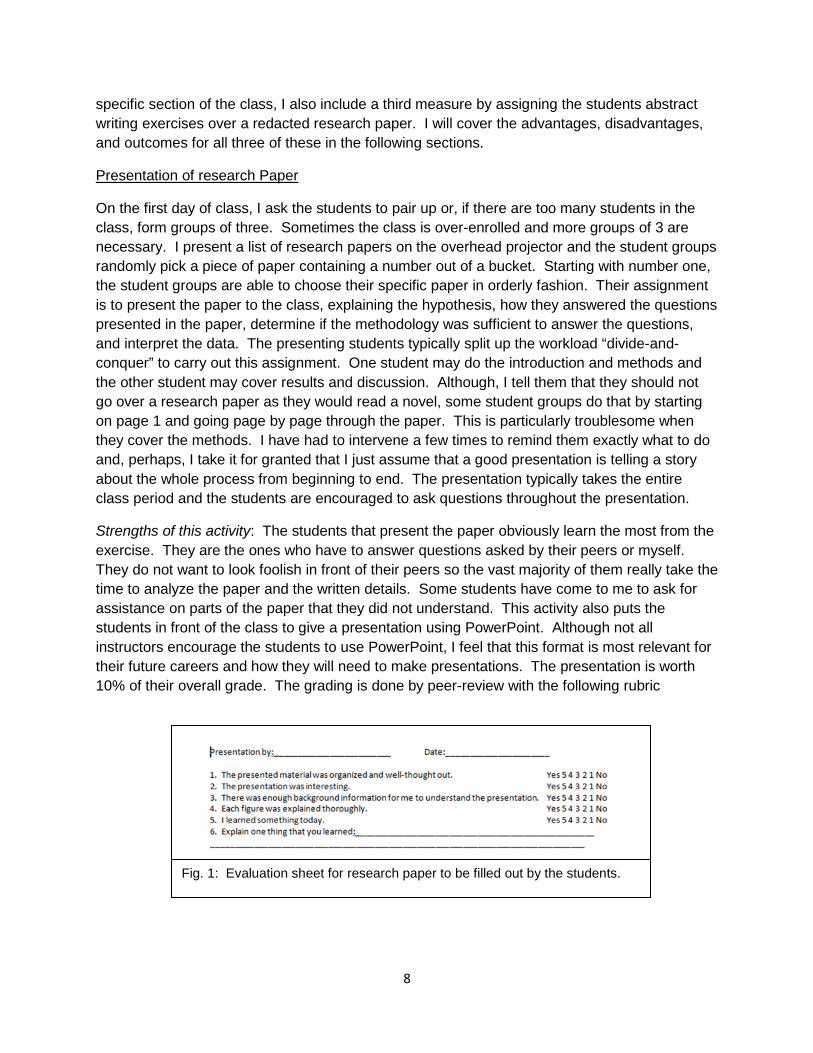
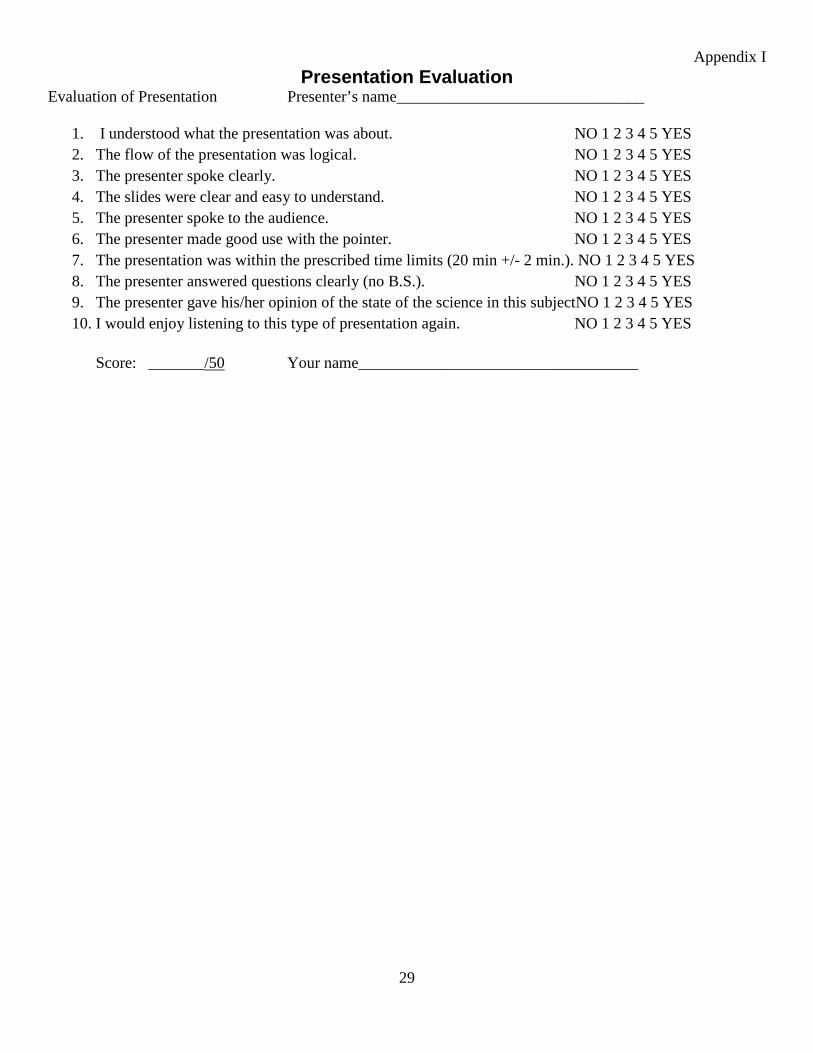
Strengths of this activity: The students that present the paper obviously learn the most from the exercise. They are the ones who have to answer questions asked by their peers or myself. They do not want to look foolish in front of their peers so the vast majority of them really take the time to analyze the paper and the written details. Some students have come to me to ask for assistance on parts of the paper that they did not understand. This activity also puts the students in front of the class to give a presentation using PowerPoint. Although not all instructors encourage the students to use PowerPoint, I feel that this format is most relevant for their future careers and how they will need to make presentations. The presentation is worth 10% of their overall grade. The grading is done by peer-review with the following rubric
Fig. 1: Evaluation sheet for research paper to be filled out by the students.
9
Weaknesses of this activity: I have found that many of the students (those who are not presenting) do not read the paper before class. They just come in and expect the presenters to teach them whatever is needed to know that day. In response, I have developed quizzes to test if they even read through the paper. Typically, I would show some data from the paper and ask them to interpret it or ask them about the model organism used and why it was used, or the hypothesis, etc. I have noticed that the quizzes have encouraged the students to at least go through the paper and understand all of the figures and basic methods and questions posed by the authors. I don’t think I will be able to get all of the students to read the entire paper unless it was a major component of an exam. As a major exam component, I think it would be counter-productive to do this since more of their time should be spent on the review paper and other activities in class.
Writing a review paper (Appendix III)
On the first day of class, my first lecture covers the differences between a research paper and review paper. The ideas behind writing a review paper is that it will force the students to go through the databases (PubMed, Google Scholar, Web of Knowledge, etc) to find papers, read them, sort through the information, and write up a paper that covers the topic area of their choice. Their choices are limited to a list of subjects I present on the first day, and like the research paper presentation, they pick a random number and then, in numerical order, pick from my list of topics. They are usually confronted with a topic they have no knowledge about or one that they don’t particularly like. In response, I tell them to make it interesting by choosing many research papers and build on themes, controversies, etc to formulate a story. Most of the topics are covered briefly in one of my twelve lectures, though they are expected to take it step further and really dive into the subject matter. The review paper consists of an abstract, introduction, three or four chapters/sections, and then conclusions/future perspectives. The paper is graded by the rubric in Appendix I.
Strengths of this activity: Those students who want a good grade really dig deep in the literature and find many papers from which to tell a good story. It is up to the students to put this paper together and make it as good as possible. To assist them, pieces of the paper are turned in over the course of the semester. For example, their outlines are due several weeks after topic selection. The introduction is due 2 weeks after the outline. Chapter 1 and the entire rough draft are submitted to me in similar manner over the course of the semester. This prevents the students from procrastinating and waiting until the very end of the semester to start working on this paper which would never work in their favor. This also allows me, as the instructor, to determine if they are on the right path or if they should change the course of the paper. The submission and return for the paper is all electronic and in MicroSoft Word. I receive the manuscripts, and use the “Track Changes” feature in Word to make corrections and suggestions for improving the paper. Usually, my instructions are received and adapted; sometimes they are not. The students are given a lot of latitude to be creative and find material that I do not know about. This happens frequently in which I am surprised that the student has taken the angle of an argument I had not considered or found an area in biotechnology that I was not aware of and made a sensible scholarly piece of work.

10
Weaknesses of this activity: It is impossible for the instructor to check all the references of each paper to ensure that the students read the paper correctly and understood the data. I suspect, and it has been confirmed by their written work, that many students read the papers superficially and pick and choose only what is needed. As a member of the research community, I am guilty of this as well in which I will only consider one figure or read just the abstract of a research paper that I will cite in my own paper or grant. Many, if not most, of these students probably do the same thing when reading papers for their review paper. I feel that many students are missing out on the opportunity to really analyze the details of papers and evaluate their merits. This is reflected in their final draft of the paper in which much of it appears to be superficial. Another weakness that I have found with the review paper is that I cannot really assess their writing until they turn in the introduction which occurs about half way through the semester. This delay limits instruction in this area.
Writing an abstract (Appendix IV)
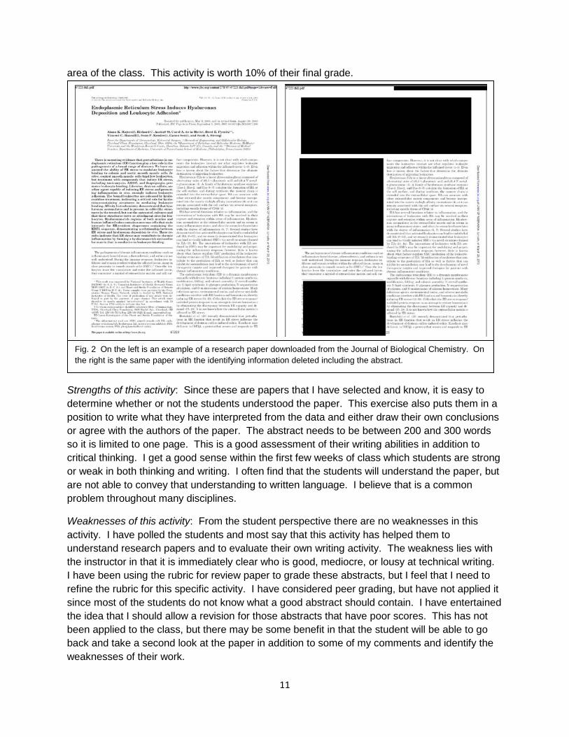
I feel that this activity is the most useful for assessing their critical thinking. During the first half of the semester, I present the students with three papers that have been redacted as a PDF document (Fig. 2). This means that all identifying information of the paper is deleted including title, authors, abstract, and journal identifiers. The students get the introduction, methods, results, and discussion. From these sections, they need to write an abstract for the paper in the paradigm that they are the author. Normally, the abstract is written last so this is a very real-world simulation for analyzing the data and writing a short, easy-to-read, and comprehensive summary. The topics of the paper are not overly complex and do not need to be in the subject
11
area of the class. This activity is worth 10% of their final grade.
Strengths of this activity: Since these are papers that I have selected and know, it is easy to determine whether or not the students understood the paper. This exercise also puts them in a position to write what they have interpreted from the data and either draw their own conclusions or agree with the authors of the paper. The abstract needs to be between 200 and 300 words so it is limited to one page. This is a good assessment of their writing abilities in addition to critical thinking. I get a good sense within the first few weeks of class which students are strong or weak in both thinking and writing. I often find that the students will understand the paper, but are not able to convey that understanding to written language. I believe that is a common problem throughout many disciplines.
Weaknesses of this activity: From the student perspective there are no weaknesses in this activity. I have polled the students and most say that this activity has helped them to understand research papers and to evaluate their own writing activity. The weakness lies with the instructor in that it is immediately clear who is good, mediocre, or lousy at technical writing. I have been using the rubric for review paper to grade these abstracts, but I feel that I need to refine the rubric for this specific activity. I have considered peer grading, but have not applied it since most of the students do not know what a good abstract should contain. I have entertained the idea that I should allow a revision for those abstracts that have poor scores. This has not been applied to the class, but there may be some benefit in that the student will be able to go back and take a second look at the paper in addition to some of my comments and identify the weaknesses of their work.
Fig. 2 On the left is an example of a research paper downloaded from the Journal of Biological Chemistry. On the right is the same paper with the identifying information deleted including the abstract.
12
Assessment and Outcomes of the Class Activities and Organization
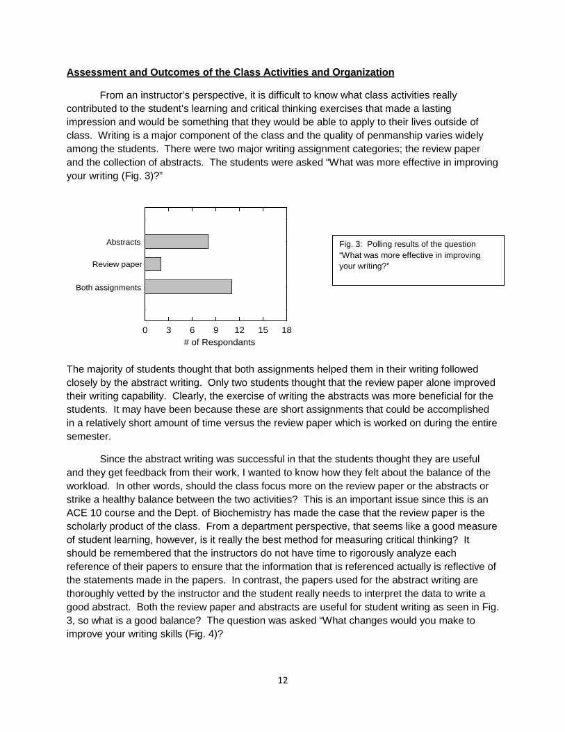
From an instructor’s perspective, it is difficult to know what class activities really contributed to the student’s learning and critical thinking exercises that made a lasting impression and would be something that they would be able to apply to their lives outside of class. Writing is a major component of the class and the quality of penmanship varies widely among the students. There were two major writing assignment categories; the review paper and the collection of abstracts. The students were asked “What was more effective in improving your writing (Fig. 3)?”
The majority of students thought that both assignments helped them in their writing followed closely by the abstract writing. Only two students thought that the review paper alone improved their writing capability. Clearly, the exercise of writing the abstracts was more beneficial for the students. It may have been because these are short assignments that could be accomplished in a relatively short amount of time versus the review paper which is worked on during the entire semester.
Since the abstract writing was successful in that the students thought they are useful and they get feedback from their work, I wanted to know how they felt about the balance of the workload. In other words, should the class focus more on the review paper or the abstracts or strike a healthy balance between the two activities? This is an important issue since this is an ACE 10 course and the Dept. of Biochemistry has made the case that the review paper is the scholarly product of the class. From a department perspective, that seems like a good measure of student learning, however, is it really the best method for measuring critical thinking? It should be remembered that the instructors do not have time to rigorously analyze each reference of their papers to ensure that the information that is referenced actually is reflective of the statements made in the papers. In contrast, the papers used for the abstract writing are thoroughly vetted by the instructor and the student really needs to interpret the data to write a good abstract. Both the review paper and abstracts are useful for student writing as seen in Fig. 3, so what is a good balance? The question was asked “What changes would you make to improve your writing skills (Fig. 4)?
# of Respondants0 3 6 9 12 15 18
Abstracts
Review paper
Both assignments
Fig. 3: Polling results of the question “What was more effective in improving your writing?”
13
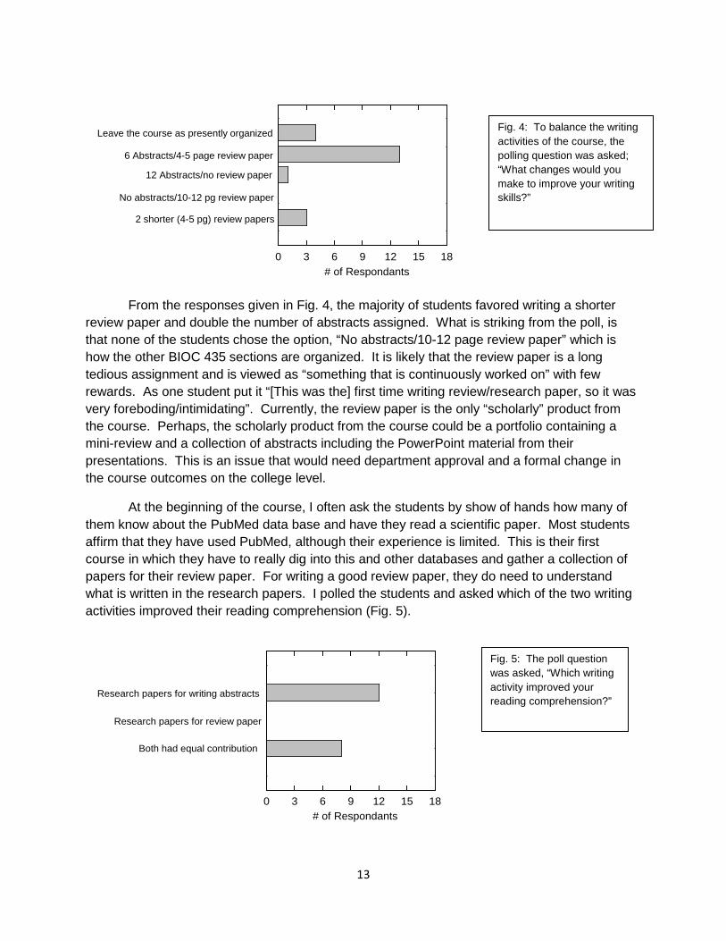
From the responses given in Fig. 4, the majority of students favored writing a shorter review paper and double the number of abstracts assigned. What is striking from the poll, is that none of the students chose the option, “No abstracts/10-12 page review paper” which is how the other BIOC 435 sections are organized. It is likely that the review paper is a long tedious assignment and is viewed as “something that is continuously worked on” with few rewards. As one student put it “[This was the] first time writing review/research paper, so it was very foreboding/intimidating”. Currently, the review paper is the only “scholarly” product from the course. Perhaps, the scholarly product from the course could be a portfolio containing a mini-review and a collection of abstracts including the PowerPoint material from their presentations. This is an issue that would need department approval and a formal change in the course outcomes on the college level.
At the beginning of the course, I often ask the students by show of hands how many of them know about the PubMed data base and have they read a scientific paper. Most students affirm that they have used PubMed, although their experience is limited. This is their first course in which they have to really dig into this and other databases and gather a collection of papers for their review paper. For writing a good review paper, they do need to understand what is written in the research papers. I polled the students and asked which of the two writing activities improved their reading comprehension (Fig. 5).
# of Respondants0 3 6 9 12 15 18
Leave the course as presently organized
6 Abstracts/4-5 page review paper
12 Abstracts/no review paper
No abstracts/10-12 pg review paper
2 shorter (4-5 pg) review papers
# of Respondants0 3 6 9 12 15 18
Research papers for writing abstracts
Research papers for review paper
Both had equal contribution
Fig. 4: To balance the writing activities of the course, the polling question was asked; “What changes would you make to improve your writing skills?”
Fig. 5: The poll question was asked, “Which writing activity improved your reading comprehension?”
14
The poll results in Fig. 5 indicate that although the abstract writing is valuable in reading comprehension, the activity of perusing many papers in the databases has value. Nearly half the students thought that both writing activities had value for improving their reading comprehension. Results from this question and the previous as shown in Fig. 4, suggest that the research paper alone is not sufficient for student reading comprehension, therefore, the combination of activities is really beneficial for the students’ critical thinking of activities.
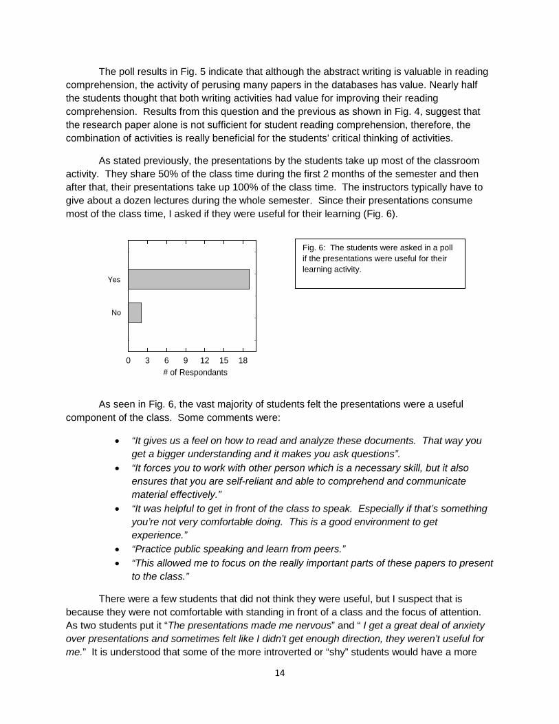
As stated previously, the presentations by the students take up most of the classroom activity. They share 50% of the class time during the first 2 months of the semester and then after that, their presentations take up 100% of the class time. The instructors typically have to give about a dozen lectures during the whole semester. Since their presentations consume most of the class time, I asked if they were useful for their learning (Fig. 6).
As seen in Fig. 6, the vast majority of students felt the presentations were a useful component of the class. Some comments were:
• “It gives us a feel on how to read and analyze these documents. That way you get a bigger understanding and it makes you ask questions”.
• “It forces you to work with other person which is a necessary skill, but it also ensures that you are self-reliant and able to comprehend and communicate material effectively.”
• “It was helpful to get in front of the class to speak. Especially if that’s something you’re not very comfortable doing. This is a good environment to get experience.”
• “Practice public speaking and learn from peers.” • “This allowed me to focus on the really important parts of these papers to present
to the class.”
There were a few students that did not think they were useful, but I suspect that is because they were not comfortable with standing in front of a class and the focus of attention. As two students put it “The presentations made me nervous” and “ I get a great deal of anxiety over presentations and sometimes felt like I didn’t get enough direction, they weren’t useful for me.” It is understood that some of the more introverted or “shy” students would have a more
# of Respondants0 3 6 9 12 15 18
Yes
No
Fig. 6: The students were asked in a poll if the presentations were useful for their learning activity.
15
difficult experience with class presentations, nevertheless, it is a useful activity and valuable for their professional career development as they will need to stand before people and speak to an audience.
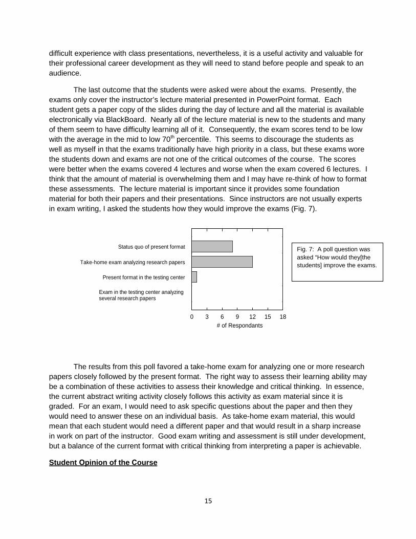
The last outcome that the students were asked were about the exams. Presently, the exams only cover the instructor’s lecture material presented in PowerPoint format. Each student gets a paper copy of the slides during the day of lecture and all the material is available electronically via BlackBoard. Nearly all of the lecture material is new to the students and many of them seem to have difficulty learning all of it. Consequently, the exam scores tend to be low with the average in the mid to low 70th percentile. This seems to discourage the students as well as myself in that the exams traditionally have high priority in a class, but these exams wore the students down and exams are not one of the critical outcomes of the course. The scores were better when the exams covered 4 lectures and worse when the exam covered 6 lectures. I think that the amount of material is overwhelming them and I may have re-think of how to format these assessments. The lecture material is important since it provides some foundation material for both their papers and their presentations. Since instructors are not usually experts in exam writing, I asked the students how they would improve the exams (Fig. 7).
The results from this poll favored a take-home exam for analyzing one or more research papers closely followed by the present format. The right way to assess their learning ability may be a combination of these activities to assess their knowledge and critical thinking. In essence, the current abstract writing activity closely follows this activity as exam material since it is graded. For an exam, I would need to ask specific questions about the paper and then they would need to answer these on an individual basis. As take-home exam material, this would mean that each student would need a different paper and that would result in a sharp increase in work on part of the instructor. Good exam writing and assessment is still under development, but a balance of the current format with critical thinking from interpreting a paper is achievable.
Student Opinion of the Course
# of Respondants0 3 6 9 12 15 18
Status quo of present format
Take-home exam analyzing research papers
Present format in the testing center
Exam in the testing center analyzing several research papers
Fig. 7: A poll question was asked “How would they[the students] improve the exams.
16
After multiple teaching experiences over the past 3 years, I am curious how the material was received since each class is different in student composition and preparedness. I gave a survey to the students and asked them what worked well (things to keep), what did not work very well (needs to stop) and which parts were the easiest and most difficult. These were all open-ended questions so the responses were variable. I will summarize what I found to be the trends.
What is working: Many students felt that all aspects of the course working together had the most benefit for them. The subject material was new and not taught in any of their previous courses so it was not just a recycling of familiar material. They also liked how the lecture material dove-tails with the research paper presentations and that the lectures also give background material for their papers. The abstracts and review paper were well-received and most students thought they were both challenging. The following quotes are from the survey.
• “I enjoy all aspects of this class and the professor is great. There is an actual focus onour learning and is centered around preparing us for the professional world. I feel thisclass prepared me more for my future than most of my other courses.”
• “I like that we turn in the final paper in segments so we get feedback on our writingthroughout the course and not just at the end.”
• “I thought the class was great, although it was challenging, it helps combine all myknowledge in one great whole.”
What is not working so well. The major complaint of the course was the exams and how difficult and detailed they were. Since this was their first exposure to this material, this opinion is valid in that maybe I demanded too much. I had noticed that when the exams covered four lectures instead of six, that the students did better and did not complain about them so much. To remedy this, I may have to go back to doing four lectures/exam instead of six/exam. The second biggest complaint is the time it took to research and write the review paper. As this is a major component of the course and the outcomes for ACE 10, I cannot realistically take this out of the course. I may be able to fine-tune it better by having them hand in more parts of it on a regular basis, though I am already having them turn it in four pieces that are marked with my comments for improvement. Concerning the research presentations, one student noted what I had suspected of these presentations. He/she had written “The group presentation over the research papers didn’t fulfill their purpose. Most of the students only read the paper they were assigned to present and class discussions didn’t really happen.” This has been a real challenge in the class in that I am trying to balance how much leadership the students take in their presentation and how much I need to get involved with leading the discussion. Since each student group is different and I do not know these students very well on a personal level, it is difficult determine when they need a bit of help and stimulation versus me taking over the class. The last thing I want to do is take their presentation away from them by leading the discussion.
What part of the course was the easiest? Many of the students felt that the presentations were the easiest, specifically, the solo presentation on their review paper. The second most popular answer was the quizzes on the research paper presentations. Other more scattered answers included the abstracts, the exams, or writing the paper.
17
What part of the course was the most difficult? The most popular answer to this question was the exams. As seen from Fig. 7, I may introduce more critical thinking exercises on the exam and ease up a bit on the lecture material. Doing this would serve the students better, though I have yet to test this. The second most popular answer was writing the research paper. Several students mentioned that they found it difficult to take a wide assortment of papers and collate them into one seamless document. This was supposed to be alleviated when they write up an outline and go over the material with me in my office in a face-to-face discussion.
Results from the Just-In-Time Teaching (JiTT). Throughout the twelve lectures, four questions were posed the night before the relevant lecture to get the students to research a question and find a suitable answer. The results of answers were given during the lecture the following day. Overall, the questions forced the students to look up material and many of them stepped up to the challenge and referenced papers in their pursuit of an answer. The four JiTT questions are as follows:
A) High Fructose corn syrup in many products that we eat and drink. Is it good for us, bad forus or neither good nor bad for us? Explain your answer.
B) What is caramelization and explain some of the chemistry that is going on?
C) How does Mucinex work?
D) Imagine that you have discovered a protein and determined the sequence. The amino acidsequence reveals that 8 sites may be modified by substrate #1. Those same 8 sites may also be modified by substrate #2. How many different species of this protein would be possible if only substrate #1 was allowed to act on the protein? How many different species of this protein would be possible of both substrates 1 and 2 are allowed to act on this protein?
Participation in each of these activities was over 80% and at least half of the students were able to get the correct answer. Interestingly, the math and biochemistry dual majors missed the 4th question, the only one that involved any mathematical knowledge. Did these activities result in a deeper understanding of the subject material? Yes, because they were more prepared for class and had some idea of what the lecture would be about. Did it help them in the exams? No. The exams did not cover the exact material and it is common for students to compartmentalize information. The students did enjoy looking up the material, although one student did dissent and asked that the participation activity cease. In future courses, I plan to develop more carefully crafted questions that require more thought and expand the number to 6 questions or about 1 per week for the first half of the semester during my 12 lectures. Details of how the class answered the JiTT questions may be found in Appendix V.
Planned Changes
Based on student feed-back and what I have noticed to be deficiencies, I plan to:
• Increase the number of abstracts to be written from 3 per semester to 5 per semester. Imay also increase the percentage of the grade for the abstract written material from
18
10% to 15%. I am still considering but not sure of adding the abstracts to a portfolio of scholarly work that the student will submit with the paper.
• Reduce the amount of questions on the exam so they may be completed within 30 min.and introduce a paper on the exam for them to interpret the data in its presented form.This type of format will still allow me to cover the necessary subject matter and assesstheir critical thinking skills. I may also reduce the exam percentage of the overall gradefrom 30% to 25%.
• Keep giving quizzes on the research paper, but include data from the paper that isneeded for interpretation. Hopefully, this will stimulate the students to at least look atthe figures in the papers and understand them. I also plan to ask a question or two onany controversial material that was presented in class as part of the studentpresentation. Currently, quizzes are worth 5% of the grade with the allowance ofdropping the two lowest scores. I will keep this format.
• The research paper format and length will not change even though students would liketo write a shorter paper. I will need to emphasize that writing the paper in a series ofsmall portions will lead to a comprehensive scholarly work by the end of the semester. Ido believe that the students just feel overwhelmed when they first see the work that isrequired at the beginning of the semester. It is all psychological.
• Expand the number of JiTT questions based on the lectures. These are always good tosee what the students know and how they tackle problems. It also serves as a guide onwhat I need to emphasize in lecture and that emphasis may increase theirunderstanding and exam scores.
• Develop a more comprehensive rubric for grading the written abstracts. So far, I haveused the rubric for the research paper, but it does not fit quite right and I have beengrading on sort of a qualitative scale. This does not seem really fair to the students andthis new rubric will be implemented before my next class.
Summary and Overall Assessment of the Portfolio Process
Throughout the process of collecting the information for this course portfolio, I have realized that I am basically on the correct track in teaching this course, but a few refinements and modifications will make this course even better and more seamless on my part. I need to exploit some of my current methodology to make a bigger impact with their learning. One way to do this is to develop better JiTT questions for the subject material. The type of question and how it is asked can really make or break this exercise. A second way for making a bigger impact for the assessment of critical thinking is to put more critical thinking exercises on the exams. Some evaluations have mentioned that my exams are too memorization intensive and that may be true, but the real emphasis of this course is critical thinking skills and not rote memorization of facts and figures. I have always been too content driven in this course and not enough emphasis has been placed in the “process” of learning. I will need to slow down in my lectures and not worry if I hit every point. Although I ask the class if there are any questions at the end of lecture, I rarely need to take a question because all of them are too afraid to ask and look “dumb” in front of their peers. Those students that I “lost” during the lecture will never speak up at the end of class, though few have asked me to re-explain something during the lecture.
19
I have realized that over the past 3 years of teaching this course that the students come in all variations of writing and reading abilities. How to manage this diversity of skill and keep everyone enthusiastic about the course has been a real challenge for me. I hate it when someone mentally drops out because it is too difficult or too easy in some cases. I implemented a wide variety of activities early on so that if a student “bombs” a test or presentation, the grade is salvageable. From my own experience, I have done poorly in some parts of a class and was relieved and encouraged that it was redeemable. I try to do that in this class, while maintaining focus on the course objectives. I don’t expect every student to exit the class as an expert technical writer, but I would like every student feel that he/she has accomplished something (i.e. the review paper) and know that through this experience they are better at technical reading and writing than when they entered the class. This is what I count as success in this course.
In revisiting my portfolio objectives from page 1, I will answer what I realized throughout this whole process. 1) how are my teaching refinements and activities improving the course? The introduction of abstract writing in the course and accountability for reading papers via quizzes is an improvement. Refinement for teaching these students how to read and write is an on-going process, but I think it is mostly complete. 2) Are my assessment methods for evaluating student learning during the semester sufficient? Yes, though I am able to improve in this area with my planned changes outlined earlier. 3) Is my course meeting all of the requirements for ACE 10? Yes it is. My methods exceed the department’s requirements, though it is more effort on my part. I feel it is my responsibility, as a professor, to prepare the students to meet the challenges in post-college life. The principles learned in this course may be transferrable to any professional career.

Appendix I
20
Biochemistry 435 Advanced Topics in Biochemistry Course Focus: Glycobiology
Spring 2014 Syllabus
Instructor: Edward N. Harris, Ph.D. N133 Beadle, 472-7468, [email protected] Office hours (Beadle N133) email/call me to set up time.
Class times: M W F 8:30 am to 9:20 am in Rm N177 Beadle
Prerequisite: Bioc 432 and major in biochemistry
Your Course Objectives • Integrate overall biochemistry knowledge and understanding with emphasis in sugar biochemistry.• Learn to read and critique the primary literature and experimental methods in biochemistry.• Become acquainted with sophisticated topics in biochemistry and able to contribute to group discussions
about the literature related to these topics.• Apply your biochemical knowledge to new areas within the life sciences.• Organize and present biochemical research data in oral and written formats.
Text (suggested as a reference only): Essentials of Glycobiology, 2nd Edition found at http://www.ncbi.nlm.nih.gov/books?term=essentials%20of%20glycobiology We will use research articles available on the Web and in the University Libraries
Activities: • There will be class discussions of assigned papers. It is assumed that each student will have carefully
read the papers and reflected on them prior to the class. • Students will be active participants in class discussions and evaluate their peers on their presentations of
scientific papers. All students will read the articles and participate in the discussion, but the presenters will lead the discussion.
• Students will present a 20 minute presentation of their chosen topic which will be a summary of theirreview paper.
• Each student will write 3 abstracts for research articles over the course of the semester.• At the end of the semester, each student will submit a review article containing an abstract, introduction,
chapters/topics of discussion, future directions, conclusion and references. The paper should containreferences from the scientific literature. Web sources are neither peer-reviewed nor stable and are notgenerally valid citations. The syllabus contains the rubric that the instructor will use in evaluating finalpaper. The paper should be at least 8 pages long (excluding references), single spaced, font size 11 inTimes New Roman or Ariel.
• Classroom participation
Grading: • Students will take 2 exams covering 6 topics during the semester. Each exam is 15% of the grade.• Students will receive numerical grades at the end of the semester for the final paper.• Short quizzes consisting of two to four questions will be administered to the class time after the
discussion of paper as indicated on the course schedule. The quizzes will cover material the research
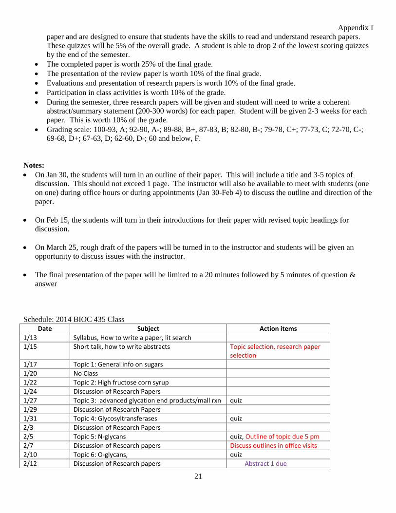
Appendix I
21
paper and are designed to ensure that students have the skills to read and understand research papers. These quizzes will be 5% of the overall grade. A student is able to drop 2 of the lowest scoring quizzes by the end of the semester.
• The completed paper is worth 25% of the final grade.• The presentation of the review paper is worth 10% of the final grade.• Evaluations and presentation of research papers is worth 10% of the final grade.• Participation in class activities is worth 10% of the grade.• During the semester, three research papers will be given and student will need to write a coherent
abstract/summary statement (200-300 words) for each paper. Student will be given 2-3 weeks for eachpaper. This is worth 10% of the grade.
• Grading scale: 100-93, A; 92-90, A-; 89-88, B+, 87-83, B; 82-80, B-; 79-78, C+; 77-73, C; 72-70, C-;69-68, D+; 67-63, D; 62-60, D-; 60 and below, F.
Notes: • On Jan 30, the students will turn in an outline of their paper. This will include a title and 3-5 topics of
discussion. This should not exceed 1 page. The instructor will also be available to meet with students (one on one) during office hours or during appointments (Jan 30-Feb 4) to discuss the outline and direction of the paper.
• On Feb 15, the students will turn in their introductions for their paper with revised topic headings fordiscussion.
• On March 25, rough draft of the papers will be turned in to the instructor and students will be given anopportunity to discuss issues with the instructor.
• The final presentation of the paper will be limited to a 20 minutes followed by 5 minutes of question &answer
Schedule: 2014 BIOC 435 Class Date Subject Action items
1/13 Syllabus, How to write a paper, lit search 1/15 Short talk, how to write abstracts Topic selection, research paper
selection 1/17 Topic 1: General info on sugars 1/20 No Class 1/22 Topic 2: High fructose corn syrup 1/24 Discussion of Research Papers 1/27 Topic 3: advanced glycation end products/mall rxn quiz 1/29 Discussion of Research Papers 1/31 Topic 4: Glycosyltransferases quiz 2/3 Discussion of Research Papers 2/5 Topic 5: N-glycans quiz, Outline of topic due 5 pm 2/7 Discussion of Research papers Discuss outlines in office visits 2/10 Topic 6: O-glycans, quiz 2/12 Discussion of Research papers Abstract 1 due
Appendix I
22
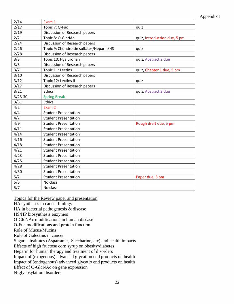
2/14 Exam 1 2/17 Topic 7: O-Fuc quiz 2/19 Discussion of Research papers 2/21 Topic 8: O-GlcNAc quiz, Introduction due, 5 pm 2/24 Discussion of Research papers 2/26 Topic 9: Chondroitin sulfates/Heparin/HS quiz 2/28 Discussion of Research papers 3/3 Topic 10: Hyaluronan quiz, Abstract 2 due 3/5 Discussion of Research papers 3/7 Topic 11: Lectins quiz, Chapter 1 due, 5 pm 3/10 Discussion of Research papers 3/12 Topic 12: Lectins II quiz 3/17 Discussion of Research papers 3/21 Ethics quiz, Abstract 3 due 3/23-30 Spring Break 3/31 Ethics 4/2 Exam 2 4/4 Student Presentation 4/7 Student Presentation 4/9 Student Presentation Rough draft due, 5 pm 4/11 Student Presentation 4/14 Student Presentation 4/16 Student Presentation 4/18 Student Presentation 4/21 Student Presentation 4/23 Student Presentation 4/25 Student Presentation 4/28 Student Presentation 4/30 Student Presentation 5/2 Student Presentation Paper due, 5 pm 5/5 No class 5/7 No class
Topics for the Review paper and presentation HA synthases in cancer biology HA in bacterial pathogenesis & disease HS/HP biosynthesis enzymes O-GlcNAc modifications in human disease O-Fuc modifications and protein function Role of Mucus/Mucins Role of Galectins in cancer Sugar substitutes (Aspartame, Saccharine, etc) and health impacts Effects of high fructose corn syrup on obesity/diabetes Heparin for human therapy and treatment of disorders Impact of (exogenous) advanced glycation end products on health Impact of (endogenous) advanced glycatio end products on health Effect of O-GlcNAc on gene expression N-glycosylation disorders

Appendix I
23
Role of Selectins in inflammation Arthritis and hyaluronan Bacterial glycomolecules and disease mechanisms Viral glycans/enzymes and disease mechanisms Protozoan glycans and disease Helminth glycans and disease Glycan storage diseases Beta-glucans and the immune system Type I diabetes (cause, mechanisms, treatment) Type II diabetes (cause, mechanisms, treatment) Honey in therapeutic treatments O-mannosylation related disorders (Muscle-eye-brain disease, Walker-warburg syndrome, etc.) C-mannosylation: What is it and what does it do? Carbohydrates in biotechnology
Special Needs Students with disabilities are encouraged to contact the instructor for a confidential discussion of their individual needs for academic accommodation. It is the policy of the University of Nebraska-Lincoln to provide flexible and individualized accommodation to students with documented disabilities that may affect their ability to fully participate in course activities or to meet course requirements. To receive accommodation services, students must be registered with the Services for Students with Disabilities (SSD) office, 132 Canfield Administration, 472-3787 voice or TTY.
Academic Honesty: Academic dishonesty includes fabrication, falsification and plagiarism and will not be tolerated by CBC faculty and should not be tolerated by CBC students. Falsification of research data or its deliberate misinterpretation is a serious offense. Cheating on an examination or assignment is an obvious form of academic dishonesty. Plagiarism is more complex and may be the result of sloppiness, rather than an intentional attempt to deceive. Plagiarism is defined as passing off someone else's ideas, words or writings as your own. Inclusion of a sentence in a paper that is copied from any source without quotation marks and citation is an example of plagiarism. Anything that you present that is under your name should be entirely your own work unless so indicated and appropriately cited. This includes ideas as well as specific quotations, artwork and figures. Some biochemistry students do not realize that they have copied whole sentences out of published work when then write papers. To help you determine if you have inadvertently done this, we can submit your written work through the Safe Assignment feature in BlackBoard.
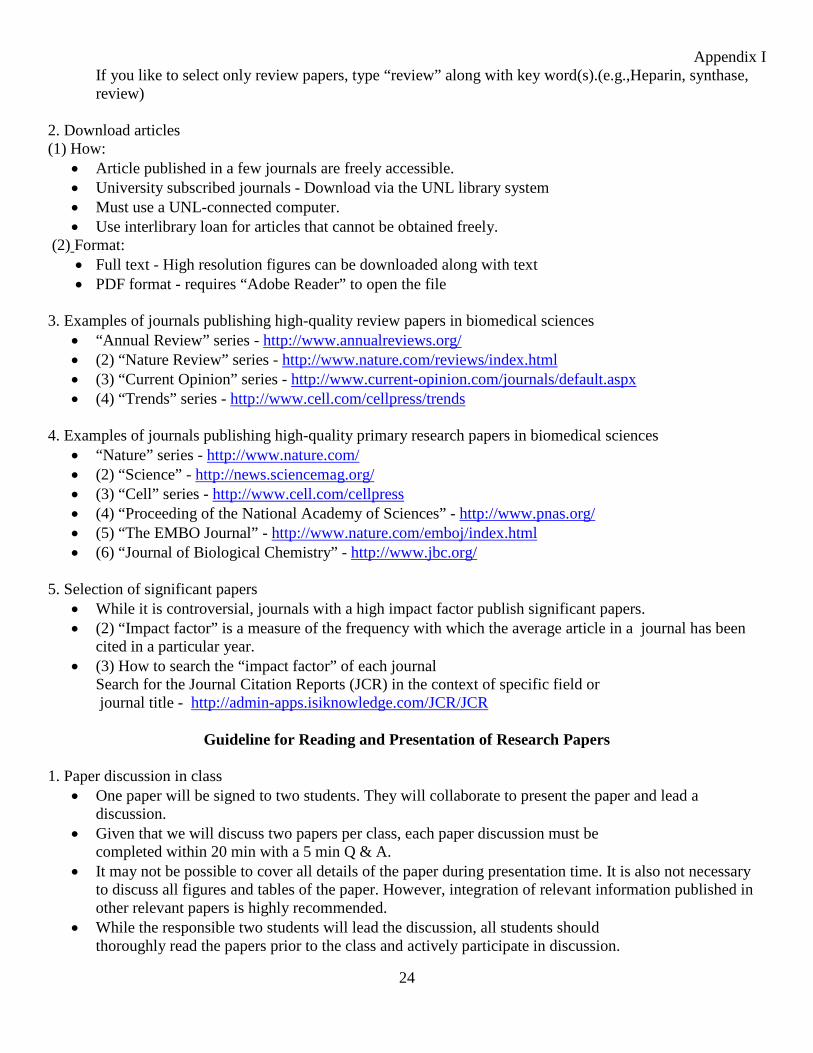
Guideline for Literature Search 1. Search literature
• Textbook – to acquire basic information and cited reference papers • PubMed (http://www.ncbi.nlm.nih.gov/pubmed) literature search database • Web of Knowledge (Google it, then bookmark it) • Visit home page of journals
(e.g., “Annual Review” series, http://www.annualreviews.org/) • Type key word(s) of your interest
Appendix I
24
If you like to select only review papers, type “review” along with key word(s).(e.g.,Heparin, synthase, review)
2. Download articles (1) How:
• Article published in a few journals are freely accessible. • University subscribed journals - Download via the UNL library system • Must use a UNL-connected computer. • Use interlibrary loan for articles that cannot be obtained freely.
(2) Format: • Full text - High resolution figures can be downloaded along with text • PDF format - requires “Adobe Reader” to open the file
3. Examples of journals publishing high-quality review papers in biomedical sciences
• “Annual Review” series - http://www.annualreviews.org/ • (2) “Nature Review” series - http://www.nature.com/reviews/index.html • (3) “Current Opinion” series - http://www.current-opinion.com/journals/default.aspx • (4) “Trends” series - http://www.cell.com/cellpress/trends
4. Examples of journals publishing high-quality primary research papers in biomedical sciences
• “Nature” series - http://www.nature.com/ • (2) “Science” - http://news.sciencemag.org/ • (3) “Cell” series - http://www.cell.com/cellpress • (4) “Proceeding of the National Academy of Sciences” - http://www.pnas.org/ • (5) “The EMBO Journal” - http://www.nature.com/emboj/index.html • (6) “Journal of Biological Chemistry” - http://www.jbc.org/
5. Selection of significant papers
• While it is controversial, journals with a high impact factor publish significant papers. • (2) “Impact factor” is a measure of the frequency with which the average article in a journal has been
cited in a particular year. • (3) How to search the “impact factor” of each journal
Search for the Journal Citation Reports (JCR) in the context of specific field or journal title - http://admin-apps.isiknowledge.com/JCR/JCR
Guideline for Reading and Presentation of Research Papers 1. Paper discussion in class
• One paper will be signed to two students. They will collaborate to present the paper and lead a discussion.
• Given that we will discuss two papers per class, each paper discussion must be completed within 20 min with a 5 min Q & A.
• It may not be possible to cover all details of the paper during presentation time. It is also not necessary to discuss all figures and tables of the paper. However, integration of relevant information published in other relevant papers is highly recommended.
• While the responsible two students will lead the discussion, all students should thoroughly read the papers prior to the class and actively participate in discussion.
Appendix I
25
2. It is critical to figure out (or extract, synthesize) the following points when you read papers.
• What are the question(s) addressed in the paper?• What are the advance(s) made by the paper?• Does the paper use appropriate methods and approaches to answer the question?• Is data collection and interpretation accurate?• Does conclusion reflect data?• Significance and impacts of the research.• You should be able to criticize the hypothesis, experimental methods and approaches, data collection
and presentation, interpretation and integration of data, and/or conclusion.• You should be able to find out remaining unanswered questions about the research topic.
3. Major issues you need to care when you lead a discussion of a paper.• Provide sufficient backgrounds.• Let everybody know about the question(s) that are addressed in the paper.• Briefly describe methods. However, if the methods are unique and/or critical for understanding
presented data, you may discuss in detail.• Interpret (what message do you extract from the data?) rather than describe (what the data is?).• Criticize presented data and interpretation by the authors.• Emphasize the significance of the study and advance(s) made by the paper.• Emphasize impacts of the data in the context of the same or related research fields.• Identify interesting unanswered questions and provide research directions of the topic.
4. A common delivery method is a PowerPoint slide; however, any creativity isappreciated, except Dance.
Useful background information has been and will be placed on the BIOC 435 BlackBoard
Biochemistry 435 is a Capstone course that meets the criteria for Area 10 of the Achievement Centered Education program for the University of Nebraska. Below are the expectations related to the ACE outcome:
SLO10: Generate a creative or scholarly product that requires broad knowledge, appropriate technical proficiency, information collection, synthesis, interpretation, presentation, and reflection.
1. Describe opportunities students should have to learn the outcome.How is the learning objective embedded in the course?
Students at the start of this capstone course generally have minimal exposure to the primary scientificliterature and to writing in a scientific style. This course focuses on one topic that cuts across the variouslife science disciplines. Students receive a few lectures of introduction, and then start reading journalarticles selected by the instructor. Then, most of the remainder of the semester is comprised of studentssearching the primary literature and collecting relevant articles with information to build a coherentpaper that proposes the direction of future research in one aspect of the general topic. Students learn tomake presentations of their selected papers and reflect on how the data reported represents an
Appendix I
26
incremental increase in knowledge and understanding. The different sections of this course taught by different instructors may have different general topics. For the section taught by Dr. Cahoon, students explore metabolic engineering for the semester. Each student is assigned one metabolic engineering target and will explore its utility, biosynthesis, and devise a metabolic engineering strategy for producing the target compound in a recombinant host. The major focus of the course is the development of the course paper, but students also give a final PowerPoint presentation to the class. Sections taught by other faculty follow a generally similar program, but the topic and specifics may vary.
2. Describe student work that will be used to assess student achievement of the outcome and explain how the students demonstrate the knowledge and skills specified by the outcome.
Students will develop a course paper on the particular aspect of the general topic for the course. The student will work on chapters that will build to form the summative paper. The chapters are turned in at three to four week intervals and receive formative assessment from the instructor based on a rubric. Students then have the ability to incorporate feedback into the development of the subsequent chapters and to rewrite the chapter already assessed for incorporation into the final paper. In addition to the continual assessment and feed-back related to the course paper, students provide self-evaluations at weeks 5 and 10 that are related to their degree of engagement with the primary literature and their contributions in the general discussions of student-presented papers that occupy most of the class sessions.
3. As part of the ACE certification process, the department/unit agrees to collect and assess a reasonable sample of students' work and provide reflections on students' achievement of the Learning Outcomes for its respective ACE-certified courses. Please comment on your plans to develop a process to collect and evaluate student work over time for the purpose of assessing student success for this ACE outcome.
The assessment will be bipartite. The instructor will save the self-evaluations and her/his comments on them as a measure of the degree to which the student's perception of their pro-active engagement in mining and presenting the primary literature is parallel to that of the instructor's perception. The second element is in archiving the students' chapters for the course paper in their original format with faculty annotations and suggestions, and the final course paper that was turned in for a final grade. The ability to compare the initial attempt to produce each chapter and the final work should provide evidence of the student's initial level of abilities at information collection, interpretation, reflection and synthesis, and the final level achieved by the end of the course. It is proposed that the instructor select the above materials for the students receiving the top two grades, the two median grades, and the two lowest grades in the course to submit to the Department for archiving.
Reinforcements According to the ACE document approved by faculty (Structural Criteria, item 9), "Every ACE course will reinforce at least one of the following skills listed below as appropriate for the discipline and as identified by the department offering the course..." Indicate skills that will be reinforced by the course by clicking on as many as apply and describe briefly how those skills will be reinforced. These areas are those OTHER THAN the one or two outcomes for which you seek ACE certification. Students will not receive ACE credit for the reinforced skills, and the reinforced skills do not need to be assessed for ACE purposes. What Outcome(s) or skill(s) will be reinforced in this course?
Appendix I
27
Writing Students will be writing in a scientific format for the first time. This may be very different from prior writing in the university. For example, it is common to use past-tense, passive-voice in scientific writing. Students will need to learn how to use and cite appropriate references from the primary literature. This will come from discussions in the classroom sessions and from feedback on the course paper chapters that are submitted during the progress of the course.
Oral Communication This will be reinforced by the presentation of relevant papers from the scientific literature during most of the class periods. Students need to learn to master the content of such papers, prioritize the important elements, and present them in a coherent fashion. The student effort receives intrinsic feedback during the process by questions and comments from the instructor and from the other students.
Critical Thinking Critical thinking can only be done once a student has mastered a significant amount of foundational literature. Students at the start of the topic will have little ability to carry out critical thinking about the course theme. As students read more of the primary literature and seek out other references to flesh out certain aspects and to reconcile contradictory reports, they will be encouraged to reflect on the epistemology of the conclusions.
Ethics Each section of the class involves one class session related to an ethical issue. This usually involves a case study that is read prior to the class and a group discussion. In some cases, it is productive to have students attempt to present differing viewpoints, but in for other topics, students seem able to grasp the diverse social impact.
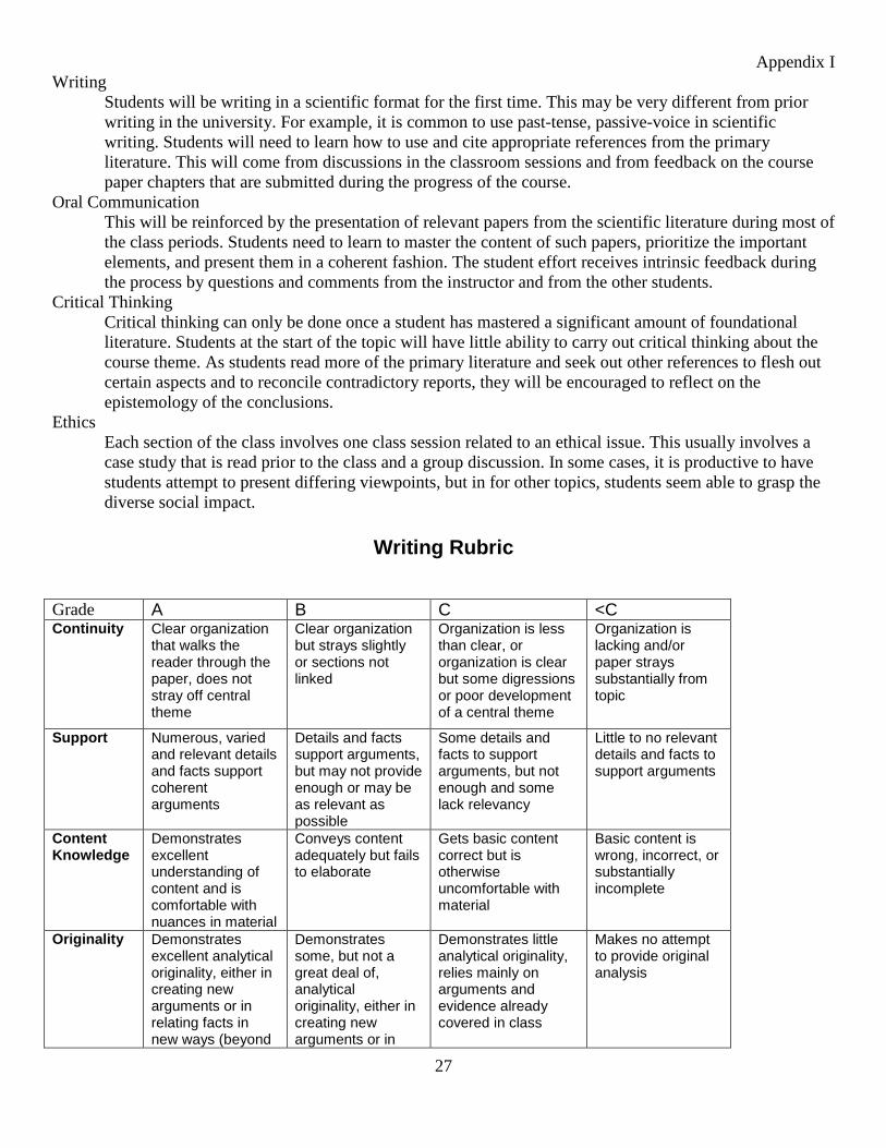
Writing Rubric
Grade A B C <C Continuity Clear organization
that walks the reader through the paper, does not stray off central theme
Clear organization but strays slightly or sections not linked
Organization is less than clear, or organization is clear but some digressions or poor development of a central theme
Organization is lacking and/or paper strays substantially from topic
Support Numerous, varied and relevant details and facts support coherent arguments
Details and facts support arguments, but may not provide enough or may be as relevant as possible
Some details and facts to support arguments, but not enough and some lack relevancy
Little to no relevant details and facts to support arguments
Content Knowledge
Demonstrates excellent understanding of content and is comfortable with nuances in material
Conveys content adequately but fails to elaborate
Gets basic content correct but is otherwise uncomfortable with material
Basic content is wrong, incorrect, or substantially incomplete
Originality
Demonstrates excellent analytical originality, either in creating new arguments or in relating facts in new ways (beyond
Demonstrates some, but not a great deal of, analytical originality, either in creating new arguments or in
Demonstrates little analytical originality, relies mainly on arguments and evidence already covered in class
Makes no attempt to provide original analysis
Appendix I
28
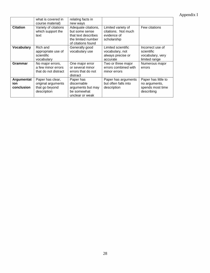
what is covered in course material)
relating facts in new ways
Citation Variety of citations which support the text
Adequate citations, but some sense that text describes the limited number of citations found
Limited variety of citations. Not much evidence of scholarship
Few citations
Vocabulary Rich and appropriate use of scientific vocabulary
Generally good vocabulary use
Limited scientific vocabulary, not always precise or accurate
Incorrect use of scientific vocabulary, very limited range
Grammar No major errors, a few minor errors that do not distract
One major error or several minor errors that do not distract
Two or three major errors combined with minor errors
Numerous major errors
Argumentation conclusion
Paper has clear, original arguments that go beyond description
Paper has discernable arguments but may be somewhat unclear or weak
Paper has arguments but often falls into description
Paper has little to no arguments, spends most time describing
Appendix I
29
Presentation Evaluation Evaluation of Presentation Presenter’s name_______________________________
1. I understood what the presentation was about. NO 1 2 3 4 5 YES 2. The flow of the presentation was logical. NO 1 2 3 4 5 YES 3. The presenter spoke clearly. NO 1 2 3 4 5 YES 4. The slides were clear and easy to understand. NO 1 2 3 4 5 YES 5. The presenter spoke to the audience. NO 1 2 3 4 5 YES 6. The presenter made good use with the pointer. NO 1 2 3 4 5 YES 7. The presentation was within the prescribed time limits (20 min +/- 2 min.). NO 1 2 3 4 5 YES 8. The presenter answered questions clearly (no B.S.). NO 1 2 3 4 5 YES 9. The presenter gave his/her opinion of the state of the science in this subjectNO 1 2 3 4 5 YES 10. I would enjoy listening to this type of presentation again. NO 1 2 3 4 5 YES
Score: _______/50 Your name___________________________________
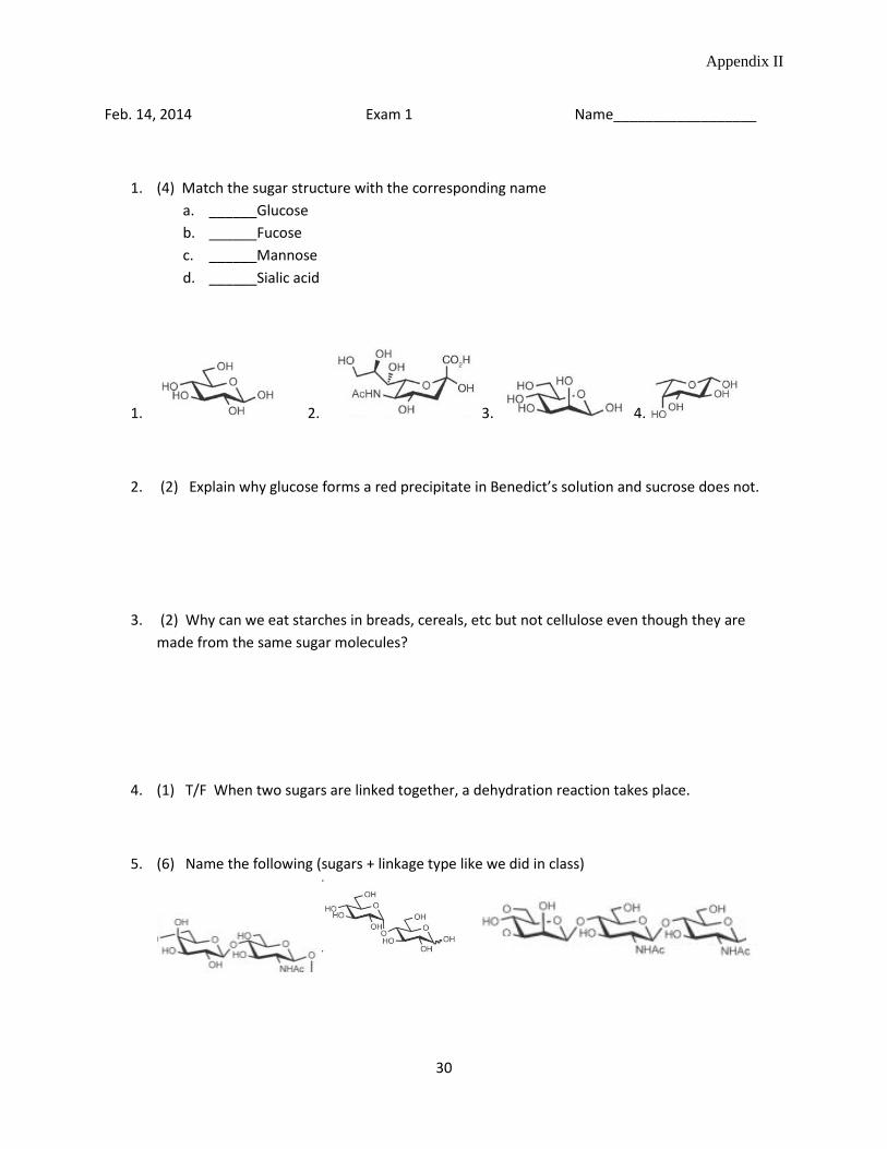
Appendix II
30
Feb. 14, 2014 Exam 1 Name__________________
1. (4) Match the sugar structure with the corresponding namea. ______Glucoseb. ______Fucosec. ______Mannosed. ______Sialic acid
1. 2. 3. 4.
2. (2) Explain why glucose forms a red precipitate in Benedict’s solution and sucrose does not.
3. (2) Why can we eat starches in breads, cereals, etc but not cellulose even though they aremade from the same sugar molecules?
4. (1) T/F When two sugars are linked together, a dehydration reaction takes place.
5. (6) Name the following (sugars + linkage type like we did in class)
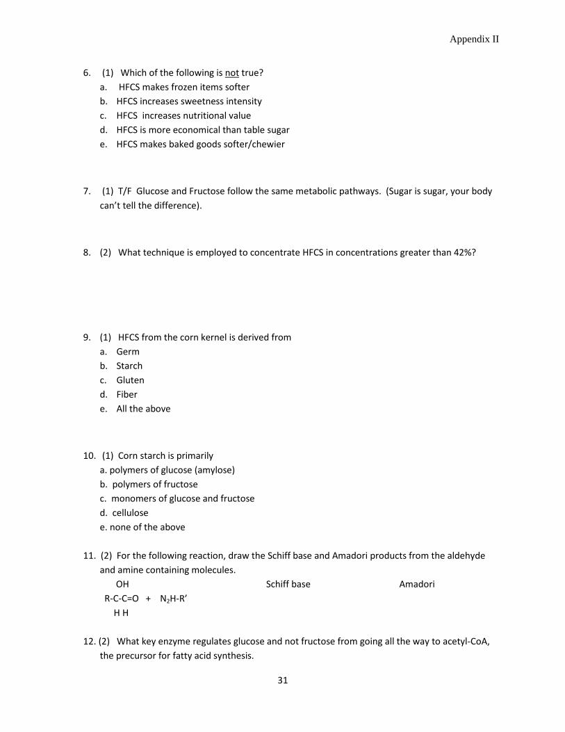
Appendix II
31
6. (1) Which of the following is not true? a. HFCS makes frozen items softer b. HFCS increases sweetness intensity c. HFCS increases nutritional value d. HFCS is more economical than table sugar e. HFCS makes baked goods softer/chewier
7. (1) T/F Glucose and Fructose follow the same metabolic pathways. (Sugar is sugar, your body can’t tell the difference).
8. (2) What technique is employed to concentrate HFCS in concentrations greater than 42%?
9. (1) HFCS from the corn kernel is derived from a. Germ b. Starch c. Gluten d. Fiber e. All the above
10. (1) Corn starch is primarily a. polymers of glucose (amylose) b. polymers of fructose c. monomers of glucose and fructose d. cellulose e. none of the above
11. (2) For the following reaction, draw the Schiff base and Amadori products from the aldehyde and amine containing molecules.
OH Schiff base Amadori R-C-C=O + N2H-R’ H H 12. (2) What key enzyme regulates glucose and not fructose from going all the way to acetyl-CoA,
the precursor for fatty acid synthesis.
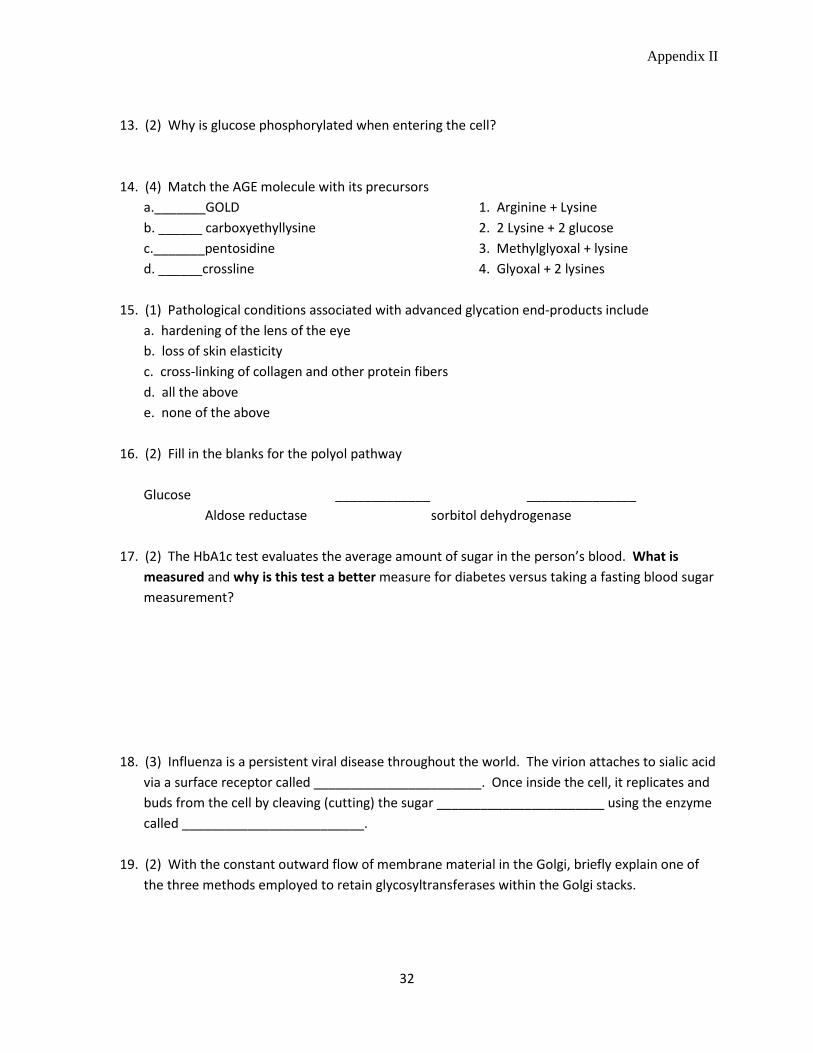
Appendix II
32
13. (2) Why is glucose phosphorylated when entering the cell? 14. (4) Match the AGE molecule with its precursors a._______GOLD 1. Arginine + Lysine b. ______ carboxyethyllysine 2. 2 Lysine + 2 glucose c._______pentosidine 3. Methylglyoxal + lysine d. ______crossline 4. Glyoxal + 2 lysines 15. (1) Pathological conditions associated with advanced glycation end-products include a. hardening of the lens of the eye b. loss of skin elasticity c. cross-linking of collagen and other protein fibers d. all the above e. none of the above 16. (2) Fill in the blanks for the polyol pathway Glucose _____________ _______________ Aldose reductase sorbitol dehydrogenase 17. (2) The HbA1c test evaluates the average amount of sugar in the person’s blood. What is
measured and why is this test a better measure for diabetes versus taking a fasting blood sugar measurement?
18. (3) Influenza is a persistent viral disease throughout the world. The virion attaches to sialic acid
via a surface receptor called _______________________. Once inside the cell, it replicates and buds from the cell by cleaving (cutting) the sugar _______________________ using the enzyme called _________________________.
19. (2) With the constant outward flow of membrane material in the Golgi, briefly explain one of
the three methods employed to retain glycosyltransferases within the Golgi stacks.
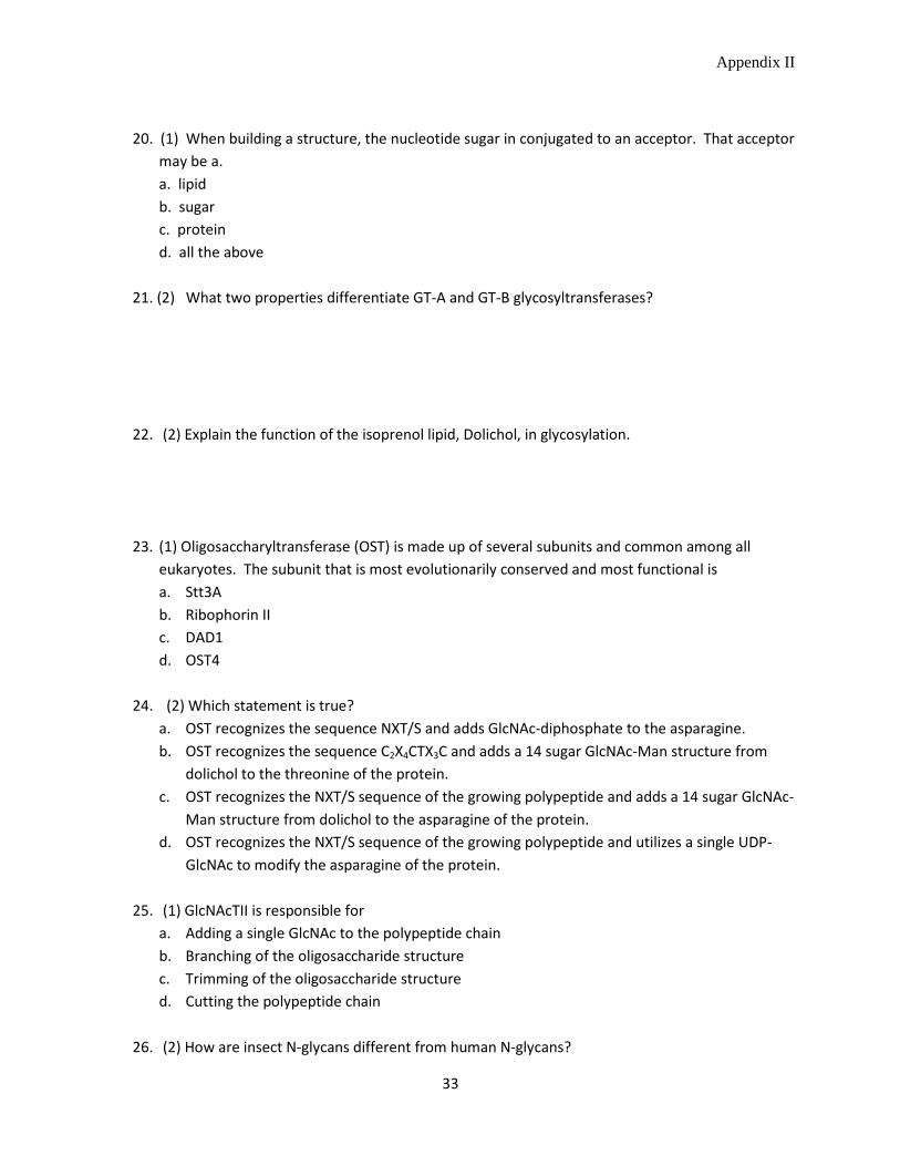
Appendix II
33
20. (1) When building a structure, the nucleotide sugar in conjugated to an acceptor. That acceptor
may be a. a. lipid b. sugar c. protein d. all the above 21. (2) What two properties differentiate GT-A and GT-B glycosyltransferases?
22. (2) Explain the function of the isoprenol lipid, Dolichol, in glycosylation.
23. (1) Oligosaccharyltransferase (OST) is made up of several subunits and common among all eukaryotes. The subunit that is most evolutionarily conserved and most functional is a. Stt3A b. Ribophorin II c. DAD1 d. OST4
24. (2) Which statement is true?
a. OST recognizes the sequence NXT/S and adds GlcNAc-diphosphate to the asparagine. b. OST recognizes the sequence C2X4CTX3C and adds a 14 sugar GlcNAc-Man structure from
dolichol to the threonine of the protein. c. OST recognizes the NXT/S sequence of the growing polypeptide and adds a 14 sugar GlcNAc-
Man structure from dolichol to the asparagine of the protein. d. OST recognizes the NXT/S sequence of the growing polypeptide and utilizes a single UDP-
GlcNAc to modify the asparagine of the protein.
25. (1) GlcNAcTII is responsible for a. Adding a single GlcNAc to the polypeptide chain b. Branching of the oligosaccharide structure c. Trimming of the oligosaccharide structure d. Cutting the polypeptide chain
26. (2) How are insect N-glycans different from human N-glycans?
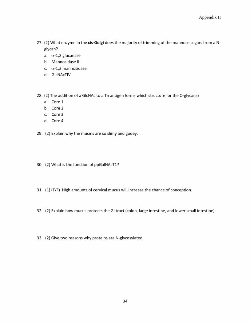
Appendix II
34
27. (2) What enzyme in the cis-Golgi does the majority of trimming of the mannose sugars from a N-glycan? a. α-1,2 glucanase b. Mannosidase II c. α-1,2 mannosidase d. GlcNAcTIV
28. (2) The addition of a GlcNAc to a Tn antigen forms which structure for the O-glycans? a. Core 1 b. Core 2 c. Core 3 d. Core 4
29. (2) Explain why the mucins are so slimy and gooey.
30. (2) What is the function of ppGalNAcT1?
31. (1) (T/F) High amounts of cervical mucus will increase the chance of conception.
32. (2) Explain how mucus protects the GI tract (colon, large intestine, and lower small intestine).
33. (2) Give two reasons why proteins are N-glycosylated.
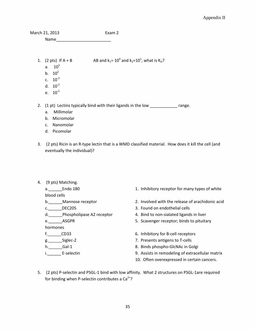
Appendix II
35
March 21, 2013 Exam 2 Name________________________
1. (2 pts) If A + B AB and k1= 104 and k2=101, what is KD? a. 103 b. 102 c. 10-3 d. 10-2 e. 10-5
2. (1 pt) Lectins typically bind with their ligands in the low ____________ range.
a. Millimolar b. Micromolar c. Nanomolar d. Picomolar
3. (2 pts) Ricin is an R-type lectin that is a WMD classified material. How does it kill the cell (and
eventually the individual)?
4. (9 pts) Matching. a.______Endo 180 1. Inhibitory receptor for many types of white blood cells b.______Mannose receptor 2. Involved with the release of arachidonic acid c.______DEC205 3. Found on endothelial cells d.______Phospholipase A2 receptor 4. Bind to non-sialated ligands in liver e.______ASGPR 5. Scavenger receptor; binds to pituitary hormones f.______CD33 6. Inhibitory for B-cell receptors g.______Siglec-2 7. Presents antigens to T-cells h.______Gal-1 8. Binds phospho-GlcNAc in Golgi i.______ E-selectin 9. Assists in remodeling of extracellular matrix 10. Often overexpressed in certain cancers.
5. (2 pts) P-selectin and PSGL-1 bind with low affinity. What 2 structures on PSGL-1are required for binding when P-selectin contributes a Ca2+?

Appendix II
36
6. (2 pts) Why is low affinity binding of the selectins advantageous for the white blood cell?
7. (2 pts) Explain the “magic bullet” concept for the treatment of acute myloid leukemia (AML).
8. (4 pts) Match a.______ Enzyme that adds a phosphate group 1. OGA b.______ Enzyme that cleaves off O-GlcNAc 2. Kinase c.______ Enzyme that cleaves off a phosphate 3. OST d.______ Enzyme that adds a O-GlcNAc 4. Phosphatase 5. OGT
9. (2 pts) Give 2 reasons for why O-GlcNAcylation of proteins was missed for so long.
10. (2pts) How does O-GlcNAcylation of proteins affect signaling pathways in cells?
11. (2 pts) Why has it been difficult to develop anti-inflammatory drugs targeting the selectins?
12. (2 pts) What causes I-cell disease and which lectin is involved?
13. (1 pt) (T/F) ERp57 associated with Calnexin is primarily responsible for forming disulfide bonds.
14. (5 pts) Matching a._____Uncovering enzyme 1. Directly involved with Pseudo-Hurler Polydystrophy b._____α-glucosidase I 2. Cleaves off the 1st glucose from the N-glycan c._____calreticulin 3. Cleaves off the 2nd glucose from the N-glycan d._____GlcNAc Phosphotransferase 4. Cleaves off GlcNAc and allow M6PR binding
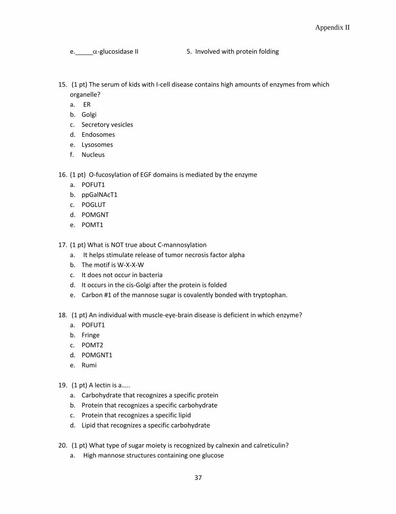
Appendix II
37
e._____α-glucosidase II 5. Involved with protein folding
15. (1 pt) The serum of kids with I-cell disease contains high amounts of enzymes from which organelle? a. ER b. Golgi c. Secretory vesicles d. Endosomes e. Lysosomes f. Nucleus
16. (1 pt) O-fucosylation of EGF domains is mediated by the enzyme
a. POFUT1 b. ppGalNAcT1 c. POGLUT d. POMGNT e. POMT1
17. (1 pt) What is NOT true about C-mannosylation
a. It helps stimulate release of tumor necrosis factor alpha b. The motif is W-X-X-W c. It does not occur in bacteria d. It occurs in the cis-Golgi after the protein is folded e. Carbon #1 of the mannose sugar is covalently bonded with tryptophan.
18. (1 pt) An individual with muscle-eye-brain disease is deficient in which enzyme?
a. POFUT1 b. Fringe c. POMT2 d. POMGNT1 e. Rumi
19. (1 pt) A lectin is a…..
a. Carbohydrate that recognizes a specific protein b. Protein that recognizes a specific carbohydrate c. Protein that recognizes a specific lipid d. Lipid that recognizes a specific carbohydrate
20. (1 pt) What type of sugar moiety is recognized by calnexin and calreticulin?
a. High mannose structures containing one glucose
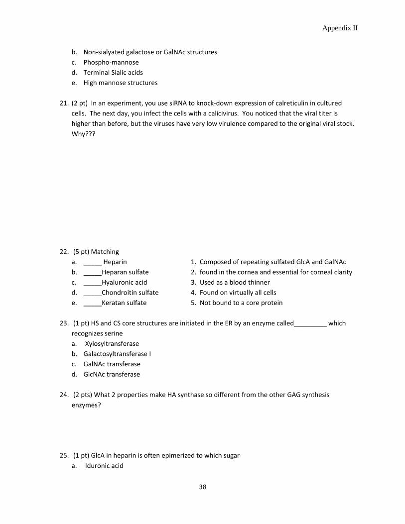
Appendix II
38
b. Non-sialyated galactose or GalNAc structures c. Phospho-mannose d. Terminal Sialic acids e. High mannose structures
21. (2 pt) In an experiment, you use siRNA to knock-down expression of calreticulin in cultured
cells. The next day, you infect the cells with a calicivirus. You noticed that the viral titer is higher than before, but the viruses have very low virulence compared to the original viral stock. Why???
22. (5 pt) Matching a. _____ Heparin 1. Composed of repeating sulfated GlcA and GalNAc b. _____Heparan sulfate 2. found in the cornea and essential for corneal clarity c. _____Hyaluronic acid 3. Used as a blood thinner d. _____Chondroitin sulfate 4. Found on virtually all cells e. _____Keratan sulfate 5. Not bound to a core protein
23. (1 pt) HS and CS core structures are initiated in the ER by an enzyme called_________ which
recognizes serine a. Xylosyltransferase b. Galactosyltransferase I c. GalNAc transferase d. GlcNAc transferase
24. (2 pts) What 2 properties make HA synthase so different from the other GAG synthesis
enzymes?
25. (1 pt) GlcA in heparin is often epimerized to which sugar a. Iduronic acid
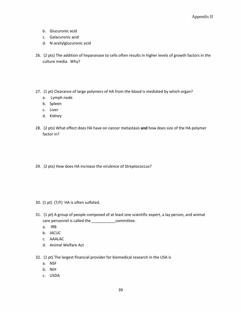
Appendix II
39
b. Glucuronic acid c. Galacuronic acid d. N-acetylglucuronic acid
26. (2 pts) The addition of heparanase to cells often results in higher levels of growth factors in the
culture media. Why?
27. (1 pt) Clearance of large polymers of HA from the blood is mediated by which organ? a. Lymph node b. Spleen c. Liver d. Kidney
28. (2 pts) What effect does HA have on cancer metastasis and how does size of the HA polymer
factor in?
29. (2 pts) How does HA increase the virulence of Streptococcus?
30. (1 pt) (T/F) HA is often sulfated.
31. (1 pt) A group of people composed of at least one scientific expert, a lay person, and animal care personnel is called the ___________committee. a. IRB b. IACUC c. AAALAC d. Animal Welfare Act
32. (1 pt) The largest financial provider for biomedical research in the USA is
a. NSF b. NIH c. USDA

Appendix II
40
d. DOE e. SNAP
Bonus question (3 pts): Notch undergoes 3 proteolytic events. Describe where this occurs and what enzyme performs each event.
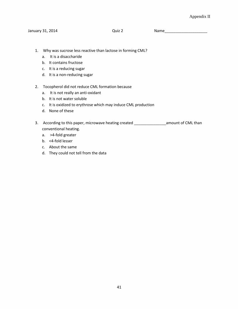
Appendix II
41
January 31, 2014 Quiz 2 Name____________________
1. Why was sucrose less reactive than lactose in forming CML? a. It is a disaccharide b. It contains fructose c. It is a reducing sugar d. It is a non-reducing sugar
2. Tocopherol did not reduce CML formation because
a. It is not really an anti-oxidant b. It is not water soluble c. It is oxidized to erythrose which may induce CML production d. None of these
3. According to this paper, microwave heating created _______________amount of CML than
conventional heating. a. >4-fold greater b. <4-fold lesser c. About the same d. They could not tell from the data

Appendix II
42
February 5, 2014 Quiz 3 Name___________________
1. Treatment of cells in culture with siRNA___________________ expression of a specific gene. a. Decreases b. Increases c. Does not change
2. Cytokines such as IL-6 _________________ expression of C1GalT1 and _________________ expression of ST6GalNAc2 in IgA nephropathy patients. a. Increase::decrease b. Decrease::increase c. Increase::increase d. Decrease::decrease
3. In which organ were immune complexes most harmful?
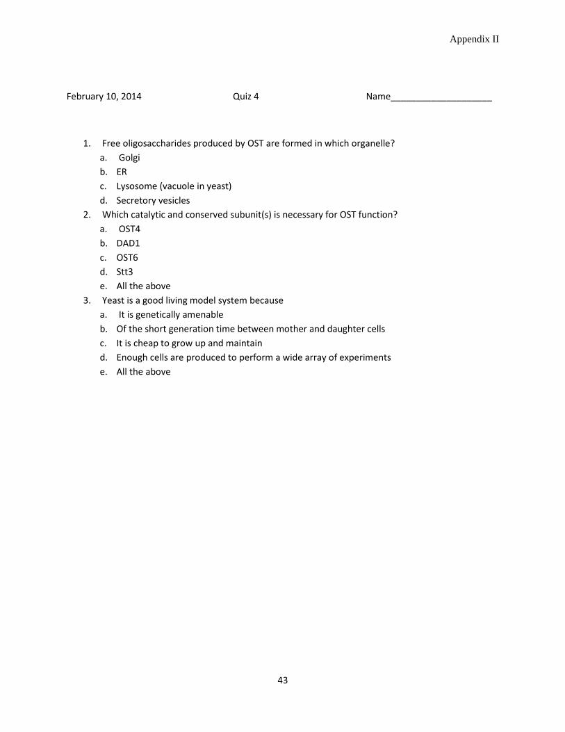
Appendix II
43
February 10, 2014 Quiz 4 Name____________________
1. Free oligosaccharides produced by OST are formed in which organelle? a. Golgi b. ER c. Lysosome (vacuole in yeast) d. Secretory vesicles
2. Which catalytic and conserved subunit(s) is necessary for OST function? a. OST4 b. DAD1 c. OST6 d. Stt3 e. All the above
3. Yeast is a good living model system because a. It is genetically amenable b. Of the short generation time between mother and daughter cells c. It is cheap to grow up and maintain d. Enough cells are produced to perform a wide array of experiments e. All the above
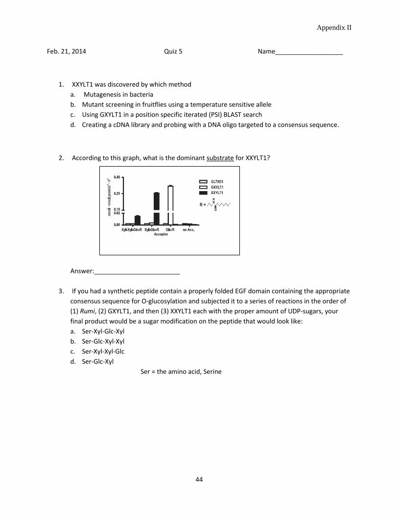
Appendix II
44
Feb. 21, 2014 Quiz 5 Name___________________
1. XXYLT1 was discovered by which method a. Mutagenesis in bacteria b. Mutant screening in fruitflies using a temperature sensitive allele c. Using GXYLT1 in a position specific iterated (PSI) BLAST search d. Creating a cDNA library and probing with a DNA oligo targeted to a consensus sequence.
2. According to this graph, what is the dominant substrate for XXYLT1?
Answer:________________________
3. If you had a synthetic peptide contain a properly folded EGF domain containing the appropriate consensus sequence for O-glucosylation and subjected it to a series of reactions in the order of (1) Rumi, (2) GXYLT1, and then (3) XXYLT1 each with the proper amount of UDP-sugars, your final product would be a sugar modification on the peptide that would look like: a. Ser-Xyl-Glc-Xyl b. Ser-Glc-Xyl-Xyl c. Ser-Xyl-Xyl-Glc d. Ser-Glc-Xyl
Ser = the amino acid, Serine
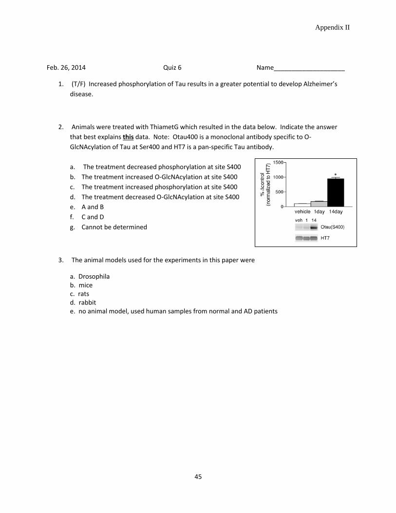
Appendix II
45
Feb. 26, 2014 Quiz 6 Name____________________
1. (T/F) Increased phosphorylation of Tau results in a greater potential to develop Alzheimer’s disease.
2. Animals were treated with ThiametG which resulted in the data below. Indicate the answer that best explains this data. Note: Otau400 is a monoclonal antibody specific to O-GlcNAcylation of Tau at Ser400 and HT7 is a pan-specific Tau antibody. a. The treatment decreased phosphorylation at site S400 b. The treatment increased O-GlcNAcylation at site S400 c. The treatment increased phosphorylation at site S400 d. The treatment decreased O-GlcNAcylation at site S400 e. A and B f. C and D g. Cannot be determined
3. The animal models used for the experiments in this paper were
a. Drosophila b. mice c. rats d. rabbit e. no animal model, used human samples from normal and AD patients

Appendix II
46
March 3, 2014 Quiz 7 Name_____________________
1. In the experiments outlined in Ramani et al., the use of heparanase III a. Promoted shedding of Syndecan-1 b. Inhibited shedding of Syndecan-1 c. Made no difference in shedding rates
2. The conclusion from the adjascent figure is:
a. Heparanase III is the cause of Syndecan-1 shedding b. Heparan sulfate addition causes Syndecan-1 shedding c. Heparin addition causes Syndecan-1 shedding d. MMPs cause Syndecan-1 shedding
3. (T/F) Heparanase and MMP-9 have increased expression
in cancer, inflammation and wound healing.
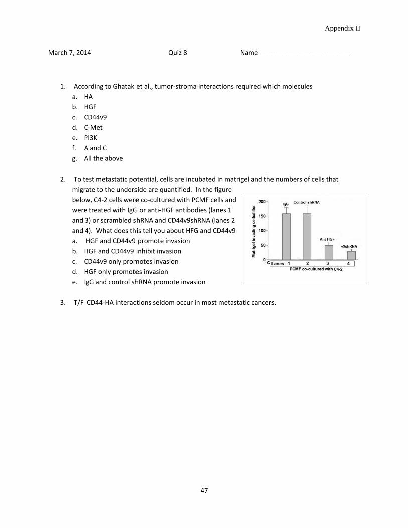
Appendix II
47
March 7, 2014 Quiz 8 Name_________________________
1. According to Ghatak et al., tumor-stroma interactions required which molecules a. HA b. HGF c. CD44v9 d. C-Met e. PI3K f. A and C g. All the above
2. To test metastatic potential, cells are incubated in matrigel and the numbers of cells that
migrate to the underside are quantified. In the figure below, C4-2 cells were co-cultured with PCMF cells and were treated with IgG or anti-HGF antibodies (lanes 1 and 3) or scrambled shRNA and CD44v9shRNA (lanes 2 and 4). What does this tell you about HFG and CD44v9 a. HGF and CD44v9 promote invasion b. HGF and CD44v9 inhibit invasion c. CD44v9 only promotes invasion d. HGF only promotes invasion e. IgG and control shRNA promote invasion
3. T/F CD44-HA interactions seldom occur in most metastatic cancers.
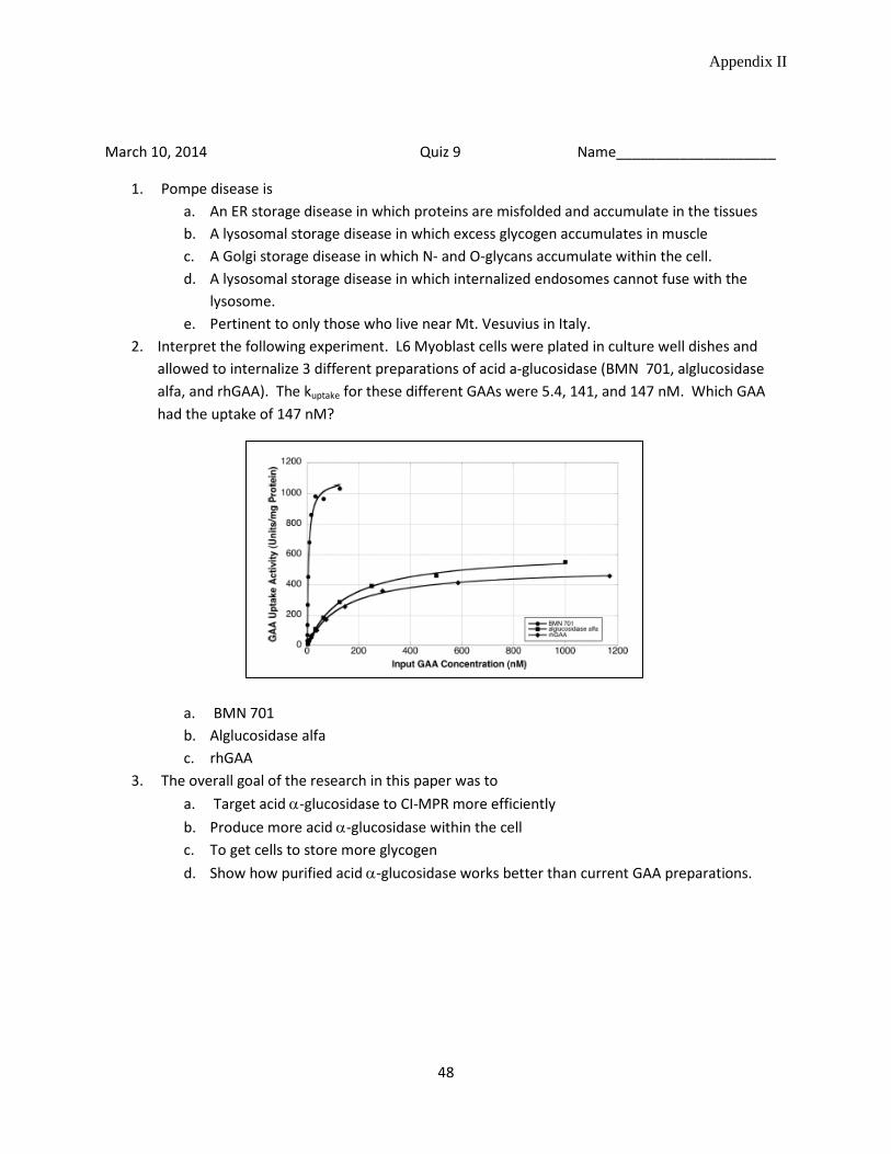
Appendix II
48
March 10, 2014 Quiz 9 Name____________________
1. Pompe disease is a. An ER storage disease in which proteins are misfolded and accumulate in the tissues b. A lysosomal storage disease in which excess glycogen accumulates in muscle c. A Golgi storage disease in which N- and O-glycans accumulate within the cell. d. A lysosomal storage disease in which internalized endosomes cannot fuse with the
lysosome. e. Pertinent to only those who live near Mt. Vesuvius in Italy.
2. Interpret the following experiment. L6 Myoblast cells were plated in culture well dishes and allowed to internalize 3 different preparations of acid a-glucosidase (BMN 701, alglucosidase alfa, and rhGAA). The kuptake for these different GAAs were 5.4, 141, and 147 nM. Which GAA had the uptake of 147 nM?
a. BMN 701 b. Alglucosidase alfa c. rhGAA
3. The overall goal of the research in this paper was to a. Target acid α-glucosidase to CI-MPR more efficiently b. Produce more acid α-glucosidase within the cell c. To get cells to store more glycogen d. Show how purified acid α-glucosidase works better than current GAA preparations.

Appendix II
49
March 19, 2014 Quiz 10 Name______________
1. PSGL-1 is a ligand for ______________ and expressed on activated _________________a. E-Selectin::myofibroblastsb. P-Selectin::Leukocytesc. L-Selectin::macrophagesd. E-Selectin::Leukocytese. P-Selectin::CHO cells
2. Knocking out the cytoplasmic domain (∆CD) of PSGL-1 resulted ina. Increased expression of PSGL-1b. Decreased expression of the mutant PSGL-1c. Increased accumulation of the PSGL-1 precursord. Increased accumulation of the PSGL-1 mature isoforme. A and Cf. B and Cg. B and D
3. T/F The authors discovered that dimerization of PSGL-1 was required for the receptor(s) to exitthe ER.
Sample of an excellent review paper Appendix III
50
Bittersweet Roles of O-Linked N-Acetylglucosamine in Human Health and Disease Department of Biochemistry, University of Nebraska-Lincoln
Key words: O-linked-N-acetylglucosamine transferase, O-linked-N-acetylglucosamine hydrolase, UDP-N-acetylglucosamine, type II diabetes, cancer, Alzheimer’s disease
Abstract
Posttranslational modification of nuclear and cytosolic proteins by the addition of O-linked N-acetylglucosamine (O-GlcNAc) is a dynamic process that is regulated by the key enzymes O-GlcNAc transferase (OGT) and O-GlcNAc hydrolase (OGA). The substrate for OGT, UDP-GlcNAc, is the final product of the hexosamine biosynthetic pathway. UDP-GlcNAc is abundant in the cytosol and acts as a metabolic sensor, reflecting the overall nutrient levels in the cell. OGT covalently modifies serine and threonine residues by attaching a single O-GlcNAc moiety, which can be cycled on and off by coordinate regulation with OGA. The substrate specificity of OGT is not well understood, the modification occurs on a myriad of proteins with no apparent consensus sequence. Importantly, O-GlcNAcylation closely resembles protein phosphorylation and the two processes are thought to be coordinately regulated in proteins such as p53 and tau, which are known to be implicated in cancer and Alzheimer’s disease, respectively.
Over the past 30 years since O-GlcNAcylation was first discovered by Dr. Gerald Hart and co-workers, this modification has been found on thousands of proteins and is associated with critical cellular functions such as facilitating protein-protein interactions, regulating transcription factors, and modulating protein stability. O-GlcNAcylation has proven challenging to study due to its dynamic nature and complex involvement in diseases associated with chronic metabolic disregulation. This review begins with a detailed look at the enzymes OGT and OGA, and then outlines recent evidence that O-GlcNAcylation plays a role in the development and progression of three major human diseases: type II diabetes, cancer, and Alzheimer’s disease.
Introduction
Posttranslational modification of serine and threonine residues with a single β-C2 O-linked N-acetylglucosamine (O-GlcNAcylation) is common in nuclear, cytosolic, and mitochondrial proteins. O-GlcNAcylation has been reported in all multicellular organisms studied including plants and fungi (reviewed in 1). O-GlcNAcylation was also found in protozoa and a limited number of bacteria (2,3). This reversible covalent modification of proteins is driven by nutrient and hormonal cues and is known to be important in regulating metabolic homeostasis.
Unlike protein phosphorylation, which is regulated by an enormous variety of kinases and phosphatases, there are only two enzymes involved in O-GlcNAcylation. O-GlcNAc transferase (OGT) transfers the UDP-GlcNAc onto an acceptor protein and O-GlcNAcase (OGA) hydrolyzes the sugar from the serine or threonine residue. Knocking
Sample of an excellent review paper Appendix III
51
out the OGT gene in mice is embryonic lethal, demonstrating its critical role in development and cellular signaling (4). There is no known consensus sequence for the polypeptides that are modified by OGT; however, there is a small group of O-GlcNAcylated proteins that share the sequence motif PV(S/T) (5). The extent of protein O-GlcNAcylation is dependent upon the concentration of substrate, UDP-GlcNAc, which is the final product of the hexosamine biosynthesis pathway.
O-GlcNAc modification of proteins acts as a metabolic rheostat within the cell due to its ability to sense and respond to altered nutrient levels through O-GlcNAc-mediated signaling. O-GlcNAcylation has been shown to severely alter the function of many important proteins by influencing substrate affinity, stability, and intracellular localization (6). Aberrant O-GlcNAcylation is associated with human metabolic diseases including diabetes mellitus type 2, cancer, and neurodegenerative diseases such as Alzheimer’s disease.
Hyperglycemia increases intracellular levels of UDP-GlcNAc, leading to hyper-O-GlcNAcylation of proteins, including those involved in insulin signaling. In type II diabetes, O-GlcNAcylation of insulin related proteins results in decreased efficiency of insulin-mediated signal transduction (7). Cancer associated changes in O-GlcNAcylation are complex and appear to be unique to each different type of cancer. In general, O-GlcNAc cycling promotes metastasis and cancer progression through the dynamic modification of several proteins. In prostate cancer, for example, O-GlcNAcylation of the oncogenic transcription factor FoxM1 leads to increased expression of matrix metalloproteinases which are involved in remodeling of the extracellular matrix, allowing for higher metastatic potential (8). Lastly, alteration of O-GlcNAc modification is strongly implicated in neurodegenerative diseases including Alzheimer’s disease, Parkinson’s disease, and Huntington’s disease. Cytoskeletal proteins such as tau have been shown to be O-GlcNAcylated, which prevents hyperphosphorylation leading to protein aggregation and improves prognosis for neurodegenerative diseases in mice models (9). Overall, it is clear that O-linked N-acetylglucosamine modification plays a complex and significant role in human health and disease.
Enzymes Involved in O-linked N-acetylglucosamine Cycling
O-GlcNAc transferase (OGT) is a cytosolic glycosyltransferase that uses α-linked uridine diphosphate N-acetylglucosamine (UDP-GlcNAc) as a substrate to transfer O-GlcNAc onto a serine or threonine residue through a β-glycosidic linkage (with inversion of anomeric configuration). UDP-GlcNAc is the final product of the hexosamine biosynthesis pathway and is considered to be a sensor for the metabolic state of the cell. UDP-GlcNAc biosynthesis integrates nucleotide, nitrogen, glucose, and fatty acid metabolism, and is reflective of the overall energy state of the cell. In most cells, UDP-GlcNAc is the second most abundant energy-containing metabolite present, second only to ATP.

Sample of an excellent review paper Appendix III
52
Figure 1. Three isoforms of OGT have different lengths of N-terminal tetratricopeptide repeats (TPR), shown in orange. The two glycosyltransferase catalytic domains are shown in blue and light blue and the hinge-like region is shown in green. This figure was taken from reference 46.
There are three different splice variants of human OGT, each with a different number of N-terminal 34 amino acid tetratricopeptide repeats (TPR). The full-length enzyme (ncOGT) is 110 kDa with 11.5 TPR repeats, followed by 103 kDa (mOGT) and 78 kDa (sOGT) (10). The full-length enzyme adds O-GlcNAc to cytosolic and nuclear proteins, while mOGT primarily modifies mitochondrial proteins. The crystal structure of human OGT (hOGT) was solved in 2004 by Jinek et al. (11). The structure showed that the TPR repeats form elongated superhelices, which are thought to facilitate protein-protein interactions. These interactions are important in recruiting substrate proteins to the OGT active site to be O-GlcNAcylated. The bacterial homolog was crystallized in 2008, leading to the identification of the active site of OGT (12). The active site is split into two different domains, with an intervening hinge-like region, which facilitates the release of the O-GlcNAcylated peptide (as seen in Figure 1). A third crystal structure, hOGT bound with UDP and a peptide substrate, was key in discovering the location of peptide substrates in the active site of OGT (13). From this structure, they found that the UDP-GlcNAc must bind first, before the polypeptide substrate, which binds on top and blocks access to the nucleotide-sugar binding pocket. Mechanistically, it is unclear which residue in the active site is serving as the general base required for OGT catalytic activity. Furthermore, there is no definitive consensus sequence for the peptide substrates, although it has been suggested that there is some preference for peptides with side chains on both sides of the O-GlcNAcylated serine or threonine (14). Identifying which protein motifs act as O-GlcNAc acceptors would help predict which proteins are targeted by O-GlcNAcylation. Bioinformatics tools have been developed to assist in predicting which proteins may be O-GlcNAcylated, including two programs that use support vector machine algorithms, OGlcNAc Scan and O-GlcNAc PRED, and one that uses a artificial neuronal network algothrim, YinOYang (15).
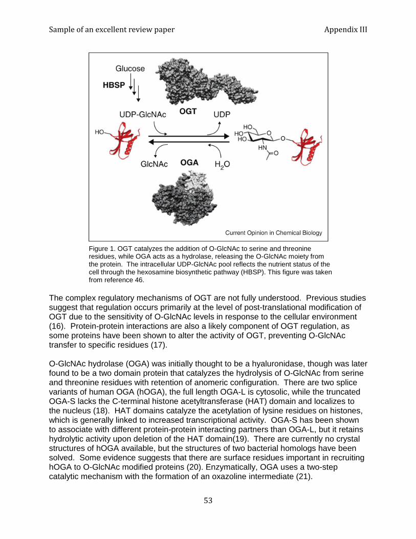
Sample of an excellent review paper Appendix III
53
Figure 1. OGT catalyzes the addition of O-GlcNAc to serine and threonine residues, while OGA acts as a hydrolase, releasing the O-GlcNAc moiety from the protein. The intracellular UDP-GlcNAc pool reflects the nutrient status of the cell through the hexosamine biosynthetic pathway (HBSP). This figure was taken from reference 46.
The complex regulatory mechanisms of OGT are not fully understood. Previous studies suggest that regulation occurs primarily at the level of post-translational modification of OGT due to the sensitivity of O-GlcNAc levels in response to the cellular environment (16). Protein-protein interactions are also a likely component of OGT regulation, as some proteins have been shown to alter the activity of OGT, preventing O-GlcNAc transfer to specific residues (17). O-GlcNAc hydrolase (OGA) was initially thought to be a hyaluronidase, though was later found to be a two domain protein that catalyzes the hydrolysis of O-GlcNAc from serine and threonine residues with retention of anomeric configuration. There are two splice variants of human OGA (hOGA), the full length OGA-L is cytosolic, while the truncated OGA-S lacks the C-terminal histone acetyltransferase (HAT) domain and localizes to the nucleus (18). HAT domains catalyze the acetylation of lysine residues on histones, which is generally linked to increased transcriptional activity. OGA-S has been shown to associate with different protein-protein interacting partners than OGA-L, but it retains hydrolytic activity upon deletion of the HAT domain(19). There are currently no crystal structures of hOGA available, but the structures of two bacterial homologs have been solved. Some evidence suggests that there are surface residues important in recruiting hOGA to O-GlcNAc modified proteins (20). Enzymatically, OGA uses a two-step catalytic mechanism with the formation of an oxazoline intermediate (21).
Sample of an excellent review paper Appendix III
54
The structural and mechanistic details of the two enzymes involved in O-GlcNAc cycling, OGT and OGA, have shed light on the importance of O-GlcNAc modification in human health and disease. One salient question that remains is how are OGT and OGA able to coordinately regulate the addition and removal of O-GlcNAc from hundreds of different peptides? As mentioned previously, post-translational modification of OGT and OGA is a likely mechanism for controlling both enzyme activity and protein-protein interaction partners. In addition, the various OGT and OGA isoforms have different catalytic efficiencies, and thus, impact overall regulation of O-GlcNAcylation. O-GlcNAcylation in Type II Diabetes Type II diabetes mellitus is a chronic metabolic disorder in which cells are unable to respond to normal levels of insulin, a peptide hormone responsible for suppressing glucose release in the liver when there is an excess of nutrients present. This results in hyperglycemia and retention of water and salt by the kidneys. Furthermore, insulin resistance leads to metabolic disregulation and alters the flux through the hexosamine biosynthetic pathway. Surprisingly, combining the results of several studies suggests that both nutrient excess and nutrient limiting conditions may cause a global increase in O-GlcNAcylation levels in cells and tissues such as erythrocytes, leukocytes, liver, and heart (22). Thus, O-GlcNAcylation may be a means of responding to the highly dynamic metabolic status of the cell, with a certain threshold level of O-GlcNAcylation required for proper signaling to occur. In a hyperglycemic state associated with type II diabetes, high glucose levels promote increased flux through the hexosamine biosynthetic pathway leading to an elevated intracellular concentration of UDP-GlcNAc (23). Experimentally, elevation of UDP-GlcNAc levels by glucosamine treatment or inhibition of OGA using O-(2-acetamido-2-deoxy-D-glucopyranosylidene) amino-N-phenyl-carbamate (PUGNAc) causes insulin resistance in 3T3-L1 adipocytes (24). Conversely, inhibition of OGA with a more selective OGA inhibitor, Thiamet G, does not result in insulin resistance in 3T3-L1 adipocytes (25). Overexpression of OGT in fat and muscle tissues results in mice with an insulin resistant phenotype, while OGA overexpression increases glucose tolerance and insulin sensitivity in insulin-resistant diabetic mice fed a high-fat diet (26, 27). In addition, it has been shown that single nucleotide polymorphisms in the OGA gene MGEA5 is associated with a significantly higher rate of developing diabetes (28).
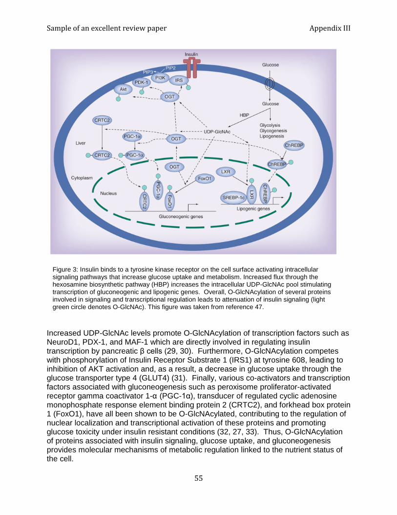
Sample of an excellent review paper Appendix III
55
Figure 3: Insulin binds to a tyrosine kinase receptor on the cell surface activating intracellular signaling pathways that increase glucose uptake and metabolism. Increased flux through the hexosamine biosynthetic pathway (HBP) increases the intracellular UDP-GlcNAc pool stimulating transcription of gluconeogenic and lipogenic genes. Overall, O-GlcNAcylation of several proteins involved in signaling and transcriptional regulation leads to attenuation of insulin signaling (light green circle denotes O-GlcNAc). This figure was taken from reference 47.
Increased UDP-GlcNAc levels promote O-GlcNAcylation of transcription factors such as NeuroD1, PDX-1, and MAF-1 which are directly involved in regulating insulin transcription by pancreatic β cells (29, 30). Furthermore, O-GlcNAcylation competes with phosphorylation of Insulin Receptor Substrate 1 (IRS1) at tyrosine 608, leading to inhibition of AKT activation and, as a result, a decrease in glucose uptake through the glucose transporter type 4 (GLUT4) (31). Finally, various co-activators and transcription factors associated with gluconeogenesis such as peroxisome proliferator-activated receptor gamma coactivator 1-α (PGC-1α), transducer of regulated cyclic adenosine monophosphate response element binding protein 2 (CRTC2), and forkhead box protein 1 (FoxO1), have all been shown to be O-GlcNAcylated, contributing to the regulation of nuclear localization and transcriptional activation of these proteins and promoting glucose toxicity under insulin resistant conditions (32, 27, 33). Thus, O-GlcNAcylation of proteins associated with insulin signaling, glucose uptake, and gluconeogenesis provides molecular mechanisms of metabolic regulation linked to the nutrient status of the cell.
Sample of an excellent review paper Appendix III
56
Another protein associated with type II diabetes that has shown to be O-GlcNAcylated is endothelial nitric oxide synthase (eNOS). eNOS plays a role in regulating vascular resistance through the production of nitric oxide. O-GlcNAcylation of eNOS prevents its phosphorylation, leading to decreased nitric oxide production and endothelial cells with vascular abnormalities (34). Chronic inhibition of eNOS activity through O-GlcNAcylation may contribute to the association between accelerated atherosclerosis and type II diabetes. While O-GlcNAcylation of key proteins plays a role in regulating signaling cascades associated with normal metabolic homeostasis, prolonged O-GlcNAcylation as a result of excess caloric intake or starvation may cause the effects to transition from protective to deleterious, as seen in type II diabetes. Therefore, understanding the molecular mechanisms of O-GlcNAc cycling has the potential to provide new therapeutic targets for treating type II diabetes. Furthermore, O-GlcNAcylation seems to be an important link between type II diabetes and other diseases associated with altered glucose metabolism, such as cancer. O-GlcNAcylation in Cancer Alteration of cellular metabolism is one of the hallmarks of cancer. Cancer cells require unusually high amounts of glucose in order to provide energy and building blocks for lipids, proteins, and nucleic acids, which are in high demand in rapidly proliferating cells. Glucose and glucosamine are the preferred sources of energy and biosynthetic precursors in cancer cells. As the major fuel sources, glucose and glucosamine also drive flux through the hexosamine biosynthetic pathway, resulting in a myriad of O-GlcNAcylated proteins, which play key roles in regulating the molecular mechanisms of cancer. Increased O-GlcNAcylation of proteins and altered OGT and OGA expression have been associated with different types of cancer including: breast, liver, colon, lung, bladder, endometrial, prostate, and chronic lymphocytic leukemia cells (35). Although each type of cancer should be considered independently, in general, cancer is most commonly associated with increased O-GlcNAcylation along with elevated expression levels of OGT. There is no apparent cancer-associated pattern of expression for OGA. O-GlcNAcylation of a wide range of proteins is associated with many essential processes in cancer cell survival, proliferation, and metastasis such as: mitogenic signaling, cell cycle regulation, transcription factor activation, and cell adhesion and migration. Modification of proteins by O-GlcNAcylation regulates cell cycle progression at each of the critical checkpoints. Most cells are in the G0 or quiescent state, and entry into the G1 stage of the cell cycle requires activation of the MAP kinase and PI3K pathways by exogenous mitogenic signals. This leads to transcription of cyclin D1, the key regulatory protein of the G1 phase. Blocking OGT activity has been shown to delay production of cyclin D1, preventing entrance of quiescent cells into the G1 phase and, therefore, inhibiting proliferation (36). Furthermore, inhibition of OGA accelerates
Sample of an excellent review paper Appendix III
57
proliferation rates, while decreasing flux through the hexosamine biosynthesis pathway corresponds to decreased cellular proliferation. This trend of increased O-GlcNAcylation promoting cell cycle progression continues at every stage except for the G1/S checkpoint, where there is a global decrease in O-GlcNAcylated proteins (37). Mutations in transcription factors leading to disregulation of gene expression is common in cancer. Posttranslational modifications are also responsible for regulation of transcription factor activity. Several cancer related transcription factors have been shown to be modified by O-GlcNAcylation including: MYC, FOXM1, p53, NF-κB, and β-catenin (35). The protein MYC is an oncogenic transcription factor often overexpressed in cancer. MYC is typically degraded upon phosphorylation of threonine 58, though extensive O-GlcNAcylation of MYC blocks phosphorylation at position 58 and stabilizes MYC, promoting proliferation (38). FOXM1 regulates transcription and is found to be associated with tumorigenesis. Although FOXM1 has not been shown to be directly modified by O-GlcNAcylation, OGT inhibition led to dramatically reduced levels of FOXM1 in breast cancer MCF-10A cells (39). The tumor suppressor protein p53 is highly implicated in cancer progression. Phosphorylation of p53 at threonine 155 targets it for proteosomal degradation when there are no signs of cellular stress. O-GlcNAcylation of p53 at serine 149 has been shown to inhibit phosphorylation at residue 155, stabilizing p53 and allowing it to act as a transcription factor involved in triggering cell cycle arrest and apoptosis (40). Therefore, O-GlcNAcylation of p53 often signals cellular stress and is essential in preventing the cell from further propagation under such conditions. β-catenin is a transcription factor that is involved in regulating proliferation and cell cycle progression. Mutations in β-catenin which increase its stability are oncogenic and are commonly found in colorectal cancer. Studies in colonic carcinoma cells show that increased flux through the hexosamine biosynthetic pathway is correlated with elevated β-catenin expression levels and an increase in global O-GlcNAcylation (41). Although O-GlcNAcylation of oncogenic and protooncogenic transcription factors is highly dependent on each individual type of cancer, this dynamic posttranslational modification appears to be an important factor linking metabolic status with control of crucial cellular processes such as transcriptional regulation, cell cycle progression, and proliferation. O-GlcNAcylation can also impact cellular adhesion and migration leading to increased metastatic potential of cancer cells. E-cadherin is a transmembrane protein involved in cell-cell adhesion. Downregulation of E-cadherin is associated with epithelial-mesenchymal transition (EMT), which is characteristic of invasion of the basement membrane by carcinoma and subsequent translocation and colonization of a secondary tumor site, otherwise known as metastasis. O-GlcNAcylation has been demonstrated to regulate expression of E-cadherin, thus influencing cancer invasiveness in breast cancer (42). The transcription factor Snail1 mediates EMT by repressing E-cadherin expression and was found to be stabilized by O-GlcNAcylation in ovarian cancer cell lines in the presence of OGT and hyperglycemic conditions (43). Lastly, matrix metalloproteases (MMPs) are enzymes involved in extracellular matrix remodeling, and thus provide conditions that support invasion of basement membrane by carcinoma. The proteolytic activity of MMPs is required for intravasation of cancerous cells from the
Sample of an excellent review paper Appendix III
58
tissue, through the basement membrane, and into the blood vessels, where the cancerous cell circulates until it undergoes extravasation into the tissue at a secondary location. Reduced O-GlcNAcylation in prostate cancer causes decreased expression of MMP2 and MMP9 as well as a decrease in FOXM1 expression (8). The same study showed that blocking OGT led to decreased metastasis of tumors in vivo. Overall, it is clear that O-GlcNAcylation is involved in regulating EMT and MMPs, both of which are critical in extracellular matrix remodeling, a key determinant of the invasiveness and metastatic potential of cancer cells. O-GlcNAcylation in Alzheimer’s Disease Improper glucose metabolism is linked to neurodegenerative diseases such as Alzheimer’s disease. There is significant evidence that O-GlcNAcylation is also implicated in Parkinson’s disease and Huntington’s disease, but in this review I will focus on Alzheimer’s disease. Studies continue to provide evidence of an increasingly strong correlation between insulin resistance, obesity, and Alzheimer’s disease. This may suggest that incessant exposure to extreme imbalance of nutrients is a risk factor for developing certain neurodegenerative diseases linked to chronic metabolic disregulation. It has been hypothesized that inefficient glucose utilization in the brain leads to lower cytosolic levels of UDP-GlcNAc, thus causing an overall decrease in O-GlcNAcylation of proteins in the brain. Alzheimer’s disease is a neurodegenerative disease characterized by accumulation of neurofibrillary tangles, which are composed of microtubule-associated tau protein monomers that assemble into paired helical filaments. The progression of the disease is characterized by the buildup of these neurofibrillary tangles. It is not well understood which factors contribute to the aggregation of tau monomers on the molecular level, although it is expected to be driven by a conformational change in the tau monomer. One widely accepted example of this is hyperphosphorylation of tau leading to increased formation of neurofibrillary tangles; however, it is estimated that there are other posttranslational modifications that may also elicit conformational changes resulting in aggregation of tau proteins. One such modification is O-GlcNAcylation. Experimental evidence shows that O-GlcNAcylation of tau occurs in a manner reciprocal to phosphorylation, and that neurofibrillary tangles consist of tau monomers that are phosphorylated, but not O-GlcNAcylated (44). This seems to support the role of O-GlcNAcylation in preventing tau aggregation by competing with phosphorylation for modification of serine and threonine residues. Treatment with the OGA inhibitor Thiamet G reduces tau phosphorylation and prevents neurodegeneration related to tau aggregation in mouse models (9). Complimentary studies have found that O-GlcNAcylation of human tau impairs the extent and overall rate of aggregation without perturbing its global conformational properties or activity related to microtubule polymerization (45). Thus, O-GlcNAcylation of tau clearly plays a role in preventing its phosphorylation and subsequent formation of neurofibrillary tangles, however, the change in conformation of tau may be more subtle than previously anticipated. O-GlcNAcylation of tau provides an important link between altered glucose metabolism
Sample of an excellent review paper Appendix III
59
and posttranslational modification of tau involved in neurodegeneration. Furthermore, O-GlcNAcylation decreases with age as the brain becomes less efficient in processing sugars, and the number one risk factor of developing Alzheimer’s disease is aging, followed by obesity and diabetes. Conclusions and Future Directions Covalent modification of proteins with O-linked N-acetylglucosamine is a dynamic, nontemplated process that serves a role in cellular signaling and reflects the metabolic status of the cell based on the concentration of UDP-GlcNAc in the cytosol. O-GlcNAcylation is predicted to target over 4,000 different proteins and has been shown to have profound impacts, ranging from activation of key transcription factors to altering protein stability to mediating protein-protein interactions. Efforts to elucidate the molecular mechanisms of the critical enzymes OGT and OGA have facilitated considerable progress in understanding O-GlcNAcylation and its regulation. Importantly, O-GlcNAc modification plays a critical role in several diseases, including type II diabetes, cancer, and Alzheimer’s disease. Detection methods continue to improve with the help of chemical biology techniques, allowing for increasingly dynamic monitoring of O-GlcNAc cycling under various cellular conditions. In the future, it will be important to characterize O-GlcNAcylation across the entire proteome in order to comprehensively understand the impact of metabolic disregulation on protein O-GlcNAcylation. Once the regulation of O-GlcNAcylation is more broadly understood for various tissues and disease states, we can begin to therapeutically target the associated enzymes, OGT and OGA. As discussed earlier, several inhibitors have already been characterized for both OGT and OGA, although at this point it is too early to tell if their mechanism of action is precise enough to target associated diseases. Moreover, there are several new areas of O-GlcNAcylation research that were not addressed earlier in this review, but have significant potential to impact the field, including the roles of O-GlcNAcylation in epigenetic regulation and stem cell biology. Epigenetic modifications such as histone acetylation, methylation, phosphorylation, and many others play an important role in integrating intracellular signaling pathways with regulation of gene expression. In particular, acetylation of histones by HAT is associated with increased transcriptional activity. As noted previously, OGA contains a HAT domain, which suggests that O-GlcNAcylation is coordinately regulated with histone acetylation. Several independent studies have shown that histones can be O-GlcNAcylated; for example, in HeLa cells, it was found that histones H2A, H2B, and H4 are dynamically O-GlcNAcylated in response to the cell cycle and cellular stress (48). Other studies discovered that O-GlcNAcylation of H2B at serine 112 depends on the glucose levels in the cell and O-GlcNAcylation promotes monoubitquination of the adjacent lysine 120, important in transcription activation (49). Lastly, OGT has been shown to associate with ten-eleven translocation proteins (TETs), which demethylate DNA, further extending the evidence for coordinate regulation by methylation, acetylation, ubiquination, phosphorylation, and now O-GlcNAcylation (50).
Sample of an excellent review paper Appendix III
60
Although O-GlcNAc signaling is implicated in diverse cellular processes including embryonic development, it is not known what role O-GlcNAc plays in embryonic stem cells (ESC) and pluripotency. Recent studies have investigated the role of O-GlcNAcylation in the regulatory network of pluripotency factors. The key transcription factors involved in regulating stem cell pluripotency are OCT4, SOX2, and NANOG (reviewed in 51). Youn and co-workers found that blocking O-GlcNAcylation disrupts the self-renewal of ESC and reprogramming of somatic cells to induced pluripotent stem cells (iPSCs) (52). In their study, they found that core pluripotency factors, OCT4 and SOX2, are O-GlcNAcylated in ESC, but the sugar is rapidly removed following differentiation. Blocking the O-GlcNAcylation of OCT4 by modifying a critical threonine residue at position 228 reduced the capacity for self-renewal in ESC and limited the reprogramming of somatic stem cells into iPSCs. In addition, there is evidence that in mice ESC, there are over 60 nuclear proteins that are O-GlcNAcylated, many of which are critical for maintaining ESC (53). Polycomb repressive complexes (PRC) are an assembly of proteins which are necessary for developmentally regulated transcriptional silencing of genes which are crucial for embryonic patterning (54). It was found that PRC2 is required in murine ESCs in order to maintain O-GlcNAcylation (53). Thus, OGT is considered a polycomb-group gene, emphasizing its importance in embryonic development and stem cell regulation. References 1. Olszewski, NE, West, CM, Sassi, SO, and Hartweck, LM. 2010. O-GlcNAc protein
modification in plants: Evolution and function. Biochim. Biophys. Acta. 1800:49-56. 2. Dieckmann-Schuppert A, Bause E, Schwarz RT. 1993. Studies on O-glycans of
Plasmodium-falciparum-infected human erythrocytes. Evidence for O-GlcNAc and O-GlcNAc-transferase in malaria parasites. Eur. J. Biochem. 216:779-788.
3. Shen, A, Kamp HD, Grundling, A, and Higgins, DE. 2006. A bifunctional O-GlcNAc transferase governs flagellar motility through anti-repression. Genes Dev. 20:3283-3295.
4. Shafi, R, Iyer, SP, Ellies LG, et al. 2000. The O-GlcNAc Transferase Gene Resides on the X Chromosome and is Essential for Embryonic Stem Cell Viability and Mouse Ontogeny. Proc Natl Acad Sci USA, 97:5737-9.
5. Vosseller, K, Trinidad JC, Chalkley RJ, et al. 2006. O-linked N-acetylglucosamine Proteomics of Postsynaptic Density Preparations Using Lectin Weak Affinity Chromatography and Mass Spectrometry. Mol. Cell Proteomics. 5:923-34.
6. Bond, MR, and Hanover, JA. 2013. O-GlcNAc Cycling: A Link Between Metabolism and Chronic Disease. Annu. Rev. Nutr. 33:205-29.
7. Forsythe ME, Love DC, Lazarus BD, et al. 2006. Caenorhabditis elegans Ortholog of a Diabetes Susceptibility Locus: oga-1 (O-GlcNAcase) Knockout Impacts O-GlcNAc
Cycling, Metabolism, and Dauer. Proc. Natl. Acad. Sci. USA.103:11952–57. 8. Lynch, TP, Ferrer, CM, Jackson, SR, et al. 2012. Critical Role of O-linked β-N-
acetylglucosamine Transferase in Prostate Cancer Invasion, Angiogenesis, and Metastasis. J. Biol. Chem. 287:11070-81.
9. Yuzwa SA, Shan X, Macauley MS, et al. 2012. Increasing O-GlcNAc Slows
Sample of an excellent review paper Appendix III
61
Neurodegeneration and Stabilizes Tau Against Aggregation. Nat. Chem.Biol. 8:393–99.
10. Kreppel LK, Blomberg MA, and Hart GW. 1997. Dynamic Glycosylation of Nuclear and Cytosolic Proteins. Cloning and Characterization of a Unique O-GlcNAc Transferase with Multiple Tetratricopeptide Repeats. J. Biol. Chem. 272:9308-9315.
11. Jinek M, Rehwinkel J, Lazarus BD, et al. 2004. The Superhelical TPR-repeat Domain of O-linked GlcNAc Transferase Exhibits Structural Similarities to Importin Alpha. Nat. Struct. Mol. Biol. 15:1001-1007.
12. Martinez-Fleites C, Macauley MS, He Y, et al. 2008. Structure of an O-GlcNAc Transferase Homolog Provides Insight into Intracellular Glycosylation. Nat. Struct. Mol. Biol. 15:764-765.
13. Lazarus MB, Nam Y, Jiang J, et al. 2011. O-GlcNAc Transferase and its Complex with a Peptide Substrate. Nature. 469:564-567.
14. Leavy TM, Bertozzi CR. 2007. A High-Throughput Assay for O-GlcNAc Transferase Detects Primary Sequence Preferences in Peptide Substrates. Bioorg. Med. Chem. Lett. 17: 3851-3854.
15. Ma J and Hart GW. 2014. O-GlcNAc profiling: from proteins to proteomes. Clin. Proteomics. 11: 1-16
16. Zachara NE, O’Donnell N, Cheung WD, et al. 2004. Dynamic O-GlcNAc Modification of Nucleocytoplasmic Proteins in Response to Stress. A Survival Response of Mammalian Cells. J. Biol. Chem. 279: 30133-30142.
17. Cheung Wd, Sakabe K, Housley MP, et al. 2008. O-Linked Beta-N-Acetylglucosaminyltransferase Substrate Specificity is Regulated by Myosin Phosphatase Targeting and Other Interacting Proteins. J Biol Chem. 283: 33935-33941.
18. Comtesse N, Maldener E, Meese E. 2001. Identification of a Nuclear Variant of MGEA5, a Cytoplasmic Hyaluronidase and a Beta-N-acetylglucosaminidase. Biochem Biophys Res Commun, 283: 634-640.
19. Keembiyehetty CN, Krzeslak A, Love DC, et al. 2011. A Lipid-Droplet-Targeted O-GlcNAcase Isoforms is a Key Regulator of the Proteasome. J Cell Sci. 124: 2851-2860.
20. Schimpl M, Schuttelkopf AW, Borodkin VS, et al. 2010. Human OGA Binds Substrates in a Conserved Peptide Recognition Groove. Biochem J. 432: 1-7.
21. Macauley MS, Whitworth GE, Debowski AW, et al. 2005. O-GlcNAcase Uses Substrate-Assisted Catalysis: Kinetic Analysis and Development of Highly Selective Mechanism-Inspired Inhibitors. J Biol Chem. 280: 25313-25322.
22. Ruan H, Singh, JP, Li M, Wu J, Yang, X. 2013. Cracking the O-GlcNAc code in metabolism. Trends Endocrin. Met. 24: 301-309.
23. Yki-Jarvinen H, Virkamaki A, DanielsMC, McClain D, Gottschalk WK. 1998. Insulin and glucosamine infusions increase O-linked N-acetyl-glucosamine in skeletal muscle proteins in vivo. Metabolism 47:449– 55
24. Vosseller, K,Wells, L, Lane, MD, Hart, GW. 2002. Elevated nucleocytoplasmic glycosylation by O-GlcNAc results in insulin resistance associated with defects in Akt activation in 3T3-L1 adipocytes. Proc. Natl. Acad. Sci. USA. 99: 5313–5318
Sample of an excellent review paper Appendix III
62
25. Macauley, MS, Bubb, A, Martinez-Fleites, C, Davies, GJ et al. 2008. Elevation of global O-GlcNAc levels in 3T3- L1 adipocytes by selective inhibition of O-GlcNAcase does not induce insulin resistance. J. Biol. Chem. 283:34687–34695
26. McClain, DA, Lubas,WA, Cooksey, RC, Hazel, M et al. 2002. Altered glycan-dependent signaling induces insulin resistance and hyperleptinemia. Proc. Natl. Acad. Sci. USA. 99:10695–10699
27. Dentin, R, Hedrick, S, Xie, J, Yates, J, III et al. 2008. Hepatic glucose sensing via the CREB coactivator CRTC2. Science. 319:1402–1405
28. Copeland, RJ, Bullen, JW, Hart, GW. 2008. Cross-talk between GlcNAcylation and phosphorylation: roles in insulin resistance and glucose toxicity. Am. J. Physiol. Endocrinol. Metab. 295:17–28
29. Andrali SS, Qian Q, Ozcan S. 2007. Glucose mediates the translocation of NeuroD1 by O-linked gly- cosylation. J. Biol. Chem. 282:15589–96
30. Gao Y, Miyazaki J, Hart GW. 2003. The transcription factor PDX-1 is post-translationally modified by O-linked N-acetylglucosamine and this modification is correlated with its DNA binding activity and insulin secretion in min6 β-cells. Arch. Biochem. Biophys. 415:155–63
31. Whelan SA, Dias WB, Thiruneelakantapillai L, Lane MD, Hart GW. 2010. Regulation of insulin recep- tor substrate 1 (IRS-1)/AKT kinase-mediated insulin signaling by O-linked beta-N-acetylglucosamine in 3T3-L1 adipocytes. J. Biol. Chem. 285:5204–11
32. Ruan, HB, Han, X, Li, MD, Singh, JP et al. 2012. O-GlcNAc transferase/host cell factor C1 complex regulates gluconeogenesis by modulating PGC-1 alpha stability. Cell Metab. 16:226–237
33. Housley, MP, Rodgers, JT, Udeshi, ND, Kelly, TJ et al. 2008. O-GlcNAc regulates FoxO activation in response to glucose. J. Biol. Chem. 283: 16283–16292
34. Du XL, Edelstein D, Dimmeler S, JuQ, Sui C, Brownlee M. 2001. Hyperglycemia inhibits endothelial nitric oxide synthase activity by posttranslational modification at the Akt site. J. Clin. Invest. 108:1341–48
35. Fardini Y, Dehennaut V, Lefebvre T, Issad T. 2013. O-GlcNAcylation: a new cancer hallmark? Front. In Endocrin. 4:1-14
36. Olivier-Van Stichelen S,Drougat L, Dehennaut V, El Yazidi-Belkoura I, Guinez C, Mir AM, et al. 2012. Serum-stimulated cell cycle entry promotes ncOGT synthesis required for cyclin D expression. Oncogenesis. 36:1-6
37. Drougat L,Olivier-VanStichelen S, Mortuaire M, Foulquier F, Lacoste AS, Michalski JC, et al. 2012. Characterization of O-GlcNAc cycling and proteomic identification of differentially O-GlcNAcylated proteins during G1/S transition. Biochim Biophys. Acta 1820:1839–1848
38. Itkonen HM, Minner S, Guldvik IJ, Sandmann MJ, Tsourlakis MC, Berge V, et al. 2013. O-GlcNAc transferase integrates metabolic pathways to regulate the stability of c-MYC in human prostate cancer. Cancer Res. 73: 5277
39. Caldwell SA, Jackson SR, Shahri- ari KS, Lynch TP, Sethi G,Walker S, et al. 2010. Nutrient sensor O-GlcNAc transferase regulates breast cancer tumorigenesis through targeting of the oncogenic transcription factor FoxM1. Oncogene. 29:2831–2842.
Sample of an excellent review paper Appendix III
63
40. Yang WH, Kim JE, Nam HW, Ju JW, Kim HS, Kim YS, et al. 2006. Modification of p53 with O-linked N-acetylglucosamine regulates p53 activity and stability. Nat Cell Biol. 8:1074–83.
41. Olivier-Van Stichelen S, Guinez C, Mir AM, Perez-Cervera Y, Liu C, Michalski JC, et al. 2012. The hexosamine biosynthetic pathway and O-GlcNAcylation drive the expression of beta-catenin and cell proliferation. Am J Physiol. Endocrinol. Metab. 302:417–424.
42. Gu Y, Mi W, Ge Y, Liu H, Fan Q, Han C, et al. 2010. GlcNAcylation plays an essential role in breast cancer metastasis. Cancer Res. 70:6344–6351
43. Jin FZ, Yu C, Zhao DZ, Wu MJ, Yang Z. 2013. A correlation between altered O-GlcNAcylation, migration and with changes in E- cadherin levels in ovarian cancer cells. Exp Cell Res. 319:1482–1490.
44. Liu F, Iqbal K, Grundke-Iqbal I, Hart GW, Gong CX. 2004. O-GlcNAcylation regulates phosphorylation of tau: a mechanism involved in Alzheimer's disease. Proc. Natl. Acad. Sci. USA.101:10804–10809.
45. Yuzwa, SA, Cheung, AH, Okon, M, McIntosh, LP, Vocadlo, DJ. 2014. O-GlcNAc modifcation of tau directly inhibits its aggregation without perturbing the conformational properties of tau monomers. J. Mol. Biol. 426:1736-1752.
46. Vocadlo, DJ. 2012. O-GlcNac processing enzymes: catalytic mechanisms, substrate specificity, and enzyme regulation. Curr. Opin. Chem. Biol. 16:488-497.
47. Ma, J, and Hart, GW. 2013. Protein O-GlcNAcylation in diabetes and diabetic complications. Expert Rev. Proteomic. 10:365-380.
48. Sakabe, K, Wang, Z, Hart, GW. 2010. Beta-N-acetylglucosamine (O-GlcNAc) is part of the histone code. Proc. Natl. Acad. Sci. USA. 107:19915–19920.
49. Fujiki, R, Hashiba, W, Sekine, H, Yokoyama, A, Chikanishi, T, Ito, S, Imai, Y, Kim, J, He, HH, Igarashi, K, Kanno, J, Ohtake, F, Kitagawa, H, Roeder, RG, Brown, M, and Kato, S. 2011. GlcNAcylation of histone H2B facilitates its monoubiquitination Nature. 480:557–560.
50. Mariappa, D, Pathak, S, and van Aalten, DM. 2013. A sweet TET-a-tete-synergy of TET proteins and O-GlcNAc transferase in transcription. EMBO J., 32:612–613.
51. Chen, L, and Daley GQ. 2008. Molecular basis of pluripotency. Hum. Mol. Gen. 17:23-27.
52. Jang, H, Kim, TW, Yoon, S, Choi, SY, Kang, TW, Kim, SY, Kwon, YW, Cho, EJ, and Youn, HD. 2012. O-GlcNAc regulates pluripotency and reprogramming by directly acting on core components of the pluripotency network. Cell Stem Cell. 11:62-74.
53. Myers, SA, Panning, B, and Burlingame, AL. 2011. Polycomb repressive complex 2 is necessary for the normal site-specific O-GlcNAc distribution in mouse embryonic stem cells. Proc. Natl. Acad. Sci. USA. 108:9490-9495.
54. Kerppola TK. 2009. Polycomb group complexes – Many combinations, many functions. Trends Cell Biol. 19:692-704.
Sample of an average paper Appendix III
64
Exogenous Advanced Glycation End Products (AGEs), their production and causation of disease
Department of Biochemistry, University of Nebraska – Lincoln
Key Words: Exogenous advanced glycation end products, diabetes, cardiovascular disease, kidney disease ABSTRACT Exogenous AGEs are produced both endogenously and exogenously. In this review exogenous AGEs will be discussed in relation to their development and then how that development causes disease. There are many AGEs that are common in many of the staples of the Western diet. This increase causes an accumulation of AGEs in the body where they are not sufficiently cleared. This clearance is important because when there are too many AGEs in the body the accumulation begins to cause greater inflammatory responses, increased oxidative stress in the cell, and greater incidence of disease for the future. The development of the disease correlates with the AGE burden. Diseases build upon one another and once one disease becomes a problem more will follow. As the body begins to have complications the accumulation of AGEs continues and the health of the patient continues to disintegrate. INTRODUCTION
Exogenous advanced glycation end products (AGEs) are found in many of the foods that are eaten every day. There are certain diets that include more AGEs and there are ways in which to reduce the amount of the AGEs that are produced in the food. The way in which the food is processed also contributes to the amount of AGEs that are in the food ingested and thus increases the amount of AGEs that accumulate in the body. Once the AGEs are in the body there is some clearance, but not a significant amount. The accumulation occurs in concordance with the endogenous AGEs that are produced in the body. If the accumulation of AGEs is more than it should be then there is a larger chance for disease to occur in the body. Reducing the amount of AGEs in the body has important health implications because AGEs add to the incidence and worsening of disease. Eating is a necessity or life, but AGE concentration needs to be taken into account when processing is occurring. Processing can occur in many different forms which may include the cooking process or the pressure that is used when preparing the food. In the modern age, more and more food is becoming processed for a variety of reasons that can include: longer shelf life, convenience, and cost of the product. Processed food makes the product look better on the shelf and extends shelf life, but the chemical treatments increase the amount of AGEs that the average person consumes (1). Over life AGEs begin to accumulate in the body and age and disease change the ability of the person to clear the AGEs out of the body. As the accumulation continues there is less chance for clearance and the AGEs will continue to build up in the tissues.
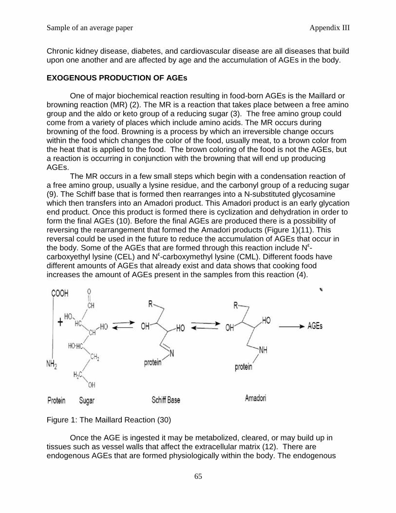
Sample of an average paper Appendix III
65
Chronic kidney disease, diabetes, and cardiovascular disease are all diseases that build upon one another and are affected by age and the accumulation of AGEs in the body. EXOGENOUS PRODUCTION OF AGEs
One of major biochemical reaction resulting in food-born AGEs is the Maillard or browning reaction (MR) (2). The MR is a reaction that takes place between a free amino group and the aldo or keto group of a reducing sugar (3). The free amino group could come from a variety of places which include amino acids. The MR occurs during browning of the food. Browning is a process by which an irreversible change occurs within the food which changes the color of the food, usually meat, to a brown color from the heat that is applied to the food. The brown coloring of the food is not the AGEs, but a reaction is occurring in conjunction with the browning that will end up producing AGEs. The MR occurs in a few small steps which begin with a condensation reaction of a free amino group, usually a lysine residue, and the carbonyl group of a reducing sugar (9). The Schiff base that is formed then rearranges into a N-substituted glycosamine which then transfers into an Amadori product. This Amadori product is an early glycation end product. Once this product is formed there is cyclization and dehydration in order to form the final AGEs (10). Before the final AGEs are produced there is a possibility of reversing the rearrangement that formed the Amadori products (Figure 1)(11). This reversal could be used in the future to reduce the accumulation of AGEs that occur in the body. Some of the AGEs that are formed through this reaction include Nε-carboxyethyl lysine (CEL) and Nε-carboxymethyl lysine (CML). Different foods have different amounts of AGEs that already exist and data shows that cooking food increases the amount of AGEs present in the samples from this reaction (4).
Figure 1: The Maillard Reaction (30) Once the AGE is ingested it may be metabolized, cleared, or may build up in tissues such as vessel walls that affect the extracellular matrix (12). There are endogenous AGEs that are formed physiologically within the body. The endogenous
Sample of an average paper Appendix III
66
AGEs add to the exogenous AGEs and this combination is a cause of the accumulation (5). There is some evidence that while there is buildup in the body there is also some clearance of the AGEs in urine (2). The clearance is not enough to outweigh the amount that ends up in the body and the health concerns result. In high AGE-containing foods there are also health benefits in these foods that may be antioxidants because of different compounds that are in the food. On the other hand problems in the body due to the AGEs themselves are their ability to promote oxidative stress and inflammation (2). For example, bread crust extract (BCE) was used to see the quantitative amount of AGEs within the extract and it was found that BCE has oxidative properties. On the other hand, decaffeinated coffee extract (CS) has both adverse and beneficial effects on the body (2). The CS can cause oxidative stress on the body, but also contain compounds that are antioxidants that are good for health (2). Melanonides are a pigment that is formed and found in processes foods. This pigment is brown in color and is heterogeneous in structure. Because of this mixture the total structure of pigment is not totally understood (6). Depending on the molecular weight of the pigment as well as the reactant causes a change in the solubility. Also, in protein rich foods there is a cross-linkage that is formed that causes the proteins to be cross-linked by carbohydrate-derived structures (1). The melanoidines may be more of a positive structure in the body, but at the same time may increase the amounts of AGEs that end up being produced. Much of the processing that occurs in the modern food, such as crackers and hot dogs, is mainly heat processing and is the primary source of food-derived AGEs (7). A reason that there is a contribution to the formation of AGEs would be the fact that foods that are high in lipid and protein concentration have free radicals that are produced from lipoxidation reactions (4). These reaction products catabolize the formation of AGES when meats and fats go through the cooking process (4).Carbohydrates usually have the lowest amount AGEs, but the amount changes greatly with increased processing. This can be seen in everyday breakfast cereals and is due to the high temperatures that are used in making the cereal (4). High temperature and high pressure are usually used together in the processing of ready-to-eat snacks and foods. The use of high of pressures can change the product. The changes include: dehydration, degradation, and changed in the chemical properties of the compound (4). Dehydration is a large problem because this allows for processing at high temperatures with low amounts of moisture that increases the amounts of AGEs that are produced in the food. The MR is used to form many of the exogenous AGEs. This reaction is used in many of the popular foods that are eaten today. AGEs can accumulate in the body because there are so many AGEs that are produced outside the body. There is not a quantitative value as to how much AGEs are too much, but there is much evidence that says that the modern person is eating too many. Figure 2 shows the amounts of AGEs that are in many common foods and how the amount is changed with certain cooking processes applied to the food.
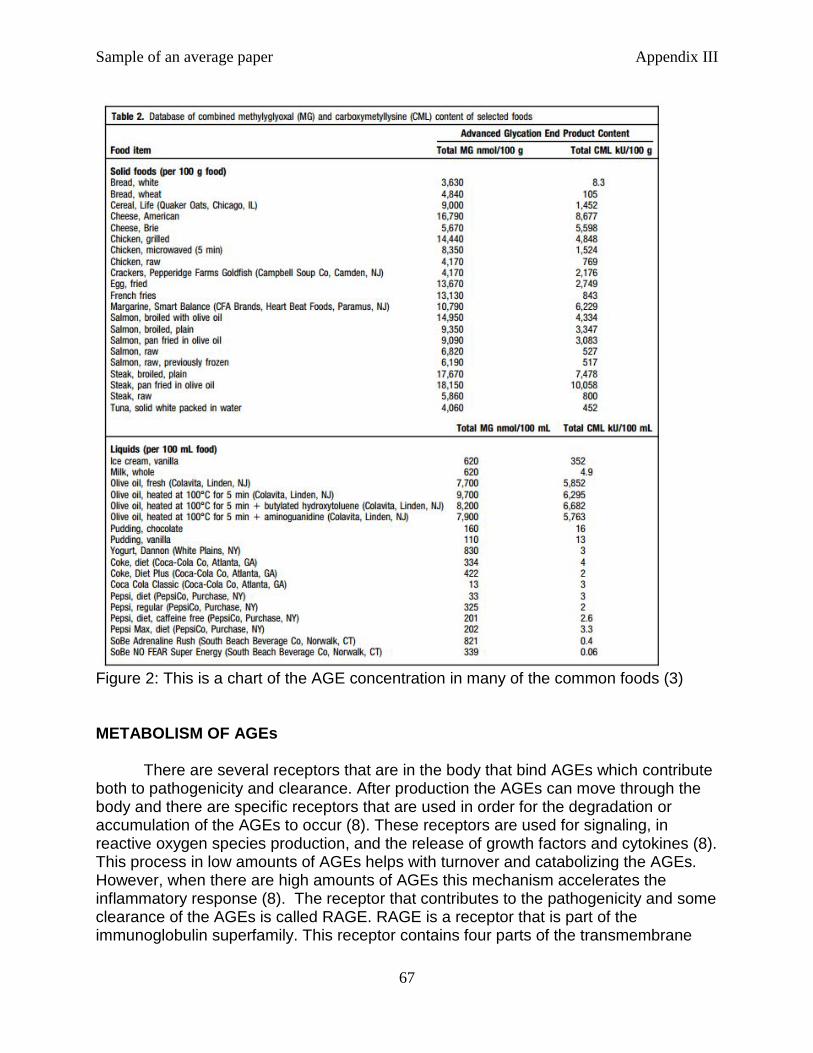
Sample of an average paper Appendix III
67
Figure 2: This is a chart of the AGE concentration in many of the common foods (3) METABOLISM OF AGEs There are several receptors that are in the body that bind AGEs which contribute both to pathogenicity and clearance. After production the AGEs can move through the body and there are specific receptors that are used in order for the degradation or accumulation of the AGEs to occur (8). These receptors are used for signaling, in reactive oxygen species production, and the release of growth factors and cytokines (8). This process in low amounts of AGEs helps with turnover and catabolizing the AGEs. However, when there are high amounts of AGEs this mechanism accelerates the inflammatory response (8). The receptor that contributes to the pathogenicity and some clearance of the AGEs is called RAGE. RAGE is a receptor that is part of the immunoglobulin superfamily. This receptor contains four parts of the transmembrane

Sample of an average paper Appendix III
68
protein which include: two constant domains, one variable domain, a transmembrane domain, and a cytoplasmic tail (13,14). Ligand-binding can occur because of the variable domain. The cytoplasmic tail is necessary for the signaling cascade that occurs when AGEs bind with RAGE (16). Activation occurs when there is an AGE that binds to RAGE, but the receptor is not specific to AGEs and can bind other molecules as well. When there is such a large variety of molecules that can attach to RAGE there are different functions that the receptor can carry out (17). RAGE is involved in the inflammatory response and in some ways there is chronic inflammation, but there is research that supports a positive function of RAGE with tissue repair (14). This different function shows how RAGE can be different because of what binds to the receptor. RAGE accepts pro-inflammatory ligands which include the following: S100 family of proteins, high mobility group protein 1 (HMGB1), β-amyloid peptide (Aβ), and macrophage antigen complex 1 (15). RAGE activates two different pathways, phosphatidylinositol-3 kinase (PI3-K) and mitogen-activated protein kinases (MAPKs), which in turn activates NF-κB (Figure 3) (30). The signaling cascade produces pro-inflammatory cytokines with the activation of NF-κB. NF-κB also causes the up regulation of RAGE which is the source of a positive feedback system which increases the amount of inflammatory promoters (31). CML, a common AGE, can also upregulate many other pro-inflammatory molecules in the cell (18). RAGE is found on cells that are a part of the immune response, especially T cells (13).
Figure 3: RAGE mechanism (32) A main protein that only has clearance function is AGER1 which is also known as OST-48 (21,34). This receptor is found in white adipose tissue, skeletal muscle, and liver tissues (35).This receptor was down regulated in disease such as diabetes which increases the accumulation of AGEs (21). When there is high amounts of oxidative stress, changes with age, or disease this receptor does not work as well and an accumulation of AGEs can occur. AGER1 and RAGE are two receptors that work against each other and negative regulation of RAGE may increase the clearance activity of AGER1. Survival factor sitruin 1 (SIRT1) is a survival factor and a NAD+ -dependent deacetylase (21, 35). The function of this protein is to remove acyl groups for many
Sample of an average paper Appendix III
69
different proteins. SIRT1 works to reduce the effect of AGEs on the body, but it works in a different way. AGER1 works as a receptor for the AGEs themselves, but SIRT1 works to control NF-κB. As was stated earlier NF-κB upregulates RAGE and helps with the activation cascade in order to increase the inflammatory response (35,18) SIRT1 decreases the inflammatory response and is tied to the AGER1 function in the cell where it has been studied in diabetes (21). This regulation is due to molecules that are inducers which are endoplasmic reticulum stress and fatty acids (22). SIRT1 works to regulate adipose tissue inflammation and this is done by regulating pro-inflammatory transcription. Through transcription regulation the SIRT1 has many mechanisms by which it can affect the inflammatory response. Some of the mechanisms that affect SIRT1 are deacetylation and methyltransferase activity. Transcriptional silencing is the result of SIRT1 which may reduce the inflammatory response (22). There are also some other receptors for the clearance of AGEs and include: 80K-H, CD36, and galectin- 3 (33). CD36 belongs to the class B scavenger family. CD36 causes endocytosis with binding of AGEs and is found in smooth muscle and macrophage cells that were found in patients with atherosclerosis (33). Galectin-3 is known as AGER3 and is a member of the lectin family and has a variety of functions. These functions include: cell to cell interactions within the cell matrix where it works to decrease adhesion, mast cell activation, some gene expression control due to splicing of pre-mRNA, and the degradation of AGEs because of the high affinity that this receptor has for them (34). 80K-H or AGER2 also has a function that independently is unknown but with these other receptors there is thought that they work as a complex and are implicated in the clearance of AGEs rather than increasing the inflammation response that RAGE is known for (34). There is both clearance and accumulation of AGEs in the body. Receptors contribute to most of the inflammatory response and the clearance of AGEs from the body. AGEs have seen to increase the amount of RAGE that is seen in the cell while there seems to be an upregulation of AGER1 when there is a lower amount of AGEs in the cell (42). This change in the receptors on the cell also affects the lifespan of the mouse that it was tested on. When there was a diet that was lower in AGEs there was an increased lifespan for the mouse than of those mice that had a diet in higher AGEs (42). The accumulation of the AGEs contributes to the incidence of disease because the RAGE receptor causes there to be a signaling pathway that allows for the disease to continue it pathogenicity in the body. The incidence of disease also increases with age, and the clearance of AGEs reduces with age which thus increases accumulation. DISEASE COMPLICATIONS There are some similarities that are evident in all of the diseases and there are complications from the diseases that build upon one another. Oxidative stress is important in the incidence of chronic kidney disease, diabetes, and cardiovascular disease. The oxidative stress affects the normal functioning of the cell and inhibits the normal protein function (40). AGEs are part of the pro-inflammatory response and in that response the oxidative stress is increased as well. Once the inflammation response is generated this causes a cascade to occur and cause more problems in the cell. This is one way in which the diseases build upon one another. The problems begin small, but
Sample of an average paper Appendix III
70
over time the health concerns compile and larger and more severe health issues become apparent. In this case there is an incidence of disease that is highly related. For example, kidney disease and diabetes are also risk factors for cardiovascular disease. The risk factors, if not treated, will cause there to be more disease and illness in the future for the patient. Also, age has an impact on the disease of a person. As the person ages the body does not work as well and the incidence of disease also reduces the ability of the body to fight off infection and decrease the health of the patient (23). Chronic Kidney Disease Chronic kidney disease is separated into indistinguishable stages that will end up causing total nephron dysfunction on the kidney (27). Diabetes will cause much of the renal failure in the United States and the renal failure can also increase the chance of having micro-vascular disease that can lead to stroke and heart attack (26). As chronic kidney disease continues there is more of an accumulation of AGEs which will continue to make the incidence of disease increase (26). When there is any amount of AGE clearance from the body it is done by the kidneys. There is some reabsorption in the proximal convoluted tubule of the nephron, but there is also some that is degraded and excreted in the urine (37). The reabsorption and degradation occurs after filtration. If filtration is compromised then there will be less excretion and the AGE will accumulate in the renal tissue (37). As the functionality of the kidneys decreases the amount of AGE accumulation continues. There is a positive correlation with AGE accumulation and oxidative stress as well as a negative correlation with AGE accumulation and glomerular filtration. Diabetes and chronic kidney disease together will increase the incidence of cardiovascular disease more than those with just chronic kidney disease (24). Asymmetric dimethylarginine (ADMA) is an endogenous nitric oxide synthase inhibitor which has seen an increase in both this disease and diabetes (26). AGEs inhibit the enzyme that breaks down this product. ADMA increases vascular injury and increases oxidative stress. ADMA that is increased in chronic kidney disease can affect other aspects of health as well including high blood pressure (27). ADMA is usually excreted from the body via the kidneys. When there is decreased or lost function of the kidneys then there is higher amounts of ADMA in the blood which impairs the function of nitric oxide synthase (NOS) (27).Nitric oxide is important in a variety of physiological functions and without the synthesis of the molecule there will be continued development of the complications. With the reduction of nitric oxide there is an increase in insulin resistance (36). This is just another example where there are diseases that all work together to cause the patient more and more complications. Type II Diabetes Diabetes is increasing its rate of development in this country for a variety of factors which include changes in diet, the increased sugar in foods, and also the amount of AGEs that are accumulated in the body. RAGE is still an important receptor when it comes to the pathogenesis in diabetic neuropathy (25). Diabetic neuropathy is nerve damage that occurs with the continuation of the disease. RAGE is higher in nerves of a person who is hypoglycemic than those who are nondiabetic. RAGE and

Sample of an average paper Appendix III
71
AGEs have been implicated in developing and continuing the disease (25). The exact mechanism by which this occurs is still unknown. However, it is thought that AGEs work to decrease the work of Schwann cells in the nervous system. Schwann cells work to myelinate nerve cells which increase the rate of the impulse moving along the axon. Both of the previous factors increase other types of disease as well (40). With a decrease in the speed of the impulse which causes there to be a degeneration of the nerve cells which can eventually cause cell death (40). ADMA is also present in higher levels in diabetes which can also lead to the incidence of cardiovascular disease because nitric oxide is used to keep the shape of vessels in the body (28). As was stated earlier a decrease in nitric oxide decreases the sensitivity to insulin which would cause the diabetes to get worse over time with the increased accumulation of AGEs. The increased amount of sugar that is in the blood in diabetic patients increases the amount of glycation that occurs. The AGEs accumulate in many tissues where there is diabetic complication such as kidney and retina and it accumulates intracellulary (38). This accumulation can interfere with the proteins within the cell and modify signaling pathways. The signaling pathways of RAGE and AGER1 were discussed above and the binding of the AGEs to different receptors causes a different response in the cell. Diabetic retinopathy is a cause of blindness in diabetic patients and is caused by the increased spreading of blood vessels in the eye and thickening of the basement membrane within the eye. AGEs are important in this aspect of diabetes because they are found to play a role in the dysfunction of retinal cells that may end up leading to the death of the cells (38). Cataracts are another cause of blindness in diabetic patients and occur when the lens becomes opaque and slowly causes the loss of vision. It has been stated that AGEs cause a change in the proteins of the cell that they are in. In the problem of diabetic cataracts, AGEs cause a change in the charge of the proteins. This change causes a disruption in the cooperation between the protein-protein and protein-water interactions that will lead to blindness (39). Cardiovascular Disease Cardiovascular disease arises from these other complications in a variety of ways and arterial stiffness is a large problem with the accumulation of AGEs. Cardiovascular disease is a wide array of problems which include blood vessel thickness, risk for clotting, and maybe heat attack or stroke (41). Accumulation of AGEs causes an increased cross-linking with collagen in the vessels which in turn causes there to be more arterial stiffness (25). This cross-linking also causes there to be problems with high blood pressure because there is less action of the enzymes to break down the collagen that is forming in the vessels (41). The nitric oxide concentrations in the blood are also a large factor in cardiovascular health. The nitric oxide is a vasodilator and ADMA inhibits the production of the nitric oxide which causes the vessels to have a higher risk for heart attack or stroke. Cardiovascular disease becomes a problem when there are many other ailments with a patient. As it has been seen when there is chronic kidney disease or diabetes the incidence of cardiovascular disease is higher.
Sample of an average paper Appendix III
72
Conclusion AGEs are a part of the body and can be ingested exogenously. The excess amount of the AGEs is what causes the problems in disease and clearance. Without sufficient clearance of the AGEs there is down regulation of receptors that help with clearance (AGER1) and increase in receptors that increase the inflammatory response (RAGE). With an increase in the RAGE receptor there is an increase in the oxidative stress that is put on the cell. With this increase there is more disease that develops. And the diseases build upon each other.
The Maillard reaction is the main reaction with food that causes there to be a production of AGEs. This reaction is found in some cooking methods and can also be found in some processing methods. Many foods, especially proteins, already have an AGE content, but frying or broiling meat can increase the amount of AGEs that are found in the body (2). There is not an amount of AGEs that have been proven to cause the adverse side effects. It is known that too much is a problem, but there is not a quantifiable amount that this refers too. Once the AGEs are in the body there are many things that can happen to them. This has to do with the type of receptor that the AGE binds to once in the cell. There is RAGE which causes there to be inflammation and a signaling cascade which will activate the cell cause more problems downstream (15). On the other hand there are receptors such as AGER1 which work to degrade and excrete the AGEs from the body so that they cannot cause any harm while they are there. The incidence of these receptors is also regulated by the amount of AGEs that are already accumulated in the cell. Even with the use of AGER1 there are not enough of the AGEs that are excreted from the body and the accumulation of them causes there to be disease in the cell (21). The diseases that are caused in the body due to the AGEs are vast and three of the common ones are chronic kidney disease, type II diabetes, and cardiovascular disease. These diseases build upon one another to cause even more problems in the cell and over time will just continue to get worse and worse. As age increases the amount of AGEs in the cell also increase as well as the incidence of disease. The main way to alleviate this problem is to reduce the amount of ingested AGEs in the diet. This could be done by reducing the amount of meat that is ingested because there is going to be a lot of AGEs that are ingested with the cooking process of meat. Protein is necessary for survival, but the protein that is eaten could affect the amount of AGEs ingested (1). Also, portion size is a factor and should be reduced in the diet. If the patient wanted to decrease the amount of AGEs they could reduce overall portions and eat a healthier diet. There is not conclusive evidence that exercise reduces AGEs, but there is evidence that a healthy and balanced diet along with exercise will create a healthier person and the impact of AGEs will be reduced.
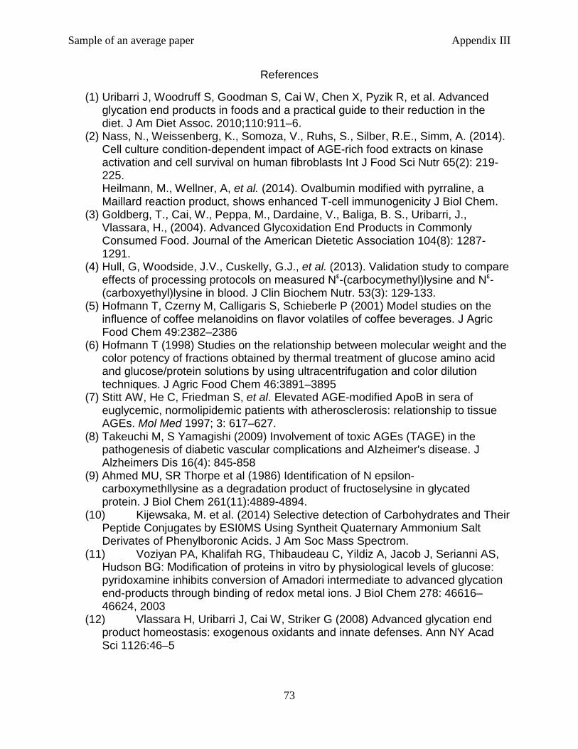
Sample of an average paper Appendix III
73
References
(1) Uribarri J, Woodruff S, Goodman S, Cai W, Chen X, Pyzik R, et al. Advanced glycation end products in foods and a practical guide to their reduction in the diet. J Am Diet Assoc. 2010;110:911–6.
(2) Nass, N., Weissenberg, K., Somoza, V., Ruhs, S., Silber, R.E., Simm, A. (2014). Cell culture condition-dependent impact of AGE-rich food extracts on kinase activation and cell survival on human fibroblasts Int J Food Sci Nutr 65(2): 219-225. Heilmann, M., Wellner, A, et al. (2014). Ovalbumin modified with pyrraline, a Maillard reaction product, shows enhanced T-cell immunogenicity J Biol Chem.
(3) Goldberg, T., Cai, W., Peppa, M., Dardaine, V., Baliga, B. S., Uribarri, J., Vlassara, H., (2004). Advanced Glycoxidation End Products in Commonly Consumed Food. Journal of the American Dietetic Association 104(8): 1287-1291.
(4) Hull, G, Woodside, J.V., Cuskelly, G.J., et al. (2013). Validation study to compare effects of processing protocols on measured Nε-(carbocymethyl)lysine and Nε-(carboxyethyl)lysine in blood. J Clin Biochem Nutr. 53(3): 129-133.
(5) Hofmann T, Czerny M, Calligaris S, Schieberle P (2001) Model studies on the influence of coffee melanoidins on flavor volatiles of coffee beverages. J Agric Food Chem 49:2382–2386
(6) Hofmann T (1998) Studies on the relationship between molecular weight and the color potency of fractions obtained by thermal treatment of glucose amino acid and glucose/protein solutions by using ultracentrifugation and color dilution techniques. J Agric Food Chem 46:3891–3895
(7) Stitt AW, He C, Friedman S, et al. Elevated AGE-modified ApoB in sera of euglycemic, normolipidemic patients with atherosclerosis: relationship to tissue AGEs. Mol Med 1997; 3: 617–627.
(8) Takeuchi M, S Yamagishi (2009) Involvement of toxic AGEs (TAGE) in the pathogenesis of diabetic vascular complications and Alzheimer's disease. J Alzheimers Dis 16(4): 845-858
(9) Ahmed MU, SR Thorpe et al (1986) Identification of N epsilon-carboxymethllysine as a degradation product of fructoselysine in glycated protein. J Biol Chem 261(11):4889-4894.
(10) Kijewsaka, M. et al. (2014) Selective detection of Carbohydrates and Their Peptide Conjugates by ESI0MS Using Syntheit Quaternary Ammonium Salt Derivates of Phenylboronic Acids. J Am Soc Mass Spectrom.
(11) Voziyan PA, Khalifah RG, Thibaudeau C, Yildiz A, Jacob J, Serianni AS, Hudson BG: Modification of proteins in vitro by physiological levels of glucose: pyridoxamine inhibits conversion of Amadori intermediate to advanced glycation end-products through binding of redox metal ions. J Biol Chem 278: 46616–46624, 2003
(12) Vlassara H, Uribarri J, Cai W, Striker G (2008) Advanced glycation end product homeostasis: exogenous oxidants and innate defenses. Ann NY Acad Sci 1126:46–5
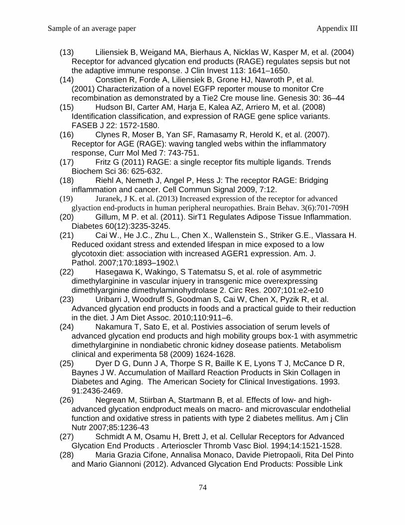
Sample of an average paper Appendix III
74
(13) Liliensiek B, Weigand MA, Bierhaus A, Nicklas W, Kasper M, et al. (2004) Receptor for advanced glycation end products (RAGE) regulates sepsis but not the adaptive immune response. J Clin Invest 113: 1641–1650.
(14) Constien R, Forde A, Liliensiek B, Grone HJ, Nawroth P, et al. (2001) Characterization of a novel EGFP reporter mouse to monitor Cre recombination as demonstrated by a Tie2 Cre mouse line. Genesis 30: 36–44
(15) Hudson BI, Carter AM, Harja E, Kalea AZ, Arriero M, et al. (2008) Identification classification, and expression of RAGE gene splice variants. FASEB J 22: 1572-1580.
(16) Clynes R, Moser B, Yan SF, Ramasamy R, Herold K, et al. (2007). Receptor for AGE (RAGE): waving tangled webs within the inflammatory response, Curr Mol Med 7: 743-751.
(17) Fritz G (2011) RAGE: a single receptor fits multiple ligands. Trends Biochem Sci 36: 625-632.
(18) Riehl A, Nemeth J, Angel P, Hess J: The receptor RAGE: Bridging inflammation and cancer. Cell Commun Signal 2009, 7:12.
(19) Juranek, J K. et al. (2013) Increased expression of the receptor for advanced glyaction end-products in human peripheral neuropathies. Brain Behav. 3(6):701-709H
(20) Gillum, M P. et al. (2011). SirT1 Regulates Adipose Tissue Inflammation. Diabetes 60(12):3235-3245.
(21) Cai W., He J.C., Zhu L., Chen X., Wallenstein S., Striker G.E., Vlassara H. Reduced oxidant stress and extended lifespan in mice exposed to a low glycotoxin diet: association with increased AGER1 expression. Am. J. Pathol. 2007;170:1893–1902.\
(22) Hasegawa K, Wakingo, S Tatematsu S, et al. role of asymmetric dimethylarginine in vascular injuery in transgenic mice overexpressing dimethlyarginine dimethylaminohydrolase 2. Circ Res. 2007;101:e2-e10
(23) Uribarri J, Woodruff S, Goodman S, Cai W, Chen X, Pyzik R, et al. Advanced glycation end products in foods and a practical guide to their reduction in the diet. J Am Diet Assoc. 2010;110:911–6.
(24) Nakamura T, Sato E, et al. Postivies association of serum levels of advanced glycation end products and high mobility groups box-1 with asymmetric dimethylarginine in nondiabetic chronic kidney dosease patients. Metabolism clinical and experimenta 58 (2009) 1624-1628.
(25) Dyer D G, Dunn J A, Thorpe S R, Baille K E, Lyons T J, McCance D R, Baynes J W. Accumulation of Maillard Reaction Products in Skin Collagen in Diabetes and Aging. The American Society for Clinical Investigations. 1993. 91:2436-2469.
(26) Negrean M, Stiirban A, Startmann B, et al. Effects of low- and high-advanced glycation endproduct meals on macro- and microvascular endothelial function and oxidative stress in patients with type 2 diabetes mellitus. Am j Clin Nutr 2007;85:1236-43
(27) Schmidt A M, Osamu H, Brett J, et al. Cellular Receptors for Advanced Glycation End Products . Arterioscler Thromb Vasc Biol. 1994;14:1521-1528.
(28) Maria Grazia Cifone, Annalisa Monaco, Davide Pietropaoli, Rita Del Pinto and Mario Giannoni (2012). Advanced Glycation End Products: Possible Link
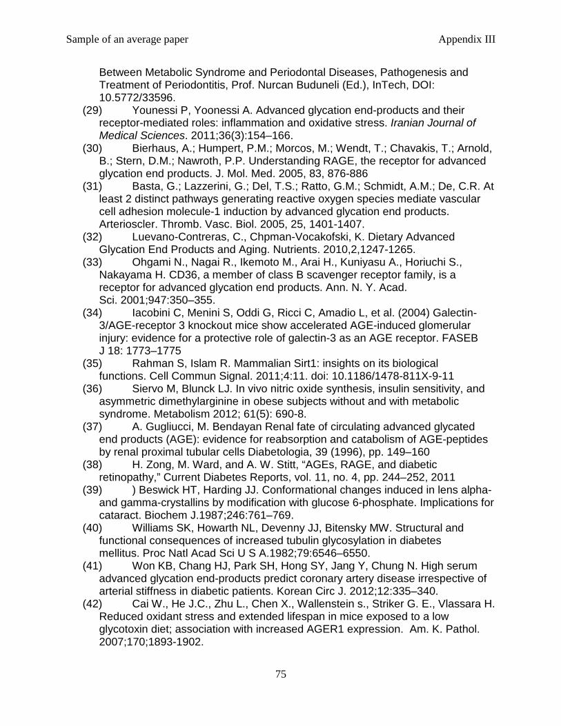
Sample of an average paper Appendix III
75
Between Metabolic Syndrome and Periodontal Diseases, Pathogenesis and Treatment of Periodontitis, Prof. Nurcan Buduneli (Ed.), InTech, DOI: 10.5772/33596.
(29) Younessi P, Yoonessi A. Advanced glycation end-products and their receptor-mediated roles: inflammation and oxidative stress. Iranian Journal of Medical Sciences. 2011;36(3):154–166.
(30) Bierhaus, A.; Humpert, P.M.; Morcos, M.; Wendt, T.; Chavakis, T.; Arnold, B.; Stern, D.M.; Nawroth, P.P. Understanding RAGE, the receptor for advanced glycation end products. J. Mol. Med. 2005, 83, 876-886
(31) Basta, G.; Lazzerini, G.; Del, T.S.; Ratto, G.M.; Schmidt, A.M.; De, C.R. At least 2 distinct pathways generating reactive oxygen species mediate vascular cell adhesion molecule-1 induction by advanced glycation end products. Arterioscler. Thromb. Vasc. Biol. 2005, 25, 1401-1407.
(32) Luevano-Contreras, C., Chpman-Vocakofski, K. Dietary Advanced Glycation End Products and Aging. Nutrients. 2010,2,1247-1265.
(33) Ohgami N., Nagai R., Ikemoto M., Arai H., Kuniyasu A., Horiuchi S., Nakayama H. CD36, a member of class B scavenger receptor family, is a receptor for advanced glycation end products. Ann. N. Y. Acad. Sci. 2001;947:350–355.
(34) Iacobini C, Menini S, Oddi G, Ricci C, Amadio L, et al. (2004) Galectin-3/AGE-receptor 3 knockout mice show accelerated AGE-induced glomerular injury: evidence for a protective role of galectin-3 as an AGE receptor. FASEB J 18: 1773–1775
(35) Rahman S, Islam R. Mammalian Sirt1: insights on its biological functions. Cell Commun Signal. 2011;4:11. doi: 10.1186/1478-811X-9-11
(36) Siervo M, Blunck LJ. In vivo nitric oxide synthesis, insulin sensitivity, and asymmetric dimethylarginine in obese subjects without and with metabolic syndrome. Metabolism 2012; 61(5): 690-8.
(37) A. Gugliucci, M. Bendayan Renal fate of circulating advanced glycated end products (AGE): evidence for reabsorption and catabolism of AGE-peptides by renal proximal tubular cells Diabetologia, 39 (1996), pp. 149–160
(38) H. Zong, M. Ward, and A. W. Stitt, “AGEs, RAGE, and diabetic retinopathy,” Current Diabetes Reports, vol. 11, no. 4, pp. 244–252, 2011
(39) ) Beswick HT, Harding JJ. Conformational changes induced in lens alpha- and gamma-crystallins by modification with glucose 6-phosphate. Implications for cataract. Biochem J.1987;246:761–769.
(40) Williams SK, Howarth NL, Devenny JJ, Bitensky MW. Structural and functional consequences of increased tubulin glycosylation in diabetes mellitus. Proc Natl Acad Sci U S A.1982;79:6546–6550.
(41) Won KB, Chang HJ, Park SH, Hong SY, Jang Y, Chung N. High serum advanced glycation end-products predict coronary artery disease irrespective of arterial stiffness in diabetic patients. Korean Circ J. 2012;12:335–340.
(42) Cai W., He J.C., Zhu L., Chen X., Wallenstein s., Striker G. E., Vlassara H. Reduced oxidant stress and extended lifespan in mice exposed to a low glycotoxin diet; association with increased AGER1 expression. Am. K. Pathol. 2007;170;1893-1902.
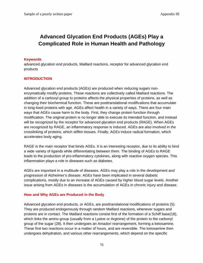
Sample of a poorly written paper Appendix III
76
Advanced Glycation End Products (AGEs) Play a
Complicated Role in Human Health and Pathology
Keywords advanced glycation end products, Maillard reactions, receptor for advanced glycation end products INTRODUCTION Advanced glycation end products (AGEs) are produced when reducing sugars non-enzymatically modify proteins. These reactions are collectively called Maillard reactions. The addition of a carboxyl group to proteins affects the physical properties of proteins, as well as changing their biochemical function. These are posttranslational modifications that accumulate in long-lived proteins with age. AGEs affect health in a variety of ways. There are four main ways that AGEs cause harm to the body. First, they change protein function through modification. The original protein is no longer able to execute its intended function, and instead will be recognized by the receptor for advanced glycation end products (RAGE). When AGEs are recognized by RAGE, an inflammatory response is induced. AGEs are also involved in the crosslinking of proteins, which stiffen tissues. Finally, AGEs induce radical formation, which accelerates body aging. RAGE is the main receptor that binds AGEs. It is an interesting receptor, due to its ability to bind a wide variety of ligands while differentiating between them. The binding of AGEs to RAGE leads to the production of pro-inflammatory cytokines, along with reactive oxygen species. This inflammation plays a role in diseases such as diabetes. AGEs are important in a multitude of diseases. AGEs may play a role in the development and progression of Alzheimer’s disease. AGEs have been implicated in several diabetic complications, mostly due to an increase of AGEs caused by higher blood sugar levels. Another issue arising from AGEs in diseases is the accumulation of AGEs in chronic injury and disease. How and Why AGEs are Produced in the Body Advanced glycation end products, or AGEs, are posttranslational modifications of proteins (5). They are produced endogenously through random Maillard reactions, whenever sugars and proteins are in contact. The Maillard reactions consist first of the formation of a Schiff base(18), which links the amino group (usually from a Lysine or Arginine) of the protein to the carbonyl group of the sugar (28). It then undergoes an Amadori rearrangement, forming a ketosamine. These first two reactions occur in a matter of hours, and are reversible. The ketosamine then undergoes dehydration, and various other rearrangements, which depend on the specific
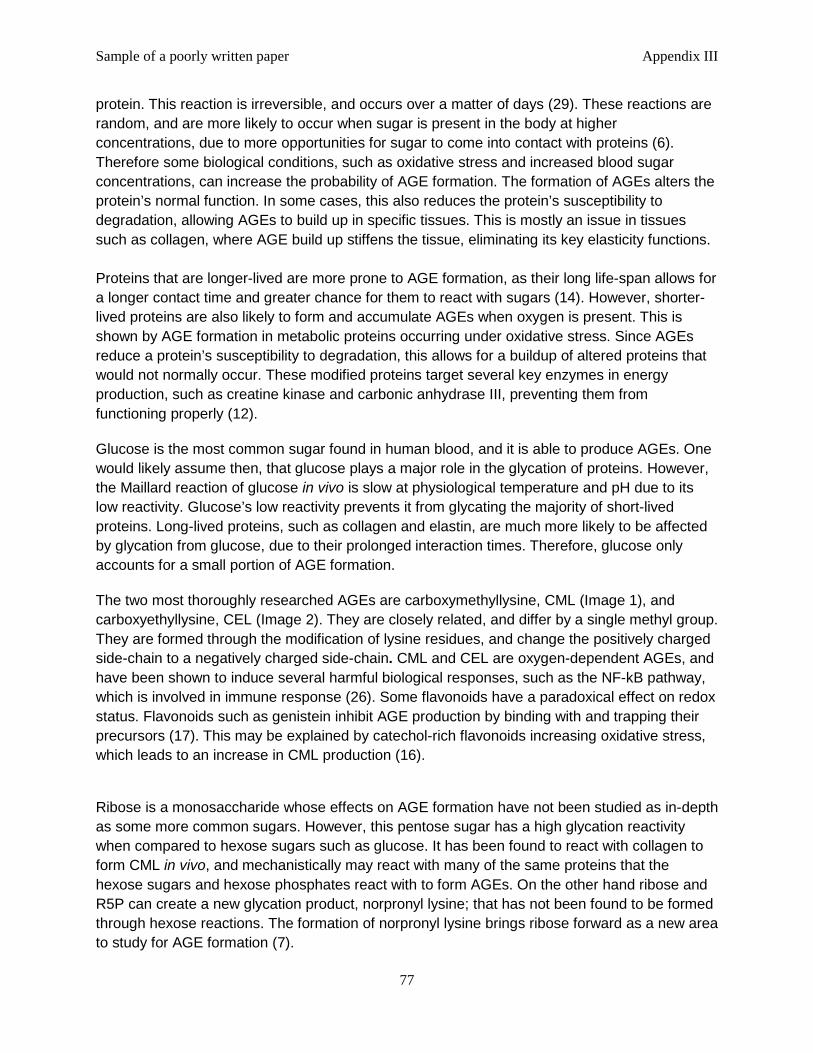
Sample of a poorly written paper Appendix III
77
protein. This reaction is irreversible, and occurs over a matter of days (29). These reactions are random, and are more likely to occur when sugar is present in the body at higher concentrations, due to more opportunities for sugar to come into contact with proteins (6). Therefore some biological conditions, such as oxidative stress and increased blood sugar concentrations, can increase the probability of AGE formation. The formation of AGEs alters the protein’s normal function. In some cases, this also reduces the protein’s susceptibility to degradation, allowing AGEs to build up in specific tissues. This is mostly an issue in tissues such as collagen, where AGE build up stiffens the tissue, eliminating its key elasticity functions. Proteins that are longer-lived are more prone to AGE formation, as their long life-span allows for a longer contact time and greater chance for them to react with sugars (14). However, shorter-lived proteins are also likely to form and accumulate AGEs when oxygen is present. This is shown by AGE formation in metabolic proteins occurring under oxidative stress. Since AGEs reduce a protein’s susceptibility to degradation, this allows for a buildup of altered proteins that would not normally occur. These modified proteins target several key enzymes in energy production, such as creatine kinase and carbonic anhydrase III, preventing them from functioning properly (12).
Glucose is the most common sugar found in human blood, and it is able to produce AGEs. One would likely assume then, that glucose plays a major role in the glycation of proteins. However, the Maillard reaction of glucose in vivo is slow at physiological temperature and pH due to its low reactivity. Glucose’s low reactivity prevents it from glycating the majority of short-lived proteins. Long-lived proteins, such as collagen and elastin, are much more likely to be affected by glycation from glucose, due to their prolonged interaction times. Therefore, glucose only accounts for a small portion of AGE formation.
The two most thoroughly researched AGEs are carboxymethyllysine, CML (Image 1), and carboxyethyllysine, CEL (Image 2). They are closely related, and differ by a single methyl group. They are formed through the modification of lysine residues, and change the positively charged side-chain to a negatively charged side-chain. CML and CEL are oxygen-dependent AGEs, and have been shown to induce several harmful biological responses, such as the NF-kB pathway, which is involved in immune response (26). Some flavonoids have a paradoxical effect on redox status. Flavonoids such as genistein inhibit AGE production by binding with and trapping their precursors (17). This may be explained by catechol-rich flavonoids increasing oxidative stress, which leads to an increase in CML production (16).
Ribose is a monosaccharide whose effects on AGE formation have not been studied as in-depth as some more common sugars. However, this pentose sugar has a high glycation reactivity when compared to hexose sugars such as glucose. It has been found to react with collagen to form CML in vivo, and mechanistically may react with many of the same proteins that the hexose sugars and hexose phosphates react with to form AGEs. On the other hand ribose and R5P can create a new glycation product, norpronyl lysine; that has not been found to be formed through hexose reactions. The formation of norpronyl lysine brings ribose forward as a new area to study for AGE formation (7).
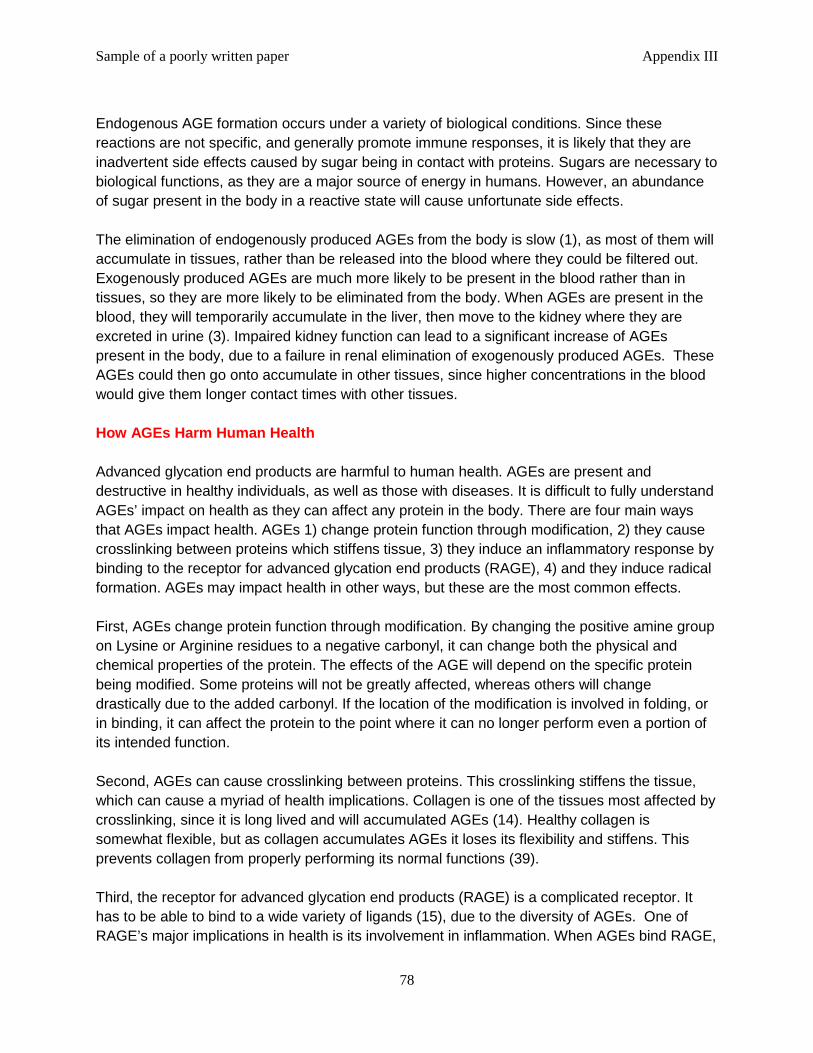
Sample of a poorly written paper Appendix III
78
Endogenous AGE formation occurs under a variety of biological conditions. Since these reactions are not specific, and generally promote immune responses, it is likely that they are inadvertent side effects caused by sugar being in contact with proteins. Sugars are necessary to biological functions, as they are a major source of energy in humans. However, an abundance of sugar present in the body in a reactive state will cause unfortunate side effects. The elimination of endogenously produced AGEs from the body is slow (1), as most of them will accumulate in tissues, rather than be released into the blood where they could be filtered out. Exogenously produced AGEs are much more likely to be present in the blood rather than in tissues, so they are more likely to be eliminated from the body. When AGEs are present in the blood, they will temporarily accumulate in the liver, then move to the kidney where they are excreted in urine (3). Impaired kidney function can lead to a significant increase of AGEs present in the body, due to a failure in renal elimination of exogenously produced AGEs. These AGEs could then go onto accumulate in other tissues, since higher concentrations in the blood would give them longer contact times with other tissues. How AGEs Harm Human Health Advanced glycation end products are harmful to human health. AGEs are present and destructive in healthy individuals, as well as those with diseases. It is difficult to fully understand AGEs’ impact on health as they can affect any protein in the body. There are four main ways that AGEs impact health. AGEs 1) change protein function through modification, 2) they cause crosslinking between proteins which stiffens tissue, 3) they induce an inflammatory response by binding to the receptor for advanced glycation end products (RAGE), 4) and they induce radical formation. AGEs may impact health in other ways, but these are the most common effects. First, AGEs change protein function through modification. By changing the positive amine group on Lysine or Arginine residues to a negative carbonyl, it can change both the physical and chemical properties of the protein. The effects of the AGE will depend on the specific protein being modified. Some proteins will not be greatly affected, whereas others will change drastically due to the added carbonyl. If the location of the modification is involved in folding, or in binding, it can affect the protein to the point where it can no longer perform even a portion of its intended function. Second, AGEs can cause crosslinking between proteins. This crosslinking stiffens the tissue, which can cause a myriad of health implications. Collagen is one of the tissues most affected by crosslinking, since it is long lived and will accumulated AGEs (14). Healthy collagen is somewhat flexible, but as collagen accumulates AGEs it loses its flexibility and stiffens. This prevents collagen from properly performing its normal functions (39). Third, the receptor for advanced glycation end products (RAGE) is a complicated receptor. It has to be able to bind to a wide variety of ligands (15), due to the diversity of AGEs. One of RAGE’s major implications in health is its involvement in inflammation. When AGEs bind RAGE,
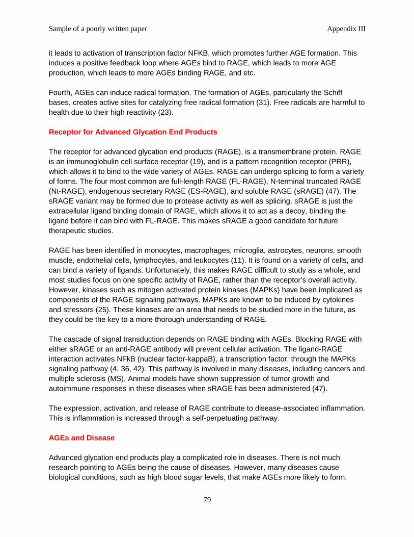
Sample of a poorly written paper Appendix III
79
it leads to activation of transcription factor NFKB, which promotes further AGE formation. This induces a positive feedback loop where AGEs bind to RAGE, which leads to more AGE production, which leads to more AGEs binding RAGE, and etc. Fourth, AGEs can induce radical formation. The formation of AGEs, particularly the Schiff bases, creates active sites for catalyzing free radical formation (31). Free radicals are harmful to health due to their high reactivity (23). Receptor for Advanced Glycation End Products The receptor for advanced glycation end products (RAGE), is a transmembrane protein. RAGE is an immunoglobulin cell surface receptor (19), and is a pattern recognition receptor (PRR), which allows it to bind to the wide variety of AGEs. RAGE can undergo splicing to form a variety of forms. The four most common are full-length RAGE (FL-RAGE), N-terminal truncated RAGE (Nt-RAGE), endogenous secretary RAGE (ES-RAGE), and soluble RAGE (sRAGE) (47). The sRAGE variant may be formed due to protease activity as well as splicing. sRAGE is just the extracellular ligand binding domain of RAGE, which allows it to act as a decoy, binding the ligand before it can bind with FL-RAGE. This makes sRAGE a good candidate for future therapeutic studies. RAGE has been identified in monocytes, macrophages, microglia, astrocytes, neurons, smooth muscle, endothelial cells, lymphocytes, and leukocytes (11). It is found on a variety of cells, and can bind a variety of ligands. Unfortunately, this makes RAGE difficult to study as a whole, and most studies focus on one specific activity of RAGE, rather than the receptor’s overall activity. However, kinases such as mitogen activated protein kinases (MAPKs) have been implicated as components of the RAGE signaling pathways. MAPKs are known to be induced by cytokines and stressors (25). These kinases are an area that needs to be studied more in the future, as they could be the key to a more thorough understanding of RAGE. The cascade of signal transduction depends on RAGE binding with AGEs. Blocking RAGE with either sRAGE or an anti-RAGE antibody will prevent cellular activation. The ligand-RAGE interaction activates NFkB (nuclear factor-kappaB), a transcription factor, through the MAPKs signaling pathway (4, 36, 42). This pathway is involved in many diseases, including cancers and multiple sclerosis (MS). Animal models have shown suppression of tumor growth and autoimmune responses in these diseases when sRAGE has been administered (47). The expression, activation, and release of RAGE contribute to disease-associated inflammation. This is inflammation is increased through a self-perpetuating pathway. AGEs and Disease Advanced glycation end products play a complicated role in diseases. There is not much research pointing to AGEs being the cause of diseases. However, many diseases cause biological conditions, such as high blood sugar levels, that make AGEs more likely to form.

Sample of a poorly written paper Appendix III
80
Since there are then more AGEs being produced in the diseased state, the AGEs are more likely to affect overall health. There has been recent evidence showing that AGEs may play a role in Alzheimer’s Disease (AD) (31, 47). It is difficult to say how involved AGEs may or may not be in the development and progression of AD, since the disease itself is not well understood. The brain has a high amount of oxidative stress, which may lead to more AGE formation. Also, glucose is the primary energy source for the brain, so it is present in higher concentrations than other areas of the body. Glucose is a reducing sugar that may form AGEs, so the higher concentrations may also lead to more AGE formation. With conditions in the brain being favorable for AGE formation, the recent studies linking AGEs and Alzheimer’s Disease are promising, but need to be studied further. One known factor is that accumulation of amyloid-beta peptide and tau protein are important to Alzheimer’s Disease (8, 32). AGE induced crosslinking may lead to this aggregation. It is also hypothesized that the amyloid precursor protein expression may be upregulated by AGEs. Also, the ApoE4 protein (an isoform of ApoE) is associated with Alzheimer’s Disease. This isoform has shown a higher binding activity to AGEs than other isoforms, which could provide a link between AGEs and AD (31). So far, the research linking AGEs and Alzheimer’s Disease looks promising, but it is by no means definitive. Diabetes is a thoroughly researched disease, and certainly the most researched in regards to AGEs (22). The focus on this research seems to focus on the increased production of AGEs due to higher blood sugar levels, along with AGEs overall health effects. AGEs have been linked to peripheral neuropathy (2), a form of nerve damage often seen in diabetics. AGEs have also been implicated in diabetic retinopathy (10), likely due to inflammation caused by AGEs binding to RAGE (35). It has previously been mentioned that sRAGE shows promise as a competitive binder of AGEs. It is important to note that sRAGE can also have a detrimental effect on the health of those with diabetes (3, 47). A higher concentration of sRAGE in the blood has been linked to an increase in inflammatory markers such as tumor necrosis factor (TNF)-α and monocyte chemo attractant protein-1 (MCP-1). Before sRAGE therapeutics can be looked upon as he future of the field, the complex interactions between AGEs, RAGE, and sRAGE needs to be better understood. Conclusion and Future Perspectives AGEs are produced both endogenously and exogenously; through random, non-enzymatic chemical reactions. These Maillard reactions are unavoidable. AGEs are harmful to human health for a variety of reasons. The most prevalent of these is the alteration of the protein preventing it from performing its intended duties. AGEs will also cause crosslinking between tissues, which stiffen the tissues and impair their normal activities. The binding of AGEs to RAGE causes a signaling cascade that proves to have many detrimental effects. Some of these include promoting tumor growth in cancer, and promoting autoimmune responses especially in diseases such as MS.
Sample of a poorly written paper Appendix III
81
The most promising areas for future studies of AGEs focus on either diseases or RAGE. AGEs have proven to be linked to Alzheimer’s disease and diabetes. However, these links are not well understood. A more thorough understanding of these links could lead to a better quality of life for people suffering from these conditions. Alternatively, studying RAGE isoforms such as sRAGE could lead to preventative therapeutics.
Image 1 CML
Image 2 CEL
Image 3 Lysine LITERATURE CITED 1. Alamir, I. et al., 2013. Digestibility of extruded proteins and metabolic transit of N ε -carboxymethyllysine in rats. Amino acids, 44(6), pp.1441–9. Available at: http://www.ncbi.nlm.nih.gov/pubmed/23160731 2. Aubert, C.E. et al., 2014. Association of peripheral neuropathy with circulating advanced glycation end products, soluble receptor for advanced glycation end products and other risk
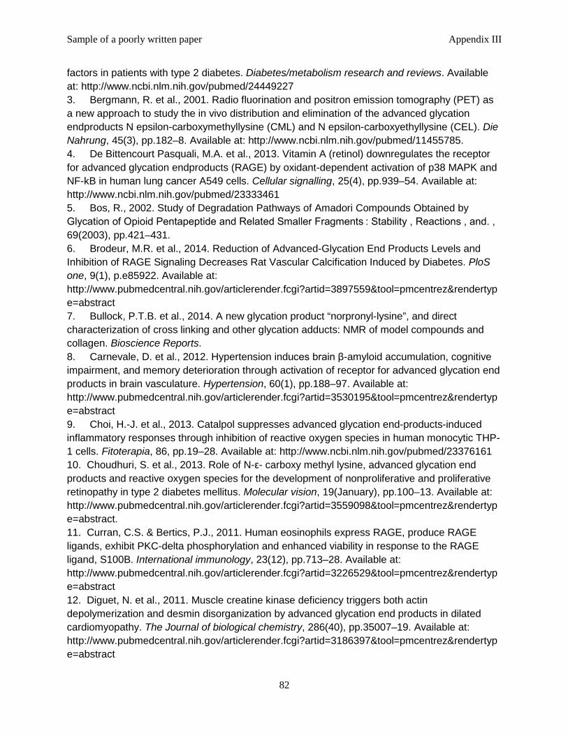
Sample of a poorly written paper Appendix III
82
factors in patients with type 2 diabetes. Diabetes/metabolism research and reviews. Available at: http://www.ncbi.nlm.nih.gov/pubmed/24449227 3. Bergmann, R. et al., 2001. Radio fluorination and positron emission tomography (PET) as a new approach to study the in vivo distribution and elimination of the advanced glycation endproducts N epsilon-carboxymethyllysine (CML) and N epsilon-carboxyethyllysine (CEL). Die Nahrung, 45(3), pp.182–8. Available at: http://www.ncbi.nlm.nih.gov/pubmed/11455785. 4. De Bittencourt Pasquali, M.A. et al., 2013. Vitamin A (retinol) downregulates the receptor for advanced glycation endproducts (RAGE) by oxidant-dependent activation of p38 MAPK and NF-kB in human lung cancer A549 cells. Cellular signalling, 25(4), pp.939–54. Available at: http://www.ncbi.nlm.nih.gov/pubmed/23333461 5. Bos, R., 2002. Study of Degradation Pathways of Amadori Compounds Obtained by Glycation of Opioid Pentapeptide and Related Smaller Fragments : Stability , Reactions , and. , 69(2003), pp.421–431. 6. Brodeur, M.R. et al., 2014. Reduction of Advanced-Glycation End Products Levels and Inhibition of RAGE Signaling Decreases Rat Vascular Calcification Induced by Diabetes. PloS one, 9(1), p.e85922. Available at: http://www.pubmedcentral.nih.gov/articlerender.fcgi?artid=3897559&tool=pmcentrez&rendertype=abstract 7. Bullock, P.T.B. et al., 2014. A new glycation product “norpronyl-lysine”, and direct characterization of cross linking and other glycation adducts: NMR of model compounds and collagen. Bioscience Reports. 8. Carnevale, D. et al., 2012. Hypertension induces brain β-amyloid accumulation, cognitive impairment, and memory deterioration through activation of receptor for advanced glycation end products in brain vasculature. Hypertension, 60(1), pp.188–97. Available at: http://www.pubmedcentral.nih.gov/articlerender.fcgi?artid=3530195&tool=pmcentrez&rendertype=abstract 9. Choi, H.-J. et al., 2013. Catalpol suppresses advanced glycation end-products-induced inflammatory responses through inhibition of reactive oxygen species in human monocytic THP-1 cells. Fitoterapia, 86, pp.19–28. Available at: http://www.ncbi.nlm.nih.gov/pubmed/23376161 10. Choudhuri, S. et al., 2013. Role of N-ε- carboxy methyl lysine, advanced glycation end products and reactive oxygen species for the development of nonproliferative and proliferative retinopathy in type 2 diabetes mellitus. Molecular vision, 19(January), pp.100–13. Available at: http://www.pubmedcentral.nih.gov/articlerender.fcgi?artid=3559098&tool=pmcentrez&rendertype=abstract. 11. Curran, C.S. & Bertics, P.J., 2011. Human eosinophils express RAGE, produce RAGE ligands, exhibit PKC-delta phosphorylation and enhanced viability in response to the RAGE ligand, S100B. International immunology, 23(12), pp.713–28. Available at: http://www.pubmedcentral.nih.gov/articlerender.fcgi?artid=3226529&tool=pmcentrez&rendertype=abstract 12. Diguet, N. et al., 2011. Muscle creatine kinase deficiency triggers both actin depolymerization and desmin disorganization by advanced glycation end products in dilated cardiomyopathy. The Journal of biological chemistry, 286(40), pp.35007–19. Available at: http://www.pubmedcentral.nih.gov/articlerender.fcgi?artid=3186397&tool=pmcentrez&rendertype=abstract

Sample of a poorly written paper Appendix III
83
13. Dinarvand, P. et al., 2014. Polyphosphate amplifies proinflammatory responses of nuclear proteins through interaction with receptor for advanced glycation end products and P2Y1 purinergic receptor. Blood, 123(6), pp.935–45. Available at: http://www.ncbi.nlm.nih.gov/pubmed/24255918 14. Ferreira, A.E.N., Ponces Freire, A.M.J. & Voit, E.O., 2003. A quantitative model of the generation of N(epsilon)-(carboxymethyl)lysine in the Maillard reaction between collagen and glucose. The Biochemical journal, 376(Pt 1), pp.109–21. Available at: http://www.pubmedcentral.nih.gov/articlerender.fcgi?artid=1223750&tool=pmcentrez&rendertype=abstract. 15. Fritz, G., 2011. RAGE: a single receptor fits multiple ligands. Trends in biochemical sciences, 36(12), pp.625–32. Available at: http://www.ncbi.nlm.nih.gov/pubmed/22019011 16. Fujiwara, Y. et al., 2011. Natural compounds containing a catechol group enhance the formation of Nε-(carboxymethyl)lysine of the Maillard reaction. Free radical biology & medicine, 50(7), pp.883–91. Available at: http://www.ncbi.nlm.nih.gov/pubmed/21195168 17. Gaens, K.H.J. et al., 2012. Endogenous formation of Nε-(carboxymethyl)lysine is increased in fatty livers and induces inflammatory markers in an in vitro model of hepatic steatosis. Journal of hepatology, 56(3), pp.647–55. Available at: http://www.ncbi.nlm.nih.gov/pubmed/21907687 18. Ghosh, M., Manna, P. & Sil, P.C., 2011. Protective role of a coumarin-derived schiff base scaffold against tertiary butyl hydroperoxide (TBHP)-induced oxidative impairment and cell death via MAPKs, NF-κB and mitochondria-dependent pathways. Free radical research, 45(5), pp.620–37. Available at: http://www.ncbi.nlm.nih.gov/pubmed/21391895 19. González, I. et al., 2013. The immunobiology of the receptor of advanced glycation end-products: trends and challenges. Immunobiology, 218(5), pp.790–7. Available at: http://www.ncbi.nlm.nih.gov/pubmed/23182709 20. Goodwin, M. et al., 2013. Advanced glycation end products augment experimental hepatic fibrosis. Journal of gastroenterology and hepatology, 28(2), pp.369–76. Available at: http://www.ncbi.nlm.nih.gov/pubmed/23173780 21. Grimm, S. et al., 2012. Advanced-glycation-end-product-induced formation of immunoproteasomes: involvement of RAGE and Jak2/STAT1. The Biochemical journal, 448(1), pp.127–39. Available at: http://www.ncbi.nlm.nih.gov/pubmed/22892029 22. Han, D. et al., 2013. Induction of receptor for advanced glycation end products by insufficient leptin action triggers pancreatic β-cell failure in type 2 diabetes. Genes to cells : devoted to molecular & cellular mechanisms, 18(4), pp.302–14. Available at: http://www.ncbi.nlm.nih.gov/pubmed/23410183 23. Han, L. et al., 2013. Hydroxyl radical induced by lipid in Maillard reaction model system promotes diet-derived N(ε)-carboxymethyllysine formation. Food and chemical toxicology : an international journal published for the British Industrial Biological Research Association, 60, pp.536–41. Available at: http://www.ncbi.nlm.nih.gov/pubmed/23959106 24. Hipkiss, A.R., 2011. Energy metabolism, proteotoxic stress and age-related dysfunction – Protection by carnosine. Molecular Aspects of Medicine, 32(4-6), pp.267–278. Available at: http://www.ncbi.nlm.nih.gov/pubmed/22020113 25. Hirasawa, Y. et al., 2011. Advanced-glycation-end-product-cholesterol-aggregated-protein accelerates the proliferation of mesangial cells mediated by transforming-growth-factor-beta 1

Sample of a poorly written paper Appendix III
84
receptors and the ERK-MAPK pathway. European journal of pharmacology, 672(1-3), pp.159–68. Available at: http://www.ncbi.nlm.nih.gov/pubmed/21989075 26. Holik, A.-K. et al., 2013. N(ε)-Carboxymethyllysine (CML), a Maillard reaction product, stimulates serotonin release and activates the receptor for advanced glycation end products (RAGE) in SH-SY5Y cells. Food & function, 4(7), pp.1111–20. Available at: http://www.ncbi.nlm.nih.gov/pubmed/23759926 27. Ibrahim, Z.A. et al., 2013. RAGE and TLRs: relatives, friends or neighbours? Molecular immunology, 56(4), pp.739–44. Available at: http://www.ncbi.nlm.nih.gov/pubmed/23954397 28. Iijima, K., Fujimoto, D. & Irie, S., 2007. Amount of N(omega)-(Carboxymethyl)arginine generated in collagen and bovine serum albumin during glycation reactions is significantly different. Connective tissue research, 48(5), pp.271–6. Available at: http://www.ncbi.nlm.nih.gov/pubmed/17882703 29. Kasper, M. & Schieberle, P., 2005. Labeling studies on the formation pathway of Nepsilon-carboxymethyllysine in maillard-type reactions. Annals of the New York Academy of Sciences, 1043, pp.59–62. Available at: http://www.ncbi.nlm.nih.gov/pubmed/16037222 30. Kim, J. et al., 2012. The role of receptor for advanced glycation end products (RAGE) in neuronal differentiation. Journal of neuroscience research, 90(6), pp.1136–47. Available at: http://www.ncbi.nlm.nih.gov/pubmed/22344976 31. Ko, S.-Y. et al., 2010. Advanced glycation end products enhance amyloid precursor protein expression by inducing reactive oxygen species. Free radical biology & medicine, 49(3), pp.474–80. Available at: http://www.ncbi.nlm.nih.gov/pubmed/20471471 32. Krautwald, M. & Münch, G., 2010. Advanced glycation end products as biomarkers and gerontotoxins - A basis to explore methylglyoxal-lowering agents for Alzheimer’s disease? Experimental gerontology, 45(10), pp.744–51. Available at: http://www.ncbi.nlm.nih.gov/pubmed/20211718 33. Li, J. et al., 2012. Advanced glycation end products and neurodegenerative diseases: mechanisms and perspective. Journal of the neurological sciences, 317(1-2), pp.1–5. Available at: http://www.ncbi.nlm.nih.gov/pubmed/22410257 34. Meneghini, V. et al., 2013. High-mobility group box-1 protein and β-amyloid oligomers promote neuronal differentiation of adult hippocampal neural progenitors via receptor for advanced glycation end products/nuclear factor-κB axis: relevance for Alzheimer’s disease. The Journal of neuroscience : the official journal of the Society for Neuroscience, 33(14), pp.6047–59. Available at: http://www.ncbi.nlm.nih.gov/pubmed/23554486 35. Miura, J. et al., 2007. Endogenous secretory receptor for advanced glycation endproducts levels are correlated with serum pentosidine and CML in patients with type 1 diabetes. Arteriosclerosis, thrombosis, and vascular biology, 27(1), pp.253–4. Available at: http://www.ncbi.nlm.nih.gov/pubmed/17185626 [ 36. Morita, M. et al., 2013. Advanced glycation end products-induced reactive oxygen species generation is partly through NF-kappa B activation in human aortic endothelial cells. Journal of diabetes and its complications, 27(1), pp.11–5. Available at: http://www.ncbi.nlm.nih.gov/pubmed/22944044 37. Qin, Q. et al., 2013. Heparanase induced by advanced glycation end products (AGEs) promotes macrophage migration involving RAGE and PI3K/AKT pathway. Cardiovascular diabetology, 12, p.37. Available at:
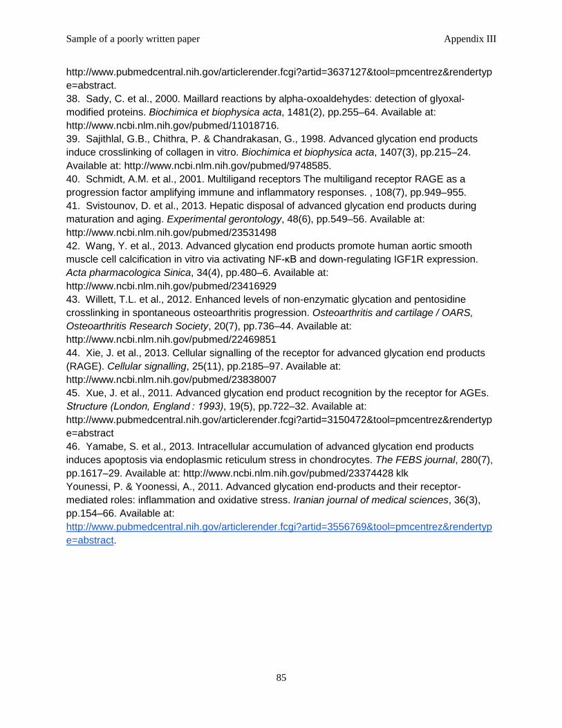
Sample of a poorly written paper Appendix III
85
http://www.pubmedcentral.nih.gov/articlerender.fcgi?artid=3637127&tool=pmcentrez&rendertype=abstract. 38. Sady, C. et al., 2000. Maillard reactions by alpha-oxoaldehydes: detection of glyoxal-modified proteins. Biochimica et biophysica acta, 1481(2), pp.255–64. Available at: http://www.ncbi.nlm.nih.gov/pubmed/11018716. 39. Sajithlal, G.B., Chithra, P. & Chandrakasan, G., 1998. Advanced glycation end products induce crosslinking of collagen in vitro. Biochimica et biophysica acta, 1407(3), pp.215–24. Available at: http://www.ncbi.nlm.nih.gov/pubmed/9748585. 40. Schmidt, A.M. et al., 2001. Multiligand receptors The multiligand receptor RAGE as a progression factor amplifying immune and inflammatory responses. , 108(7), pp.949–955. 41. Svistounov, D. et al., 2013. Hepatic disposal of advanced glycation end products during maturation and aging. Experimental gerontology, 48(6), pp.549–56. Available at: http://www.ncbi.nlm.nih.gov/pubmed/23531498 42. Wang, Y. et al., 2013. Advanced glycation end products promote human aortic smooth muscle cell calcification in vitro via activating NF-κB and down-regulating IGF1R expression. Acta pharmacologica Sinica, 34(4), pp.480–6. Available at: http://www.ncbi.nlm.nih.gov/pubmed/23416929 43. Willett, T.L. et al., 2012. Enhanced levels of non-enzymatic glycation and pentosidine crosslinking in spontaneous osteoarthritis progression. Osteoarthritis and cartilage / OARS, Osteoarthritis Research Society, 20(7), pp.736–44. Available at: http://www.ncbi.nlm.nih.gov/pubmed/22469851 44. Xie, J. et al., 2013. Cellular signalling of the receptor for advanced glycation end products (RAGE). Cellular signalling, 25(11), pp.2185–97. Available at: http://www.ncbi.nlm.nih.gov/pubmed/23838007 45. Xue, J. et al., 2011. Advanced glycation end product recognition by the receptor for AGEs. Structure (London, England : 1993), 19(5), pp.722–32. Available at: http://www.pubmedcentral.nih.gov/articlerender.fcgi?artid=3150472&tool=pmcentrez&rendertype=abstract 46. Yamabe, S. et al., 2013. Intracellular accumulation of advanced glycation end products induces apoptosis via endoplasmic reticulum stress in chondrocytes. The FEBS journal, 280(7), pp.1617–29. Available at: http://www.ncbi.nlm.nih.gov/pubmed/23374428 klk Younessi, P. & Yoonessi, A., 2011. Advanced glycation end-products and their receptor-mediated roles: inflammation and oxidative stress. Iranian journal of medical sciences, 36(3), pp.154–66. Available at: http://www.pubmedcentral.nih.gov/articlerender.fcgi?artid=3556769&tool=pmcentrez&rendertype=abstract.
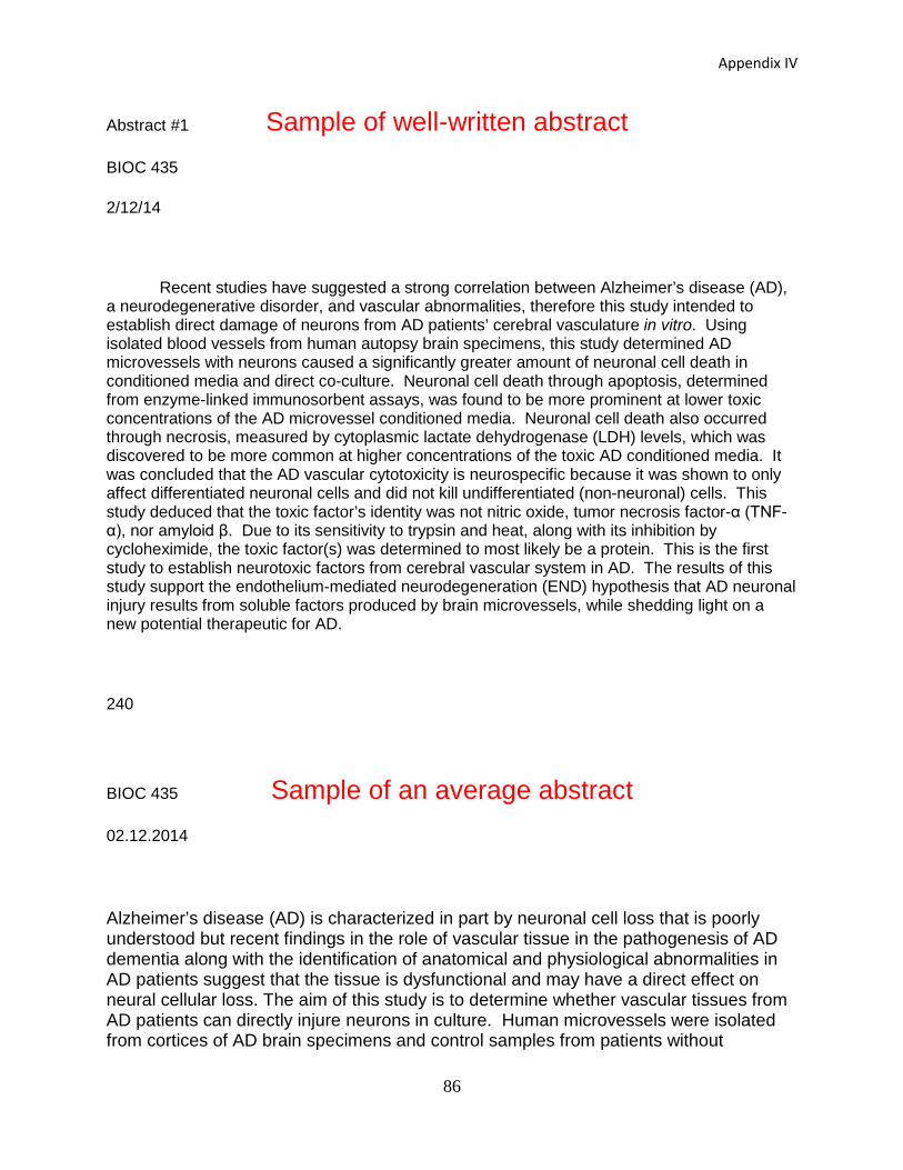
Appendix IV
86
Abstract #1 Sample of well-written abstractBIOC 435
2/12/14
Recent studies have suggested a strong correlation between Alzheimer’s disease (AD), a neurodegenerative disorder, and vascular abnormalities, therefore this study intended to establish direct damage of neurons from AD patients’ cerebral vasculature in vitro. Using isolated blood vessels from human autopsy brain specimens, this study determined AD microvessels with neurons caused a significantly greater amount of neuronal cell death in conditioned media and direct co-culture. Neuronal cell death through apoptosis, determined from enzyme-linked immunosorbent assays, was found to be more prominent at lower toxic concentrations of the AD microvessel conditioned media. Neuronal cell death also occurred through necrosis, measured by cytoplasmic lactate dehydrogenase (LDH) levels, which was discovered to be more common at higher concentrations of the toxic AD conditioned media. It was concluded that the AD vascular cytotoxicity is neurospecific because it was shown to only affect differentiated neuronal cells and did not kill undifferentiated (non-neuronal) cells. This study deduced that the toxic factor’s identity was not nitric oxide, tumor necrosis factor-α (TNF-α), nor amyloid β. Due to its sensitivity to trypsin and heat, along with its inhibition by cycloheximide, the toxic factor(s) was determined to most likely be a protein. This is the first study to establish neurotoxic factors from cerebral vascular system in AD. The results of this study support the endothelium-mediated neurodegeneration (END) hypothesis that AD neuronal injury results from soluble factors produced by brain microvessels, while shedding light on a new potential therapeutic for AD.
240
BIOC 435 Sample of an average abstract02.12.2014
Alzheimer’s disease (AD) is characterized in part by neuronal cell loss that is poorly understood but recent findings in the role of vascular tissue in the pathogenesis of AD dementia along with the identification of anatomical and physiological abnormalities in AD patients suggest that the tissue is dysfunctional and may have a direct effect on neural cellular loss. The aim of this study is to determine whether vascular tissues from AD patients can directly injure neurons in culture. Human microvessels were isolated from cortices of AD brain specimens and control samples from patients without
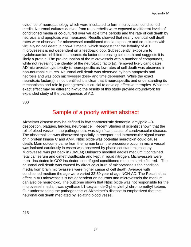
Appendix IV
87
evidence of neuropathology which were incubated to form microvessel-conditioned media. Neuronal cultures derived from rat cerebella were exposed to different levels of conditioned media or co-cultured over variable time periods and the rate of cell death by necrosis and apoptosis was measured. Results showed that nearly identical cell death rates were observed for microvessel-conditioned media exposure and co-cultures with virtually no cell death in non-AD media, which suggest that the lethality of AD microvessels is not dependent on a feedback loop. Subsequently, exposure to cyclohexamide inhibited the neurotoxic factor decreasing cell death and suggests it is likely a protein. The pre-incubation of the microvessels with a number of compounds, while not revealing the identity of the neurotoxic factor(s), removed likely candidates. AD microvessel cytotoxicity is neurospecific as low rates of cell death was observed in non-neuronal cultures. Neuronal cell death was observed by both apoptosis and necrosis and was both microvessel dose- and time dependent. While the exact neurotoxic factor(s) is not identified it is clear that it neurospecific and understanding its mechanisms and role in pathogenesis is crucial to develop effective therapies. While the exact effect may be different in-vivo the results of this study provide groundwork for expanded study of the pathogenesis of AD.
300
Sample of a poorly written abstract Alzheimer disease may be defined in few characteristic dementia, amylpoid –B- despostion, plaques, tangles, neuronal cell. Recent Studies of scientist shown that the roll of blood vessel in the pathogenesis was significant cause of cerebvascular disease. The abnormalities was discovered specially in receptor and intravascular signal cause of in protein kinase C and AMP. Nitric oxide was potential neurotoxin could cause death. Main outcome came from the human brain the procedure occur in micro vessel was isolated cautiously in essen was observed by phase constant microscopy. Microvessel was put back in (DMEM) Dulbucco modified eagles medium it contained fetal calf serum and dimethylsulfoxide and kept in liquid nitrogen. Microvessels were then incubated in CO2 incubator, centrifuged conditioned medium sterile filtered. The neuronal cell death was caused by direct co culture of micorvasssels the condition media from brain microvassels were higher cause of cell death. Average with conditioned medium the age were varied 32-59 year of age NON-AD. The Result lethal effect in AD micovessels is not dependent on neurons and microvessels the medium can also be neurotoxic. The outcome shown that Nitric oxide was not responsible for the microvessel media it was synthase L1-tosylamide-2-phenylethyl chromomethyl ketone. Our understanding the pathogenesis of Alzheimer’s disease to emphasized that the neuronal cell death mediated by isolating blood vessel.
215
Class format 1. I give 12 lectures.2. Students present a paper between each lecture on topic that I discussed in prev. day.3. Students work on review paper and writing assignments throughout semester.4. After spring break, all class time is used for student presentations (2/day).
ACE 10 course
Appendix V

Why use JITT???
• Most topics in the course have not been covered in previous courses
• Most topics are very relevant for the world around them
• Importance of biochemistry in daily life
Appendix V
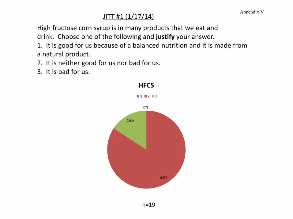
High fructose corn syrup is in many products that we eat and drink. Choose one of the following and justify your answer. 1. It is good for us because of a balanced nutrition and it is made froma natural product. 2. It is neither good for us nor bad for us.3. It is bad for us.
JITT #1 (1/17/14) Appendix V
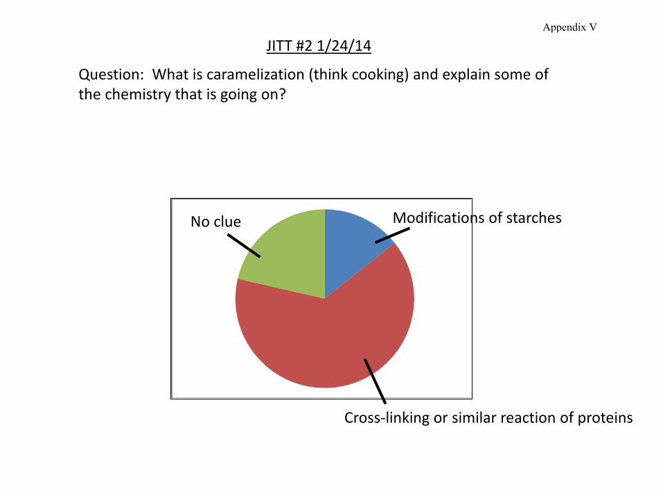
Question: What is caramelization (think cooking) and explain some of the chemistry that is going on?
Cross-linking or similar reaction of proteins
No clue Modifications of starches
JITT #2 1/24/14 Appendix V
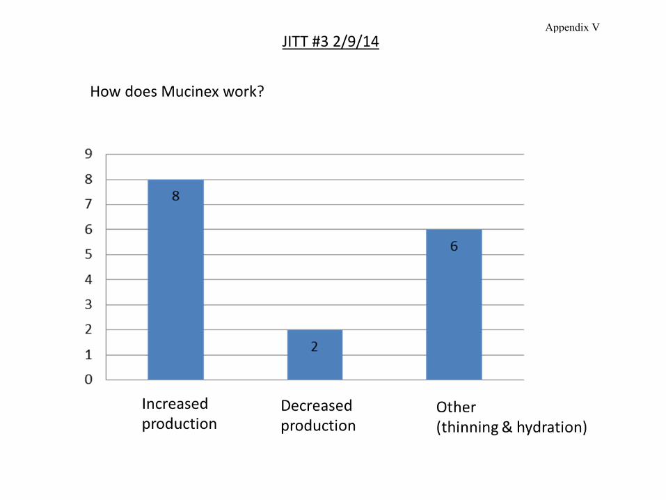
How does Mucinex work?
JITT #3 2/9/14 Appendix V
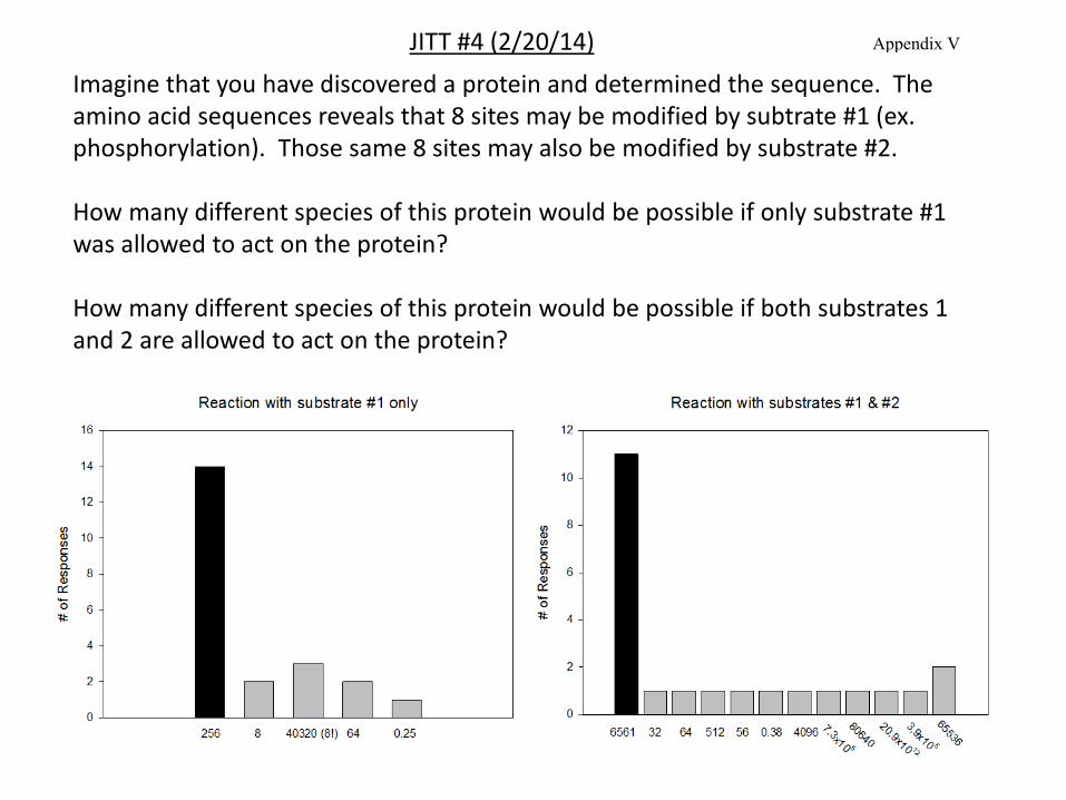
Imagine that you have discovered a protein and determined the sequence. The amino acid sequences reveals that 8 sites may be modified by subtrate #1 (ex. phosphorylation). Those same 8 sites may also be modified by substrate #2.
How many different species of this protein would be possible if only substrate #1 was allowed to act on the protein?
How many different species of this protein would be possible if both substrates 1 and 2 are allowed to act on the protein?
JITT #4 (2/20/14) Appendix V
Outcomes
• Over 75% of the students thought the JITT helped them better prepare for thelecture.
• 1/24 asked it to stop and thought it was just busy work.
• Very weak to no relevance for exam scores (n=1).
• Students want answers explained so plan to spend time on that.
Appendix V