bioactive implants in cervical spine injury – original
TRANSCRIPT

Global Journal of Medical research: J Dentistry and Otolaryngology Volume 14 Issue 1 Version 1.0 Year 2014 Type: Double Blind Peer Reviewed International Research Journal Publisher: Global Journals Inc. (USA) Online ISSN: 2249-4618 & Print ISSN: 0975-5888
Bioactive Implants in Cervical Spine Injury – Original Research(From 1995 To 2011)
By M.Filip, F. Šámal, P.Linzer, P.Jurek & J.Strnad Neurosurgical department KNTB Zlín, Czech Republic Abstract- Objectives: The paper deals with the development and clinical evaluation of a new bioactive implant designed for anterior cervical interbody fusion (ACIF) in the surgical treatment of unstable injury in subaxial part of cervical spine (type A2, 3 and B3 fractures according to Aebi and Nazarian classification). Significance of the topic: In the middle of the nineties of the last century the glass-ceramic prosthesis BAS-0 made it possible to gain the first experiences in materials replacing allografts for ACIF. Its major disadvantage lay in insufficient resistance. Given these complications, we searched for a stronger material while maintaining the bioactive properties of the glass-ceramics. Bioactive titanium with a special surface treatment by the company LASAK proved to be such a material. New Implant suitable for ACIF was developed in the year 2003. This type was introduced into clinical practice in 2004 after experimental mathematical verification of the design and cadaver testing. Brief methodology: The new implant has a basic shape of a full truncated prism narrowed by 1 degree towards the spinal canal; its length is 13-15 mm with a graded height of 8-5 mm and width of 13 mm. We have used this implant successfully in the treatment of patients with cervical spine injury in unstable fractures. It was indicated the anterior decompression of the spinal canal with interbody fusion together with plate systems.
GJMR-J Classification : NLMC Code:WE 725
BioactiveImplantsinCervicalSpineInjuryOriginalResearchFrom1995To2011
Strictly as per the compliance and regulations of:
© 2014. M.Filip, F. Šámal, P.Linzer, P.Jurek & J.Strnad. This is a research/review paper, distributed under the terms of the Creative Commons Attribution-Noncommercial 3.0 Unported License http://creativecommons.org/licenses/by-nc/3.0/), permitting all non-commercial use, distribution, and reproduction inany medium, provided the original work is properly cited.
Bioactive Implants in Cervical Spine Injury –Original Research(From 1995 To 2011)
M.Filip α, F. Šámal σ, P.Linzer ρ P.J urek Ѡ & J.Strnad ¥
Abstract- Objectives: The paper deals with the development and clinical evaluation of a new bioactive implant designed for anterior cervical interbody fusion (ACIF) in the surgical treatment of unstable injury in subaxial part of cervical spine (type A2, 3 and B3 fractures according to Aebi and Nazarian classification).
Significance of the topic: In the middle of the nineties of the last century the glass-ceramic prosthesis BAS-0 made it possible to gain the first experiences in materials replacing allografts for ACIF. Its major disadvantage lay in insufficient resistance. Given these complications, we searched for a stronger material while maintaining the bioactive properties of the glass-ceramics. Bioactive titanium with a special surface treatment by the company LASAK proved to be such a material. New Implant suitable for ACIF was developed in the year 2003. This type was introduced into clinical practice in 2004 after experimental mathematical verification of the design and cadaver testing.
Brief methodology: The new implant has a basic shape of a full truncated prism narrowed by 1 degree towards the spinal canal; its length is 13-15 mm with a graded height of 8-5 mm and width of 13 mm. We have used this implant successfully in the treatment of patients with cervical spine injury in unstable fractures. It was indicated the anterior decompression of the spinal canal with interbody fusion together with plate systems.
Results: During the years 2006 – 2011 we operated 26 patients with unstable fractures in subaxial cervical spine. We performed successful surgery using new bioactive titanium implant in 12 patients. The outcomes were evaluated according to the standard criteria used in this kind of operations (clinical scoring schemes, radiological imaging) with a follow-up of at least 1 year.
Conclusion: When comparing the operation techniques using different types of implants to our implants we found one significant difference. Thanks to the new shape and bioactive properties of the surface it is not necessary to fill it with further material.
I. Introduction
njuries of the lower cervical spine occurs as monotrauma or compound injury. They are rarely caused by only direct force on the spinal structures.
Typically there is an indirect injury of spinal segment due to non-physiological forces (compression, flexion, extension or rotation). Cervical spine injuries result in the spine segment instability which poses a threat to the Author α σ ρ Ѡ: Neurosurgical department KNTB Zlín, Czech Republic. Author ¥: Research and Development Center for Dental Implantology and Tissue Regeneration, LASAK Ltd Prague, Czech Republic. e-mail: [email protected]
nerve structures of the spinal canal (spinal cord, roots) (Aebi 1991, Bohlman 1979, Caspar 1989). Modern classifications of lower cervical spine injuries respect these pathological anatomical characteristics and determine the level of injury severity and the prognosis. Detailed and frequently used classification by Aebi and Nazarian (Aebi 1987) divides injuries into type A, type B and type C and into groups and subgroups 1 to 3, and respects the extent of traumatic instability or residual stability, distinguishes anterior and posterior column of the spine and differentiates between mostly osseous, mostly ligamentous, and combined injury. Conventional X-ray and CT examinations are needed for the determination of injury classification. In many cases it is also necessary to add MRI examination to determine the damage to the soft tissues – ligaments, joint capsules and intervertebral discs. Depending on whether the injury is classified as stable or unstable, a decision is made about the management (surgery/conservative therapy). Surgical intervention is required for unstable spine injuries (Bohlman 1992, Fehlings 2005. Kandziora 2005, Osti 1989, Štulík 2003 ). It allows stabilization and decompression of the spinal cord and reconstruction of the anatomical structures of the spine to prevent secondary damage to the spinal cord and late post-traumatic changes. It is not possible to heal the "unstable" type of injury using conservative manag-ement. The most common surgical technique in ligamentous (A3, B3, C3) and osteoligamentous injuries (A2, C2) is anterior approach using a plate and the anterior cervical interbody fusion (ACIF) similarly as in degenerative cervical spine disease ( Norrell 1970, Perret 1968, Caspar 1989, Connoly 1996 ).
In 1960 Bailey (Bailey 1960) and then Robinson and Southwick published their first experience with surgical treatment of lower cervical spine injuries using the anterior approach technique described between 1955 and1958 by Robinson and Cloward for the treatment of degenerative diseases (Cloward 1958 ). Standard surgical procedure includes decompression of the spinal canal (reduction of luxation, removal of damaged intervertebral disk, etc.), anterior cervical interbody fusion using bone grafts and fixating the operated segment with a plate and monocortical or bicortical screws. Because of problems associated mainly with bone graft harvest (Banvart 1994, Hrabálek 2007) implants designed for use in ACIF made of various materials (glass-ceramics, titanium, PEEK,
I -
Globa
l Jo
urna
l of M
edical R
esea
rch
15
Volum
e XIV
Issu
e I V
ersio
n I
© 2014 Global Journals Inc. (US)
(DDDD)
Year
J20
14

polylactide) have been developed since the 80´s of the last century (Yamamuro 1995, Matge 1998, Filip 2000, Cho D 2002, McConnell 2003,Vaccaro 2002). They should eliminate the problems inherent to bone grafts and copy as much as possible the biological properties of bone tissue. Based on biomechanical studies we have developed an implant made of bioactive glass-ceramics in the first half of 1990´s. Its strength parameters and bioactive properties simulated bone tissue (Kokubo 1982, Bienik 1991, Urban1992). In clinical practice it gradually replaced bone grafts in surgical treatment of degenerative disease (Filip 2000 ) and unstable, mainly osteoligamentous injuries to the lower cervical spine. At the Neurosurgical Department of the University Hospital in Ostrava we operated 10 patients with cervical spine injuries using this implant supplemented with a plate fixed by screws during the period of 1997 to 1999. Neurological findings improved by one grade on the Frankel scale in 3 patients. According to imaging examinations (RTG, CT) no dislocation of glass-ceramics implant occurred after a period of one year and more since the time of the operation. After two months, we observed in two operated that a screw in the plate became partially loose without a need for re-operation. The main advantages for the patients included mainly shorter time of the operation and elimination of complications associated with the bone graft harvest. Bioactive properties of the surface contributed to bone fusion without supplementing additional material. Implant fragility was the main disadvantage (Filip 2000). During the application there was a risk of damage to the implant by the contact with metal instruments. In 2003–2004 we eliminated this disadvantage by developing an implant made of a new material – bioactive titanium. It has shown several times higher strength while retaining its bioactive properties as a result of a special surface treatment [Strnad 2001]. We have gradually implemented this to the clinical practice for the same indications as in case of the preceding glass-ceramics implant. In 2007–2011 we used it at Neurosurgical Department of KNTB Zlín in 12 of 34 patients who underwent anterior approach surgery due to unstable injury to the lower cervical spine. Compared to the glass ceramic implant the new implant handling during the surgical procedure was easier without a risk of damage. Its shape and bioactive properties contributed to bone fusion without the need of additional material (Filip 2005, Filip 2010). In the monitored post-operative period of at least one year neither any dislocation nor deterioration in the clinical condition was observed in a set of all 34 operated patients. Cage implants made of polylactide or PEEK (Vaccaro 2002, Hacker 2000, ChoD 2002, Matge 2002, Suchomel 2004) were applied to the remaining 22 patients who were operated in the same period. Their cavity needed to be filled with additional material (bone, BCP, TCP) to initiate interbody fusion. Compared with
the application of a titanium implant with bioactive surface, cage implants with filling material are more demanding with regards to their insertion, which prolongs the duration of the operation.
II. Material and Methodology a)
Implant for use in ACIF made of glass-ceramics BAS-O (1996–1999)
In 1997 we used an implant made of bioactive glass-ceramics for
ACIF in unstable injury to the lower
cervical spine as an equivalent replacement of autologous bone drafts (Kokubo 1982,Urban 1992,Yamamuro 1995). It imitated bone tissue properties by its mechanical strength and bioactive properties. In vertical compression glass-ceramics exceeded twice the strength of cortical bone tissue and it was identical in bending strength. Disadvantage of BAS-O glass-ceramics is its fragility causing problems in optimizing the implant shape during biomechanical modelling. Based on mathematical studies we have retained the implant´s shape as a tapered prism with the following dimensions: length 15mm, height 7.8mm ventrally and 6.9mm dorsally, and width 13mm. Strength parameters of this shape exceeded the strength of an autograft (see Figure 1, Figure 2).
Figure 1 : Drawing of a cervical implant for use in ACIF made of bioactive ceramics (1994)
Figure 2 : Cervical implant for use in ACIF in clinical
practice (1996) Its bioactive properties were the result of an
active chemical bond initiated by hydroxyapatite surface with surrounding bone tissue that developed within 48 hours and then the migration of osteoblasts over the surface to create bone fusion within 2 to 3 months by
Bioactive Implants in Cervical Spine Injury – Original Research(From 1995 To 2011)Globa
l Jo
urna
l of M
edical R
esea
rch
© 2014 Global Journals Inc. (US)
Volum
e XIV
Issu
e I V
ersio
n I
Year
()
2014
J
connecting the surrounding vertebral bodies (see Figure 3).
16
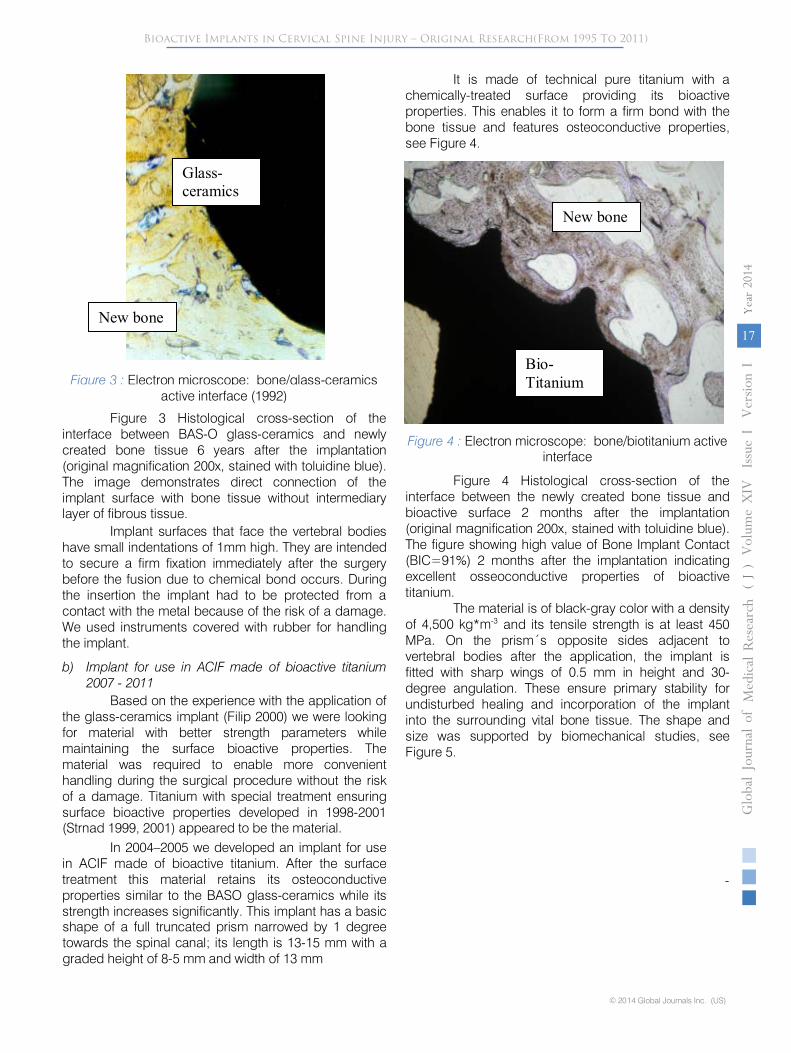
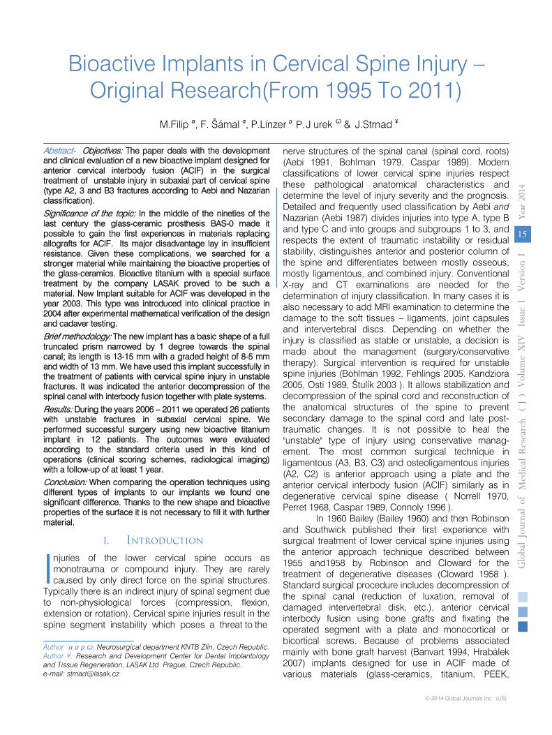
Figure 3 :
Electron microscope: bone/glass-ceramics active interface (1992)
Figure 3
Histological cross-section of the interface between BAS-O glass-ceramics and newly created bone tissue 6 years after the implantation (original magnification 200x, stained with toluidine blue). The image demonstrates direct connection of the implant surface with bone tissue without intermediary layer of fibrous tissue.
Implant surfaces that face the vertebral bodies
have small indentations of 1mm high. They are intended to secure a firm fixation immediately after the surgery before the fusion due to chemical bond occurs. During the insertion the implant had to be protected
from a contact with the metal because of the risk of a damage. We used instruments covered with rubber for handling the implant.
b)
Implant for use in ACIF made of bioactive titanium 2007 -
2011
Based on the experience with the application of
the glass-ceramics implant (Filip 2000) we were looking for material with better strength parameters while maintaining the surface bioactive properties. The material was required to enable more convenient handling during the surgical procedure without the risk
of a damage. Titanium with special treatment ensuring surface bioactive properties developed in 1998-2001 (Strnad 1999, 2001) appeared to be the material.
In 2004–2005 we developed an implant for use in ACIF made of bioactive titanium. After the surface treatment this material retains its osteoconductive properties similar to the BASO glass-ceramics while its strength increases significantly. This implant has a basic shape of a full truncated prism narrowed by 1 degree towards the spinal canal; its length is 13-15 mm with a graded height of 8-5 mm and width of 13 mm
It is made of technical pure titanium with a chemically-treated surface providing its bioactive properties. This enables it to form a firm bond with the bone tissue and features osteoconductive
properties, see Figure 4.
Figure 4 :
Electron microscope: bone/biotitanium active interface
Figure 4
Histological cross-section of the interface between the newly created bone tissue and bioactive surface 2 months after the implantation (original magnification 200x, stained with toluidine blue). The figure showing high value of Bone Implant Contact (BIC=91%) 2 months after the implantation indicating excellent osseoconductive properties of bioactive titanium.
The material is of black-gray color with a density of 4,500 kg*m-3
and its tensile strength is at least 450 MPa. On the prism´s opposite sides adjacent to vertebral bodies after the application, the implant is fitted with sharp wings of 0.5 mm in height and 30-degree angulation. These ensure primary stability for undisturbed healing and incorporation of the implant into the surrounding vital bone tissue. The shape and size was supported by biomechanical studies, see Figure 5.
New bone
Glass-ceramics
Bio-Titanium
New bone
Bioactive Implants in Cervical Spine Injury – Original Research(From 1995 To 2011)
-
Globa
l Jo
urna
l of M
edical R
esea
rch
Volum
e XIV
Issu
e I V
ersio
n I
© 2014 Global Journals Inc. (US)
(DDDD)
Year
J20
14
17

Figure 5
: Model evaluation of shape and strength of wings (2003)
Figure 6 : Cervical implant made of bioactive titanium (2004)
III. Results
In both types of implants (glass-ceramics/bioactive titanium) developed by us we indicated patients for the operation according to the instability of the injured lower spine defined in the preoperational stage according to the imaging methods (X-ray, MRI, CT) and using the classification according
to Aebi and Nazarian and according to the neurological findings using the Frankel scale. We carried out the surgery by the Caspar technique (Caspar 1989, Klézl 1999). Under general anesthesia from the prevertebral
incision and after exposing anterior surface of the veretbral bodies we removed the structures compressing the spinal canal (intervertebral disk, posterior ligament residues, fragments of the edges of
Bioactive Implants in Cervical Spine Injury – Original Research(From 1995 To 2011)Globa
l Jo
urna
l of M
edical R
esea
rch
© 2014 Global Journals Inc. (US)
Volum
e XIV
Issu
e I V
ersio
n I
Year
()
2014
J
These studies supplemented by clinical experience with the glass-ceramics implant resulted inthe creation of a new implant for use in ACIF, see Figure 6.
18

the vertebral bodies, haematoma, etc.) using an operating microscope. Then we prepared a bed for inserting the implant into the interbody space. We removed the endplates from the vertebral bodies and
exposed cancellous bone. In traction and using the Caspar´s instrumentarium we inserted the implant into the interbody space under the control of X-ray, see Figure 7.
Figure 7 :
Inserting the bio-titanium implant into the interbody space C5/6 using the X-ray control
After releasing traction and checking the
position on X-ray we fixed the impaired segment by a plate secured with monocortical or bicortical screws into the neighboring vertebral bodies. Surgical procedure is
similar for both the glass-ceramics implant and
the bio-titanium implant. We used the same surgical procedure for other types of implants as well (polyactide/PEEK).
Figure 8
: Implant for use in ACIF (PEEK/TCP) 2010
We carried out verticalization in operated patients in case of all implants on the first post-operative day in a collar for a period of 6 weeks until the expected bone fusion occurrence.
At Neurosurgery Department of the University Hospital in Ostrava we operated 10 patients with unstable injury to the lower cervical spine using glass-ceramics implants between 1997 and 1999, see Figures 9 and 10.
Bioactive Implants in Cervical Spine Injury – Original Research(From 1995 To 2011)
Globa
l Jo
urna
l of M
edical R
esea
rch
Volum
e XIV
Issu
e I V
ersio
n I
© 2014 Global Journals Inc. (US)
(DDDD)
Year
J20
14
19

Figure 9 : X-ray after fixation of C6/7 due to unstable injury (Aebi-Nazarian – A3) 12 months after the surgery – glass-ceramics BAS-O (1997)
Figure 10 : CT after fixation of C6/7 12 months following the surgery – glass-ceramics BASO (1998) The implant for use in ACIF made of glass-
ceramics fulfilled our expectations. It removed complications associated with bone graft harvest and due to its shape and bioactive properties it enabled a chemical bond with surrounding osseous tissue to create bone fusion without a need for filling with other material (Bienik 1991,Madawi 1996, Filip 2000). Its disadvantages included fragility in contact with metal and threshold bending strength. These disadvantages were eliminated by a new implant made of bio-titanium that we introduced into clinical practice for identical indications in 2004. In years 2007-2011 at the Neurosurgery Department of KNTB Zlín we operated 34 patients with unstable lower cervical spine injury. In 12 patients we used a bio-titanium implant in ACIF (Figures 11 and 12). In 22 patients we used an implant made of different materials (Figure 13).
Bioactive Implants in Cervical Spine Injury – Original Research(From 1995 To 2011)Globa
l Jo
urna
l of M
edical R
esea
rch
© 2014 Global Journals Inc. (US)
Volum
e XIV
Issu
e I V
ersio
n I
Year
()
2014
J
20

Figure 11 : Unstable injury C6/7 (Aebi-Nazarian – A3) (2009)
Trauma MRI- T2, X-ray 6 months after the surgery – bioactive titanium
Figure 12 : Unstable injury C6/7 (Aebi-Nazarian –
C3) (2010)
Trauma MRI-T X-ray 12 months after the surgery – bioactive titanium
Figure 13
: X-ray image 6 months after the surgery on C6/7 –PEEK/BCP (2011)
Bioactive Implants in Cervical Spine Injury – Original Research(From 1995 To 2011)
Globa
l Jo
urna
l of M
edical R
esea
rch
21
Volum
e XIV
Issu
e I V
ersio
n I
© 2014 Global Journals Inc. (US)
(DDDD)
Year
J20
14

In our own set of patients we evaluated the neurological finding according to the Frankel scale with a finding from the imaging methods (X-ray, CT, MRI) preoperative and 2, 6 and 12 months after the surgery.
We indicated the actual surgical approach (ACIF + plate) according to the type of traumatic instability from the imaging examinations evaluated according to Aebi-Nazarian (Table 1). Table 1
Classification according to Aebi-Nazarian in our patients
Glass-ceramics + Aesculap plate
(1996–1999)
Bioactive titanium + plate (Zephire,Venture, Reflex, Eagle)
(2007–2011)
Polylactide/BCP + plate (Zephire, Eagle)
(2007–2011)
PEEK/TCP + plate (Zephire, Reflex,
Eagle) (2007–2011)
A2
1
3
1
2 A3
2
2
3
3
C2
3
2
4
1 C3
4
5
3
5
The most common type of unstable injury operated using the ACIF approach with a plate and all types of implants was diagnosed as osteoligamentous
injury type A (about 35%) and type C (about 65%). We evaluated the neurological finding
according to the Frankel scale (A– Complete lesions, B
– Preserved sensitivity only,
C –
Preserved non-
functional motorics, D –
Preserved sensitivity and
functional motorics, E –
No lesions) before the surgery and 12 months after the surgery (Table 2).
Table 2 shows that improvement in the
neurological finding 12 months after the surgery occurred regardless of the implant type in 30% of patients (28–32%) by at least one grade of the Frankel scale, most frequently in incomplete spinal lesions.
Table 2
Neurological lesions according to the Frankel system
preoperative/12 months postoperative
Glass-ceramics (1996–1999)
Bioactive titanium (2007–2011)
Polylactide/BCP (2007–2011)
PEEK/TCP (2007–2011)
A
2/2
2/1
1/1
2/1 B
3/1
2/2
2/1
2/2
C
2/3
2/1
3/3
3/3 D
1/2
3/4
4/4
2/3
E
2/2
3/3
1/2
2/2 Number of improved
3 (30%)
4 (32%)
3 (28%)
3 (28%)
In addition to the neurological finding we also evaluated findings from imaging examinations performed 2, 6 and 12 months after the surgery.
Here we focused on a change in the implant position (ventral or dorsal dislocation and sinking into the vertebral bodies) and signs of instability (reduced density of bone tissue surrounding the implant, plate loosening).
Using postoperative imaging methods (X-ray, CT) we did not observe any dislocation or instability signs in the used implants in the entire group of patients. In two
patients
(glass
- ceramics) partial
loosening of screws in the plate was observed without the implant or the plate being dislocated. Steady position of fixation on images correlates with postoperative evaluation of neurological lesion according to the Frankel scale (30% of improved patients).
Complications associated with the surgical procedure (secondary healing of surgical wound, temporary paresis of the recurrent laryngeal nerve, permanent partial paresis of the recurrent laryngeal nerve) which we observed in our group is shown in
Bioactive Implants in Cervical Spine Injury – Original Research(From 1995 To 2011)Globa
l Jo
urna
l of M
edical R
esea
rch
© 2014 Global Journals Inc. (US)
Volum
e XIV
Issu
e I V
ersio
n I
Year
()
2014
J
22
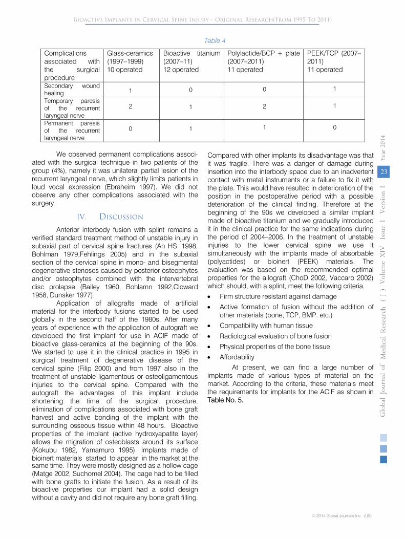
Table 4
Complications associated with the surgical procedure
Glass-ceramics (1997–1999) 10 operated
Bioactive titanium (2007–11) 12 operated
Polylactide/BCP + plate (2007–2011) 11 operated
PEEK/TCP (2007–2011) 11 operated
Secondary wound healing 1
0
0 1
Temporary paresis of the recurrent laryngeal nerve
2
1
2
1
Permanent paresis of the recurrent laryngeal nerve
0
1
1
0
We observed permanent complications associ-
ated with the surgical technique in two patients of the group (4%), namely it was unilateral partial lesion of the recurrent laryngeal nerve, which slightly limits patients in loud vocal expression (Ebraheim 1997). We did not observe any other complications associated with the surgery.
IV. Discussion
Anterior interbody fusion with splint remains a verified standard treatment method of unstable injury in subaxial part of cervical spine fractures (An HS. 1998, Bohlman 1979,Fehlings 2005) and in the subaxial section of the cervical spine in mono- and bisegmental degenerative stenoses caused by posterior osteophytes and/or osteophytes combined with the intervertebral disc prolapse (Bailey 1960, Bohlamn 1992,Cloward 1958, Dunsker 1977).
Application of allografts made of artificial material for the interbody fusions started to be used globally in the second half of the 1980s. After many years of experience with the application of autograft we developed the first implant for use in ACIF made of bioactive glass-ceramics at the beginning of the 90s. We started to use it in the clinical practice in 1995 in surgical treatment of degenerative disease of the cervical spine (Filip 2000) and from 1997 also in the treatment of unstable ligamentous or osteoligamentous injuries to the cervical spine. Compared with the autograft the advantages of this implant include shortening the time of the surgical procedure, elimination of complications associated with bone graft harvest and active bonding of the implant with the surrounding osseous tissue within 48 hours. Bioactive properties of the implant (active hydroxyapatite layer) allows the migration of osteoblasts around its surface (Kokubu 1982, Yamamuro 1995). Implants made of bioinert materials started to appear in the market at the
Compared with other implants its disadvantage was that it was fragile. There was a danger of damage during insertion into the interbody space due to an inadvertent contact with metal instruments or a failure to fix it with the plate. This would have resulted in deterioration of the position in the postoperative period with a possible deterioration of the clinical finding. Therefore at the beginning of the 90s we developed a similar implant made of bioactive titanium and we gradually introduced it in the clinical practice for the same indications during the period of 2004–2006. In the treatment of unstable injuries to the lower cervical spine we use it
simultaneously with the implants made of absorbable (polyactides) or bioinert (PEEK) materials. The evaluation was based on the recommended optimal properties for the allograft (ChoD 2002, Vaccaro 2002) which should, with a splint, meet the following criteria.
•
Firm structure resistant against damage
•
Active formation of fusion without the addition of other materials (bone, TCP, BMP. etc.)
•
Compatibility with human tissue
•
Radiological evaluation of bone fusion
•
Physical properties of the bone tissue
•
Affordability
At present, we can find a large number of implants made of various types of material on the market. According to the criteria, these materials meet the requirements for implants for the ACIF as shown in Table No. 5.
Bioactive Implants in Cervical Spine Injury – Original Research(From 1995 To 2011)
Globa
l Jo
urna
l of M
edical R
esea
rch
Volum
e XIV
Issu
e I V
ersio
n I
© 2014 Global Journals Inc. (US)
(DDDD)
Year
J20
14
same time. They were mostly designed as a hollow cage (Matge 2002, Suchomel 2004). The cage had to be filled with bone grafts to initiate the fusion. As a result of its bioactive properties our implant had a solid design without a cavity and did not require any bone graft filling.
23

Table 5 : Comparison of material properties for ACIF Material properties PEEK Glass-
ceramics
Polylactide Biotitanium
1. Rigid support +/- - +/- + 2.Active formation of fusion (Osteoconduction) - + - + 3. Compatibility with human tissue + + + + 4. Radiological rating of fusion + + + - 5. Physical and biochemical properties of bones + +/- +/- +/- 6. Affordability +/- + +/- +
From the table above it follows that, when
compared to other materials, the properties of bioactive titanium make it a very-close-to-optimum material for ACIF.
Out of all the properties, the emphasis must be on the bioactivity of the overall surface of the biotitanium implant specified in point 2 of the table. Bioactivity enables the osteoconduction of bone cells at the implant/bone interface with their subsequent migration over the implant surface (Strnad 1999,2001 Filip 2010). Most of the other implants do not have this property. Only glass-ceramics have similar bioactive properties, however without sufficient strength parameters. The active formation of fusion is enabled by the surface treatment of the titanium using the technology, as mentioned in the Material and Methodology section. It enables the new formation of bone cells and their migration on the implant surface, as we have verified using the CT, see Figure 14.
Figure 14 : CT after 12 months with signs of migration of
osteoblasts along the anterior and lateral walls of the implant, section C5/6
Hence there is no necessity to fill the implant inside with supplementary material (bone / artificial
material) as is the case with the other implants (Hacker
2000, Matge 2002, Suchomel 2004,Kandziora 2005). Its application is, therefore, made easier and the state of
the operated-on patient is not impaired when expanding
the surgery time by taking an autograft or preparing an implant with filling. This results in a lower surgical burden and better affordability. The other implants do not have this property. They are in the shape of hollow cages increasing only mechanical strength without any bioactivity of the material itself. To develop fusion the hollow of the cage must be filled with one of bioactive materials (BCP, TCP, BMP).
The chemical bond and the subsequent interbody fusion develop only in the contact area of bone/supplementary material outside the implant itself. If, for various reasons, the filling homogeneity is impaired, the fusion formation may be slowed down or stopped with the development of later instability in the operated-on region. Regarding the other properties, biotitanium is not significantly different from the other materials as seen in table No. 5.
Another benefit of our implant compared to the other ones is its shape of a full truncated prism in different sizes with surface treatment on the opposite sides. This provides primary stability minimizing the danger of migration in all directions. It gives a better chance to maintain the cervical spine lordosis in the postoperative period compared to some other implants of a shape without truncation. Implant dislocation endangers the operated-on patient by new instability with compression of the spinal canal and by worsening of the clinical findings. Due to its surface bioactivity, our implant has no hollow in the shape of an oval or square. When comparing the operation techniques using different types of implants to our implants we did not find any significant differences. Always the Smith-Robinson technique with splint with Caspar instrumentation is used. The only difference is seen in simpler handling during the surgery. Thanks to the bioactive properties of the surface it is not necessary to fill it with further material. This shortens the surgery time as well as the surgery burden on the operated-on patient.
New bone on the lateral wall of biotitanium implant (space C5/6)
Bioactive Implants in Cervical Spine Injury – Original Research(From 1995 To 2011)Globa
l Jo
urna
l of M
edical R
esea
rch
© 2014 Global Journals Inc. (US)
Volum
e XIV
Issu
e I V
ersio
n I
Year
()
2014
J
24

V. Conclusion
It follows from the results above that the our implant from bioactive titanium is a good alternative for operation treatment of unstable injury in subaxial part of cervical spine to the anterior cervical interbody fusion with splint. Regarding the quality and price it successfully competes with the other products for ACIF. This has been proven by clinical evaluation of our group by Frankel scale (30%) improve surgery within the interval of 12 months after surgery in all types of implants supplemented by imaging examinations (X-ray, CT).
VI. Bibliography
1. Aebi M, Zuber K, Marchesi D. Treatment of Cervical Spine Injuries with Anterior Plating; Indications, Techniques, and Results. Spine 1991; 16(Suppl. 3): 38-45.
2. Aebi M., Nazarian S. Classification of injuries of the cervical spine. Orthopäde 1987; 16(1): 27-36.
3. An HS. Cervical Spine Trauma. Spine 1998; 23: 2713-2729.
4. Bailey RW., Badgley CE. Stabilization of the Cervical Spine by Anterior Fusion. J Bone Jt Surg 1960; 42-A: 565-594
5. BANVART, J., ASHER, M., HASSANEIN, R.: Iliac crest bone harvest donor site morbidity; a statistical evaluation. Spine. 1994;20:1055-1060
6. Bienik J,Swiecki Z.Porous and porous-compact ceramics in orthopedics.Clin Orthop 1991;27:88-94
7. Bohlman HH. Acute fractures and dislocations of the cervical spine. An analysis of three hundred hospitalized patients and review of the literature. J Bone Joint Surg Am 1979; 61 (8): 1119-1142.
8. Bohlman HH, Anderson PA. Anterior Decompression and Arthrodesis of the Cervical Spine: Long-Term Motor Improvement. J Bone Jt Surg 1992;74-A, 671-682.
9. Capen DA, Garland DE, Waters RL. Surgical Stabilization of the Cervical Spine. A Comparative Analysis of Anterior and Poterior Spine Fusions. Clin Orthop 1985; 196: 229-237.
10. Caspar W, Barbier DD, Klara PM. Anterior Cervical Fusion and Caspar Plate Stabilization for Cervical Trauma. Neurosurgery 1989; 25: 491-502.
11. Cloward RB: The anterior approach for removal of ruptured cervical discs. J Neurosurg. 1958;15: 602-617,.
12. Connoly PJ, Esses SI, Kostiuk JP. Anterior Cervical Fusion: Outcome Analysis of Patients Fused with and without Anterior Cervical Plates. J Spinal Disord 1996; 9: 202-206.
13. Dunsker SB: Anterior cervical discectomy with and without fusion. Clin Neurosurg. 24: 516-520, 1977.
14. Ebraheim NA, Lu J, Skie M, Heck B, Yeasting RA. Vulnerability of the Reccurent Laryngeal Nerve in the
Anterior Approach to the Lower Cervical Spine. Spine 1997; 22: 2664-2667.
15. Fehlings MG, Perrin RG. The role and timing of early decompression for cervical spinal cord injury: Update with a review of recent clinical evidence. Injury 2005; 36: 13-26.
16. Filip M., Veselský P., Paleček T., Wolný E. Glass-ceramics Prosthesis of an intervertebral disc in the degenerative diseases of the cervical spine – initial experiences. Čes a Slov Neurol a Neurochir 2000; 1: 31-36
17. Filip M, Veselský P, Mrůzek M, Paleček T, Strnad Z, Strnad J: Bioactive cage Implaspin in treatment of degenerative disease of cervical spine – First experiences. NEURO3 01:A01, 2005
18. Filip M.,Linzer P.,Šámal F.,Jurek P.,Strnad Z.,Strnad J.: Bioactive titan cage Implaspin in treatment of degenerative disease of the cervical spine-the results from 2007till 2008 Chirurgia narzadów ruchu i Ortopedia Polska 75 ( 1 ),69-73,2010
19. Hacker RJ, Cauthen JC, Gilbert TJ, Griffith SL: A prospective randomized multicenter clinical evaluation of an anterior cervical fusion cage. Spine. 25:2646-2655, 2000.
20. Hrabálek L., Vaverka M., Křupka B., Houdek M: Komplikace operací z předního přístupu pro degenerativní onemocnění krční páteře. Česká a slovenská neurologie a neurochirurgie, 70/103 (2), 23-28, 2007
21. Cho D, Liau W, Lee W, et al: Preliminary experience using a polyetheretherketone (PEEK) cage in the treatment of cervical disk disease. Neurosurgery. 51:1343-1350, 2002
22. Kandziora F, Pflugmacher R, Scholz M, Schnake K, Putzier, M, Khodadadyan-Klostermann C, Hass NP. Treatment of traumatic cervical spine instability with interbody fusion cages: prospective controlled study with a 2-year follow-up. Injury 2005; 36: 27-35.
23. KOKUBO, T., SHIGEMATSU, M., NAGHASHIMA, Y.: Apatite wollastonite-containing glass-ceramic for prosthetic application. Bull. Inst. Chem. Res., Kyoto Univ., 60: 260-268, 1982
24. Madawi AA, Powell M, Crockard HA: Biocompatible osteoconductive polymer versus iliac graft. Spine. 21:2123-2130, 1996
25. Matge G: Anterior interbody fusion with the BAK-cage in cervical spondylosis. Acta Neurochir. 140:18, 1998
26. Matge G: Cervical cage fusion with 5 different implants: 250 cases. Acta Neurochir. 2002; 144:539-550.
27. McConnell J, Freeman B, Dabnath U, et al: A prospective randomized comparison of coralline hydroxyapatite with autograft in cervical interbody fusion. Spine. 28:317-323, 2003
Bioactive Implants in Cervical Spine Injury – Original Research(From 1995 To 2011)
-
Globa
l Jo
urna
l of M
edical R
esea
rch
Volum
e XIV
Issu
e I V
ersio
n I
© 2014 Global Journals Inc. (US)
(DDDD)
Year
J20
14
25

28. McLellen D, Tew J, Mayfield FH: Complications of surgery of the anterior spine. Clin Neurosurg. 23:424-434,1976
29. Klézl Z, Fousek J, Pěkný I. Technika přední instrumentované spondylodézy krční páteře. Acta Chir orthop Traum čech 1999; 66: 158-164.
30. Norrell H, Wilson CB. Early Anterior Fusion for Injuries of the Cervical Portion of the Spine. J Am Med Assoc 1970; 214: 525-530.
31. Osti OL, Fraser RD, Griffiths ER. Reduction and Stabilization of Cervical Dislocations: An Analysis of 167 Cases. J Bone Joint Surg Br 1989; 71B: 275-282.
32. Perret G, Greene J. Anterior Interbody Fusion in the Treatment of Cervical Fracture Dislocation. Arch Surg 1968; 96: 530-539.
33. Strnad J.et al: Způsob úpravy povrchu titanových implantátů. Pat.CZ 291685;(2001)
34. Strnad J. et al: Secondary Stability Assessment of Titanium Implants with Alkali-Etched Surface: A Resonance Frequency Analysis Study in Beagle Dogs The International Journal of Oral & Maxillofacial Implants (2008) 23,502
35. Strnad J., Helebrant A., Mráz R. Effect of titanium processing on the bioactivity of sodium titanite gel layer, in Proceedings Euromat 99, Materials for Medical engineering, 1999; 2: 967
36. Suchomel P, BarsaP, Buchvald P, Svobodník A,Vaničková E.Autologus versus allogenic bone grafts in instrumented anterior cervical discectomy and fusion:a prospective study with respect to a bone union pattern.Eur Spine J. 13:510-515, 2004.
37. Štulík J, Krbec M, Vyskočil T. Poranění dolní krční páteře – monokortikální technika stabilizace. Acta Chir orthop Traum čech 2003; 70: 226-232.
38. Robinson RA, Smith GW: Anterolateral cervical disc removal and interbody fusion for cervical disc syndrome. Bull John Hopkins Hosp. 96: 223-224, 1955.
39. Yamamuro T: AW Glass-Ceramic in Spinal Repair, Bioceramics. Vol.8,p.123, Ed.:J.Wilson, L.Henche, D.Greenspan , Ponte Verda,Florida,USA,November 1995
40. Urban, K., Strnad, J.: Clinical Application of the Bioactive Glass-Ceramics BAS-O in Orthopaedics, Lékařské zprávy LF UK, Hradec Králové 37, 1992.
41. Vaccaro AR, Madigan L: Spinal applications of bioabsorbable implants. J Neurosurg. 2002 ; 97[Suppl 4]:407 - 412,
Bioactive Implants in Cervical Spine Injury – Original Research(From 1995 To 2011)Globa
l Jo
urna
l of M
edical R
esea
rch
© 2014 Global Journals Inc. (US)
Volum
e XIV
Issu
e I V
ersio
n I
Year
()
2014
J
26