bi-fi: an embedded sensor/system architecture for remote biological monitoring
TRANSCRIPT

IEEE TRANSACTIONS ON INFORMATION TECHNOLOGY IN BIOMEDICINE, VOL. 11, NO. 6, NOVEMBER 2007 611
Bi-Fi: An Embedded Sensor/System Architecture forRemote Biological Monitoring
Shahin Farshchi, Student Member, IEEE, Aleksey Pesterev, Paul H. Nuyujukian, Istvan Mody,and Jack W. Judy, Senior Member, IEEE
Abstract—Wireless-enabled processor modules intended forcommunicating low-frequency phenomena (i.e., temperature, hu-midity, and ambient light) have been enabled to acquire and trans-mit multiple biological signals in real time, which has been achievedby using computationally efficient data acquisition, filtering, andcompression algorithms, and interfacing the modules with bio-logical interface hardware. The sensor modules can acquire andtransmit raw biological signals at a rate of 32 kb/s, which is near thehardware limit of the modules. Furthermore, onboard signal pro-cessing enables one channel, sampled at a rate of 4000 samples/sat 12-bit resolution, to be compressed via adaptive differential-pulse-code modulation (ADPCM) and transmitted in real time.In addition, the sensors can be configured to filter and transmitindividual time-referenced “spike” waveforms, or to transmit thespike height and width for alleviating network traffic and increas-ing battery life. The system is capable of acquiring eight channelsof analog signals as well as data via an asynchronous serial connec-tion. A back-end server archives the biological data received vianetworked gateway sensors, and hosts them to a client applicationthat enables users to browse recorded data. The system also ac-quires, filters, and transmits oxygen saturation and pulse rate via acommercial-off-the-shelf interface board. The system architecturecan be configured for performing real-time nonobtrusive biologicalmonitoring of humans or rodents. This paper demonstrates thatlow-power, computational, and bandwidth-constrained wireless-enabled platforms can indeed be leveraged for wireless biosignalmonitoring.
Index Terms—Brain–Machine interface, mote, neural record-ing, smart dust, spike compression, spike filtering, stimulation,telemetry, TinyOS, wireless.
I. INTRODUCTION
AMAJOR challenge to realize a remote biological-monitoring system is the creation of miniature wireless
biological sensors that serve as the interface between the patientand the network infrastructure. These wireless biological sen-sors must be capable of sensing, amplifying, and transmittingbiological signals that range from the order of tens of microvoltsto several milivolts, while being nonobtrusive (i.e., compact) andconsuming low power (i.e., sufficient battery life). The vast ma-jority of the power used by the wireless sensor is dedicated tothe radio transmitter for signal transmission. Therefore, the addi-tion of local data-processing capabilities can prolong battery life
Manuscript received July 26, 2006; revised February 9, 2007.S. Farshchi, P. H. Nuyujukian, A. Pesterev, and J. W. Judy are with the
Department of Electrical Engineering, University of California at Los Angeles,Los Angeles, CA 90025 USA (e-mail: [email protected]).
I. Mody is with the Department of Neurology, University of California at LosAngeles, Los Angeles, CA 90025 USA (e-mail: [email protected]).
Digital Object Identifier 10.1109/TITB.2007.897600
significantly, due to the elimination of the requirement for con-stant high-throughput wireless data transmission. In addition,the combination of onboard signal-processing capabilities and areceiver can enable user-defined multimode operation, such asallowing the observer to switch between low-power event detec-tion and variable rates of real-time biological-signal transmis-sion. Therefore, a system that combines bidirectional communi-cations with onboard computational abilities would be superiorto a simple transmitter/receiver unit. Possible approaches forimplementing a wireless biological sensor range from assem-bling commercial-off-the-shelf PC (COTS-PC) components [1]to custom-fabricating integrated circuits (ICs) [2]. COTS-PCcomponents yield large power-intensive units with powerfulcommunications and signal-processing capabilities, while cus-tom ICs yield very specialized, compact, and power-efficientsolutions. Unfortunately, investing in the development of cus-tom ICs for digital signal acquisition, processing, and communi-cation for nonstandardized applications (e.g., biological-signalrecording), unlike cellular phones that operate on strict interna-tional standards (e.g., GSM, Bluetooth, etc.) may not be eco-nomically feasible. The overlay of a biological-recording systemupon an embedded wireless sensing and communications plat-form has been reported in [3]. This wireless communicationsplatform uses ultraminiature computers as sensor nodes (some-times referred to as “motes”), which are capable of digital-signalprocessing and two-way wireless communication. The signifi-cance of this paper is that it is a novel attempt to enable a mote tocommunicate neural signals in real time, which is an applicationthat requires the mote to operate at or near the hardware limit,as opposed to other approaches that use hardware with highdegree of communications and digital-signal-processing capa-bilities that are not fully utilized, at the expense of unnecessarypower consumption. The motes operate on a component-basedoperating system called TinyOS [4]. The TinyOS-based motes(i.e., MICA2) have been used as a foundation for an electroen-cephalograms (EEG) recording system that is capable of trans-mitting up to six channels of EEG at an aggregate rate of up to2400 8-bit samples/s (1200 8-bit samples/s for the MICA2DOT)among all channels. The system was later enhanced by using alater generation of motes (the MICAz) and TinyOS build and iscapable of capturing and transmitting neural signals at a rate of45 kb/s for a single channel of spike recording at approximately5600 samples/s or eight channels of EEG [4]. Unfortunately, thehigh power consumption required to continuously sample andtransmit data-limited battery life to approximately 5 h [5] . Thispaper leverages the direct memory access (DMA) capability of-fered in the more modern TelosB mote platform to expand on
1089-7771/$25.00 © 2007 IEEE

612 IEEE TRANSACTIONS ON INFORMATION TECHNOLOGY IN BIOMEDICINE, VOL. 11, NO. 6, NOVEMBER 2007
Fig. 1. Bi-Fi Mote, including the Smiths Medical PM.3044 finger oximeter,Erlich Senser SS TM10 thermistor, and Discount Disposables TD429 EKGleads.
the previous work reported in [3] and [5] by: 1) adding signal-processing capabilities to lower bandwidth, hence, increasingbattery life and 2) pulse oximetry and temperature sensing. Thewireless sensor (depicted in Fig. 1) combines a TelosB motewith biological interface circuitry and probes, thus, yielding amatchbox-sized form factor.
Biological signals of interest include temperature, local-field potentials (LFP), and electrocardiograms (EKG). In addi-tion, spike identification is required for many brain–computer-interface (BCI) applications [6]. Furthermore, the system mustbe capable of bidirectional communications to enable remoteconfiguration of the sensor (e.g., adjustable gain and recordingbandwidth) that is embedded on the patient or test subject. Re-mote system configurability enables the investigator to achievea compromise between the granularity of the received informa-tion and battery life, as the radio is the largest power-consumingcomponent of the device. For example, transmitting a patient’svital signs (EKG, pulse oximetry, and temperature) in real timeresults in a battery life of 100 h from a pair of AA alkalinebatteries. The telehealthcare provider could choose to have thesensor evaluate vital-sign data locally to arrive at a modifiedearly warning score (MEWS) [7], which is transmitted period-ically (e.g., hourly, weekly, monthly, etc.), or only in the eventthat there is a change in calculated score, which requires thesystem to be recharged approximately once every ten weeks(as microcontroller power dissipation dominates in this mode ofoperation). Brain–computer-interface developers desire a recordof the time at which neural spikes occur and an identification ofthe cells from which they originate, which enables dual-channelrecording and results in a battery life of approximately threedays from a single 3-V coin cell [5]. Neuroscientists may notdesire any sort of filtering to be performed whatsoever in orderto have a record of the actual electrophysiological signal, requir-ing a constant data sampling and transmission rate of 32 kb/s fora single channel, which results in a battery life of approximately5 h from a coin cell [5].
II. DEFINITION OF A NEW APPROACH
A. Existing Miniature-Scale Wireless Biosignal-MonitoringSystems
A thorough review of existing approaches toward developingwireless biological sensors has been covered in [5]. These ap-proaches can be divided into two major categories: fully analogand microcontroller-based. Fully analog systems are either: 1)fully integrated amplifiers and transceivers or 2) assembled mul-tichip modules. Although the fully integrated devices demon-strated in [2], [8]–[10], and feature very small size (5–100 mm)and low power consumption (approximately 2–14 mWs), theydo not provide digital-signal filtering or bidirectional communi-cations (except for the system described in [10], which can mod-ulate the inductive power link as a carrier signal for communica-tion from the base station to the sensor). In addition, large rein-tegration efforts could be required for even minor upgrades andimprovements (e.g., channel count, signal bandwidth, etc.). Theassembled multichip modules, which are composed of COTSICs for the amplifier and the transmitter (i.e., [11]–[13]), haveperformance characteristics similar to those that use custom ICs,but have a much shorter development time, greater size, moremass, and increased power consumption. In order to comparethe performance and capabilities of the fully analog systems,it would be useful to define a figure of merit as the analog-telemetry-efficiency factor (ATEF)
ATEF =BW d
mP(1)
where BW is the aggregate communications bandwidth (ex-pressed in kilohertz), d is the maximum telemetry distance(expressed in meters), m is the mass of the sensor (expressedin grams), and P is the average power dissipation (expressedin miliwatts). Table I presents several recently reported fullyanalog biological sensors and compares them on the basisof the ATEF figure of merit. Although a microcontroller-based system with ample signal-processing and bidirectional-communications abilities has been demonstrated [14], its size,weight, and power consumption is significantly greater thanthe aforementioned integrated systems. Other microcontroller-based systems have been introduced [15]–[17]; however, theirdata throughput and processing performance were not nearthe potential limits of their underlying hardware (i.e., theydo not perform digital-signal filtering). The underutilizationof such powerful hardware leads to unnecessarily high powerconsumption, which will be outlined in Sections VI and VII.In order to compare the performance and capabilities of themicrocontroller-based systems, it would be useful to define afigure of merit as the digital-telemetry-efficiency factor (DTEF)
DTEF =BReff d
mP(2)
where BReff is the effective bitrate (expressed in bits per sec-ond) that takes into account any form of compression (e.g., acompression efficiency of 2 will double the effective bitrate), dis the maximum telemetry distance (expressed in meters), m isthe mass of the sensor (expressed in grams), and P is the average

FARSHCHI et al.: BI-FI: AN EMBEDDED SENSOR/SYSTEM ARCHITECTURE FOR REMOTE BIOLOGICAL MONITORING 613
TABLE ICOMPARISON WITH FULLY ANALOG SYSEMS
TABLE IICOMPARISON WITH MICROPROCESSOR-BASED SYSTEMS
power dissipation of the sensor (expressed in miliwatts). Table IIpresents several recently reported microcontroller-based biolog-ical sensors and compares them on the basis of the DTEF figureof merit.
B. TinyOS and the Mica-Based Sensor Network
The type of mote used in this paper is the TelosB mote pro-duced by Crossbow Technology, Inc. (San Jose, CA, USA) andMoteiv (El Cerrito, CA, USA). Data is processed by a mi-crocontroller (MSP430, Texas Instruments, Dallas, TX, USA)with 10 kB of RAM. The TI MSP430 has eight analog in-put channels that are time-multiplexed onto a single analog-to-digital converter (ADC). Data transmission is handled by aZigBee-compliant (IEEE 802.15.4) 2.4-GHz transceiver (Chip-con CC2420, Oslo, Norway). An antenna embedded on theprinted-circuit board is used for wireless communication. Whentwo 1.5-V batteries (Panasonic Industrial AA, Secaucus, NJ,USA) are installed, the TelosB mote becomes approximatelythe size of a matchbox (65 mm × 31 mm ×21 mm). Users havethe option of using more compact 3-V batteries that may be moresuitable for their application (e.g., coin cells for experiments in-volving rodents). One TelosB mote, running a signal acquisition,filtering, and transmission framework [18], has been interfacedwith the test subject via a biological interface (depicted in Fig. 1).A second TelosB mote interfaced with a gateway module, whichin this experiment is a laptop (Thinkpad X21, IBM, Armonk,NY, USA) running a modified version of Emstar [19] to emulatea Stargate Gateway (Crossbow Technology, Inc.), wirelessly re-
ceives and forwards sensor readings over the network (Ethernet).The gateway module also provides sensors with configurationdata for remote adjustment of filter properties. This paper hasbeen directed toward investigating software filters, biologicalinterfaces, and a back-end server architecture to enable chronic,multichannel, and wireless biosignal recording.
III. SYSTEM DESIGN
A. Hardware
Two types of biological interface circuits are used in thispaper. For capturing raw EEG, an instrumentation amplifier(AD627, Analog Devices Norwood, MA, USA) is used. TheAD627 is preceded by a high-pass filter with its f-3dB pointset to 1 Hz, followed by a low-pass filter with its f-3dB pointset to 200 Hz. The gain of the AD627 is set by an externalresistor to 200. This circuit has been described in greater detailin [5]. A three-lead EKG circuit has been implemented withan instrumentation amplifier (INA321, Texas Instruments) anda quad op-amp (OPA4336, Texas Instruments). The details ofthis circuit layout can be found in the INA321 datasheet. Theoutputs of these circuits are interfaced directly with the ADCinputs of the TelosB mote. For capturing oxygen saturation andpulse rate, an oximeter (Smiths Medical PM 31392B1, Wauke-sha, WI, USA) is interfaced with the serial input on the TelosB(UART0) in a manner similar to [20], albeit with a TelosBrather than a MICAz mote. Temperature sensing is performedwith the temperature sensor thermistor (Senser SS TM10, Erlich

614 IEEE TRANSACTIONS ON INFORMATION TECHNOLOGY IN BIOMEDICINE, VOL. 11, NO. 6, NOVEMBER 2007
Fig. 2. Neural-spike parameters of interest (adapted from [21]).
Industrial Development Corporation, Charlotte, NC, USA) aspart of a resistor bridge circuit followed by an INA321 instru-mentation amplifier. Although the present form factor of thetemperature probe is not suitable for biological temperaturesensing, a thermistor with a more suitable form factor could beused in its place and would only require different resistor valuesin the resistive bridge sensing circuit.
B. Software
To best leverage the limited processing capabilities of theTelosB for the purpose of improving battery life, computation-ally efficient filters were designed for acquiring neural spikesto minimize the amount of raw data being transmitted fromthe radio while still providing useful biological information.These filters were incorporated as part of a modular frameworkfor signal acquisition, filtering, and transmission. The frame-work operates at the hardware level, thus minimizing overheadand achieving the maximum performance that can be providedby the direct memory access (DMA)-enabled microcontrollerand radio. Details regarding this framework are beyond thescope of this paper, and can be found in [18]. An excellentexample of where sensor-level signal processing can yield highbandwidth and, hence, power savings is detecting and classify-ing single-neuron firings when investigating single-unit activ-ity. Raw neural recording and transmission normally requiresa bandwidth in excess of 40 kb/s per channel [21]. Numerousmethods have been investigated for detecting a neural spike (ordischarge of a single neuron) to ease network traffic and band-width requirements. The memory and computational resourcesrequired by each spike-detection algorithm vary from requir-ing powerful desktop PCs [22] to simple analog circuits [23].Obeid and Wolf [14] have performed an evaluation of neural-spike-detection algorithms, and concluded that for systems withlimited computational resources, taking the absolute value of theneural signal before applying a threshold (in combination with arefractory period) is just as effective as applying more elaborateenergy-based detectors. In addition, basic spike sorting can beachieved by measuring the width and height of each individualspike waveform [21]. The spike features that neuroscientists useto categorize the spikes are illustrated in Fig. 2.
For detecting the spikes, an adaptive spike-detection algo-rithm is used to detect neural spikes in the presence of varyingbackground noise [24]. This approach avoids the occurrence offalse positives due to occasional increases in background noisepower. If the baseline level of the high-pass filtered neural signalis regarded as band-limited white Gaussian noises, then the prob-ability of exceeding a threshold set at one standard deviation,or root mean square (rms) of the baseline is 15.9%. Setting thethreshold at three standard deviations of the baseline noise levelresults in spikes being detected reliably without false positives,as Gaussian noise rarely exceeds three times its rms value. Thelowest multiple of standard deviations of the baseline at whichthe threshold can be set depends on the signal-to-noise ratio(SNR) of the spike waveform. An in-depth analysis of spike-detection effectiveness as a function of threshold and SNR forabsolute-threshold-based spike-detection algorithms has beenpresented in [14].
The algorithm continuously buffers the signal until its abso-lute value exceeds a user-defined number of standard deviationsof the baseline noise, which is calculated via a sliding-windowalgorithm or a user-defined threshold. To avoid interference(such as movement artifacts) from being classified as neuralspikes, the height, width, and trough depths of the detectedspike (see Fig. 2) are also measured against a range of accept-able values predetermined by the user. If the measured spikeparameters fit within these ranges, the spike is accepted by thefilter. The data points representing the spike are compressed viaadaptive differential-pulse-code modulation (ADPCM) [25] andmarked for transmission over the radio. This filtering methodprovides users with a time-reference record of the individualspike waveforms. A second filter passes the height and widthof each spike along with its time of occurrence, which enablesthe client to statistically categorize the spikes based on theirfeatures [21].
EKG-feature extraction has been recently attempted on em-bedded sensors for sensor-level analysis [26], using a modifiedversion of the real-time QRS-detection algorithm [27] in sim-ulation. Implementing feature-detection algorithms as well asa classifier [28] would enable the TelosB mote to detect var-ious types of arrhythmia. This capability would eliminate theneed for sending the real-time EKG signal, and only requirethe sensor to transmit information regarding an arrhythmia ifone should be detected. However, in the present system, the rawwaveform is simply compressed by ADPCM and transmittedover the radio. EKG feature extraction and classification willbe investigated in future work. Local-field potentials are rhyth-mically varying electrical impulses of large neural populationsthat vary at rates below 100 Hz [29]. Local-field potential en-ergy has been correlated to specific arm movement and reachparameters such as direction, distance, and speed [30]. The en-ergy of the signal is derived by obtaining its rms value overa user-programmable window. For calculating the square rootin a computationally efficient manner, a lookup-table-based al-gorithm has been employed that only uses bit-shifting for thesquare-root computation. The 31392B1 pulse oximeter trans-mits 60 4-byte packets/s via its serial connection. The packetscontain pulse rate and blood oxygen saturation, both digitized
FARSHCHI et al.: BI-FI: AN EMBEDDED SENSOR/SYSTEM ARCHITECTURE FOR REMOTE BIOLOGICAL MONITORING 615
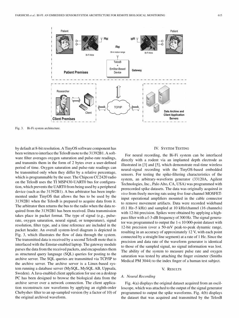
Fig. 3. Bi-Fi system architecture.
by default at 8-bit resolution. A TinyOS software component hasbeen written to interface the TelosB mote to the 31392B1. A soft-ware filter averages oxygen saturation and pulse-rate readings,and transmits them in the form of 2 bytes over a user-definedperiod of time. Oxygen saturation and pulse-rate readings canbe transmitted only when they differ by a relative percentage,which is programmable by the user. The Chipcon CC2420 radioon the TelosB uses the TI MSP430-UART0 bus for configura-tion, which prevents the UART0 from being used by a peripheraldevice (such as the 31392B1). A bus arbitrator has been imple-mented under TinyOS that allows the bus to be used by the31392B1 when the TelosB is prepared to acquire data from it.The arbitrator then returns the bus to the radio when the data re-quired from the 31392B1 has been received. Data transmissiontakes place in packet format. The type of signal (e.g., pulse-rate, oxygen saturation, neural signal, or temperature), signalresolution, filter type, and a time reference are included in thepacket header. An overall system-level diagram is depicted inFig. 3, which illustrates the flow of data through the system.The transmitted data is received by a second TelosB mote that isinterfaced with the Emstar-enabled laptop. The gateway moduleparses the data from the received packets, and encapsulates themas structured query language (SQL) queries for posting to thearchive server. The SQL queries are transmitted via TCP/IP tothe archive server. The archive server is a Linux-based sys-tem running a database server (MySQL, MySQL AB, Uppsala,Sweden). A Java-enabled client application for use on a desktopPC has been designed to browse the biological data from thearchive server over a network connection. The client applica-tion reconstructs raw waveforms by applying an eighth-orderChebyshev filter to an up-sampled version (by a factor of 10) ofthe original archived waveform.
IV. SYSTEM TESTING
For neural recording, the Bi-Fi system can be interfaceddirectly with a rodent via an implanted depth electrode asillustrated in [3] and [5], which demonstrate real-time wirelessneural-signal recording with the TinyOS-based embeddedsensors. For testing the spike-filtering characteristics of thesystem, an arbitrary-waveform generator (33120A, AgilentTechnologies, Inc., Palo Alto, CA, USA) was programmed withprerecorded spike datasets. The data was originally acquired invivo from freely moving rats using five four-channel MOSFET-input operational amplifiers mounted in the cable connectorto remove movement artifacts. Data were recorded wideband(0.1 Hz–5 kHz) and sampled at 10 kHz/channel (16 channels)with 12-bit precision. Spikes were obtained by applying a high-pass filter with a f-3-dB frequency of 300 Hz. The signal genera-tor was programmed to output the 1-s 10 000-point dataset with12-bit precision (over a 50-mV peak-to-peak dynamic range,resulting in an accuracy of approximately 12 V, with each pointconnected by a straight line segment) at a rate of 1 Hz. Since theprecision and data rate of the waveform generator is identicalto those of the sampled signal, no signal information was lost.The ability of the system to measure pulse rate and oxygensaturation was tested by attaching the finger oximeter (SmithsMedical PM 3044) to the index finger of a human test subject.
V. RESULTS
A. Neural Recording
Fig. 4(a) displays the original dataset acquired from an oscil-loscope, which was attached to the output of the signal generatorprogrammed to output the spike waveforms. Fig. 4(b) displaysthe dataset that was acquired and transmitted by the TelosB

616 IEEE TRANSACTIONS ON INFORMATION TECHNOLOGY IN BIOMEDICINE, VOL. 11, NO. 6, NOVEMBER 2007
Fig. 4. Neural signal (a) applied to the mote, (b) ADPCM-compressed, (c) filtered waveforms, (d) spike parameters, and the bandwidth required for transmittingthem. To demonstrate the ability of the filter to accept spikes while rejecting unwanted noise (such as motion artifacts, which result in spike-like patterns in theinput signal), the filter parameters (i.e., window of acceptable spike heights, widths, and trough depths, which are user programmable) were programmed to rejectthe sixth-spike waveform in the dataset—as though it were noise—as it has a very low trough depth.
mote at 4000 12-bit samples/s followed by ADPCM compres-sion. Fig. 4(c) displays the transmitted signal when the motewas programmed to acquire the neural signal at 8000 12-bitsamples, and then to detect and transmit time-referenced spikesusing an adaptive absolute-value-thresholding algorithm. Thealgorithm sets two thresholds (one positive and one negative)that are defined by the user as multiples in the rms value ofthe baseline noise. In the event of a positive-threshold crossing,the algorithm anticipates a negative-threshold crossing within auser-defined period of time (set to 500 s). If a negative-thresholdcrossing occurs in this time window, the time at which the spikeoccurred, as well as its data points (or the peak-trough heightand peak-trough width, depending on the mode of operation)are recorded and marked for transmission over the radio. Thisalgorithm is discussed in greater detail in [31]. To illustratethe ability of the filter to accept spikes in the present of un-wanted noise (such as motion artifacts, which could result insignals that resemble spikes), the filter parameters were chosensuch that the required signal-trough depth exceeded that of thesixth spike; hence, the spike was rejected as though it were un-wanted noise. The signal parameters extracted from the spikewaveform are listed in Fig. 4(d). The amount of data through-put necessary for transmitting each waveform is also labeledin Fig. 4. Transmitting the spike parameters only (e.g., spiketime, peak height, and trough depth) requires only 48 bits perspike, thus, lowering the required bandwidth for transmittingthe 1-s signal to only 288 bits. The normalized correlation ofthe received ADPCM-compressed raw spike signal to the auto-correlated original waveform is over 99%.
B. Pulse Oximetry and Heart Rate
Transmitting the raw heart-rate and pulse-oximetry signalswould require a total data rate of 1.92 kb/s. However, program-
ming the TelosB mote to only transmit the heart rate and pulseoximetry when heart rate varies by 5 beats/min, or when oxygensaturation changed by 2%, can significantly lower the requiredbandwidth and power consumption depending on the activitylevel of the test subject.
C. EKG
A power saving of 75% is realized by applying ADPCMto EKG signals (output from the signal generator) obtainedat a rate of 200 12-bit samples/s. The normalized correlationof the received signal with respect to the autocorrelated inputwaveform is over 99%.
VI. DISCUSSION
To put the system in perspective, we compare it with: 1) fullyanalog and 2) microcontroller-based solutions. The fully ana-log solutions do not provide onboard digital-signal processingor compression, and can, thus, be regarded as analog telemetrysystems. However, these analog systems are only a fraction ofthe size and consume far less power than the system outlined inthis paper. These analog systems also require custom receivers,and are not capable of communicating on any standard protocol(e.g., Health Level 7 [32]–[34], and IEEE [35]. Table I comparesthe key performance specifications of the system presented inthis paper against those reported for several fully analog wirelessbiological signal-acquisition systems (covered in detail in [31]).The key parameters of interest are raw data throughput, range,power dissipation, size, and weight, as the fully analog natureof the integrated systems inhibits their ability to perform signalprocessing, although analog-domain signal processing is beinginvestigated [30]. Table II compares the system presented inthis paper with several recently reported microcontroller-based

FARSHCHI et al.: BI-FI: AN EMBEDDED SENSOR/SYSTEM ARCHITECTURE FOR REMOTE BIOLOGICAL MONITORING 617
wireless biological monitoring platforms. The system presentedin [1] is an AMD-based 66-MHz PC-class device that communi-cates through an 802.11b wireless adapter. The system coveredin [36] is a similar TelosB-based system that acquires ECG, yetdoes not perform onboard signal processing to lower power dis-sipation. The system covered in [15] is a PIC-enabled device thatcommunicates over Bluetooth, which at a data transmission rateof 300 Hz, does not fully leverage the data throughput capabilityof its underlying hardware. The system covered in [17] is an-other Bluetooth-based system that acquires ECG, yet consumespower of the order of watts, as does [16], which is a PC-classdevice that communicates ECG over Bluetooth. Although allof the platforms compared in Table II could potentially pro-vide a level of digital-signal processing capabilities, the com-putational capabilities of the microcontrollers have been solelyused to implement communications protocols (e.g., Bluetooth,etc.), rather than biological-signal compression and interpreta-tion (e.g., spike detection). Table II illustrates that the systemreported in this paper provides similar signal-communicationsabilities (i.e., regarding channel count, data multiplexing, andstandards-based communications protocols), while also demon-strating data compression and signal analysis, at a fraction ofthe size, weight, and power dissipation of the other reportedmicrocontroller-based systems. An EKG monitor based on aTelosB mote [36], and a wireless pulse oximeter based ona MICA2 mote have been reported [20]; however, neither ofthese systems provide any onboard signal compression or eventdetection.
VII. CONCLUSION
In this paper, we have demonstrated an embedded sen-sor/system architecture for wireless biosignal recording. Thewireless biological sensors leverage the limited signal-filteringcapabilities of the COTS TelosB wireless-enabled processormodules on which they are based. By applying efficient filters,which were designed based on existing methods for interpretingbiological signals, the power efficiency of the embedded systemhas been improved by a factor of over 400. After taking intoaccount the standby power dissipation and communications-protocol overhead, a 360% improvement in battery life canbe expected. Furthermore, onboard signal-processing capabil-ity has removed the bandwidth bottleneck imposed by thetransceiver, thereby enabling signals to be sampled at doublethe rate at which they could be transmitted in raw form. There-fore, performing local signal processing not only lowers thepower dissipated by the radio, but also enables the observer tosample data at rates that would be prohibitive if they were tobe transmitted in raw format. We have demonstrated this byperforming spike detection on data sampled at a rate of 800012-bit samples/s, which by no means could be supported by theradio. Two figures of merit have been introduced, i.e., ATEF forfully analog telemetry systems and DTEF for microcontroller-based telemetry systems. We calculated the ATEF and DTEF ofour system, and compared it against those of several recentlyreported fully analog and microcontroller-based telemetry sys-tems, respectively. The ATEF of the system presented in this
paper is poor compared to the other fully analog telemetry sys-tems, because it is significantly heavier and more power inten-sive. However, the DTEF of the system presented in this paper isorders-of-magnitude better than the microcontroller-based sys-tems that have been reported in recent literature. For biomedi-cal monitoring applications that require data rates of up to 32kb/s, with the option of event detection, the system presented inthis paper efficiently exploits an appropriate level of hardwarecapabilities.
ACKNOWLEDGMENT
The authors would like to thank Dr. A. Bragin for providingus with the raw neural recordings.
REFERENCES
[1] I. Obeid, M. Nicolelis, and P. Wolf, “A multichannel telemetry system forsingle unit neural recordings,” J. Neurosci. Methods, vol. 133, no. 1-2,pp. 33–38, 2004.
[2] P. Irazoqui-Pastor, I. Mody, and J. Judy, “Transcutaneous RF-poweredneural recording device,” in Proc. 24th Annu. EMBS/BMES Conf. Eng.Med. Biol. 2002, vol. 3, pp. 2105–2106
[3] S. Farshchi, A. Pesterev, P. Nuyujukian, I. Modi, and J. Judy, “A TinyOS-enabled MICA2-based wireless neural interface,” IEEE Trans. Biomed.Eng., vol. 53, no. 7, pp. 1416–1424, Jul. 2006.
[4] J. Hill, R. Szewczyk, A. Woo, S. Hollar, D. Culler, and K. Pister, “Sys-tem architecture directions for network sensors,” presented at ASPLOS,Cambridge, MA, Oct. 2000.
[5] S. Farshchi, P. Nuyujukian, A. Pesterev, I. Mody, and J. Judy, “A TinyOS-based wireless neural sensing, archiving, and hosting system,” in Proc.2nd Int. IEEE EMBS Conf. Neural Eng., 2005, pp. 671–674.
[6] K. Wise, D. Anderson, J. Hetke, D. Kipke, and K. Najafi, “Wireless im-plantable microsystems: High-density electronic interfaces to the nervoussystem,” Proc. IEEE, vol. 92, no. 1, pp. 76–97, Jan. 2004.
[7] C. Subbe, M. Kruger, P. Rutherford, and L. Gemmel, “Validation of amodified early warning score in medical admissions,” QJM, vol. 94,pp. 521–526, 2001.
[8] H. Song, D. Allee, and K. Speed, “Single chip system for bio-data acqui-sition, digitization and telemetry,” in Proc. 1997 IEEE Int. Symp. CircuitsSyst., Hong Kong, vol. 3, pp. 1848–1851.
[9] P. Mohseni, K. Najafi, and S. Eliades, “Wireless multichannel biopotentialrecording using an integrated FM telemetry circuit,” IEEE Trans. NeuralSyst. Rehabil. Eng., vol. 13, no. 3, pp. 263–271, Sep. 2005.
[10] J. Parramon, P. Doguet, D. Marin, M. Verleyssen, R. Munoz, L. Leija,E. Valderrama, and B. Cnm, “ASIC-based batteryless implantable teleme-try microsystem forrecording purposes,” in Proc. 19th Annu. Int. Conf.IEEE Eng. Med. Biol. Soc. 1997, Chicago, IL, vol. 5, pp. 2225–2228
[11] A. Nieder, “Miniature stereo radio transmitter for simultaneous record-ing of multiple single-neuron signals from behaving owls,” J. Neurosci.Methods, vol. 101, no. 2, pp. 157–64, 2000.
[12] S. Takeuchi and I. Shimoyama, “A three-dimensional shape memory alloymicroelectrode with clipping structure for insect neural recording,” J.Microelectromech. Syst., vol. 9, no. 1, pp. 24–31, Feb. 2000.
[13] M. Modarreszadeh and R. Schmidt, “Wireless, 32-channel, EEG andepilepsy monitoring system,” in Proc. 19th Annu. Int. Conf. IEEE Eng.Med. Biol. Soc. 1997, Chicago, IL, vol. 3, pp. 1157–1160
[14] I. Obeid and P. Wolf, “Evaluation of spike-detection algorithms fora brain-machine interface application,” IEEE Trans. Biomed. Eng., vol. 51, no. 6,pp. 905–911, Jun. 2004.
[15] C. Mundt, K. Montgomery, U. Udoh, V. Barker, G. Thonier, A. Tellier,R. Ricks, R. Darling, Y. Cagle, N. Cabrol et al., “A multiparameter wear-able physiologic monitoring system for space and terrestrial applications,”IEEE Trans. Inf. Technol. Biomed., vol. 9, no. 3, pp. 382–391, Sep. 2005.
[16] M. Rasid and B. Woodward, “Bluetooth telemedicine processor for mul-tichannel biomedical signal transmission via mobile cellular networks,”IEEE Trans. Inf. Technol. Biomed., vol. 9, no. 1, pp. 35–43, Mar. 2005.
[17] J. Yao, R. Schmitz, and S. Warren, “A wearable point-of-care system forhome use that incorporates plug-and-play and wireless standards,” IEEETrans. Inf. Technol. Biomed., vol. 9, no. 3, pp. 363–371, Sep. 2005.

618 IEEE TRANSACTIONS ON INFORMATION TECHNOLOGY IN BIOMEDICINE, VOL. 11, NO. 6, NOVEMBER 2007
[18] B. Greenstein, A. Pesterev, C. Mar, E. Kohler, J. Judy, S. Farshchi, andD. Estrin, “Capturing high-frequency phenomena using a bandwidth-limited sensor network,” in Proc. ACM SenSys, 2006.
[19] L. Girod, T. Stathopoulos, N. Ramanathan, J. Elson, D. Estrin,E. Osterweil, and T. Schoellhammer, “A system for simulation, emu-lation, and deployment of heterogeneous sensor networks,” in Proc. 2ndInt. Conf. Embedded Netw. Sensor Syst., 2004, pp. 201–213.
[20] K. Lorincz, D. Malan, T. Fulford-Jones, A. Nawoj, A. Clavel, V. Shnayder,G. Mainland, M. Welsh, and S. Moulton, “Sensor networks for emergencyresponse: Challenges and opportunities,” IEEE Pervasive Comput., vol. 3,no. 4, pp. 16–23, Dec. 2004.
[21] M. Nicolelis, Methods for Neural Ensemble Recordings. Boca Raton,FL: CRC, 1999.
[22] M. Lewicki, “A review of methods for spike sorting: The detection andclassification of neural action potentials,” Netw: Comput. Neural Syst.,vol. 9, no. 4, pp. 53–78, 1998.
[23] R. Harrison, P. Watkins, R. Kier, R. Lovejoy, D. Black, R. Normann, andF. Solzbacher, “A low-power integrated circuit for a wireless 100-electrodeneural recording system,” in IEEE ISSCC 2006 Tech. Dig., pp. 554–555.
[24] R. Harrison, “A low-power integrated circuit for adaptive detection ofaction potentials in noisy signals,” in Proc. 25th Annu. Int. Conf. IEEEEMBS, 2003, pp. 3325–3328.
[25] P. Cummiskey, N. Jayant, and J. Flanagan, “Adaptive quantization indifferential PCM coding of speech,” Bell Syst. Tech. J., vol. 52, no. 7,p. 1105, 1973.
[26] R. Jafari, H. Noshadi, M. Sarrafzadeh, and S. Ghiasi, “Adaptive medi-cal feature extraction for resource constrained distributed embedded sys-tems,” in Proc. 4th Annu. IEEE Int. Conf. Pervasive Comput. Commun.Workshops, 2006, pp. 506–511.
[27] J. Pan and W. Tompkins, “A real-time QRS detection algorithm,” IEEETrans. Biomed. Eng., vol. 32, no. 3, pp. 230–236, Mar. 1985.
[28] Y. Hu, W. Tompkins, J. Urrusti, and V. Afonso, “Applications of arti-ficial neural networks for ECG signal detection and classification,,” J.Electrocardiol., vol. 26, pp. 66–73, 1993.
[29] G. Buzsaki, M. Penttonen, Z. Nadasdy, and A. Bragin, “Pattern andinhibition-dependent invasion of pyramidal cell dendrites by fast spikes inthe hippocampus in vivo,” Proc. Nat. Sci. USA, vol. 93, no. 18, pp. 9921–9925, 1996.
[30] P. Watkins, G. Santhanam, K. Shenoy, and R. Harrison, “Validation ofadaptive threshold spike detector for neural recording,” in Proc. 26thAnnu. Int. Conf. IEEE Eng. Med. Biol. Soc., 2004, vol. 2, pp. 4079–4082.
[31] S. Farshchi, W. Pesterev, A. Ho, I. Mody, and J. Judy, “Acquiring high-rate neural spike data with hardware-constrained embedded sensors,” inProc. 28th Annu. Int. Conf. IEEE Eng. Med. Biol. Soc., 2006, pp. 903–907.
[32] Hl7 United States. [Online]. Available: http://www.hl-7.org[33] CEN/TC251-European standardization of health informatics. Nen-Dutch
Standardization Inst. [Online]. Available: http://www.centc251.org[34] Digital imaging and communications in medicine. Nat. Elect. Manuf.
Assoc. [Online]. Available: http://medical.nema.org/dicom.html[35] IEEE 1073 Gen. Committee. IEEE 1073 medical device communications.
[Online]. Available: http://www.ieee1073.org[36] T. Fulford-Jones, G. Wei, and M. Welsh, “A portable, low-power, wireless
two-lead EKG system,” in Proc. 26th Annu. Int. Conf. IEEE Eng. Med.Biol. Soc., 2004, vol. 1, pp. 2141–2144.
Shahin Farshchi (S’03) was born in Berkeley, CA, in1978. He received the B.S. (honors) degree in electri-cal engineering and computer science from the Uni-versity of California, Berkeley, in 2002, and the M.S.and Ph.D. degrees in electrical engineering from theUniversity of California, Los Angeles, in 2005 and2006, respectively.
He is currently a Postdoctoral Fellow with theUniversity of California, Los Angeles. His currentresearch interests include developing wireless neural-signal acquisition, processing, and communications
systems.
Aleksey Pesterev is currently working toward theB.S. degree in computer science and electrical engi-neering at the University of California, Los Angeles.
Paul H. Nuyujukian is currently working towardthe B.S. degree in cybernetics at the University ofCalifornia, Los Angeles.
Istvan Mody received the Ph.D. degree in physiol-ogy (neurophysiology) from the University of BritishColumbia, Vanouver, BC, Canada.
He was a Postdoctoral Researcher first with theMax-Planck Institute, Munich, Germany, and thenwith the Playfair Neuroscience Center, University ofToronto, Toronto, Ontario, ON, under an Izaak Wal-ton Killam and a Canadian MRC Fellowhip. He waswith the Department of Neurology and NeurologicalSciences, Stanford University, California. He is cur-rently the Tony Coelho Professor of Neurology and
Professor of Physiology at the David Geffen School of Medicine, Departmentof Neurology, University of California, Los Angeles. His research interests in-clude ligand-gated ion channels, inhibition in the brain, basic mechanisms ofepilepsies, and the regulation of intraneural calcium.
Dr. Mody has recived several awards, including the Alfred Hauptmann Prize,the Michael Prize, the Basic Scientist Award from the AES/Milken Foundation,and the Javits Award from the National Institutes of Health/NINDS. He is amember of the Hungarian Academy of Sciences.
Jack W. Judy (S’87–M’96–SM’02) received theB.S.E.E. (summa cum laude) degree from the Uni-versity of Minnesota, Minneapolis, in 1989, and theM.S. and Ph.D. degrees in electrical engineering fromthe University of California, Berkeley, in 1994 and1996, respectively.
From 1996 to 1997, he was with Silicon LightMachines, Inc., Sunnyvale, CA. Since 1997, he hasbeen a Faculty with the Department of ElectricalEngineering, University of California, Los Angeles(UCLA), where he is currently an Associate Pro-
fessor. At UCLA, he is the Chair of the MEMS and Nanotechnology MajorField of the Electrical Engineering Department and the Director of the UCLANeuroEngineering Training Program. He is the Director of the UCLA Nano-electronics Research Facility as well as the Director of the UCLA Microfabri-cation Laboratory. His research interests include novel ferromagnetic MEMS(e.g., MEMS magnetometers, reconfigurable frequency-selective surfaces, andimplantable magnetic microactuators for biomedical applications), nanomag-netomechanical systems, chemical sensors, micromachined sensors for plasmaresearch, wireless sensor networks, microprobes for Parkinson’s disease re-search, implantable and flexible 3-D microelectrode arrays, dynamic modelingof 3-D microelectrodes, microelectrode arrays for high-density neural stimula-tion and retinal prostheses, simulation of the synthetic vision provided by retinalprostheses, micromachined systems to manipulate individual cells and to per-form high-impedance patch-clamp recording, microactuators to solve the hydro-cephalus shunt-clogging problem, wireless neural transceivers, brain–computerinterfaces, and developing neural-control strategies and systems for functionaldeficits (e.g., spinal cord injury, ocular motility, and deep-brain stimulation). Hehas developed and improved graduate-level training programs in the multidisci-plinary engineering fields of microelectromechanical systems, nanotechnology,and neuroengineering.