bdnf mrna expression in rat hippocampus and prefrontal cortex: effects of neonatal ventral...
TRANSCRIPT

BDNF mRNA expression in rat hippocampus andprefrontal cortex: effects of neonatal ventral hippocampaldamage and antipsychotic drugs
Barbara K. Lipska, Zin Z. Khaing, Cynthia Shannon Weickert and Daniel R. WeinbergerClinical Brain Disorders Branch, Intramural Research Program, National Institute of Mental Health, Bethesda, MD,
20892-1385 USA
Keywords: animal model, clozapine, haloperidol, neurotrophin, schizophrenia
Abstract
Brain-derived neurotrophic factor (BDNF) plays an important role in development, synapse remodelling and responses to stress
and injury. Its abnormal expression has been implicated in schizophrenia, a neuropsychiatric disorder in which abnormal neural
development of the hippocampus and prefrontal cortex has been postulated. To clarify the effects of antipsychotic drugs used inthe therapy of schizophrenia on BDNF mRNA, we studied its expression in rats treated with clozapine and haloperidol and in rats
with neonatal lesions of the ventral hippocampus, used as an animal model of schizophrenia. Both antipsychotic drugs reduced
BDNF expression in the hippocampus of control rats, but did not signi®cantly lower its expression in the prefrontal cortex. The
neonatal hippocampal lesion itself suppressed BDNF mRNA expression in the dentate gyrus and tended to reduce its expressionin the prefrontal cortex. These results indicate that, unlike antidepressants, antipsychotics down-regulate BDNF mRNA, and
suggest that their therapeutic properties are not mediated by stimulation of this neurotrophin. To the extent that the lesioned rat
models some pathophysiological aspects of schizophrenia, our data suggest that a neurodevelopmental insult might suppressexpression of the neurotrophin in certain brain regions.
Introduction
Brain-derived neurotrophic factor (BDNF) promotes a variety of
neuromodulatory processes in the brain including neuronal survival,
neurite outgrowth and synapse formation (Altar & DiStefano, 1998).
Consistent with these roles, BDNF mRNA is abundantly expressed
throughout life in highly plastic regions of the rat and human brain,
e.g. the neocortex, dentate gyrus and CA1±4 regions of the
hippocampus (Phillips et al., 1990). It is involved in promoting
neuronal function during development, in response to injury and
stress, in supporting functional activity of dopaminergic, cholinergic,
glutaminergic and serotonergic neurons (Altar, 1999), and in
regulating activity of mature neurons in the hippocampus (Zafra
et al., 1990; Ballarin et al., 1991; Kang & Schuman, 1995; Levine
et al., 1995) and the neocortex (Desai et al., 1999; Marty et al., 1997).
As abnormal development of these brain regions has been frequently
implicated in the pathogenesis of schizophrenia (Weinberger, 1999;
Lewis & Gonzalez-Burgos, 2000), it is possible that BDNF is
abnormally regulated in schizophrenia (Weickert & Weinberger,
1998). Indeed, recent postmortem studies have shown that BDNF
mRNA is reduced in the hippocampus and prefrontal cortex of
patients with schizophrenia (Brouha et al., 1996; Kittel et al., 1997).
However, another recent study reported elevated levels of BDNF
protein in the anterior cingulate cortex and hippocampus of schizo-
phrenic patients (Takahashi et al., 2000). The potential role of BDNF
in schizophrenia is further complicated by the fact that the impact of
antipsychotic drugs on BDNF mRNA expression or BDNF protein
levels is unclear.
To determine whether treatment with typical and atypical
antipsychotics in¯uences BDNF mRNA in brain regions implicated
in schizophrenia, we studied the effects of acute (a single intra-
peritoneal injection) and chronic (28 days) treatment with haloperidol
(0.5 and 1 mg/kg) and clozapine (10 mg/kg) on BDNF mRNA
expression in the rat prefrontal cortex and the hippocampus using an
in situ hybridization method that, with some modi®cations, has been
employed in our laboratory to measure BDNF mRNA in human tissue
(Brouha et al., 1996; Kittel et al., 1997). We have also assessed
BDNF mRNA expression in drug-free and neuroleptic-treated rats
with a neonatal excitotoxic lesion of the ventral hippocampus. We
have shown previously that the neonatally lesioned animals display in
adulthood a variety of abnormalities reminiscent of schizophrenia and
are used as an animal model of this disorder (Lipska et al., 1993,
1995a; Sams-Dodd et al., 1997; for review see Lipska & Weinberger,
2000). We hypothesized that, consistent with cognitive impairments
(Moghaddam et al., 1999) and molecular and morphological changes
in the prefrontal cortex of the neonatally lesioned animals (Bertolino
et al., 1999; Lipska & Weinberger, 2000), the neonatal ventral
hippocampal insult might impair BDNF mRNA expression in the
prefrontal cortex of adult animals. We have also tested whether the
lesion altered BDNF mRNA expression in other parts of the
hippocampal formation which are relatively spared by the lesion
(dentate gyrus and CA3 area) and which normally show high
expression of BDNF mRNA. Based on some of the previous reports
of increased BDNF mRNA after clozapine (Chlan-Fourney et al.,
1998), we also hypothesized that compromised BDNF mRNA
Correspondence: Dr B.K. Lipska, as above.E-mail: [email protected]
Received 15 January 2001, revised 3 April 2001, accepted 3 May 2001
European Journal of Neuroscience, Vol. 14, pp. 135±144, 2001 ã Federation of European Neuroscience Societies

136 B. K. Lipska et al.
ã 2001 Federation of European Neuroscience Societies, European Journal of Neuroscience, 14, 135±144
expression in the lesioned animals may be normalized by chronic
treatment with clozapine, an atypical antipsychotic drug.
Materials and methods
Surgery
All procedures were performed in accordance with the National
Institutes of Health guidelines for use and care of laboratory animals.
Animals were on a 12-h light : 12-h dark cycle (lights on at 06.00 h)
in a temperature-controlled environment and with ad lib. access to
food and water. Female Sprague-Dawley rats were purchased
pregnant at 14 days of gestation (Taconic, Germantown, NY, USA)
and housed individually in breeding cages. Litters of four to eight
male pups were formed (total n = 80 pups). Pups were randomly
assigned to lesion (n = 42) or sham (n = 38) status. Surgery was
performed as previously described (Lipska et al., 1993). Pups (7 days
of age, weight 15±18 g) were anaesthetized by hypothermia (placed
on wet ice for 10±20 min) and then immobilized by taping onto a
styrofoam platform ®xed to a stereotaxic Kopf instrument (David
Kopf Instruments, Tujunga, CA, USA). An incision was made in the
skin overlying the skull and 0.3 mL (3 mg) of ibotenic acid (in the
lesioned rats; Sigma Chemical Co., St Louis, MO, USA) or arti®cial
cerebrospinal ¯uid (in the sham-operated rats) was injected bilater-
ally, using an infusion pump through a Hamilton needle, into the
ventral hippocampus at a rate of 0.15 mL/min (AP, 3.0 mm relative to
bregma; ML, 63.5 mm; DV, 5.0 mm). The needle was left in place
for 4 min, to prevent diffusion of neurotoxin along the needle track,
and then withdrawn. The skin incision was closed with staples. The
pups were placed under a heating lamp and then returned to their
mothers. At postnatal day 24, the rats were weaned, separated by
lesion status, and housed two to three per cage.
Drug preparation
Ibotenic acid (Sigma) was dissolved in arti®cial cerebrospinal ¯uid
(5 mg in 500 mL) with the addition of 2 mL of 10 M NaOH, and
further neutralized to pH 7.4 by adding » 6 mL of 1 M NaOH.
Aliquots of 50 mL were stored at ±80 °C for up to several months. A
stock solution of haloperidol (Research Biochemicals Inc., Natick,
MA, USA) 20 mg/mL was prepared in 1% lactic acid by heating up
200 mg of haloperidol in 10 mL of 1% lactic acid until dissolved. To
obtain solutions of 0.5 and 1.0 mg/mL, a stock solution was diluted
with distilled water to a ®nal volume of 20 mL. Approximately
30 mL of 1 M NaOH was added to adjust the ®nal solutions to pH 5.3.
Clozapine (a generous gift from Sandoz Research Institute Berne Ltd,
Berne, Switzerland) was prepared as folows: 100 mg was dissolved in
a small amount of 0.1 M HCl (400 mL), diluted with distilled water to
40 mL and neutralized with 1 M NaOH to pH 5.2. Vehicle was
prepared by adding 2 mL of 1% lactic acid to 100 mL distilled water.
Treatment with antipsychotic drugs
Drug treatment started 2 months after the lesion (at postnatal day 56).
Rats were given intraperitoneal injections of haloperidol (0.5 and
1 mg/kg) or clozapine (10 mg/kg) either acutely (a drug administered
on the last day of a 27-day regimen of vehicle injections) or
chronically (once daily for 28 days). A control group for both acute
and chronic treatment was injected with vehicle for 28 days. All rats
were killed 7 h after the last injection. Brains were frozen and
sectioned into 20-mm slices which were collected onto gelatin-coated
slides (two slices per slide) and stored at ±80 °C. Every 10th slide
was Nissl-stained and used for anatomical matching. The hippo-
campal slices were used for veri®cation of the lesion. Matched slides
through the prefrontal cortex and the hippocampus (four consecutive
slides per region per rat corresponding to ®gs 9 and 38 for prefrontal
cortex and hippocampus, respectively, of the atlas of Paxinos &
Watson, 1986) were used in the in situ hybridization study.
In situ hybridization histochemistry
An 35S-labelled riboprobe was synthesized using a clone containing
1095 base pairs of the rat BDNF cDNA sequence (Maisonpierre et al.,
1991). To produce an antisense ribonucleotide probe, the Bluescript
SK-vector was linearized with BamH1 restriction enzyme (New
England BioLabs, Beverly, MA, USA); 200 ng (1 mL) of linearized
plasmid was labelled using 150 mCi of [35S]UTP (NEN, Boston, MA,
USA) and 1 mL (5 units) T7 RNA polymerase (Stratagene, CA,
USA). For a sense probe, HindIII enzyme and T3 polymerase
(Promega, Madison, WI, USA) were used. The total volume of this
reaction was 10 mL: 1 mL each of 10 mM rATP, rCTP, rGTP
(Promega), 2 mL of 53 transcription buffer, 1 mL of 100 mM
dithiothreitol, 1 mL of RNasin and 1 mL diethyl pyrocarbonate
(DEPC)-treated water. After incubation for 15 min at 37 °C, labelled
RNA was digested with DNase (3 mL, 10 U/mL) and precipitated
with ethanol in the presence of yeast tRNA and ammonium acetate.
After precipitation, the probe was washed with 70% ethanol (speci®c
activity 2.2 3 109 dpm/mg), resuspended in DEPC water and added
to the hybridization cocktail containing 5 mL of hybridization buffer
(see below), 5 mL formamide, 20 mL 50% sodium thiosulphate,
200 mL 5 M dithiothreitol and 100 mL 10% sodium dodecyl sulphate
(Research Genetics). The hybridization buffer consisted of 1200 nM
NaCl, 20 mM Tris-HCl, 0.04% Ficoll, 0.04% bovine serum albumin
(Pierce, Rockford, IL, USA), 0.04% polyvinylpyrrolidone (PVP),
2 mM EDTA (Research Genetics, Huntsville, AL, USA), pH = 8,
0.02% salmon sperm DNA, 0.1% total yeast RNA, 0.01% yeast tRNA
(Invitrogen, Rockville, MD, USA) and 20% Dextran SO4. This
yielded a ®nal concentration of 5 ng/mL of the riboprobe, corres-
ponding to » 0.5 3 106 dpm per section (per 25 mL of hybridization
cocktail). The sections were ®xed in 4% formaldehyde, acetylated
using acetic anhydride, dehydrated in ethanol and defatted in
chloroform. In situ hybridization was carried out overnight at 55 °C
in humidi®ed chambers. After removing the coverslips in 23 sodium
chloride±sodium citrate (SSC), the sections were treated with RNase
A (20 mg/L for 30 min at 37 °C), washed in RNase A-free buffer for
30 min at 37 °C, washed in 23 SSC for 15 min at room temperature,
twice in 23 SSC for 1 h at 50 °C, once in 0.23 SSC for 1 h at 55 °C,
once in 0.23 SSC for 30 min at 60 °C and rinsed with 0.23 SSC at
room temperature. The sections were then dehydrated with 50±100%
ethanol solutions with ammonium acetate (300 mM), air-dried and
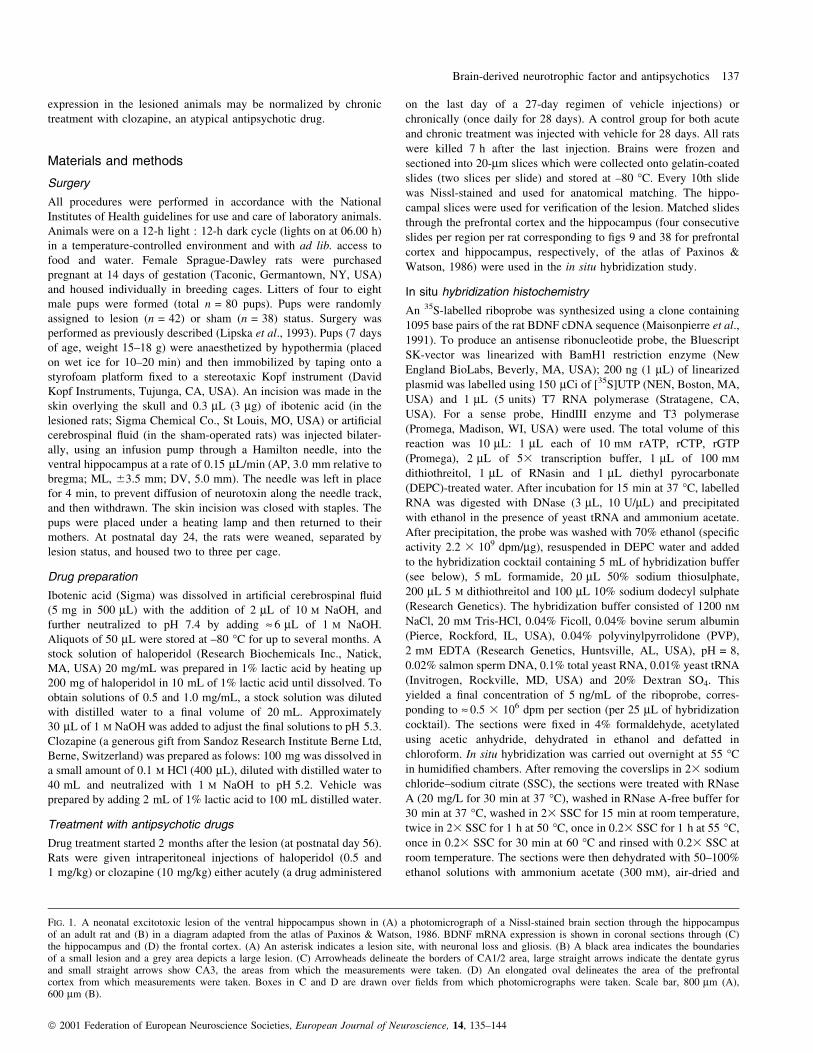
FIG. 1. A neonatal excitotoxic lesion of the ventral hippocampus shown in (A) a photomicrograph of a Nissl-stained brain section through the hippocampusof an adult rat and (B) in a diagram adapted from the atlas of Paxinos & Watson, 1986. BDNF mRNA expression is shown in coronal sections through (C)the hippocampus and (D) the frontal cortex. (A) An asterisk indicates a lesion site, with neuronal loss and gliosis. (B) A black area indicates the boundariesof a small lesion and a grey area depicts a large lesion. (C) Arrowheads delineate the borders of CA1/2 area, large straight arrows indicate the dentate gyrusand small straight arrows show CA3, the areas from which the measurements were taken. (D) An elongated oval delineates the area of the prefrontalcortex from which measurements were taken. Boxes in C and D are drawn over ®elds from which photomicrographs were taken. Scale bar, 800 mm (A),600 mm (B).
Brain-derived neurotrophic factor and antipsychotics 137
ã 2001 Federation of European Neuroscience Societies, European Journal of Neuroscience, 14, 135±144
apposed with the 14C standards to BioMax Kodak ®lm for 11 days.
DEPC, sodium thiosulphate, salmon sperm DNA, ammonium acetate,
diethyl pyrocarbonate and total yeast RNA were purchased from
Sigma.
Analysis of autoradiographic images was performed using NIH
Image software. The regions analysed (medial prefrontal cortex,
CA1/2 and CA3 areas of the hippocampus, dentate gyrus) are
depicted in Fig. 1. In the lesioned rats, only those parts of the CA1/2
and CA3 areas of the hippocampus and dentate gyrus depicted in
Fig. 1C that were visibly spared by the lesion (i.e. contained intact
neurons, no gliosis as shown in Fig. 1A) were measured (the
hippocampal regions damaged by the lesion showed no expression of
BDNF and no neurons in Nissl-stained sections). Optical density was
converted to dpm/mm2 using 14C standards (Miller, 1991). Four
measurements were obtained per region per rat from four slides
containing two sections each, and averaged. The sense riboprobe did
not yield any signi®cant hybridization signal (not shown).
Statistical analysis
The densitometric data were analysed using a two-way analysis of
variance (ANOVA) with Drug and Lesion as independent factors.
Acute and chronic groups were analysed separately. A Fisher PLSD
test was used for post hoc comparisons.
Results
Lesion veri®cation
Analysis of Nissl-stained 20-mm brain sections through the
hippocampus of the lesioned rats showed that damage was restricted
to the ventral hippocampus (Fig. 1A and B). Consistent with our
previous reports (Lipska et al., 1993, 1995a; Lipska & Weinberger,
1995), neuronal loss, gliosis and some cavitation was mostly evident
in cytoarchitectural subdivisions CA1/2 of the hippocampal forma-
tion and in the ventral parts of the subiculum, whereas only minimal
damage was detected in the ventral dentate gyrus and the ventral CA3
sub®eld.
Hippocampal formation
BDNF mRNA expression was found in all cytoarchitectural divisions
of the hippocampus. The hybridization silver grain signal was located
over pyramidal (CA1±3) and granule (dentate gyrus) cell layers but
was not evenly distributed (Fig. 1C). The strongest hybridization
signal was observed in the dentate dyrus and the CA3 area, whereas
the BDNF mRNA expression over CA1/2 sub®elds was modest. As
previously noted (Hofer et al., 1990), the signal was not seen in all
cells of the hippocampal subregions; whilst most cells were strongly
labelled, occasional cells were often almost completely devoid of
silver grains (Fig. 3).
Both antipsychotic drugs markedly reduced BDNF mRNA expres-
sion in the CA1/2 sub®eld of the hippocampus and in the dentate
gyrus of control rats (Fig. 2A and B). Moreover, lesioned vehicle-
treated animals displayed profoundly attenuated BDNF mRNA
expression in parts of CA1/2 and in the dentate gyrus apparently
spared by the lesion as compared with sham-operated animals. In the
CA1/2 area after acute treatment (Fig. 2A), an ANOVA revealed a
signi®cant effect of Lesion (F1,35 = 6.91, P < 0.05) and Drug
(F2,35 = 7.37, P < 0.01, but no signi®cant Lesion±Drug interaction
(F2,35 = 2.15, P > 0.1). Post hoc tests showed that acute injections of
haloperidol and clozapine signi®cantly reduced BDNF levels in CA1/
2 of control rats (by 34 and 57%, respectively; P < 0.01) but had no
signi®cant effect in the lesioned rats whose basal (after vehicle
injections) BDNF mRNA expression was signi®cantly attenuated as
compared with sham-operated controls (by 41%; P < 0.01; Fig. 3A±
D).
Chronic treatment with antispychotics produced effects similar to
acute treatment in CA1/2 (Fig. 2A). An ANOVA revealed a signi®cant
effect of Lesion (F1,47 = 22.6, P < 0.0001) and Drug (F3,47 = 9.01,
P < 0.001), and a signi®cant Lesion±Drug interaction (F3,47 = 6.50,
P < 0.01). Both doses of haloperidol (0.5 and 1.0 mg/kg) and
clozapine attenuated BDNF mRNA expression in control rats (by 25,
31 and 55%, respectively; P < 0.01). In the lesioned animals, whose
basal levels of BDNF mRNA expression were signi®cantly attenuated
as compared to sham controls (by 41%; P < 0.001; Fig. 3), only a
high dose of haloperidol further reduced BDNF mRNA expression in
CA1/2 (by 54% as compared with the lesion vehicle group; P < 0.01;
Fig. 3E±H).
The effects of antipsychotic drugs were slightly less pronounced in
the dentate gyrus than in CA1/2, but the neonatal lesion had an even
more profound effect in reducing BDNF mRNA in the dentate gyrus
than in CA1/2 (Fig. 2B). For acute treatment, an ANOVA showed a
signi®cant effect of Lesion (F1,35 = 163.83, P < 0.0001) and Drug
(F2,35 = 11.93, P < 0.001) and a signi®cant Lesion±Drug interaction
(F2,35 = 12.65, P < 0.0001). Post hoc comparisons showed that
haloperidol and clozapine signi®cantly reduced BDNF mRNA
expression in the dentate gyrus (by 21 and 55%, respectively;
P < 0.0.01) in control rats. Neither drug had an effect in the lesioned
animals whose basal (after vehicle injections) BDNF mRNA levels
were signi®cantly attenuated as compared to the sham-operated rats
(by 83%; P < 0.0001; Fig. 3I±L). For chronic treatment, an ANOVA
revealed a signi®cant lesion effect (F1,47 = 179.28, P < 0.05), no
signi®cant Drug effect (F3,47 = 2.40, P = 0.08), and a signi®cant
Lesion±Drug interaction (F3,47 = 3.3, P = 0.03). Post hoc compari-
sons showed that both doses of haloperidol and clozapine signi®-
cantly reduced BDNF mRNA expression in the dentate gyrus of
sham-operated rats (by 19, 33 and 30%, respectively). The effects of
antipsychotic drugs were not detected in the lesioned rats whose basal
(after vehicle) levels of BDNF mRNA were very low as compared
with vehicle- or antipsychotic drug-treated rats (P < 0.001 for all
sham vs. lesion comparisons; Fig. 2B).
The effects of antipsychotic drugs and the lesion differed in the
CA3 area of the hippocampus from those in the dentate gyrus and the
CA1/2 region (Fig. 2C). For acute treatment, ANOVA showed a main
effect of Lesion (F1,35 = 5.0, P < 0.05), but no effect of Drug
(F2,35 = 2.1, P = 0.13) and no signi®cant Lesion±Drug interaction
(F2,35 = 1.8, P = 0.17). In contrast to other hippocampal sub®elds,
there was no signi®cant difference in BDNF mRNA expression
between sham and lesioned vehicle-treated animals in the CA3 area.
In chronically treated rats, ANOVA showed a signi®cant effect of Drug
(F3,47 = 3.7, P < 0.02) but the effect of Lesion (F1,47 = 0.68,
P = 0.4) and the Drug±Lesion interaction (F3,47 = 0.27, P = 0.8)
were not signi®cant. Post hoc comparisons revealed that both doses
of chronic haloperidol signi®cantly attenuated BDNF mRNA expres-
sion in control and lesioned rats (P < 0.05), whereas clozapine had no
effect in either group of animals (Fig. 2C).
Prefrontal cortex
The area comprising the medial prefrontal cortex is depicted in
Fig. 1D. The hybridization signal was present throughout all layers of
the prefrontal cortex. This ®nding is in agreement with the study of
Yan et al. (1997) who reported the presence of BDNF protein in
layers I±III and V±VI of the neocortex. Clusters of silver grains were
overlying lightly Nissl-stained large cells, putatively pyramidal
neurons (not shown). Neither acute nor chronic treatment with
138 B. K. Lipska et al.
ã 2001 Federation of European Neuroscience Societies, European Journal of Neuroscience, 14, 135±144

haloperidol or clozapine signi®cantly altered expression of BDNF
mRNA expression in the medial prefrontal cortex of the sham or
lesioned animals (Fig. 2D). ANOVAs for acutely or chronically treated
animals revealed no signi®cant effects of Lesion (F1,35 = 0.23,
P > 0.5 and F1,47 = 1.1, P > 0.3, respectively) or Drug (F2,35 = 1.40,
P > 0.2 and F3,47 = 1.0, P > 0.3, respectively). Lesion±Drug inter-
FIG. 2. Expression of BDNF mRNA (mean 6 SD) in (A) the hippocampal CA1/2 area, (B) the dentate gyrus, (C) the hippocampal CA3 area and (D) theprefrontal cortex of sham-operated rats (Sham) and rats with the neonatal lesion of the ventral hippocampus (Lesion). Rats were acutely (Acute) orchronically (Chronic; 28 days) treated with vehicle (Veh), haloperidol (Hal; 0.5 or 1.0 mg/kg) and clozapine (Cloz; 10 mg/kg). *P < 0.05 vs. a vehicle-treatedgroup of the same lesion status, #P < 0.05 vs. vehicle-treated sham animals.
Brain-derived neurotrophic factor and antipsychotics 139
ã 2001 Federation of European Neuroscience Societies, European Journal of Neuroscience, 14, 135±144

FIG. 3. Light (A, C, E, G, I, K) and dark(B, D, F, H, J, L) ®eld autoradiographicimages of BDNF mRNA expression in (A±H)the hippocampus CA1/2 area and (I±L) thedentate gyrus. (A and B) CA1 area of acontrol rat treated with vehicle. (C and D)Corresponding region of a vehicle-treated ratwith a neonatal lesion in the ventralhippocampus. The lesioned rats showedsigni®cantly less BDNF mRNA expression inthe CA1/2 area of the hippocampus than didcontrols. (E and F) CA1 area of a control rattreated chronically with haloperidol (1.0 mg/kgi.p.). (G and H) Corresponding region of a ratwith a neonatal lesion in the ventralhippocampus, treated chronically withhaloperidol 1.0 mg/kg i.p.The haloperidol-treated lesioned rats showedsigni®cantly less BDNF mRNA expression inthe CA1/2 area of the hippocampus than didhaloperidol-treated controls. (I and J) Dentategyrus of a control rat treated with vehicle. (Kand L) Corresponding region of a rat with aneonatal lesion in the ventral hippocampus,treated with vehicle. Expression of BDNFmRNA was signi®cantly reduced in the dentategyrus of the lesioned rats. Scale bar, 25 mm.
140 B. K. Lipska et al.
ã 2001 Federation of European Neuroscience Societies, European Journal of Neuroscience, 14, 135±144

actions were also not signi®cant (F2,35 = 0.42, P > 0.5 and
F3,47 = 0.21, P > 0.5, for acute and chronic treatment, respectively).
The results suggest, however, that BDNF mRNA expression levels
tended to be lower in the medial prefrontal cortex of control rats
treated chronically with clozapine as compared with chronic vehicle
injections (by 34%; Fig. 2D). BDNF mRNA expression was also
lower in the lesioned vehicle-treated rats as compared with sham-
operated vehicle-treated animals although the difference did not reach
statistical signi®cance (by 18%; Fig. 2D).
Discussion
Effects of antipsychotic drugs
The results of the current study revealed that antipsychotic drugs
attenuated the expression of BDNF mRNA in the normal rat
hippocampus but did not signi®cantly lower its expression in the
prefrontal cortex. The extent of the change depended on the speci®c
subregion, type of drug, dosage and length of administration. In
particular, both haloperidol and clozapine after acute and chronic
administration markedly down-regulated expression of BDNF mRNA
in the dentate gyrus and CA1/2 sub®elds of the rat hippocampus. In
the CA3 sub®eld, only chronic treatment with haloperidol (but not
clozapine) signi®cantly reduced BDNF mRNA expression in control
rats. In contrast, haloperidol had no signi®cant effect in the medial
prefrontal cortex, whereas chronic treatment with clozapine tended to
lower BDNF mRNA expression in this region.
The ®nding of reduced BDNF mRNA in the hippocampus of
haloperidol- and clozapine-treated rats contrasts markedly with the
action of antidepressants that elevate BDNF mRNA in this brain
region (Nibuya et al., 1995; Russo-Neustadt et al., 1999). The
antidepressant-induced BDNF elevations have been attributed to
increased synaptic levels of the neurotransmitters serotonin and
noradrenaline in the forebrain (Celada et al., 1996; for review see
Altar, 1999) and enhanced growth of serotonergic and noradrenergic
®bers (Mamounas et al., 2000). These effects may be mediated
through 5-hydroxytryptamine (5-HT)2A and b-adrenoreceptor sub-
types (Vaidya et al., 1997). Although the exact mechanisms of
boosting the BDNF levels by antidepressants are not well understood,
stimulation of intracellular pathways involving cyclic adenosine
monophosphate (cAMP) (Fujimaki et al., 2000) and cAMP response
element binding protein (CREB) (Duman et al., 1997) have been
implicated.
Our ®ndings of reduced BDNF mRNA expression in the hippo-
campal formation by typical and atypical antipsychotics are in
agreement with one recently published study that assessed BDNF
protein levels after a 29-day treatment with haloperidol and
risperidone (Angelucci et al., 2000), which, however, also found a
signi®cant reduction of BDNF in the frontal cortex after haloperidol,
and with a report by Chlan-Fourney et al. (1998) who found reduced
BDNF mRNA. Our ®ndings additionally indicate that single doses of
haloperidol and clozapine are suf®cient to produce a signi®cant
suppression of BDNF mRNA in the hippocampal areas CA1/2 and in
the dentate gyrus, suggesting that these drugs can alter BDNF mRNA
synthesis by short-term changes in synaptic transmission in addition
to possible long-term adaptive mechanisms involving synaptic
plasticity and rearrangement of connections. It is unclear, however,
which neurotransmitter systems might be involved.
Haloperidol and clozapine were shown to down-regulate D1 and
D4 dopamine receptor mRNA in the hippocampus (D'Souza et al.,
1997; Ritter & Meador-Woodruff, 1997), suggesting that blockade of
these dopamine receptor subtypes might interfere with BDNF gene
expression. Clozapine is also a potent 5-HT2A inhibitor and was
shown to in¯uence the serotonergic system by decreasing the
expression of 5-HT6 receptors in all hippocampal sub®elds
(Frederick & Meador-Woodruff, 1999) and increasing the densities
of 5-HT transporters in the ventral hippocampus (Ase et al., 1999).
Moreover, both haloperidol and clozapine reduce serotonin concen-
trations in the hippocampus (Burnet et al., 1996). Because enhanced
serotonergic transmission has been associated with the up-regulation
of BDNF mRNA in the hippocampus (Nibuya et al., 1995), it is
conceivable that the suppression of 5-HT function by antipsychotics
might lead to the reduction of BDNF mRNA. The glutamatergic
system may also play a role in mediating the effects of antipsychotics
because both drugs, although in complex and differential fashions,
alter the expression of certain a-amino-3-hydroxy-5-methyl-4-
isoxazolepropionic acid (AMPA)- and kainate-receptor subunits in
the hippocampus (Eastwood et al., 1996; Meador-Woodruff et al.,
1996), and glutamate potently stimulates expression of BDNF mRNA
in the hippocampus (Zafra et al., 1990; Wetmore et al., 1994). In the
CA3 region, however, only chronic treatment with haloperidol had a
signi®cant effect on BDNF down-regulation, and clozapine had no
effect at all.
In contrast to the hippocampus, neither drug had a signi®cant effect
in the prefrontal cortex, although chronic clozapine showed a trend
for a reduction BDNF mRNA levels. It has been suggested that the
activation of 5-HT2A receptors in the neocortex results in enhanced
presynaptic release of glutamate, stimulation of AMPA receptors and
a subsequent increase of BDNF mRNA synthesis (Vaidya et al.,
1997). Because clozapine antagonizes 5-HT2A receptors, which are
abundant in the prefrontal cortex (Pompeiano et al., 1994; Jakab &
Goldman-Rakic, 1998), it is conceivable that it could reduce BDNF
mRNA via this mechanism. Moreover, clozapine and other atypical
drugs, but not haloperidol, have been shown to induce internalization
of 5-HT2A receptors after chronic treatment and their subsequent loss
from apical dendrites of pyramidal neurons in the medial prefrontal
cortex (Willins et al., 1999).
BDNF is important in long-term potentiation of hippocampal
neurons and in learning and memory (Jankowsky & Patterson, 1999;
Hall et al., 2000; Liu et al., 2000), and its loss has been implicated in
the failure of cognitive functions in dementia and Alzheimer's
disease (Altar & DiStefano, 1998; Phillips et al., 1991). Whatever the
mechanisms responsible for the down-regulation of BDNF mRNA
following antipsychotic treatment, our ®ndings suggest that anti-
psychotic drugs, unlike antidepressants, would not improve cognition
by stimulating BDNF production. This notion is partially supported
by the observations that chronic haloperidol reduces spine density in
the prefrontal cortex and the striatum (Benes et al., 1985; Kelley et al.,
1997), although no effect on spine density was observed in the
hippocampus (Uranova et al., 1991). Thus, to the extent that BDNF
and synaptic density play a role in cognition (Elston, 2000), we would
not expect these drugs to bene®t cognitive abilities via these
mechanisms (Weinberger & Gallhofer, 1997).
Effects of the neonatal hippocampal lesion
We also found that the neonatal lesion of the ventral hippocampus
profoundly attenuated expression of BDNF mRNA in areas of the
hippocampus outside the lesion site, particularly in the dentate gyrus.
The lesion had no signi®cant effect in the prefrontal cortex although
some reduction in the BDNF mRNA expression was also seen there.
This raises the possibility that the hippocampal subregions may also
be directly damaged by the neurotoxin, although the assessment of
Nissl-stained sections through these regions does not reveal
discernable losses of neurons or gliosis. Our recent data suggest,
Brain-derived neurotrophic factor and antipsychotics 141
ã 2001 Federation of European Neuroscience Societies, European Journal of Neuroscience, 14, 135±144

however, that hippocampal sub®elds outside the ventral hippocampus
suffer DNA damage in response to neonatal insult (Khaing et al.,
2000). It is also possible, however, as the hippocampus and prefrontal
cortex develop extensively after birth, that the restricted lesion in the
ventral aspects of the hippocampal areas CA1/2 had dramatic effects
on synaptogenesis and neuronal development in other parts of the
hippocampal formation and, although in a much more subtle way,
affected development of the prefrontal cortex to which the ventral
hippocampus projects (Jay & Witter, 1991; Carr & Sesack, 1996).
Reduced levels of N-acetylaspartate, a marker of neuronal metabolic
activity, in the prefrontal cortex of adult rats with the neonatal
hippocampal lesions may support this notion (Bertolino et al., 1999).
The neonatal ventral hippocampal lesion might have disturbed
function of interconnected brain regions by producing loss of neural
targets for some neurons (as in the case of the dentate gyrus neurons
that send projections to areas CA3, CA1/2) or loss of neuronal inputs
in other areas (as in the case of the prefrontal cortex that would lose
glutamatergic projections from the ventral hippocampus and may also
suffer reduced glutamatergic excitation from the thalamus, which is
deafferented by the hippocampal lesion). It is noteworthy in this
regard that unilateral electrolytic lesions of the hippocampus in
neonatal (postnatal day 1) rats, which produce severe spatial learning
disabilities, attenuate BDNF mRNA expression in the contralateral
hippocampus for up to 20 weeks postlesion without producing other
neurochemical changes, such as in choline-acetyltransferase or
GABA decarboxylase activity (van Praag et al., 1998). Thus,
unilateral damage of the hippocampus has long-lasting adverse
effects on neurotrophin synthesis on the seemingly healthy side that
can possibly have detrimental effects on development of the
contralateral hippocampus, and possibly on learning and memory.
Another possibility for reduced BDNF mRNA in rats with the
neonatal ventral hippocampal lesion may be their increased vulner-
ability to stress. We have previously shown that behavioural
responses to stressful stimuli are exaggerated (Lipska et al., 1993),
that stress-induced dopamine release is attenuated (Lipska et al.,
1995b; Lillrank et al., 1999) and that BDNF mRNA response to acute
restraint stress is blunted in the prefrontal cortex of the lesioned rats
(Molteni et al., 2001). Repeated stress decreases BDNF mRNA in the
hippocampus and the dentate gyrus (Smith et al., 1995) and up-
regulates TrkB receptors in all hippocampal sub®elds (Nibuya et al.,
1999). Thus, it is conceivable that stress experienced chronically by
the neonatally lesioned rats may contribute to the loss of BDNF
mRNA in vulnerable brain regions. However, we did not observe
diminished BDNF mRNA in the CA3 sub®eld, the region whose
pyramidal neurons have been reported to be most sensitive to stress
and to atrophy following repeated stressful events (McEwen, 1999),
and which show elevations in TrkB mRNA following chronic stress
(Nibuya et al., 1999).
Finally, it should be noted that, similarly to the normal animals, we
did not ®nd evidence for a bene®cial effect of antipsychotic drugs on
the BDNF mRNA expression in the neonatally lesioned rats.
Antipsychotics had either no effect or further lowered BDNF
mRNA levels in the brains of the lesioned rats.
In summary, our results indicate that antipsychotic drugs exert
suppressive effects on BDNF gene expression in the brain regions
studied. Thus, the results raise the possibility that reduced BDNF
mRNA levels observed in brain tissue obtained from patients with
schizophrenia might re¯ect the consequences of long-term treatment
with antipsychotic drugs and may not be necessarily related to the
pathogenesis of the disease. However, to the extent that rats with the
neonatal lesion of the hippocampus model some pathophysiological
aspects of schizophrenia, our data also suggest that a neurodevelop-
mental insult affecting the ventral portion of the hippocampus might
suppress BDNF production in distant sites.
Acknowledgements
We would like to thank Mr Michael Valentine for his technical assistance.
Abbreviations
AMPA, a-amino-3-hydroxy-5-methyl-4-isoxazolepropionic acid; BDNF,brain-derived neurotrophic factor; DEPC, diethyl pyrocarbonate; 5-HT,5-hydroxytryptamine (serotonin); SSC, sodium chloride±sodium citrate.
References
Altar, C.A. (1999) Neurotrophins and depression. Trends Pharmacol. Sci., 20,59±61.
Altar, C.A. & DiStefano, P.S. (1998) Neurotrophin traf®cking by anterogradetransport. Trends Neurosci., 21, 433±437.
Angelucci, F., Mathe, A.A. & Aloe, L. (2000) Brain-derived neurotrophicfactor and tyrosine kinase receptor TrkB in rat brain are signi®cantly alteredafter haloperidol and risperidone administration. J. Neurosci. Res., 60,783±794.
Ase, A.R., Amdiss, F., Hebert, C., Huang, N., van Gelder, N.M. & Reader,T.A. (1999) Effects of antipsychotic drugs on dopamine and serotonincontents and metabolites, dopamine and serotonin transporters, andserotonin1A receptors. J. Neural Transm., 106, 75±105.
Ballarin, M., Ernfors, P., Lindefors, N. & Persson, H. (1991) Hippocampaldamage and kainic acid injection induce a rapid increase in mRNA forBDNF and NGF in the rat brain. Exp. Neurol., 114, 35±43.
Benes, F.M., Paskevich, P.A., Davidson, J. & Domesick, V.B. (1985) Synapticrearrangements in medial prefrontal cortex of haloperidol-treated rats. BrainRes., 348, 15±20.
Bertolino, A., Roffman, J.L., Lipska, B.K., Van Gelderen, P., Olson, A. &Weinberger, D.R. (1999) Postpubertal emergence of prefrontal neuronalde®cits and altered dopaminergic behaviors in rats with neonatalhippocampal lesions. Soc. Neurosci. Abstr., 25, 1294.
Brouha, A.K., Shannon Weickert, C., Hyde, T.M., Herman, M.M., Murray,A.M., Bigelow, L.B., Weinberger, D.R. & Kleinman, J.E. (1996)Reductions in brain derived neurotrophic factor mRNA in thehippocampus of patients with schizophrenia. Soc. Neurosci. Abstr, 22, 1680.
Burnet, P.W., Chen, C.P., McGowan, S., Franklin, M. & Harrison, P.J. (1996)The effects of clozapine and haloperidol on serotonin-1A-2A and -2Creceptor gene expression and serotonin metabolism in the rat forebrain.Neuroscience, 7, 531±540.
Carr, D.B. & Sesack, S.R. (1996) Hippocampal afferents to the rat prefrontalcortex: synaptic targets and relation to dopamine terminals. J. Comp.Neurol., 369, 1±15.
Celada, P., Siuciak, J.A., Tran, T.M., Altar, C.A. & Tepper, J.M. (1996) Localinfusion of brain-derived neurotrophic factor modi®es the ®ring pattern ofdorsal raphe serotonergic neurons. Brain Res., 712, 293±298.
Chlan-Fourney, J., Li, X.-M., Juorio, A.V., Ashe, P. & Boulton, A.A. (1998)Differential regulation of brain derived neurotrophic factor (BDNF) mRNAin rat brain by chronic haloperidol, risperidone and clozapine treatment.Soc. Neurosci. Abstr., 24, 754.
D'Souza, U., McGuf®n, P. & Buckland, P.R. (1997) Antipsychotic regulationof dopamine D1, D2 and D3 receptor mRNA. Neuropharmacology, 36,1689±1696.
Desai, N.S., Rutherford, L.C. & Turrigiano, G.G. (1999) BDNF regulates theintrinsic excitability of cortical neurons. Learn. Mem., 6, 284±291.
Duman, R.S., Heninger, G.R. & Nestler, E.J. (1997) A molecular and cellulartheory of depression. Arch. Gen. Psychiatry, 54, 597±606.
Eastwood, S.L., Porter, R.H. & Harrison, P.J. (1996) The effect of chronichaloperidol treatment on glutamate receptor subunit (GluR1, GluR2, KA1,KA2, NR1) mRNAs and glutamate binding protein mRNA in rat forebrain.Neurosci. Lett., 212, 163±166.
Elston, G.N. (2000) Pyramidal cells of the frontal lobe: all the more spinous tothink with. J. Neurosci., 20, RC95, 1±4.
Frederick, J.A. & Meador-Woodruff, J.H. (1999) Effects of clozapine andhaloperidol on 5-HT6 receptor mRNA levels in rat brain. Schizophr. Res.,38, 7±12.
142 B. K. Lipska et al.
ã 2001 Federation of European Neuroscience Societies, European Journal of Neuroscience, 14, 135±144

Fujimaki, K., Morinobu, S. & Duman, R.S. (2000) Administration of a cAMPphosphodiesterase 4 inhibitor enhances antidepressant-induction of BDNFmRNA in rat hippocampus. Neuropsychopharmacology, 22, 42±51.
Hall, J., Thomas, K.L. & Everitt, B.J. (2000) Rapid and selective induction ofBDNF expression in the hippocampus during contextual learning. NatureNeurosci., 3, 533±535.
Hofer, M., Pagliusi, S.R., Hohn, A., Leibrock, J. & Barde, Y.A. (1990)Regional distribution of brain-derived neurotrophic factor mRNA in theadult mouse brain. EMBO J., 9, 2459±2464.
Jakab, R.L. & Goldman-Rakic, P.S. (1998) 5-Hydroxytryptamine2A serotoninreceptors in the primate cerebral cortex: possible site of action ofhallucinogenic and antipsychotic drugs in pyramidal cell apical dendrites.Proc. Natl. Acad. Sci. USA, 95, 735±740.
Jankowsky, J.L. & Patterson, P.H. (1999) Cytokine and Growth FactorInvolvement in Long-Term Potentiation. Mol. Cell. Neurosci., 14, 273±286.
Jay, T.M. & Witter, M.P. (1991) Distribution of hippocampal CA1 andsubicular efferents in the prefrontal cortex of the rat studied by means ofanterograde transport of Phaseolus vulgaris-leucoagglutinin. J. Comp.Neurol., 313, 574±586.
Kang, H. & Schuman, E.M. (1995) Long-lasting neurotrophin-inducedenhancement of synaptic transmission in the adult hippocampus. Science,267, 1658±1662.
Kelley, J.J., Gao, X.M., Tamminga, C.A. & Roberts, R.C. (1997) The effect ofchronic haloperidol treatment on dendritic spines in the rat striatum. Exp.Neurol., 146, 471±478.
Khaing, Z.Z., Shannon Weickert, C., Weinberger, D.R. & Lipska, B.K. (2000)DNA damage after neonatal and adult damage of the rat ventralhippocampus. Eur. J. Neurosci., 12, 4424±4433.
Kittell, D.A., Shannon Weickert, C., Hyde, T.M., Herman, M.M. & Kleinman,J.E. (1997) Reduction of BDNF mRNA in the prefrontal cortex of patientswith schizophrenia. American College on NeuropsychopharmacologyAbstract.
Levine, E.S., Dreyfus, C.F., Black, I.B. & Plummer, M.R. (1995) Brain-derived neurotrophic factor rapidly enhances synaptic transmission inhippocampal neurons via postsynaptic tyrosine kinase receptors. Proc. Natl.Acad. Sci. USA, 92, 8074±8077.
Lewis, D.A. & Gonzalez-Burgos, G. (2000) Intrinsic excitatory connections inthe prefrontal cortex and the pathophysiology of schizophrenia. Brain Res.Bull., 52, 309±317.
Lillrank, S.M., Lipska, B.K., Kolachana, B. & Weinberger, D.R. (1999)Altered levels of extracellular dopamine and 5-HIAA after restraintstress and amphetamine in rats with neonatal ventral hippocampaldamage. An in vivo microdialysis study in awake rats. J. Neur.Transm., 106, 183±196.
Lipska, B.K., Chrapusta, S.J., Egan, M.F. & Weinberger, D.R. (1995b)Neonatal excitotoxic ventral hippocampal damage alters dopamine responseto mild chronic stress and haloperidol treatment. Synapse, 20, 125±130.
Lipska, B.K., Jaskiw, G.E. & Weinberger, D.R. (1993) Postpubertalemergence of hyperresponsiveness to stress and to amphetamine afterneonatal hippocampal damage: a potential animal model of schizophrenia.Neuropsychopharmacology, 9, 67±75.
Lipska, B.K., Swerdlow, N.R., Geyer, M.A., Jaskiw, G.E., Braff, D.L. &Weinberger, D.R. (1995a) Neonatal excitotoxic hippocampal damage in ratscauses postpubertal changes in prepulse inhibition of startle and itsdisruption by apomorphine. Psychopharmacology, 122, 35±43.
Lipska, B.K. & Weinberger, D.R. (1995) Genetic variation in vulnerability tothe behavioral effects of neonatal hippocampal damage in rats. Proc. Natl.Acad. Sci. USA, 92, 8906±8910.
Lipska, B.K. & Weinberger, D.R. (2000) To model a psychiatric disorder inanimals. Schizophrenia as a reality test. Neuropsychopharmacology, 23,223±239.
Liu, D., Diorio, J., Day, J.C., Francis, D.D. & Meaney, M.J. (2000) Maternalcare, hippocampal synaptogenesis and cognitive development in rats.Nature Neurosci., 3, 799±806.
Maisonpierre, P.C., Le Beau, M.M., Espinosa, R., 3rd., Ip, N.Y., Belluscio, L.,de la Monte, S.M., Squinto, S., Furth, M.E. & Yancopoulos, G.D. (1991)Human and rat brain-derived neurotrophic factor and neurotrophin-3: genestructures, distributions, and chromosomal localizations. Genomics, 10,558±568.
Mamounas, L.A., Altar, C.A., Blue, M.E., Kaplan, D.R., Tessarollo, L. &Lyon, W.E. (2000) BDNF promotes the regenerative sprouting, but notsurvival, of injured serotonergic axons in the adult rat brain. J. Neurosci.,20, 771±782.
Marty, S., Berzaghi, Mda, P. & Berninger, B. (1997) Neurotrophins and
activity-dependent plasticity of cortical interneurons. Trends Neurosci., 20,198±202.
McEwen, B.S. (1999) Stress and hippocampal plasticity. Annu. Rev. Neurosci.,22, 105±122.
Meador-Woodruff, J.H., King, R.E., Damask, S.P. & Bovenkerk, K.A. (1996)Differential regulation of hippocampal AMPA and kainate receptor subunitexpression by haloperidol and clozapine. Mol. Psychiatry, 1, 41±53.
Miller, J.A. (1991) The calibration of 35S or 32P with 14C-labeled brain pasteor 14C-plastic standards for quantitative autoradiography using LKBUltra®lm or Amersham Hyper®lm. Neurosci. Lett., 121, 211±214.
Moghaddam, B., Aultman, J., Weinberger, D.R. & Lipska, B.K. (1999)Neonatal damage of the rat ventral hippocampus impairs acquisition of aworking memory task. Soc. Neurosci. Abstr., 25, 1891.
Molteni, R., Lipska, B.K., Figini, A., Khaing, Z.Z., Weinberger, D.R.,Racagni, G. & Riva, M.A. (2001) Developmental and stress-inducedchanges of neurotrophic factor expression in an animal model ofschizophrenia. Mol. Psychiatry, 6, 285±292.
Nibuya, M., Morinobu, S. & Duman, R.S. (1995) Regulation of BDNF andtrkB mRNA in rat brain by chronic electroconvulsive seizure andantidepressant drug treatment. J. Neurosci., 15, 7539±7547.
Nibuya, M., Takahashi, M., Russell, D.S. & Duman, R.S. (1999) Repeatedstress increases catalytic TrkB mRNA in rat hippocampus. Neurosci. Lett.,267, 81±84.
Paxinos, G. & Watson, C. (1986). The Rat Brain in Stereotaxic Coordinates,2nd edn. Academic Press, San Diego, CA.
Phillips, H.S., Hains, J.M., Armanini, M., Laramee, G.R., Johnson, S.A. &Winslow, J.W., (1991) BDNF mRNA is decreased in the hippocampus ofindividuals with Alzheimer's disease. Neuron, 7, 695±702.
Phillips, H.S., Hains, J.M., Laramee, G.R., Rosenthal, A. & Winslow, J.W.(1990) Widespread expression of BDNF but not NT3 by target areas ofbasal forebrain cholinergic neurons. Science, 250, 290±294.
Pompeiano, M., Palacios, J.M. & Mengod, G. (1994) Distribution of theserotonin 5-HT2 receptor family mRNAs: comparison between 5-HT2Aand 5-HT2C receptors. Brain Res. Mol. Brain Res., 23, 163±178.
van Praag, H., Qu, P.M., Elliott, R.C., Wu, H., Dreyfus, C.F. & Black,I.B. (1998) Unilateral hippocampal lesions in newborn and adult rats:effects on spatial memory and BDNF gene expression. Behav. BrainRes., 92, 21±30.
Ritter, L.M. & Meador-Woodruff, J.H. (1997) Antipsychotic regulation ofhippocampal dopamine receptor messenger RNA expression. Biol.Psychiatry, 42, 155±164.
Russo-Neustadt, A., Beard, R.C. & Cotman, C.W. (1999) Exercise,antidepressant medications, and enhanced brain derived neurotrophicfactor expression. Neuropsychopharmacology, 21, 679±682.
Sams-Dodd, F., Lipska, B.K. & Weinberger, D.R. (1997) Neonatal lesions ofthe rat ventral hippocampus result in hyperlocomotion and de®cits in socialbehaviour in adulthood. Psychopharmacology, 132, 303±310.
Smith, M.A., Makino, S., Kvetnansky, R. & Post, R.M. (1995) Stress andglucocorticoids affect the expression of brain-derived neurotrophic factorand neurotrophin-3 mRNAs in the hippocampus. J. Neurosci., 15,1768±1777.
Takahashi, M., Shirakawa, O., Toyooka, K., Kitamura, N., Hashimoto, T.,Maeda, K., Koizumi, S., Wakabayashi, K., Takahashi, H., Someya, T. &Nawa, H. (2000) Abnormal expression of brain-derived neurotrophic factorand its receptor in the corticolimbic system of schizophrenic patients. Mol.Psychiatry, 5, 293±300.
Uranova, N.A., Orlovskaya, D.D., Apel, K., Klintsova, A.J., Haselhorst, U. &Schenk, H. (1991) Morphometric study of synaptic patterns in the ratcaudate nucleus and hippocampus under haloperidol treatment. Synapse, 7,253±259.
Vaidya, V.A., Marek, G.J., Aghajanian, G.K. & Duman, R.S. (1997) 5-HT2Areceptor-mediated regulation of brain-derived neurotrophic factor mRNA inthe hippocampus and the neocortex. J. Neurosci., 17, 2785±2795.
Weickert, C.S. & Weinberger, D.R. (1998) A candidate molecule approach tode®ning developmental pathology in schizophrenia. Schizophr. Bull., 24,303±316.
Weinberger, D.R. (1999) Cell biology of the hippocampal formation inschizophrenia. Biol. Psychiatry, 45, 395±402.
Weinberger, D.R. & Gallhofer, B. (1997) Cognitive function in schizophrenia.Int. Clin. Psychopharmacol., 12 (Suppl. 4), S29±S36.
Wetmore, C., Olson, L. & Bean, A.J. (1994) Regulation of brain-derivedneurotrophic factor (BDNF) expression and release from hippocampalneurons is mediated by non-NMDA type glutamate receptors. J. Neurosci.,14, 1688±1700.
Willins, D.L., Berry, S.A., Alsayegh, L., Backstrom, J.R., Sanders-Bush, E.,
Brain-derived neurotrophic factor and antipsychotics 143
ã 2001 Federation of European Neuroscience Societies, European Journal of Neuroscience, 14, 135±144

Friedman, L. & Roth, B.L. (1999) Clozapine and other 5-hydroxy-tryptamine-2A receptor antagonists alter the subcellular distribution of 5-hydroxytryptamine-2A receptors in vitro and in vivo. Neuroscience, 91,599±606.
Yan, Q., Rosenfeld, R.D., Matheson, C.R., Hawkins, N., Lopez, O.T.,Bennett, L. & Welcher, A.A. (1997) Expression of brain-derived
neurotrophic factor protein in the adult rat central nervous system.Neuroscience, 78, 431±448.
Zafra, F., Hengerer, B., Leibrock, J., Thoenen, H. & Lindholm, D. (1990)Activity dependent regulation of BDNF and NGF mRNAs in the rathippocampus is mediated by non-NMDA glutamate receptors. EMBO J., 9,3545±3550.
144 B. K. Lipska et al.
ã 2001 Federation of European Neuroscience Societies, European Journal of Neuroscience, 14, 135±144