basic 12 lead ecg
TRANSCRIPT

9/26/2014
1
Basic 12 Lead ECG Debbie Haswell, RN, MA, MS, PhD
Nursing Education and Professional Development
Objectives
Recognize the location and
complications of cardiac muscle
injury based on changes seen on 12
lead ECG.
Analyze actual 12 lead ECG’s
depicting acute and evolving
changes.
What is a 12 lead ECG?
Records the electrical activity of
the heart (depolarization and
repolarization of the myocardium
Views the surfaces of the left
ventricle from 12 different angles
9/26/2014
2
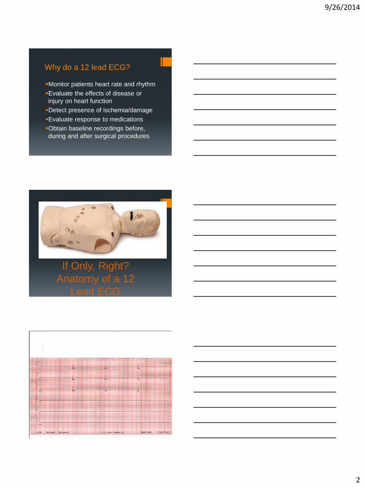
Why do a 12 lead ECG?
Monitor patients heart rate and rhythm
Evaluate the effects of disease or
injury on heart function
Detect presence of ischemia/damage
Evaluate response to medications
Obtain baseline recordings before,
during and after surgical procedures
If Only, Right?
Anatomy of a 12
Lead ECG
9/26/2014
3
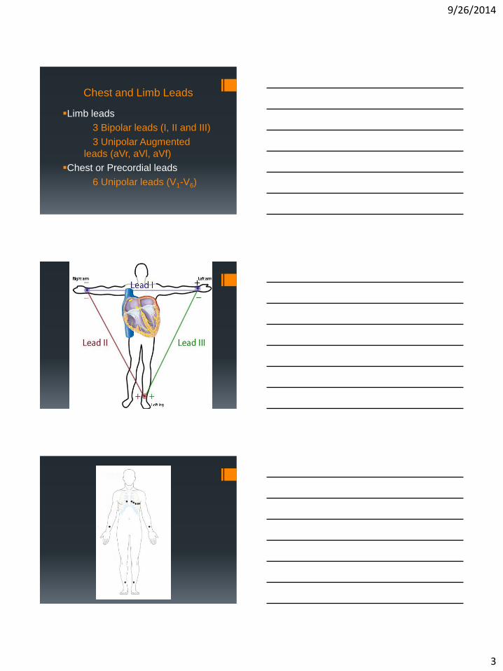
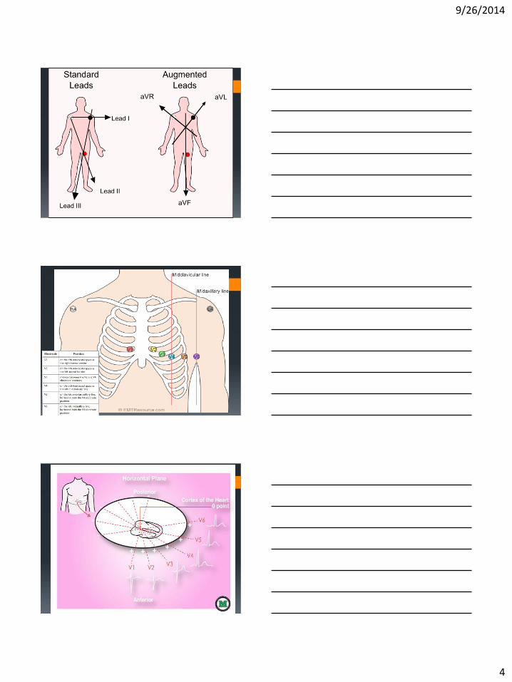
Chest and Limb Leads
Limb leads
3 Bipolar leads (I, II and III)
3 Unipolar Augmented
leads (aVr, aVl, aVf)
Chest or Precordial leads
6 Unipolar leads (V1-V6)
9/26/2014
4
9/26/2014
5
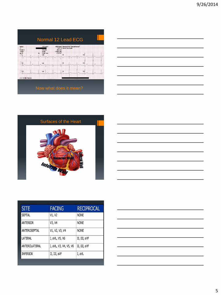
Normal 12 Lead ECG
Now what does it mean?
Surfaces of the Heart

9/26/2014
6
Ischemia, Injury or Infarction?
9/26/2014
7
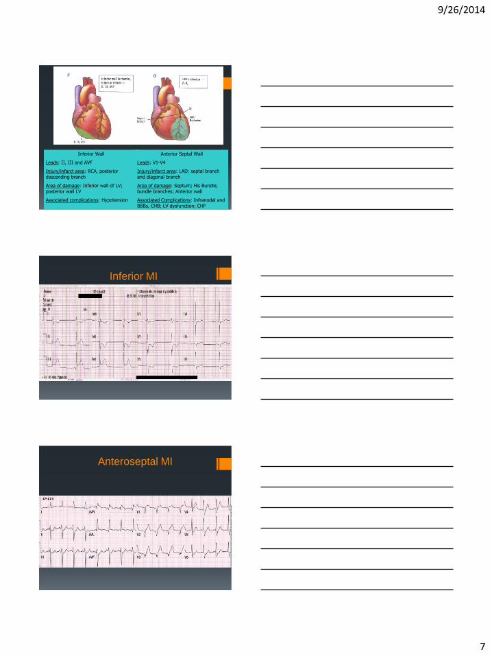
Inferior Wall
Leads: II, III and AVF
Injury/infarct area: RCA, posterior descending branch
Area of damage: Inferior wall of LV; posterior wall LV
Associated complications: Hypotension
Anterior Septal Wall
Leads: V1-V4
Injury/infarct area: LAD: septal branch and diagonal branch
Area of damage: Septum; His Bundle; bundle branches; Anterior wall
Associated Complications: Infranodal and BBBs, CHB; LV dysfunction; CHF
Inferior MI
Anteroseptal MI
9/26/2014
8
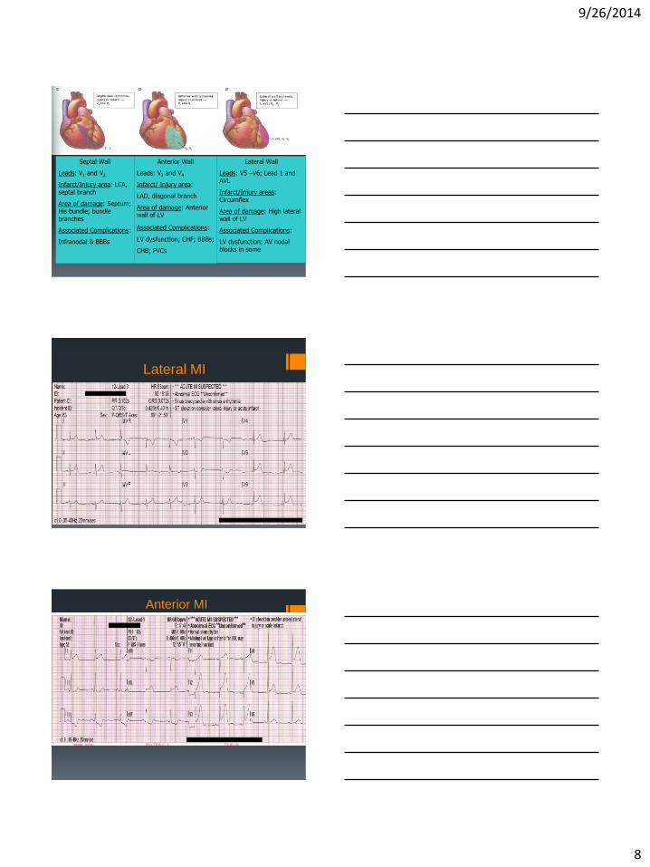
Septal Wall
Leads: V1 and V2
Infarct/Injury area: LCA, septal branch
Area of damage: Septum; His bundle; bundle branches
Associated Complications:
Infranodal & BBBs
Anterior Wall
Leads: V3 and V4
Infarct/ Injury area:
LAD, diagonal branch
Area of damage: Anterior wall of LV
Associated Complications:
LV dysfunction; CHF; BBBs;
CHB; PVCs
Lateral Wall
Leads: V5 –V6; Lead 1 and AVL
Infarct/Injury areas: Circumflex
Area of damage: High lateral wall of LV
Associated Complications:
LV dysfunction; AV nodal blocks in some
Lateral MI
Anterior MI
9/26/2014
9
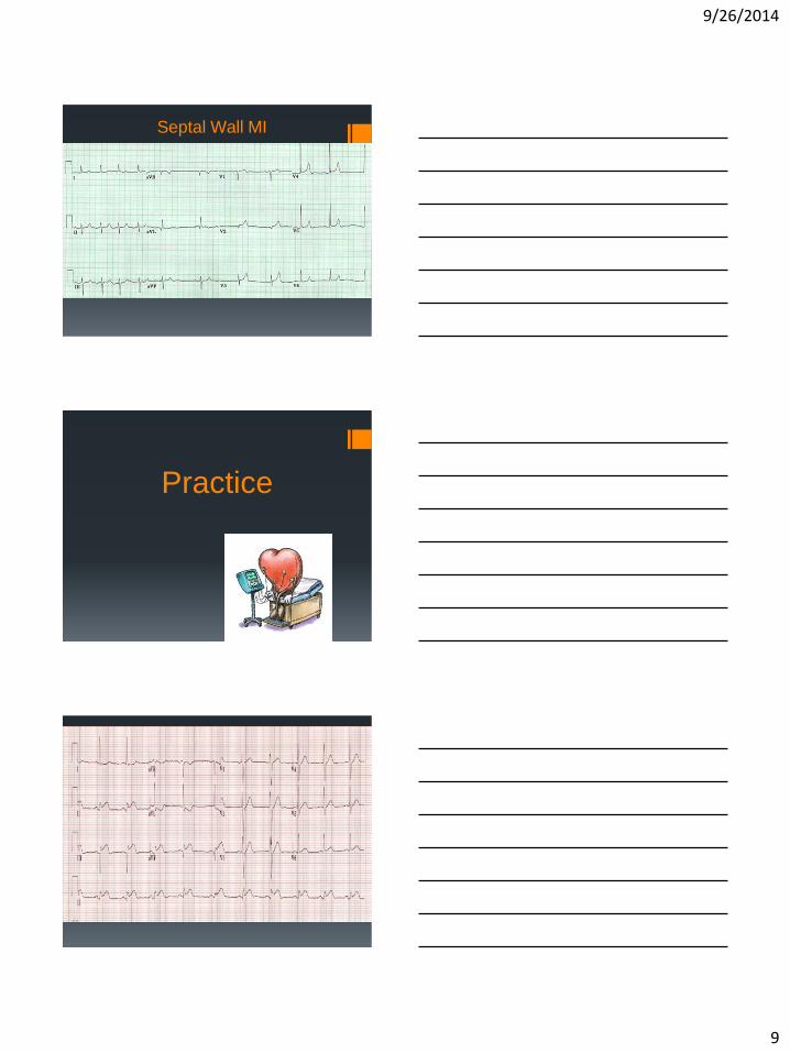
Septal Wall MI
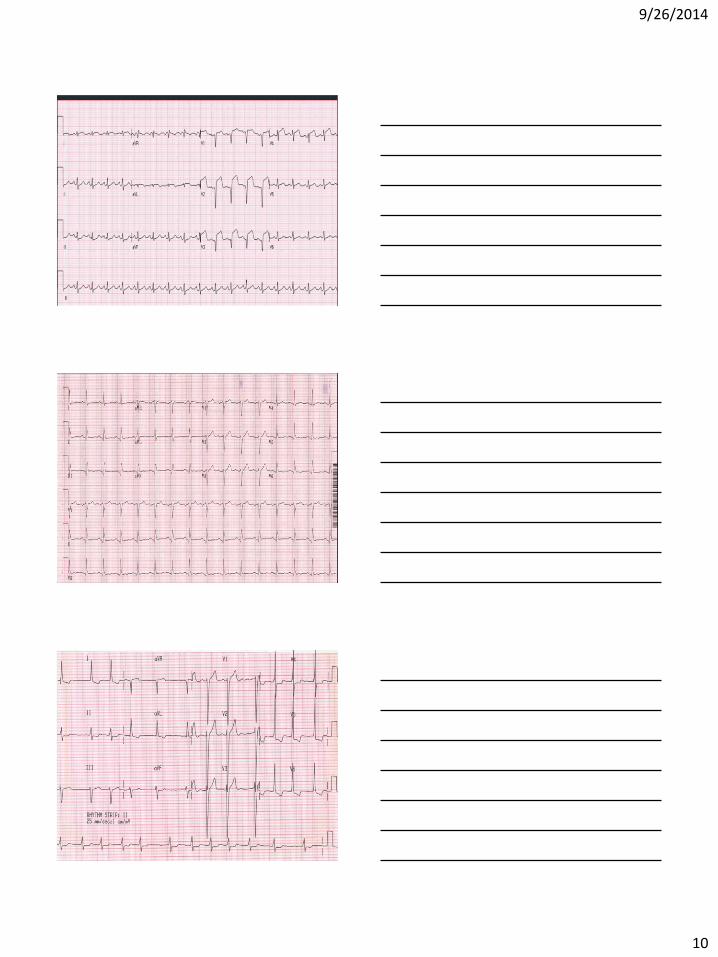
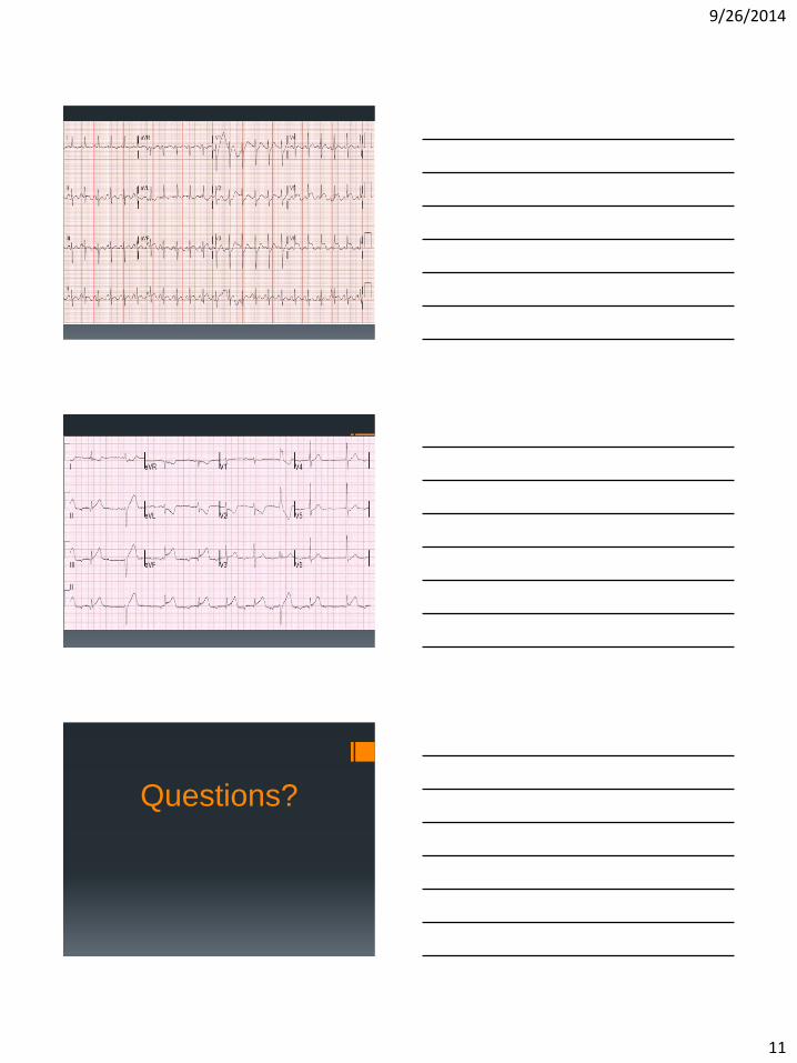
Practice
9/26/2014
10
9/26/2014
11
Questions?
9/26/2014
12
References
Ellis, Karen M. (2007) ECG Plain and Simple. Upper Saddle River, NJ: Pearson/Prentice Hall Publishers
ECG Interpretation Made Incredibly Easy, 2nd Ed. (2002). Springhouse Publishing Company
Geiter, Jr. Henry B. (2007). E-Z ECG Rhythm Interpretation. Philadelphia: F.A. Davis Company
Thaler, Malcolm S. (2010). The Only EKG Book You’ll Ever Need, 6th Ed. Baltimore, et al: Lippincott Williams & Wilkins
“Learning about 12 Lead ECGs” Power Point module: University of Detroit/Mercy. Accessed September 2014 http://urbanhealth.udmercy.edu/ekg/learn.html
Bouthillet, Tom. 12 Lead ECG and Stemi Basics. (2010). Presentation for Hilton Head EMS/Fire and Rescue. Accessed September 2014