baicalein restrains proliferation, migration, and invasion ... · baicalein restrains...
TRANSCRIPT

Baicalein restrains proliferation, migration, andinvasion of human malignant melanoma cells by
down-regulating colon cancer associated transcript-1
Xiaoliang Yang0000-0000-0000-00001, Jinjie Jiang0000-0000-0000-0000
1, Chunyan Zhang0000-0000-0000-00002, and Yinghao Li0000-0000-0000-0000
1
1Department of Burn and Plastic Surgery, Qingdao Central Hospital, The Affiliated Central Hospital of Qingdao University,Qingdao, Shandong, China
2Department of Traditional Chinese Medicine, Qingdao Central Hospital, The Affiliated Central Hospital of Qingdao University,Qingdao, Shandong, China
Abstract
Baicalein (BAI) is an acknowledged flavonoids compound, which is regarded as a useful therapeutic pharmaceutical fornumerous cancers. However, its involvement in melanoma is largely unknown. This study aimed to examine the anti-melanomafunction of BAI and unraveled the regulatory mechanism involved. A375 and SK-MEL-28 were treated with BAI for 24 h. Then,CCK-8 assay, flow cytometry, and transwell assay were carried out to investigate cell growth, migration, and invasion. RT-qPCRwas applied to detect the expression of colon cancer associated transcript-1 (CCAT1) in melanoma tissues and cells. Thefunctions of CCAT1 in melanoma cells were also evaluated. Western blot was utilized to appraise Wnt/b-catenin or MEK/ERKpathways. BAI restrained cell proliferation and stimulated cell apoptotic capability of melanoma by suppressing cleaved-caspase-3 and cleaved-PARP. Cell migratory and invasive abilities were restrained by BAI via inhibiting MMP-2 and vimentin.CCAT1 was over-expressed in melanoma tissues and down-regulated by BAI in melanoma cells. Overexpressed CCAT1reversed the BAI-induced anti-growth, anti-migratory, and anti-invasive effects. Furthermore, BAI inhibited Wnt/b-catenin andMEK/ERK pathways-axis via regulating CCAT1. Our study indicated that BAI blocked Wnt/b-catenin and MEK/ERK pathwaysvia regulating CCAT1, thereby inhibiting melanoma cell proliferation, migration, and invasion.
Key words: Malignant melanoma; Colon cancer associated transcript-1; Wnt/b-catenin; MEK/ERK
Introduction
Melanoma evolves from skin mucosa or pigmentedmembrane and is the most common cancer with highmetastatic potential (1). Malignant melanoma, caused bythe abnormal transformation of normal melanocytes, isone of the fastest growing malignant tumors with anannual growth rate of 3–5% (2). To date, surgery andchemotherapy combined with immunotherapy are the mostcommon endorsed therapeutic approaches to melanoma(3,4). Nevertheless, the biggest disadvantage of thesetherapies is toxicity. Thus, there is research focused onnatural products towards cell metastasis of melanoma (5).However, the potential value of traditional Chinese medi-cine in the treatment of melanoma has not been assessed.
Scutellaria baicalensis Georgi is a kind of traditionalChinese medicine containing several flavonoids. One ofthe ingredients is baicalein (BAI), which is commonlyregarded as useful adjuvant therapeutic pharmaceuticalfor various diseases (6). Thus far, a number of research-ers tested the efficacy of BAI on malignant tumors, such
as breast carcinoma (7), non-small-cell lung carcinoma(8), cervical carcinoma (9), and carcinoma of urinarybladder (10). Moreover, previous research indicated thatBAI impeded cell proliferation and melanogenesis ofB16F10 mouse melanoma cells (11,12). What is not yetclear is the functional mechanism of BAI on humanmalignant melanoma.
Long noncoding RNAs (lncRNAs) are RNA segmentswith no fewer than 200 nucleotides in length that do notencode proteins (13). lncRNAs are closely linked tomiscellaneous regulations, functioning as regulators ofgene transcription, RNA splicing, and miRNA regulatorysystems (14,15). A number of investigators reported thatlncRNAs SLNCR1 and HEIH interfered with the mela-noma cell proliferative potential, migratory status, andinvasive ability via regulating corresponding downstreamtargets (16,17). Colon cancer associated transcript-1(CCAT1), an innovative tumor-related lncRNA, plays anessential role in tumor progression, being up-regulated
Correspondence: Yinghao Li: <[email protected]>
Received June 15, 2019 | Accepted September 9, 2019
Braz J Med Biol Res | doi: 10.1590/1414-431X20198934
Brazilian Journal of Medical and Biological Research (2019) 52(12): e8934, http://dx.doi.org/10.1590/1414-431X20198934ISSN 1414-431X Research Article
1/9

in malignancies (18). However, the extent to whichCCAT1 is related to malignant melanoma remains poorlyunderstood.
Here, we demonstrated a crucial role of BAI ininhibiting cell growth and motility by mediating CCAT1as well as the underlying mechanism of BAI-inducedsignaling pathways in human melanoma cells. Our findingsmight provide new insights into the application of traditionalChinese medicine and feasible therapies for malignantmelanoma.
Material and Methods
Clinical tissuesTwenty-two pairs of human melanoma tissues and
corresponding paracancerous skin specimens were col-lected from patients at Qingdao Central Hospital (Qing-dao, Shandong) from January 2017 to July 2018. Thirteencases were from males and 9 were from females, who didnot receive any radiation or chemotherapy before surgery.Participants signed an authorization and the EthicsCommittee of Qingdao Central Hospital approved theprocedures and the study.
Cell culture and treatmentThe malignant melanoma cell lines A375 and
SK-MEL-28, which were cultured in DMEM (Gibco, USA)enriched with 10% fetal bovine serum (FBS, Gibco), wereobtained from ATCC (USA). The conditions for cell culturewere 5% CO2 and 37°C. BAI was obtained from NanjingZeLang Medical Technology Co. Ltd. (#ZL100708, China).BAI was diffused in DMSO as a storage concentration anddiluted using DMEM to work concentrations (100, 50, 20,and 10 mM). The cells were treated with BAI for 24 h.
Cell transfectionThe entire length of CCAT1 was concatenated into
the pcDNA3.1 vector (GenePharma, China). The recom-bination plasmid was termed as pCCAT1. The lipofecta-mine 2000 reagent (Life Technology, USA) was used forthe cells transfection. The stably transfected cells werecultured in DMEM combined with 0.5 mg/mL G418(Solarbio, China). Four weeks later, stable transfectedcells were formed.
Cell viability assayCells (5� 103/well) were seeded into 96-well plates
and were raised for 48 h. After treatment with BAI, 10 mLCell Counting Kit-8 (CCK-8, Dojindo, USA) solutions wereadded to the cultures. Then, cultures were incubated for1 h at 37°C. Microplate Reader (Bio-Rad, USA) wasemployed to evaluate the cell viability at 450 nm.
Bromodeoxyuridine (BrdU) assayCell proliferation was determined using BrdU (Sigma-
Aldrich, USA). After treatment of BAI, BrdU (1 mg/mL)
was added to the cells for 3 h. Then, immunofluorescenceassay was carried out to estimate the BrdU-tagged cells,providing the cell proliferation rate.
Cell migration and invasion assaysCell migratory capacity and invasive potential were
assessed by transwell culture chamber (Corning Cosatar,USA), which consists of 8-mm pore polycarbonatemembrane. Firstly, 200 mL of 1�104 cells, which werecultured in DMEM without FBS, were seeded into the topchamber, which had been covered with Matrigel matrix(Becton Dickinson, USA) for invasion assay or keptuncovered for migration assay. Consequently, 800 mLmedium was injected to the lower chamber. After 24 h, themigratory cells were fixed with methyl alcohol and dyedwith 0.5% crystal violet liquid (Solarbio). Then, the relativemigration rates were calculated. After 48 h, the invadingcells were processed in the above same manner and thenumber of invading cells was counted.
Apoptosis assayApoptotic cells proportion was measured utilizing PI/
FITC-Annexin V staining kit (Invitrogen, USA). In brief,cells (1�106/well) were cultured into 6-well plates andstarved in FBS-free medium for 12 h. Next, PI andAnnexin V-FITC solutions were added to the cell cultures.Flow cytometry was performed with FACScan (BectonDickinson). The apoptosis ratio was calculated usingFlowJo software (Becton Dickinson).
Reverse transcription and quantitative real-time PCR(RT-qPCR)
Trizol reagent (Life Technologies Corporation, USA)was utilized to isolate total RNA of tissue samples and cellcultures. Reverse transcription of RNA was implementedutilizing SuperRT cDNA Synthesis Kit (Cwbio, China).SYBRs Green PCR Kit (Qiagen, Germany) was employedfor qPCR analysis to detect CCAT1 expression. qPCR wasexecuted on iQ5 real-time PCR Detection system (Bio-Rad). The mRNA expression of CCAT1 was normalizedwith b-actin. The relative quantification of CCAT1 in tumortissues and cells was calculated using the equation:amount of target = 2-DDCt (19).
Western blotTotal proteins were extracted from cells utilizing RIPA
lysis buffer (Cwbio), which contains phenylmethylsulfonylfluoride (PMSF, Solarbio). Proteins were quantified by theSuper-Bardford Pritein Assay Kit (Cwbio). The extractionswere loaded into 12% polyacrylamide gel on the Bis-TrisGel system (Bio-Rad). The products were transferred ontopolyvinylidene fluoride (PVDF) membranes, which werethen cultivated at 4°C overnight with primary antibodies.The primary antibodies included anti-cleaved-caspase-3(#ab2303, Abcam, USA), anti-cleaved-PARP (#ab3246,Abcam), anti-MMP-2 (#ab37150, Abcam), anti-vimentin
Braz J Med Biol Res | doi: 10.1590/1414-431X20198934
Role of baicalein in human malignant melanoma 2/9

(#ab92547, Abcam), anti-Wnt3a (#ab219412, Abcam),anti-b-catenin (#ab32572, Abcam), anti-t-MEK (#9126,Cell Signaling Technology, USA), anti-p-MEK (#9154, CellSignaling Technology), anti-t-ERK (#9102, Cell SignalingTechnology), anti-p-ERK (#4370, Cell Signaling Technol-ogy), and anti-b-actin (#ab179467, Abcam). Then, thePVDF membranes were rinsed and incubated withhorseradish peroxidase-conjugated goat anti-rabbit IgG(#ab6721, Abcam) and goat anti-mouse IgG (#ab205719,Abcam) for 1 h at 20°C. After washing, the PVDFmembranes were treated with ChemiDoct XRS system(Bio-Rad), and the intensity of bands was finallyevaluated with ImageJ software (NIH, USA).
Statistical analysisEach experiment was repeated three times. Graphpad
6.0 software (USA) was utilized for statistical analysis.Data are reported as means±SD. Analysis of variance(ANOVA) and Student’s t-test were applied to calculateP values. A P value o0.05 was regarded as significant.
Results
BAI attenuated cell proliferation and promoted cellapoptosis of malignant melanoma cells
Figure 1A presents the inhibition of BAI on cell viability.Cells were sensitive to 20 mM BAI compared with theuntreated group (Po0.01). Cell viability was impeded byBAI with an inhibitory concentration of 50% (50 mM).Therefore, 50 mM was considered to be an acceptableconcentration for the next proliferation and apoptosisassay. Figure 1B shows that BAI (50 mM) significantlyinhibited the cell proliferation of A375 and SK-MEL-28 cells compared to the untreated cells (Po0.001).Reversely, flow cytometry using PI/FITC-Annexin Vindicated that BAI promoted cell apoptosis compared withthe untreated group (Po0.01, Figure 1C). We analyzedthe expression of cleaved-caspase-3, which acted in cellapoptosis and participated in the cleavage of repairenzymes, such as PARP (20). The protein expressionanalysis was consistent with the result of flow cytometry.
Figure 1. Baicalein (BAI) attenuated cell proliferation and strengthened cell apoptotic capacity of malignant melanoma cells. A, Cellviability of A375 and SK-MEL-28 cells followed by 24-h treatment with BAI (0, 10, 20, 50, and 100 mM) was assessed by CCK-8. B, Cellproliferation of melanoma cells was examined by bromodeoxyuridine (BrdU) assay. C, Flow cytometry was utilized to assess theapoptotic rate of melanoma cells. D, Expression of cleaved-caspase-3 and cleaved-PARP was tested by western blot assay. The relativeexpression of protein was normalized by b-actin. Data are reported as mean±SD. **Po0.01, ***Po0.001 (ANOVA). ns: not significant;CTRL: control.
Braz J Med Biol Res | doi: 10.1590/1414-431X20198934
Role of baicalein in human malignant melanoma 3/9
BAI treatment accelerated cleaved-caspase-3 and cleaved-PARP expression compared with the untreated cells(Po0.001, Figure 1D). The experiments detected someevidence for the inhibitory effect of BAI on the growth ofmalignant melanoma cells.
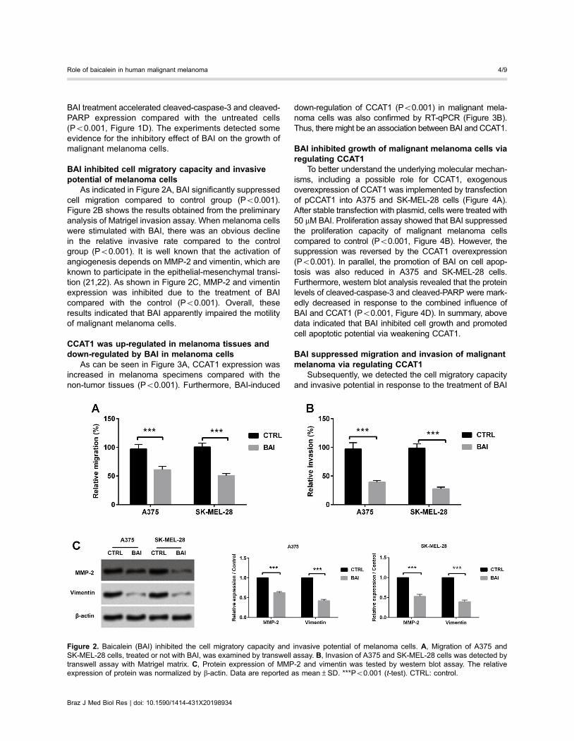
BAI inhibited cell migratory capacity and invasivepotential of melanoma cells
As indicated in Figure 2A, BAI significantly suppressedcell migration compared to control group (Po0.001).Figure 2B shows the results obtained from the preliminaryanalysis of Matrigel invasion assay. When melanoma cellswere stimulated with BAI, there was an obvious declinein the relative invasive rate compared to the controlgroup (Po0.001). It is well known that the activation ofangiogenesis depends on MMP-2 and vimentin, which areknown to participate in the epithelial-mesenchymal transi-tion (21,22). As shown in Figure 2C, MMP-2 and vimentinexpression was inhibited due to the treatment of BAIcompared with the control (Po0.001). Overall, theseresults indicated that BAI apparently impaired the motilityof malignant melanoma cells.
CCAT1 was up-regulated in melanoma tissues anddown-regulated by BAI in melanoma cells
As can be seen in Figure 3A, CCAT1 expression wasincreased in melanoma specimens compared with thenon-tumor tissues (Po0.001). Furthermore, BAI-induced
down-regulation of CCAT1 (Po0.001) in malignant mela-noma cells was also confirmed by RT-qPCR (Figure 3B).Thus, there might be an association between BAI and CCAT1.
BAI inhibited growth of malignant melanoma cells viaregulating CCAT1
To better understand the underlying molecular mechan-isms, including a possible role for CCAT1, exogenousoverexpression of CCAT1 was implemented by transfectionof pCCAT1 into A375 and SK-MEL-28 cells (Figure 4A).After stable transfection with plasmid, cells were treated with50 mM BAI. Proliferation assay showed that BAI suppressedthe proliferation capacity of malignant melanoma cellscompared to control (Po0.001, Figure 4B). However, thesuppression was reversed by the CCAT1 overexpression(Po0.001). In parallel, the promotion of BAI on cell apop-tosis was also reduced in A375 and SK-MEL-28 cells.Furthermore, western blot analysis revealed that the proteinlevels of cleaved-caspase-3 and cleaved-PARP were mark-edly decreased in response to the combined influence ofBAI and CCAT1 (Po0.001, Figure 4D). In summary, abovedata indicated that BAI inhibited cell growth and promotedcell apoptotic potential via weakening CCAT1.
BAI suppressed migration and invasion of malignantmelanoma via regulating CCAT1
Subsequently, we detected the cell migratory capacityand invasive potential in response to the treatment of BAI
Figure 2. Baicalein (BAI) inhibited the cell migratory capacity and invasive potential of melanoma cells. A, Migration of A375 andSK-MEL-28 cells, treated or not with BAI, was examined by transwell assay. B, Invasion of A375 and SK-MEL-28 cells was detected bytranswell assay with Matrigel matrix. C, Protein expression of MMP-2 and vimentin was tested by western blot assay. The relativeexpression of protein was normalized by b-actin. Data are reported as mean±SD. ***Po0.001 (t-test). CTRL: control.
Braz J Med Biol Res | doi: 10.1590/1414-431X20198934
Role of baicalein in human malignant melanoma 4/9

Figure 3. CCAT1 was up-regulated in melanoma tissues and down-regulated by baicalein (BAI) in melanoma cells. A, Expression ofCCAT-1 in malignant melanoma tissues (n=22) and non-tumor skin specimens (n=22) was analyzed by RT-qPCR. B, Expression ofCCAT-1 in malignant melanoma cells after treating with BAI or not was determined by RT-qPCR. Data are reported as mean±SD.***Po0.001 (t-test). CTRL: control.
Figure 4. Baicalein (BAI) inhibited the growth of malignant melanoma cells via regulating CCAT1. A, RT-qPCR assay was used toestimate CCAT1 expression in A375 and SK-MEL-28 after transfection with pCCAT1. B and C, BradU assay and flow cytometry assayswere utilized to evaluate overexpression of CCAT1 and BAI on cell proliferation and apoptosis. D, Western blot assay evaluatedthe relative expression levels of cleaved-caspase-3 and cleaved-PARP. The relative expression of protein was normalized by b-actin.Data are reported as mean±SD. *Po0.05, ***Po0.001 (ANOVA). CTRL: control.
Braz J Med Biol Res | doi: 10.1590/1414-431X20198934
Role of baicalein in human malignant melanoma 5/9
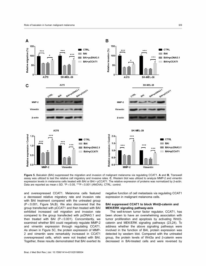
and overexpressed CCAT1. Melanoma cells featureda decreased relative migratory rate and invasion ratewith BAI treatment compared with the untreated group(Po0.001, Figure 5A,B). We also discovered that thegroup transfected with pCCAT1 and then treated with BAIexhibited increased cell migration and invasion ratecompared to the group transfected with pcDNA3.1 andthen treated with BAI (Po0.001). Concomitantly, weexamined whether BAI could negatively regulate MMP-2and vimentin expression through regulating CCAT1.As shown in Figure 5C, the protein expression of MMP-2 and vimentin were remarkably increased in CCAT1overexpressed cells, which were not treated with BAI.Together, these results demonstrated that BAI exerted its
negative function of cell metastasis via regulating CCAT1expression in malignant melanoma cells.
BAI suppressed CCAT1 to block Wnt/b-catenin andMEK/ERK signaling pathway-axis
The well-known tumor factor regulator, CCAT1, hasbeen shown to have an overwhelming association withtumor proliferation and apoptosis by activating Wnt/b-catenin and MEK/ERK signaling pathways (23,24). Toaddress whether the above signaling pathways wereinvolved in the function of BAI, protein expression wasdetected by western blot. Compared with the untreatedgroup, the protein levels of Wnt3a and b-catenin weredecreased in BAI-treated cells and were reversed by
Figure 5. Baicalein (BAI) suppressed the migration and invasion of malignant melanoma via regulating CCAT1. A and B, Transwellassay was utilized to test the relative cell migratory and invasive rates. C, Western blot was utilized to analyze MMP-2 and vimentinexpression levels in melanoma cells treated with BAI or BAI+pCCAT1. The relative expression of proteins was normalized by b-actin.Data are reported as mean±SD. *Po0.05, ***Po0.001 (ANOVA). CTRL: control.
Braz J Med Biol Res | doi: 10.1590/1414-431X20198934
Role of baicalein in human malignant melanoma 6/9

exogenous CCAT1 (Po0.001, Figure 6A). Similarly, theprotein levels of p-MEK and p-ERK were also suppressedby BAI (Po0.001, Figure 6B). The results indicated thatBAI blocked Wnt/b-catenin or MEK/ERK pathways bynegatively regulating CCAT1.
Discussion
Numerous active components extracted from tradi-tional Chinese medicinal plants exert multiple pharmaco-logical effects (25). Among these, perhaps the most
Figure 6. Baicalein (BAI) suppressed CCAT1 to block Wnt/b-catenin and MEK/ERK signaling pathway. A, The expression of Wnt3a andb-catenin and, B, of p-MEK, p-ERK, t-MEK, and t-ERK were examined by western blot. The relative expression of proteins wasnormalized by b-actin. Data are reported as mean±SD. ***Po0.001 (ANOVA). CTRL: control.
Braz J Med Biol Res | doi: 10.1590/1414-431X20198934
Role of baicalein in human malignant melanoma 7/9
unexpected finding was that BAI induced growth of HeLacells via mitochondrial and death receptor pathways (9).Although it has been reported that BAI could act as anessential anti-tumor modulator, leading to amelioratedbiological processes, such as programmed cell death andangiogenesis in the B16F10 cells (26), the underlyingmolecular mechanisms remained to be fully demon-strated. Our study found that there were intricate regulat-ing effects between BAI and the progression of malignantmelanoma.
BAI is a vigorous herbal medicine that exerts indis-pensable functions towards the cardiovascular systemand hepatoma (27,28). The function of BAI mainly displaysas two aspects: anti-oxidative and inhibitory action oncell growth. Chou et al. (11) showed that BAI caused areduction in cellular viability of melanoma cells throughgenerating ROS scavengers. Existing research recog-nized that BAI inhibited tumor growth via activation ofcleaved-caspase-3 (26). The results of our study were inline with the above previous experiments. We found thatBAI alleviated cell growth, and migratory and invasiveability in malignant melanoma. Our findings indicated thatBAI exerted indispensable functions as tumor suppressor.
It was reported that abnormal expression of lncRNAsmight be related to a wide spectrum of tumor biologicalprocesses (29). Reports such as that conducted by Wu etal. (30) show that overexpression of CCAT1 significantlyelicit cell proliferation and invasion and inhibit cell cycle inclear cell renal cell carcinoma. Beyond that, Lv et al. (31)verified that CCAT1 served as an oncogenic factor inmelanoma genesis, accumulating cell proliferation, migra-tion, and invasion abilities. However, there is a relativepaucity of literature concerning CCAT1 involvement inregulating the effects of BAI on melanoma biological
processes. In this study, we measured CCAT1 expressionlevel and found that CCAT1 was up-regulated in mela-noma. We showed for the first time that BAI inhibited cellproliferation, migration, and invasion of malignant mela-noma via regulating CCAT1.
Wnt3a is a key activator of Wnt pathway, generallytriggering the acknowledged Wnt/b-catenin signalingpathway (32) and is related to diversified biologicalprocesses, such as cell growth and migration (33,34).The MAPK/ERK signaling pathway regulates cell prolif-eration and differentiation in many tumor cells (35,36) andis associated with melanin synthesis (37). Debates havebeen raised about the signaling pathway involved in theprogression of malignant melanoma. Results from earlierstudies demonstrated that BAI inhibited melanogenesisthrough activation of the ERK signaling pathway but didnot induce AKTactivation (12). Recent investigators foundthat BAI impeded the migratory and invasive potentialof B16F10 cells through the suppression of PI3K /AKTsignaling pathway (38). The present experiments uncov-ered the down-regulated protein expression of Wnt3a,b-catenin, p-MEK, and p-ERK in malignant melanomacells treated with BAI. The restraint was reversed byexogenous expressed CCAT1. Therefore, we speculatedthat BAI blocked Wnt/b-catenin or MEK/ERK signalingpathways by regulating CCAT1.
Overall, our study indicated that BAI hindered Wnt/b-catenin and MEK/ERK signaling pathways by regulatingCCAT1, thereby inhibiting proliferation, migration, andinvasion of melanoma cells. The present study demon-strated a pivotal role of BAI in tumor regulation, whichmight provide new light on the development of therapeuticstrategies against malignant melanoma. Comprehensivein vivo experiments are crucial for future research.
References
1. Cao C, Su Y, Gao Y, Luo C, Yin L, Zhao Y, et al. Ginkgobiloba exocarp extract inhibits the metastasis of B16-F10melanoma involving PI3K/Akt/NF-kappaB/MMP-9 signalingpathway. Evid Based Complement Alternat Med 2018; 2018:4969028, doi: 10.1155/2018/4969028.
2. Trotter SC, Sroa N, Winkelmann RR, Olencki T, Bechtel M.A global review of melanoma follow-up guidelines. J ClinAesthet Dermatol 2013; 6: 18–26.
3. Robert C, Karaszewska B, Schachter J, Rutkowski P,Mackiewicz A, Stroiakovski D, et al. Improved overallsurvival in melanoma with combined dabrafenib andtrametinib. N Engl J Med 2015; 372: 30–39, doi: 10.1056/NEJMoa1412690.
4. Puzanov I, Flaherty KT. Targeted molecular therapy inmelanoma. Semin Cutan Med Surg 2010; 29: 196–201, doi:10.1016/j.sder.2010.06.005.
5. Luke JJ, Flaherty KT, Ribas A, Long GV. Targeted agentsand immunotherapies: optimizing outcomes in melanoma.Nat Rev Clin Oncol 2017; 14: 463–482, doi: 10.1038/nrclinonc.2017.43.
6. Liu H, Dong Y, Gao Y, Du Z, Wang Y, Cheng P, et al. Thefascinating effects of baicalein on cancer: a review. Int J MolSci 2016; 17. pii: E1681, doi: 10.3390/ijms17101681.
7. Chen Y, Chen L, Hong D, Chen Z, Zhang J, Fu L, et al.Baicalein inhibits fibronectin-induced epithelial-mesenchy-mal transition by decreasing activation and upregulation ofcalpain-2. Cell Death Dis 2019; 10: 341, doi: 10.1038/s41419-019-1572-7.
8. Zhao Z, Liu B, Sun J, Lu L, Liu L, Qiu J, et al. Baicaleininhibits orthotopic human non-small cell lung cancerxenografts via Src/Id1 pathway. Evid Based ComplementAlternat Med 2019; 2019: 9806062, doi: 10.1155/2019/9806062.
9. Peng Y, Guo C, Yang Y, Li F, Zhang Y, Jiang B, et al.Baicalein induces apoptosis of human cervical cancer HeLacells in vitro. Mol Med Rep 2015; 11: 2129–2134, doi:10.3892/mmr.2014.2885.
10. Yang Y, Liu K, Yang L, Zhang G. Bladder cancer cellviability inhibition and apoptosis induction by baicaleinthrough targeting the expression of anti-apoptotic genes.
Braz J Med Biol Res | doi: 10.1590/1414-431X20198934
Role of baicalein in human malignant melanoma 8/9

Saudi J Biol Sc 2018; 25: 1478–1482, doi: 10.1016/j.sjbs.2017.03.014.
11. Chou DS, Hsiao G, Lai YA, Tsai YJ, Sheu JR. Baicaleininduces proliferation inhibition in B16F10 melanoma cells bygenerating reactive oxygen species via 12-lipoxygenase.Free Radic Biol Med 2009; 46: 1197–1203, doi: 10.1016/j.freeradbiomed.2009.01.024.
12. Li X, Guo L, Sun Y, Zhou J, Gu Y, Li Y. Baicalein inhibitsmelanogenesis through activation of the ERK signalingpathway. Int J Mol Med 2010; 25: 923–927, doi: 10.3892/ijmm_00000423.
13. Peng WX, Koirala P, Mo YY. LncRNA-mediated regulation ofcell signaling in cancer. Oncogene 2017; 36: 5661–5667,doi: 10.1038/onc.2017.184.
14. Valadkhan S, Gunawardane LS. lncRNA-mediated regula-tion of the interferon response. Virus Res 2016; 212: 127–136, doi: 10.1016/j.virusres.2015.09.023.
15. Liz J, Esteller M. lncRNAs and microRNAs with a role incancer development. Biochim Biophys Acta 2016; 1859:169–176, doi: 10.1016/j.bbagrm.2015.06.015.
16. Zhao H, Xing G, Wang Y, Luo Z, Liu G, Meng H. Longnoncoding RNA HEIH promotes melanoma cell proliferation,migration and invasion via inhibition of miR-200b/a/429.Biosci Rep 2017; 37. pii. BSR20170682, doi: 10.1042/BSR20170682.
17. Schmidt K, Joyce CE, Buquicchio F, Brown A, Ritz J, DistelRJ, et al. The lncRNA SLNCR1 mediates melanoma invasionthrough a conserved SRA1-like region. Cell Rep 2016; 15:2025–2037, doi: 10.1016/j.celrep.2016.04.018.
18. Wang N, Yu Y, Xu B, Zhang M, Li Q, Miao L. Pivotalprognostic and diagnostic role of the long noncoding RNAcolon cancerassociated transcript 1 expression in humancancer (Review). Mol Med Rep 2019; 19: 771–782.
19. Livak KJ, Schmittgen TD. Analysis of relative gene expres-sion data using real-time quantitative PCR and the 2�DDCT method. Methods 2001; 25: 402–408, doi: 10.1006/meth.2001.1262.
20. Fernandes-Alnemri T, Litwack G, Alnemri ES. CPP32, anovel human apoptotic protein with homology to Caenor-habditis elegans cell death protein Ced-3 and mammalianinterleukin-1 beta-converting enzyme. J Biol Chem 1994;269: 30761–30764.
21. Liang X, Sun R, Zhao X, Zhang Y, Gu Q, Dong X, et al.Rictor regulates the vasculogenic mimicry of melanoma viathe AKT-MMP-2/9 pathway. J Cel Mol Med 2017; 21: 3579–3591, doi: 10.1111/jcmm.13268.
22. Ma H, Qiu P, Xu H, Xu X, Xin M, Chu Y, et al. The inhibitoryeffect of propylene glycol alginate sodium sulfate onfibroblast growth factor 2-mediated angiogenesis and inva-sion in murine melanoma B16-F10 cells in vitro. Mar Drugs2019; 17. pii: E257, doi: 10.3390/md17050257.
23. Gao JZY. CCAT-1 promotes proliferation and inhibitsapoptosis of cervical cancer cells via the Wnt signalingpathway. Oncotarget 2017; 8: 68059–68070, doi: 10.18632/oncotarget.19155.
24. Gao R, Zhang R, Zhang C, Zhao L, Zhang Y. Longnoncoding RNA CCAT1 promotes cell proliferation andmetastasis in human medulloblastoma via MAPK pathway.Tumori 2018; 104: 43–50, doi: 10.5301/tj.5000662.
25. Dai SX, Li WX, Han FF, Guo YC, Zheng JJ, Liu JQ, et al. Insilico identification of anti-cancer compounds and plantsfrom traditional Chinese medicine database. Sci Rep 2016;6: 25462, doi: 10.1038/srep25462.
26. Park YG, Choi J, Jung HK, Kim B, Kim C, Park SY, et al.Baicalein inhibits tumor progression by inhibiting tumor cellgrowth and tumor angiogenesis. Oncol Rep 2017; 38: 3011–3018, doi: 10.3892/or.2017.6007.
27. Huang Y, Tsang SY, Yao X, Chen ZY. Biological properties ofbaicalein in cardiovascular system. Curr Drug TargetsCardiovasc Haematol Disord 2005; 5: 177–184, doi:10.2174/1568006043586206.
28. Motoo Y, Sawabu N. Antitumor effects of saikosaponins,baicalin and baicalein on human hepatoma cell lines.Cancer Lett 1994; 86: 91–95, doi: 10.1016/0304-3835(94)90184-8.
29. Salmena L, Poliseno L, Tay Y, Kats L, Pandolfi PP.A ceRNA hypothesis: the Rosetta Stone of a hidden RNAlanguage? Cell 2011; 146: 353–358, doi: 10.1016/j.cell.2011.07.014.
30. Wu Y, Tan C, Weng WW, Deng Y, Zhang QY, Yang XQ, et al.Long non-coding RNA Linc00152 is a positive prognosticfactor for and demonstrates malignant biological behavior inclear cell renal cell carcinoma. Am J Cancer Res 2016; 6:285–299.
31. Lv L, Jia JQ, Chen J. The lncRNA CCAT1 upregulatesproliferation and invasion in melanoma cells via suppressingmiR-33a. Oncol Res 2018; 26: 201–208, doi: 10.3727/096504017X14920318811749.
32. Fuster-Matanzo A, Manferrari G, Marchetti B, Pluchino S.Wnt3a promotes pro-angiogenic features in macrophages invitro: Implications for stroke pathology. Exp Biol Med 2018;243: 22–28, doi: 10.1177/1535370217746392.
33. Lie DC, Colamarino SA, Song HJ, Desire L, Mira H,Consiglio A, et al. Wnt signalling regulates adult hippocam-pal neurogenesis. Nature 2005; 437: 1370–1375, doi:10.1038/nature04108.
34. Miller JR. The Wnts. Genome Biol 2002; 3: REVIEWS3001.35. Weng J, Tu M, Wang P, Zhou X, Wang C, Wan X, et al.
Amiodarone induces cell proliferation and myofibroblastdifferentiation via ERK1/2 and p38 MAPK signaling infibroblasts. Biomed Pharmacother 2019; 115: 108889, doi:10.1016/j.biopha.2019.108889.
36. Wang JR, Shen GN, Luo YH, Piao XJ, Zhang Y, Wang H,et al. 2-(4-methoxyphenylthio)-5,8-dimethoxy-1,4-naphtho-quinone induces apoptosis via ROS-mediated MAPK andSTAT3 signaling pathway in human gastric cancer cells.J Chemother 2019; 214–226, doi: 10.1080/1120009X.2019.1610832.
37. Englaro W, Bertolotto C, Busca R, Brunet A, Pages G,Ortonne JP, et al. Inhibition of the mitogen-activated proteinkinase pathway triggers B16 melanoma cell differentiation.J Biol Chem 1998; 273: 9966–9970, doi: 10.1074/jbc.273.16.9966.
38. Choi EO, Cho EJ, Jeong JW, Park C, Hong SH, Hwang HJ,et al. Baicalein inhibits the migration and invasion of B16F10Mouse melanoma cells through inactivation of the PI3K/Aktsignaling pathway. Biomol Ther (Seoul) 2017; 25: 213–221,doi: 10.4062/biomolther.2016.094.
Braz J Med Biol Res | doi: 10.1590/1414-431X20198934
Role of baicalein in human malignant melanoma 9/9