bacillus ii mbbs dr ekta chourasia microbiology. 28/02/2008dr ekta chourasia, microbiology...
TRANSCRIPT

Bacillus
II MBBS
Dr Ekta Chourasia
Microbiology
28/02/2008 Dr Ekta Chourasia, Microbiology
Introduction Sporing rod shaped bacteria: 2 groups
1. Aerobic – Bacillus2. Anaerobic – Clostridia
Important Bacillus species: Bacillus anthracis Bacillus cereus Bacillus stearothermophilus
28/02/2008 Dr Ekta Chourasia, Microbiology
Bacillus: key words Sporing Gram+ve bacilli
Bacillus anthracis Anthrax Hide porter’s disease Wool sorter’s disease Malignant pustule Eschar M’fadyean’s reaction Bamboo stick
appearance Medusa head colony
String of pearl’s reaction PLET medium Ascoli’s test Duckering Anthrax vaccine Bioterrorism
Bacillus cereus Gastroenteritis
Bacillus stearothermophilus
28/02/2008 Dr Ekta Chourasia, Microbiology
History of Bacillus anthracis
1st pathogenic bacterium to be seen under microscope – Pollender, 1849
1st communicable disease shown to be transmitted by inoculation of infected blood – Davaine, 1850
1st bacillus to be isolated in pure culture & shown to possess spores – Koch, 1876
1st bacterium used for the preparation of an attenuated vaccine – Pasteur, 1881
28/02/2008 Dr Ekta Chourasia, Microbiology
Pathogenicity Anthrax – zoonotic disease
primarily involves cattle & sheep. Animals – infected by ingestion of
spores present in the soil Large no of bacilli are shed in
discharges from the mouth, nose & rectum - sporulate in the soil.
Human anthrax – contracted from animals, directly or indirectly.

28/02/2008 Dr Ekta Chourasia, Microbiology
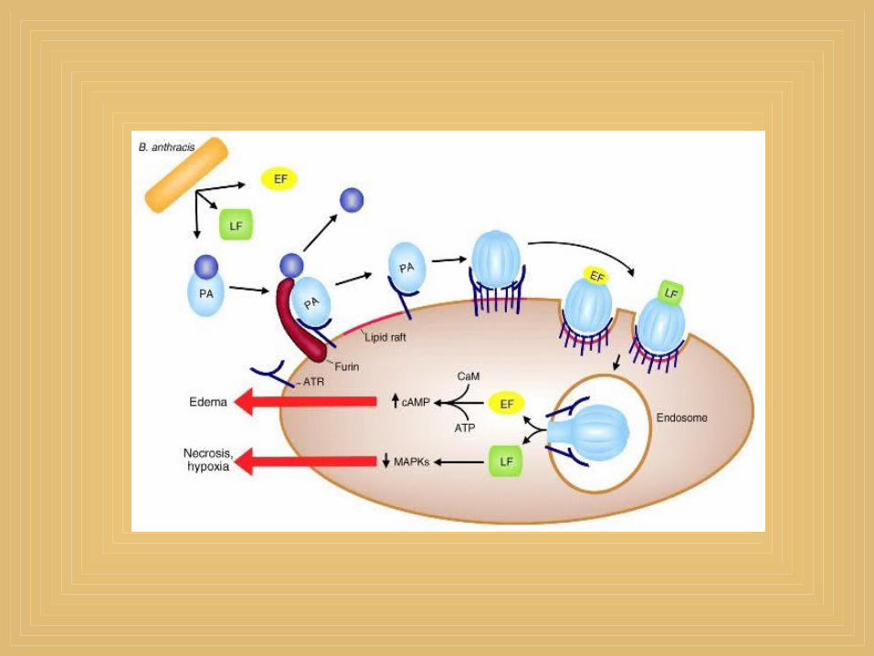
Pathogenicity: virulence factors
Two virulence factors – Capsular polysaccharide – inhibits
phagocytosis, encoded by a plasmid Anthrax toxin : made up of 3 fractions1. Edema factor (EF or Factor I)2. Protective antigen factor (PA or Factor II)3. Lethal factor (LF or Factor III) * They are not toxic individually, the
whole complex produces local edema & generalised shock. Toxin production is plasmid mediated
28/02/2008 Dr Ekta Chourasia, Microbiology
Human Anthrax The disease may be1. Cutaneous2. Pulmonary, or3. Intestinal
* All types lead to fatal septicemia
28/02/2008 Dr Ekta Chourasia, Microbiology
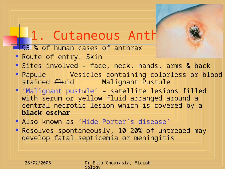
1. Cutaneous Anthrax 95 % of human cases of anthrax Route of entry: Skin Sites involved – face, neck, hands, arms & back Papule Vesicles containing colorless or blood
stained fluid Malignant Pustule ‘Malignant pustule’ – satellite lesions filled with
serum or yellow fluid arranged around a central necrotic lesion which is covered by a black eschar
Also known as ‘Hide Porter’s disease’ Resolves spontaneously, 10-20% of untreaed may
develop fatal septicemia or meningitis
28/02/2008 Dr Ekta Chourasia, Microbiology
2. Pulmonary Anthrax Also called ‘Wool Sorter’s disease’ –
common in workers in wool factories
A life- threatening hemorrhagic pneumonia caused by Inhalation of spores

28/02/2008 Dr Ekta Chourasia, Microbiology
3. Gastrointestinal Anthrax Rare By ingestion of inadequately cooked meat containing B. anthracis spores
* Human anthrax can be Industrial – in meat packing or wool factories Nonindustrial – frequent association with
animals like butchers, veterinarians, farmers
28/02/2008 Dr Ekta Chourasia, Microbiology
Laboratory Diagnosis Specimen Fluid or pus from local lesion, blood,
sputum Microscopy Culture In septicemic anthrax, blood culture
should be done Serological test Animal inoculation
28/02/2008 Dr Ekta Chourasia, Microbiology
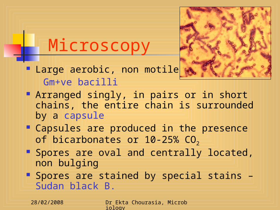
Microscopy Large aerobic, non motile, Gm+ve bacilli Arranged singly, in pairs or in short chains,
the entire chain is surrounded by a capsule Capsules are produced in the presence of
bicarbonates or 10-25% CO2
Spores are oval and centrally located, non bulging
Spores are stained by special stains – Sudan black B.
28/02/2008 Dr Ekta Chourasia, Microbiology
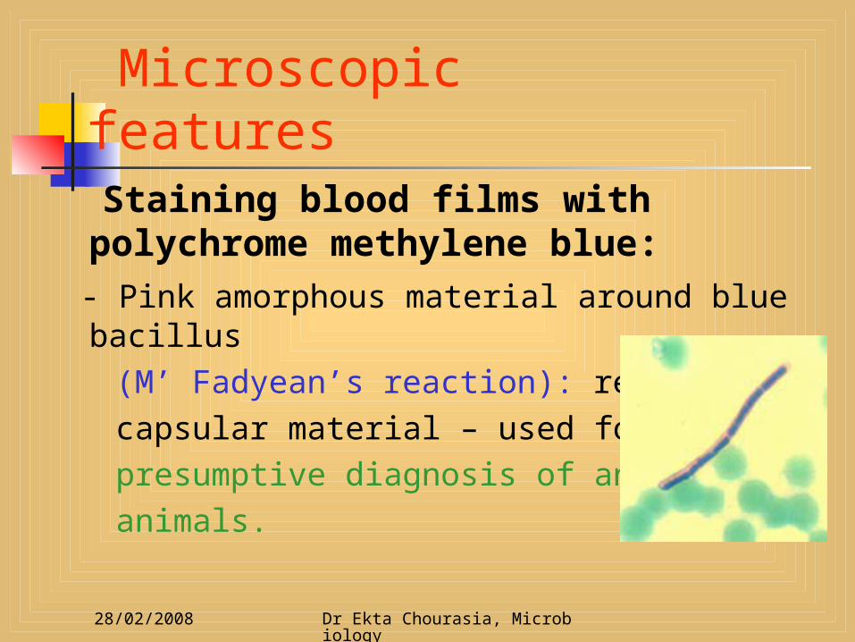
Microscopic features Staining blood films with
polychrome methylene blue: - Pink amorphous material around blue
bacillus (M’ Fadyean’s reaction): represents capsular material – used for the presumptive diagnosis of anthrax in animals.

28/02/2008 Dr Ekta Chourasia, Microbiology
Cultural Characteristics Grow on blood or nutrient agar, at 37°C Irregular, round, raised, dull, opaque,
greyish white colonies with a frosted glass appearance.
Low power – edge of the colony is composed of long, interlacing chains of bacilli, resembling locks of matted hair – “Medusa Head Appearance”
Gelatin stab culture – “inverted fir tree” appearance, with slow liquefaction starting from top.
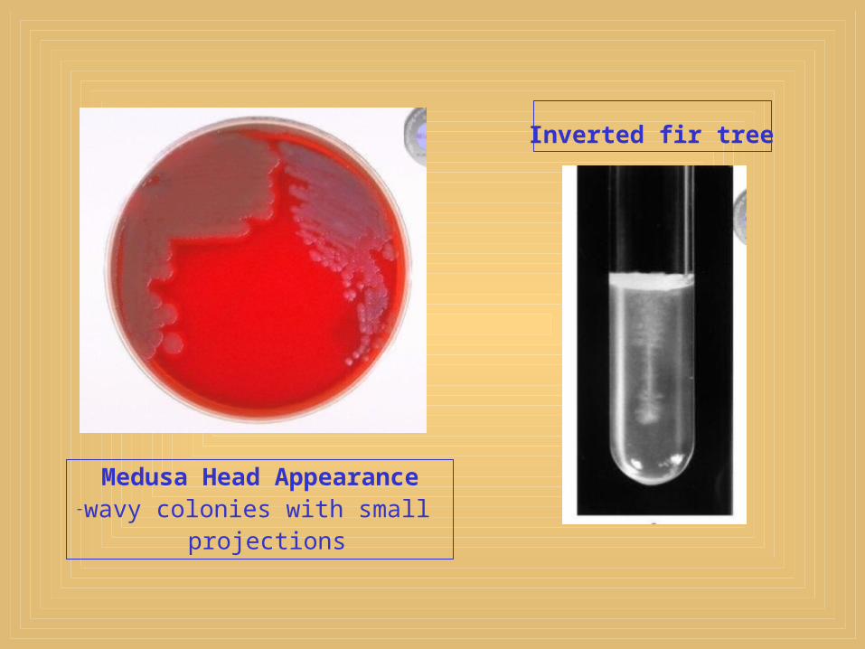
Medusa Head Appearance-wavy colonies with small
projections
Inverted fir tree
28/02/2008 Dr Ekta Chourasia, Microbiology
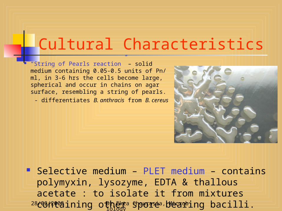
Cultural Characteristics “String of Pearls reaction” – solid
medium containing 0.05-0.5 units of Pn/ ml, in 3-6 hrs the cells become large, spherical and occur in chains on agar surface, resembling a string of pearls.
- differentiates B. anthracis from B. cereus
Selective medium – PLET medium – contains polymyxin, lysozyme, EDTA & thallous acetate : to isolate it from mixtures containing other spore bearing bacilli.
28/02/2008 Dr Ekta Chourasia, Microbiology
Smear from colony
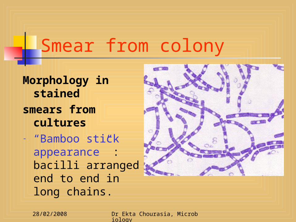
Morphology in stained
smears from cultures
- “Bamboo stick appearance” : bacilli arranged end to end in long chains.

28/02/2008 Dr Ekta Chourasia, Microbiology
Laboratory Diagnosis Animal inoculation By rubbing contaminated tissues over
shaven skin of a guinea pig Serology1. Ascoli’s thermoprecipitation test – to
demonstrate anthrax Ag in tissue extracts
2. EIA (using purified anthrax toxin Ag)3. PCR to detect anthrax contamination
of animal & agricultural products
28/02/2008 Dr Ekta Chourasia, Microbiology
Resistance Bacilli destroyed at 60°C in 30 mins. Animal carcasses – bacilli remain viable
in BM for a wk & in skin for 2 wks. Spores – highly resistant, survive in soil
for 60 yrs Spores can be destroyed by1. 4% KMnO4 in 15 mins2. ‘Duckering’ – using formaldehyde
solution for animal products imported into non endemic countries

28/02/2008 Dr Ekta Chourasia, Microbiology
Duckering For disinfection of wool – 2% soln of
formaldehyde at 30- 40°C for 20 mins
Animal hair & bristles – 0.25% at 60°C for 6 hrs
28/02/2008 Dr Ekta Chourasia, Microbiology
Prophylaxis General methods of prevention1. Improvement of factory hygiene2. Proper sterilisation of animal products3. Animal carcasses to be buried deep in
quicklime or cremated

28/02/2008 Dr Ekta Chourasia, Microbiology
Prophylaxis Active immunisation of 1. Domestic animals with live attenuated
spore vaccines
2. Persons with occupational risk (butchers, farmers, veterinarians) with a cell- free vaccine containing purified protective antigen as immunogen. 3 doses IM with annual booster injections.
* Anthrax infection in humans give life long permanent immunity & secondary infections are very rare.
28/02/2008 Dr Ekta Chourasia, Microbiology
Anthrax vaccines Original anthrax vaccine – developed by
Pasteur – live attenuated bacilli vaccine – strain rendered avirulent by the loss of plasmids which encodes anthrax toxin
Live attenuated anthrax spore vaccine 1. Sterne vaccine – contains spores of a
noncapsulated avirulent mutant strain - loss of plasmid which controls capsule production
2. Mazucchi vaccine – contains spores of stable attenuated Carbazoo strain

28/02/2008 Dr Ekta Chourasia, Microbiology
Biological warfare Large epidemics (occasionally)1. In 1979 – former Soviet Union: due to
accidental release of spores from a military facility engaged in biological research
2. In 1980s – Zimbabwe: affected 10,000 persons.
* Hence the need to develop better human vaccine.

28/02/2008 Dr Ekta Chourasia, Microbiology
Treatment
Bacillus anthracis is sensitive to: - Penicillin - Doxycycline - Ciprofloxacin

28/02/2008 Dr Ekta Chourasia, Microbiology
Anthracoid bacilli Belongs to the genus Bacillus Occasionally cause human infections Includes B. cereus, B. subtilis, B.
licheniformis & other species. These and a variety of non pathogenic
aerobic spore bearing bacilli appear as laboratory contaminants & resemble anthrax bacilli – Pseudoanthrax or Anthracoid bacilli.

28/02/2008 Dr Ekta Chourasia, Microbiology
Differences b/n Anthrax & Anthracoid bacilli
Anthrax bacilli Nonmotile Capsulated Grow in long chains Medusa head colony No growth in Pn agar
(10units/ml) Weak or no hemolysis Inverted fir tree growth
& slow gelatin liquefaction
No growth at 45C
Anthracoid bacilli Generally motile Noncapsulated Grow in short chains Not present Grow usually
Hemolysis well marked
Rapid liquefaction
Usually grows
28/02/2008 Dr Ekta Chourasia, Microbiology
Bacillus cereus Readily isolated from soil, vegetables
and a wide variety of foods including milk, cereals, spices, poultry & meat.
Causes foodborne gastroenteritis – 2 patterns of disease (diarrhoeal & emetic); both types are mild & self limited, requiring no specific therapy.

Gastroenteritis
Gastroenteritis
Bacillus cereus clinical presentation
Incubation period < 6 hoursSevere vomitingLasts 8-10 hours
Incubation period > 6 hoursDiarrhoea
Lasts 20-36 hours
EMETIC FORM DIARRHOEAL FORM

28/02/2008 Dr Ekta Chourasia, Microbiology
Types of GastroenteritisType I
Wide range of foods including cooked meat & vegetables
Diarrhoea & abdominal pain develops 8 –16 hrs after consumption
Few bacilli seen in fecal specimens
Caused by serotypes 2,6,8,9,10 or 12.
Enterotoxin resembles LT of E.coli
Type II Chinese fried rice
exclusively.
Acute nausea & vomiting 1-5 hrs after meals, diarrhoea rare
Large no of bacilli in cooked rice & fecal samples.
Caused by serotypes 1,3 or 5
Toxin resembles staphylococcal enterotoxin
28/02/2008 Dr Ekta Chourasia, Microbiology
Diagnosis Primarily depends on clinical diagnosis
& food sources Laboratory Diagnosis1. Specimen – stool, vomitus, food, blood2. Microscopy – not of much help3. Culture 4. Test for toxin – to differentiate from
staphylococcal food poisoning.
28/02/2008 Dr Ekta Chourasia, Microbiology
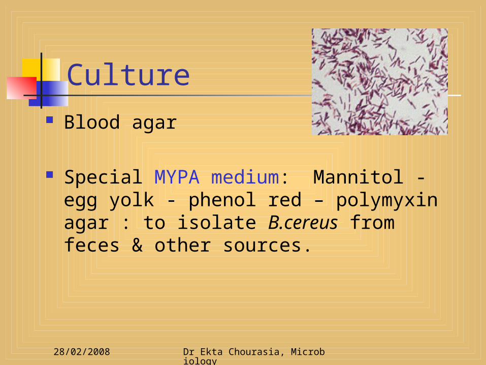
Culture Blood agar
Special MYPA medium: Mannitol - egg yolk - phenol red – polymyxin agar : to isolate B.cereus from feces & other sources.

28/02/2008 Dr Ekta Chourasia, Microbiology
Treatment Rehydration Antibiotics – in systemic infections