avascular necrosis of stapes crura in one case of … · and radiologically by x-ray mastoid ......
TRANSCRIPT
193ACTA MEDICA (Hradec Králové) 2012; 55(4): 193–197
AVASCUlAR NECROSIS OF STAPES CRURA IN ONE CASE OF OPERATED OTOSClEROSIS
Milan Erdoglija1, Jelena Sotirovic1, Violeta Jacimovic1, Biserka Vukomanovic2
Department of Otorhinolaryngology, Otology Unit, Military Medical Academy, Belgrade, Serbia1; institute of Pathology, Military Medical Academy, Belgrade, Serbia2
Summary: Unilateral otosclerosis combined with avascular necrosis of stapes crura is a rare entity. it should be considered in a case of high grade otosclerosis. Symptoms are the same as in patients who suffer from common otosclerosis. Patients complain on progressive hearing loss and tinnitus. The diagnosis is made clinically by conventional audiologic evaluation and radiologically by x-ray mastoid Schuller’s view and CT scan. HRCT scan makes visible all parts of ossicular chain and gives surgeon some information about ossicular chain damage. Surgery with stapedotomy and stapes prosthesis implan-tation in a case of otosclerosis with avascular necrosis of stapes crura can be success therapy to improve patient’s hearing
Key words: Otosclerosis; Avascular necrosis; Stapes crura; Hearing loss; Tinnitus; Pure-tone audiometry; Tympanometry; Stapedotomy
introduction
Otosclerosis is a special progressive bone remodeling disorder restricted to the otic capsule, following with stapes fixation and resulting in conductive hearing loss and tinni-tus. Etiopathogenesis of otosclerosis is controversial. The uncontrolled and increased osteoid turnover results in oste-olytic foci with sharply defined, irregular margins, mostly in the anterior pole of the stapes footplate. The disease leads to stapes fixation and conductive deafness through many years. Otosclerosis usually begins in teenage period of life, devel-ops bilateral (70–80%), two times more frequently in female population and leads to severe deafness in the 5th decade (2). There is mild hearing loss in the beginning off this illness with thickened stapes footplate (grade i otosclerosis). Histological appearance of otosclerosis is characterized by hypervascularized focus filled with numerous multinucle-ated osteoclasts, hypercellular fibrous stroma and plump, distorted osteoblasts. The process develops to severe deaf-ness after many years and finishes with endstage or grade iV otosclerosis corresponding to a remodeled lamellar bone structure with hypocellularity or acellularity on wide pseu-do-vascular spaces of bones (1, 2). The diagnosis of oto-sclerosis can be established by clinical examination, hearing tests and radiological findings. Anamnesis must be sufficient and precisely. Otoscopy and otomycroscopy examination is normal in patients with otosclerosis. Pure tone audiometry shows conductive or mixed hearing loss. Tympanometry is mostly normal and acoustic stapedial reflex is absent in patients with otosclerosis. X-ray mastoid Schuller’s view shows normally pneumatized mastoid air cells in patients with otosclerosis. Surgery is the only therapeutic treatment
for otosclerosis. Stapedotomy with stapedial prosthesis is most successful surgery preformed nowadays.
Avascular necrosis is a bones disease resulting from the temporary or permanent loss of the blood supply to the bones. There are no oxygen and nutrition for bone tissue. Bone tissue collapse and dies in area without blood supply-ing. Avascular necrosis of the bone tissue leads to pathologi-cal fractures of the bone. There are many causes of avascular necrosis such as alcoholism, excessive steroid use, trauma, Caisson disease (decompression sickness), osteogenesis imperfecta, vascular compression, hypertension, vasculitis, thrombosis, radiation, bisphosphonates (particularly in the mandible) (11) and (very rarely) sickle cell anaemia. Avas-cular necrosis can happen in any bone, it may affect just one bone, more than one bone at the same time, or more than one bone at different times, almost in ages 30–50 years. Clinical avascular necrosis most commonly affects the ends (epiphy-sis) of long bones such as the femur and humerus or joints of the hip, knee, and shoulder. Avascular necrosis is rare entity for other bones, including bones in the middle ear. There are tree little bones in the middle ear (malleus, incus and stapes) which are not long bones. There were many described cases of avascular necrosis of long process of the incus caused by poor blood supply which was a result of different kind of pathological process or result of stapes prosthesis (3, 12). Avascular necrosis of stapes crura potentially can happen as it happens with long bones femur or humerus, for example. There are no articles about avascular necrosis of stapes cru-ra. Displacement of stapes crura is described in some other pathology process like congenital anomalies, chronic sup-purative otitis media, cholesteatoma etc. Fracture of stapes crura occurs rarely, following head injures, isolated or asso-ciated with temporal bone fractures (15).
CASE REPORTS
Acta Medica 4/12 2802.indd 193 22.2.13 13:46
194
Case report
37-year-old male patient was admitted in Clinic for ear, nose and throat at the Military Medical Academy because of evaluation of hearing loss and tinnitus on his right ear. He has been suffering from progressive hearing loss and tinnitus on his right ear for 15 years, which became especially dete-riorated during the year before his admission. He noticed that his hearing was temporarily better during Valsalva test. He has never had previous injures of the head or ear, ear pain or ear infection and vertigo as well. He was in good physical condition, without some systemic diseases. There was no evidence of problems with some other bones previ-ously. He was not in a habit of smoking or drinking alcohol. There were no cases of otosclerosis in his family. Patient did not show any risk factors for avascular necrosis at all.
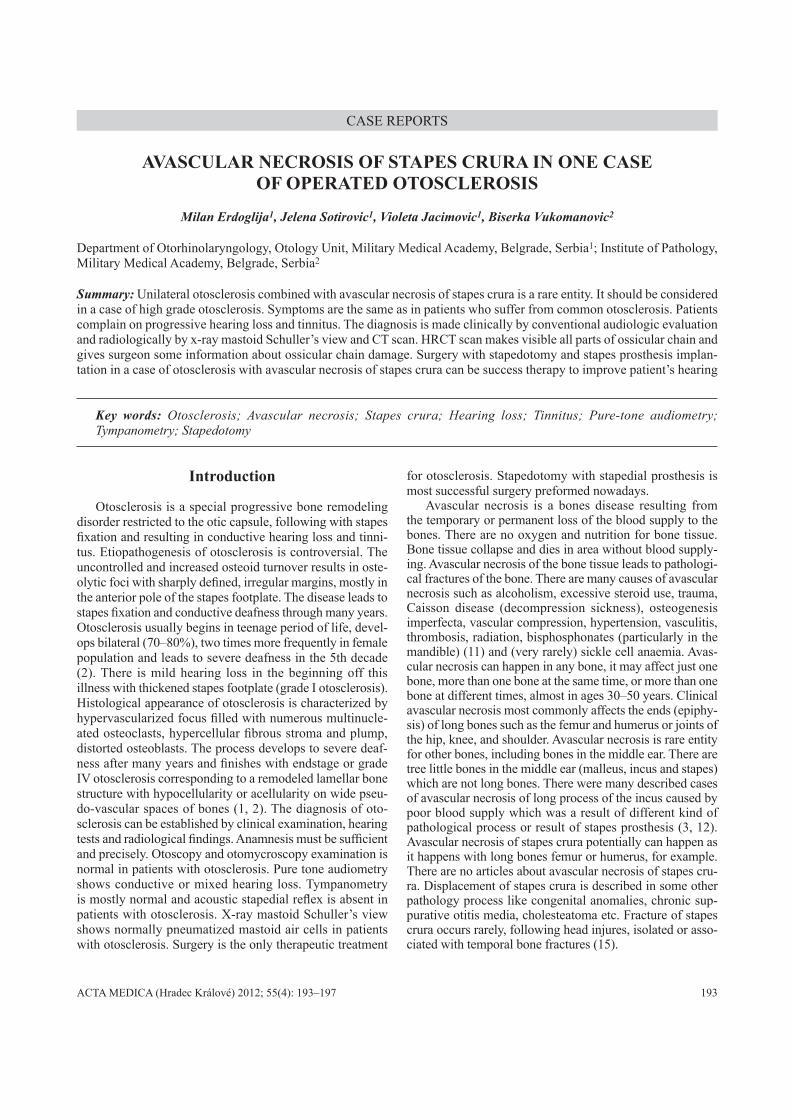
Fig. 1: Pure-tone audiometry before surgery – mixed hear-ing loss on the right ear
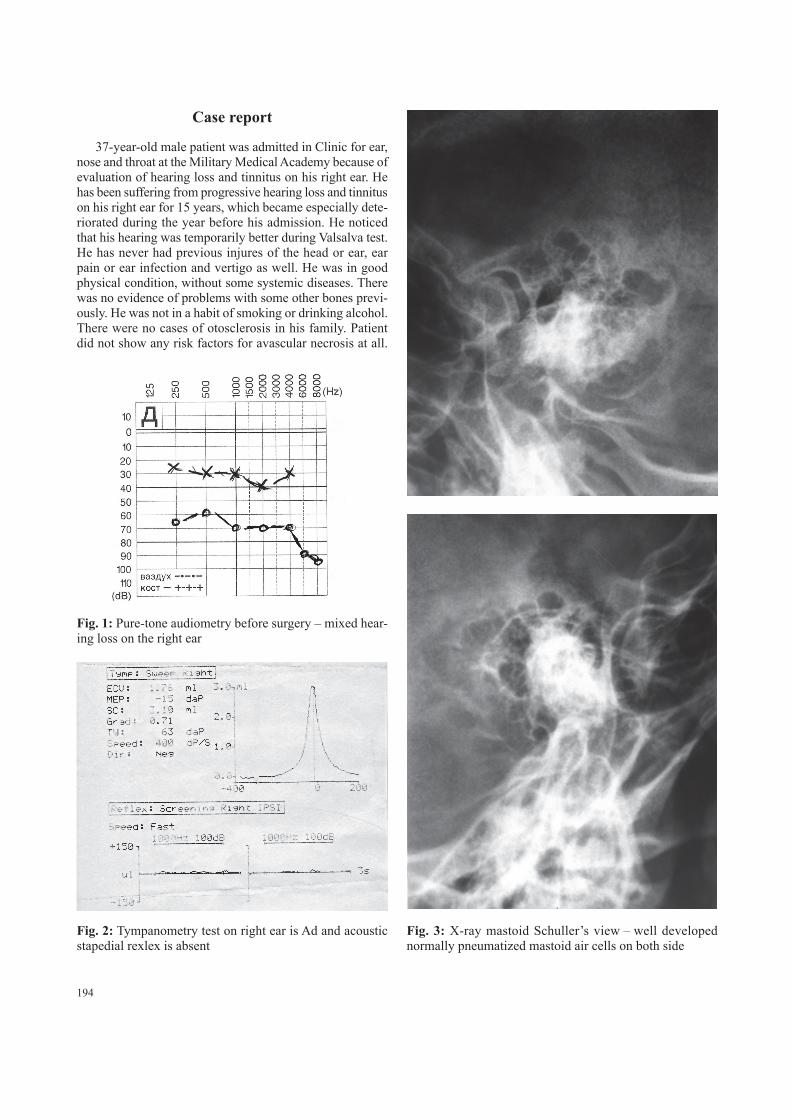
Fig. 2: Tympanometry test on right ear is Ad and acoustic stapedial rexlex is absent
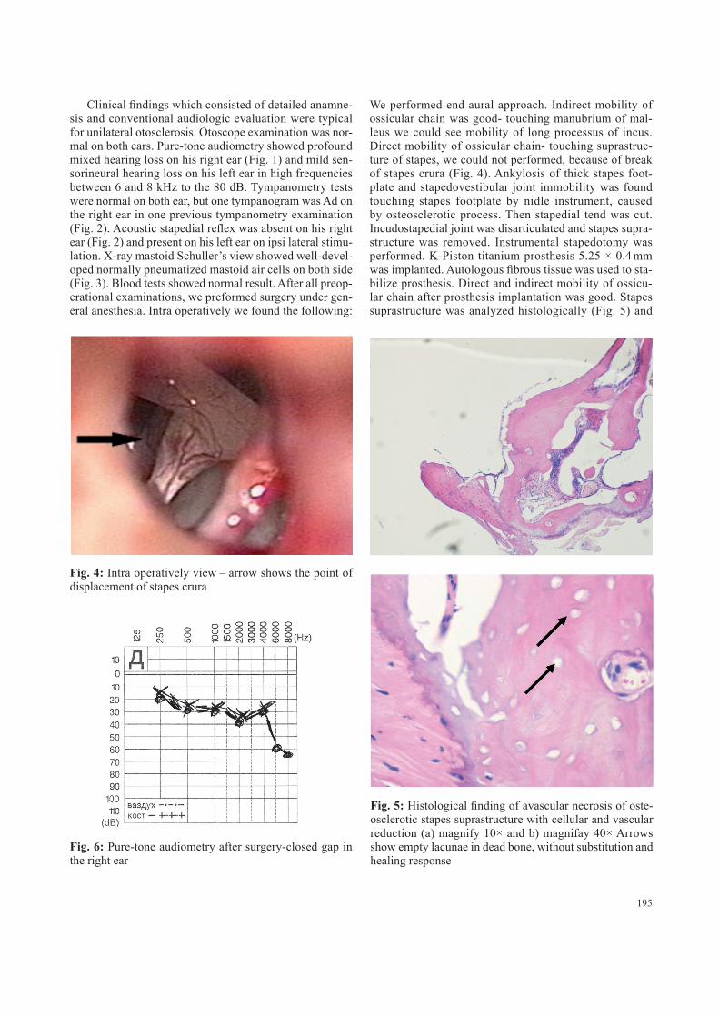
Fig. 3: X-ray mastoid Schuller’s view – well developed normally pneumatized mastoid air cells on both side
Acta Medica 4/12 2802.indd 194 22.2.13 13:46
195
Clinical findings which consisted of detailed anamne-sis and conventional audiologic evaluation were typical for unilateral otosclerosis. Otoscope examination was nor-mal on both ears. Pure-tone audiometry showed profound mixed hearing loss on his right ear (Fig. 1) and mild sen-sorineural hearing loss on his left ear in high frequencies between 6 and 8 kHz to the 80 dB. Tympanometry tests were normal on both ear, but one tympanogram was Ad on the right ear in one previous tympanometry examination (Fig. 2). Acoustic stapedial reflex was absent on his right ear (Fig. 2) and present on his left ear on ipsi lateral stimu-lation. X-ray mastoid Schuller’s view showed well-devel-oped normally pneumatized mastoid air cells on both side (Fig. 3). Blood tests showed normal result. After all preop-erational examinations, we preformed surgery under gen-eral anesthesia. intra operatively we found the following:
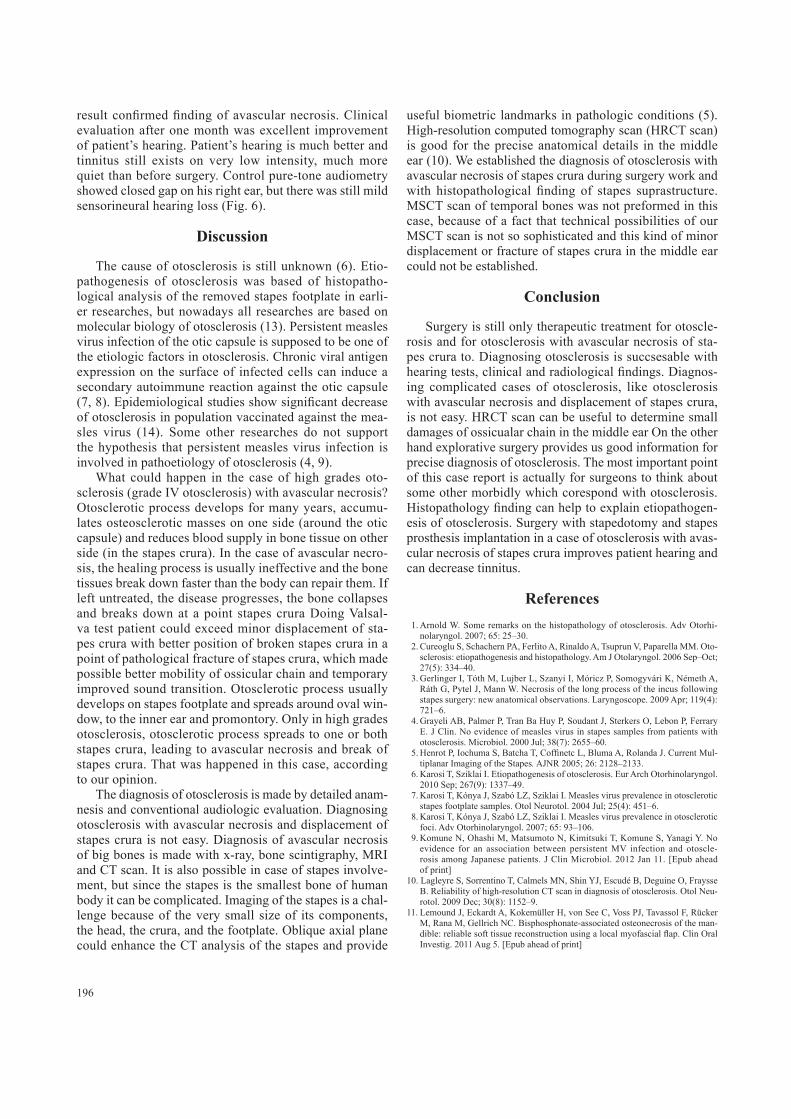
we performed end aural approach. indirect mobility of ossicular chain was good- touching manubrium of mal-leus we could see mobility of long processus of incus. Direct mobility of ossicular chain- touching suprastruc-ture of stapes, we could not performed, because of break of stapes crura (Fig. 4). Ankylosis of thick stapes foot-plate and stapedovestibular joint immobility was found touching stapes footplate by nidle instrument, caused by osteosclerotic process. Then stapedial tend was cut. incudostapedial joint was disarticulated and stapes supra-structure was removed. instrumental stapedotomy was performed. K-Piston titanium prosthesis 5.25 × 0.4 mm was implanted. Autologous fibrous tissue was used to sta-bilize prosthesis. Direct and indirect mobility of ossicu-lar chain after prosthesis implantation was good. Stapes suprastructure was analyzed histologically (Fig. 5) and
Fig. 4: intra operatively view – arrow shows the point of displacement of stapes crura
Fig. 5: Histological finding of avascular necrosis of oste-osclerotic stapes suprastructure with cellular and vascular reduction (a) magnify 10× and b) magnifay 40× Arrows show empty lacunae in dead bone, without substitution and healing response
Fig. 6: Pure-tone audiometry after surgery-closed gap in the right ear
Acta Medica 4/12 2802.indd 195 22.2.13 13:46
196
result confirmed finding of avascular necrosis. Clinical evaluation after one month was excellent improvement of patient’s hearing. Patient’s hearing is much better and tinnitus still exists on very low intensity, much more quiet than before surgery. Control pure-tone audiometry showed closed gap on his right ear, but there was still mild sensorineural hearing loss (Fig. 6).
discussion
The cause of otosclerosis is still unknown (6). Etio-pathogenesis of otosclerosis was based of histopatho-logical analysis of the removed stapes footplate in earli-er researches, but nowadays all researches are based on molecular biology of otosclerosis (13). Persistent measles virus infection of the otic capsule is supposed to be one of the etiologic factors in otosclerosis. Chronic viral antigen expression on the surface of infected cells can induce a secondary autoimmune reaction against the otic capsule (7, 8). Epidemiological studies show significant decrease of otosclerosis in population vaccinated against the mea-sles virus (14). Some other researches do not support the hypothesis that persistent measles virus infection is involved in pathoetiology of otosclerosis (4, 9).
what could happen in the case of high grades oto-sclerosis (grade iV otosclerosis) with avascular necrosis? Otosclerotic process develops for many years, accumu-lates osteosclerotic masses on one side (around the otic capsule) and reduces blood supply in bone tissue on other side (in the stapes crura). in the case of avascular necro-sis, the healing process is usually ineffective and the bone tissues break down faster than the body can repair them. if left untreated, the disease progresses, the bone collapses and breaks down at a point stapes crura Doing Valsal-va test patient could exceed minor displacement of sta-pes crura with better position of broken stapes crura in a point of pathological fracture of stapes crura, which made possible better mobility of ossicular chain and temporary improved sound transition. Otosclerotic process usually develops on stapes footplate and spreads around oval win-dow, to the inner ear and promontory. Only in high grades otosclerosis, otosclerotic process spreads to one or both stapes crura, leading to avascular necrosis and break of stapes crura. That was happened in this case, according to our opinion.
The diagnosis of otosclerosis is made by detailed anam-nesis and conventional audiologic evaluation. Diagnosing otosclerosis with avascular necrosis and displacement of stapes crura is not easy. Diagnosis of avascular necrosis of big bones is made with x-ray, bone scintigraphy, MRi and CT scan. it is also possible in case of stapes involve-ment, but since the stapes is the smallest bone of human body it can be complicated. imaging of the stapes is a chal-lenge because of the very small size of its components, the head, the crura, and the footplate. Oblique axial plane could enhance the CT analysis of the stapes and provide
useful biometric landmarks in pathologic conditions (5). High-resolution computed tomography scan (HRCT scan) is good for the precise anatomical details in the middle ear (10). we established the diagnosis of otosclerosis with avascular necrosis of stapes crura during surgery work and with histopathological finding of stapes suprastructure. MSCT scan of temporal bones was not preformed in this case, because of a fact that technical possibilities of our MSCT scan is not so sophisticated and this kind of minor displacement or fracture of stapes crura in the middle ear could not be established.
conclusion
Surgery is still only therapeutic treatment for otoscle-rosis and for otosclerosis with avascular necrosis of sta-pes crura to. Diagnosing otosclerosis is succsesable with hearing tests, clinical and radiological findings. Diagnos-ing complicated cases of otosclerosis, like otosclerosis with avascular necrosis and displacement of stapes crura, is not easy. HRCT scan can be useful to determine small damages of ossicualar chain in the middle ear On the other hand explorative surgery provides us good information for precise diagnosis of otosclerosis. The most important point of this case report is actually for surgeons to think about some other morbidly which corespond with otosclerosis. Histopathology finding can help to explain etiopathogen-esis of otosclerosis. Surgery with stapedotomy and stapes prosthesis implantation in a case of otosclerosis with avas-cular necrosis of stapes crura improves patient hearing and can decrease tinnitus.
references 1. Arnold w. Some remarks on the histopathology of otosclerosis. Adv Otorhi-
nolaryngol. 2007; 65: 25–30. 2. Cureoglu S, Schachern PA, Ferlito A, Rinaldo A, Tsuprun V, Paparella MM. Oto-
sclerosis: etiopathogenesis and histopathology. Am J Otolaryngol. 2006 Sep–Oct; 27(5): 334–40.
3. Gerlinger i, Tóth M, Lujber L, Szanyi i, Móricz P, Somogyvári K, Németh A, Ráth G, Pytel J, Mann w. Necrosis of the long process of the incus following stapes surgery: new anatomical observations. Laryngoscope. 2009 Apr; 119(4): 721–6.
4. Grayeli AB, Palmer P, Tran Ba Huy P, Soudant J, Sterkers O, Lebon P, Ferrary E. J Clin. No evidence of measles virus in stapes samples from patients with otosclerosis. Microbiol. 2000 Jul; 38(7): 2655–60.
5. Henrot P, Iochuma S, Batcha T, Coffinetc L, Bluma A, Rolanda J. Current Mul-tiplanar imaging of the Stapes. AJNR 2005; 26: 2128–2133.
6. Karosi T, Sziklai i. Etiopathogenesis of otosclerosis. Eur Arch Otorhinolaryngol. 2010 Sep; 267(9): 1337–49.
7. Karosi T, Kónya J, Szabó LZ, Sziklai I. Measles virus prevalence in otosclerotic stapes footplate samples. Otol Neurotol. 2004 Jul; 25(4): 451–6.
8. Karosi T, Kónya J, Szabó LZ, Sziklai I. Measles virus prevalence in otosclerotic foci. Adv Otorhinolaryngol. 2007; 65: 93–106.
9. Komune N, Ohashi M, Matsumoto N, Kimitsuki T, Komune S, Yanagi Y. No evidence for an association between persistent MV infection and otoscle-rosis among Japanese patients. J Clin Microbiol. 2012 Jan 11. [Epub ahead of print]
10. Lagleyre S, Sorrentino T, Calmels MN, Shin YJ, Escudé B, Deguine O, Fraysse B. Reliability of high-resolution CT scan in diagnosis of otosclerosis. Otol Neu-rotol. 2009 Dec; 30(8): 1152–9.
11. Lemound J, Eckardt A, Kokemüller H, von See C, Voss PJ, Tavassol F, Rücker M, Rana M, Gellrich NC. Bisphosphonate-associated osteonecrosis of the man-dible: reliable soft tissue reconstruction using a local myofascial flap. Clin Oral Investig. 2011 Aug 5. [Epub ahead of print]
Acta Medica 4/12 2802.indd 196 22.2.13 13:46

197
12. Mangham CA Jr. Long-term impact of incus necrosis on revision stapes sur-gery: incus versus malleus reconstruction. Otol Neurotol. 2009 Dec; 30(8): 1145–51.
13. McKenna MJ, Kristiansen AG. Molecular biology of otosclerosis. Adv Otorhi-nolaryngol. 2007; 65: 68–74.
14. Niedermeyer HP, Arnold w. Otosclerosis and measles virus – association or causation? ORL J Otorhinolaryngol Relat Spec. 2008; 70(1): 63–9.
15. Singh S, Salib RJ, Oates J. Traumatic fracture of the stapes suprastructure fol-lowing minor head injury. The Journal of Laryngology & Otology 2002; 116: 457–459.
Received: 20/05/2012Accepted in revised form: 15/11/2012
Corresponding author:
Milan Erdoglija, Clinic for Ear, Nose and Throat, Military Medical Academy, Belgrade, Serbia; e-mail: milan.erdoglija @gmail.com
Acta Medica 4/12 2802.indd 197 22.2.13 13:46