autoimmune liver disease key points - ucsf · pdf filetype iii sla/lp indistinguishable from...
TRANSCRIPT
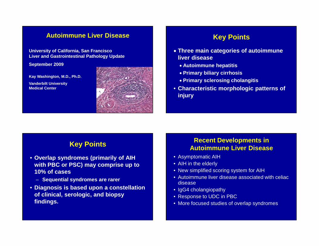
Autoimmune Liver Disease
Kay Washington, M.D., Ph.D.
Vanderbilt University Medical Center
University of California, San Francisco Liver and Gastrointestinal Pathology Update
September 2009
Key Points
• Three main categories of autoimmune liver disease • Autoimmune hepatitis• Primary biliary cirrhosis• Primary sclerosing cholangitis
• Characteristic morphologic patterns of injury
Key Points
• Overlap syndromes (primarily of AIH with PBC or PSC) may comprise up to 10% of cases– Sequential syndromes are rarer
• Diagnosis is based upon a constellation of clinical, serologic, and biopsy findings.
Recent Developments in Autoimmune Liver Disease
• Asymptomatic AIH• AIH in the elderly• New simplified scoring system for AIH• Autoimmune liver disease associated with celiac
disease• IgG4 cholangiopathy• Response to UDC in PBC• More focused studies of overlap syndromes
Autoimmune Hepatitis: Topics
• Definition and Epidemiology• Etiology and Immunology
• Clinical Features• Serology• Histologic Features• Special Problems
• Differential Diagnosis
Autoimmune Hepatitis: Definition
• Unresolving hepatitis • Increased IgG levels• Tissue directed
autoantibodies• Responds to
immunosuppression
Autoimmune Hepatitis: Epidemiology
• Prevalence of ~ 1 per 100,000 in North America
• ~20% of chronic hepatitis
• Associated with HLA A1-B8-DR3 in European populations and DR4 in Japan
• More common in women• Wide age distribution
Immunology of AIH
• Loss of self-tolerance• Cause usually unclear• ? Viral trigger- anecdotal reports of AIH following
HAV, HBV, HSV, EBV infections• Unlikely to be triggered by HCV: anti-LKM
antibodies in HCV are directed against different epitopes
• Small heritable component; complex genetic trait
Autoimmune Hepatitis:Clinical Features
• Presentation is highly variable (asymptomatic to fulminant hepatic failure)
• 1/3 are cirrhotic at presentation• 1/3 have acute presentation mimicking viral
hepatitis• 1/3 have prodromal phase• 20% are asymptomatic• ~50% will have other autoimmune disorders
Asymptomatic Presentation of AIH
• ~20 to 25% are asymptomatic at diagnosis• Discovered on routine liver testing
• Older patients• Less activity in liver biopsy• 25% will develop symptoms upon follow up• Survival is similar to patients with symptomatic
presentation
Two Groups of Asymptomatic Patients
• “Burned out” cirrhosis and nearly normal liver tests– 36%; similar to that seen in
symptomatic patients– Less favorable outcome (62%
vs. 94% 10-year survival)
• Mild hepatitis without cirrhosis
AIH in the Elderly
• Over 20% are diagnosed after age 60
• More likely to be cirrhotic at presentation
• Responds to corticosteroid therapy, even in setting of cirrhosis
• HLA DR4 more common
Diagnosis of AIH: Serum StudiesAutoantibody Target Antigen Frequency
ANA and/or SMA
ANA-ds DNA & histones; laminin; SMA- actin
70-80%
LKM1 Cytochrome p450 (CYP) 2D6 (endoplasmic reticulum)
3-4%
SLA/LP UDA suppressor serine tRNA-protein complex
10-30%
p-ANCA Multiple targets; actin 60-90%
Autoimmune Hepatitis: Classification
Type Autoantibodies Comment
Type I ANA +/- SMA Most common
Type II LKM1 Young women with severe disease
Type III SLA/LP Indistinguishable from Type I
Autoimmune Hepatitis:Histologic Features
• Chronic hepatitis pattern of injury– Interface hepatitis
– Lobular activity– Prominent plasma cells (IgG+,
rarely IgM+)
• Hepatocyte regeneration• Centrilobular necroinflammatory
activity• Bile duct injury not rare
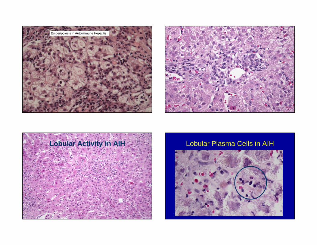
Emperipolesis in Autoimmune Hepatitis
Lobular Activity in AIH Lobular Plasma Cells in AIH
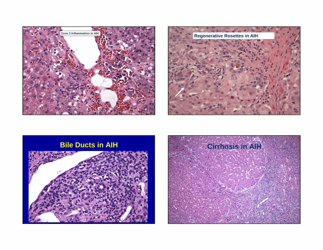
Zone 3 Inflammation in AIHRegenerative Rosettes in AIH
Bile Ducts in AIH Cirrhosis in AIH
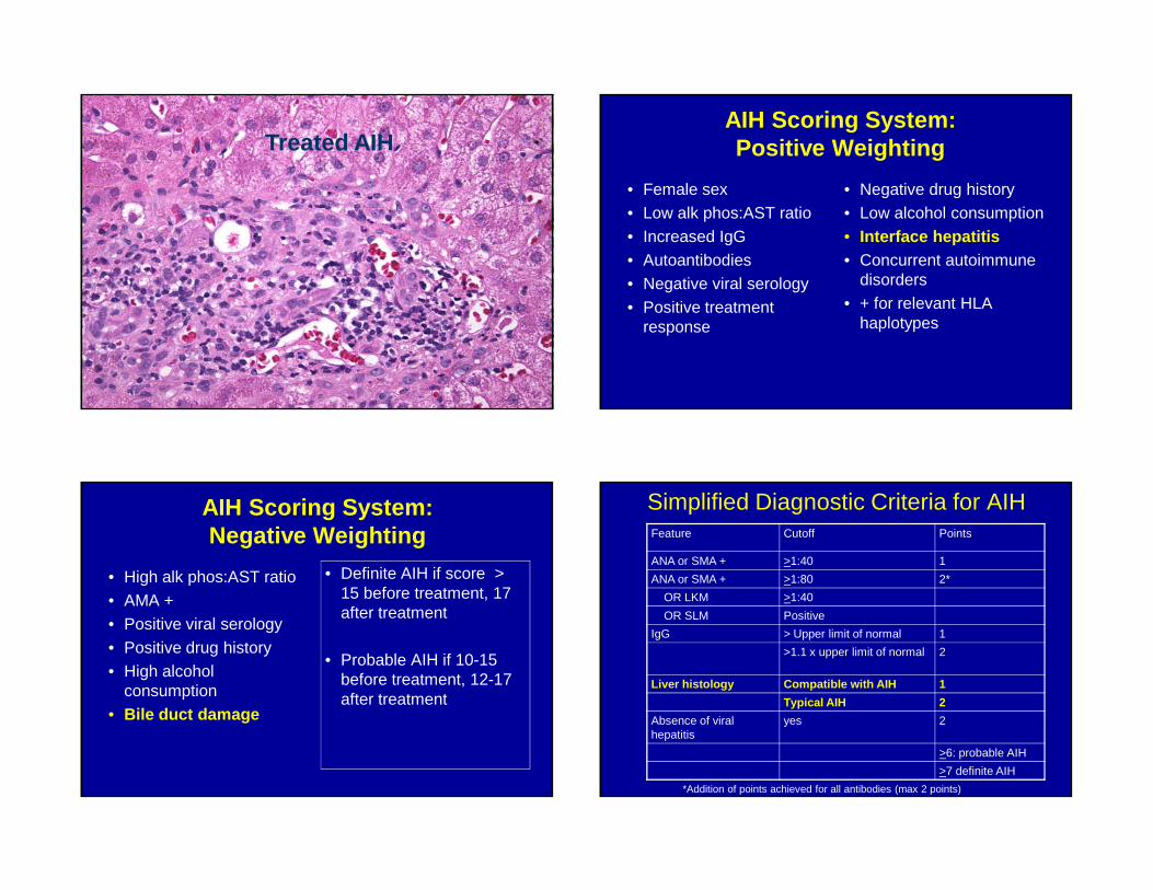
Treated AIHAIH Scoring System: Positive Weighting
• Female sex• Low alk phos:AST ratio• Increased IgG• Autoantibodies• Negative viral serology• Positive treatment
response
• Negative drug history• Low alcohol consumption• Interface hepatitis• Concurrent autoimmune
disorders• + for relevant HLA
haplotypes
AIH Scoring System: Negative Weighting
• High alk phos:AST ratio• AMA +• Positive viral serology• Positive drug history• High alcohol
consumption• Bile duct damage
• Definite AIH if score > 15 before treatment, 17 after treatment
• Probable AIH if 10-15 before treatment, 12-17 after treatment
Simplified Diagnostic Criteria for AIHFeature Cutoff Points
ANA or SMA + >1:40 1
ANA or SMA + >1:80 2*
OR LKM >1:40
OR SLM Positive
IgG > Upper limit of normal 1
>1.1 x upper limit of normal 2
Liver histology Compatible with AIH 1
Typical AIH 2
Absence of viral hepatitis
yes 2
>6: probable AIH
>7 definite AIH
*Addition of points achieved for all antibodies (max 2 points)
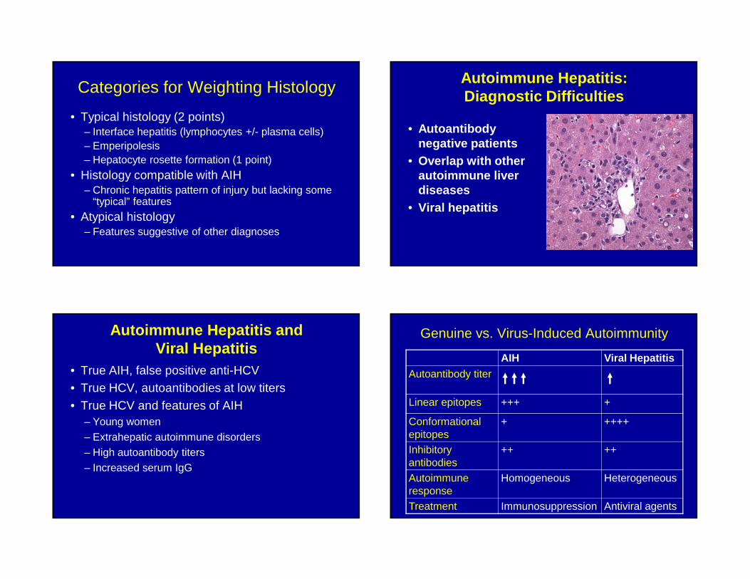
Categories for Weighting Histology
• Typical histology (2 points)– Interface hepatitis (lymphocytes +/- plasma cells)– Emperipolesis– Hepatocyte rosette formation (1 point)
• Histology compatible with AIH– Chronic hepatitis pattern of injury but lacking some
“typical” features
• Atypical histology– Features suggestive of other diagnoses
Autoimmune Hepatitis: Diagnostic Difficulties
• Autoantibody negative patients
• Overlap with other autoimmune liver diseases
• Viral hepatitis
Autoimmune Hepatitis and Viral Hepatitis
• True AIH, false positive anti-HCV• True HCV, autoantibodies at low titers
• True HCV and features of AIH– Young women– Extrahepatic autoimmune disorders– High autoantibody titers– Increased serum IgG
Genuine vs. Virus-Induced Autoimmunity
AIH Viral Hepatitis
Autoantibody titer
Linear epitopes +++ +
Conformational epitopes
+ ++++
Inhibitory antibodies
++ ++
Autoimmune response
Homogeneous Heterogeneous
Treatment Immunosuppression Antiviral agents
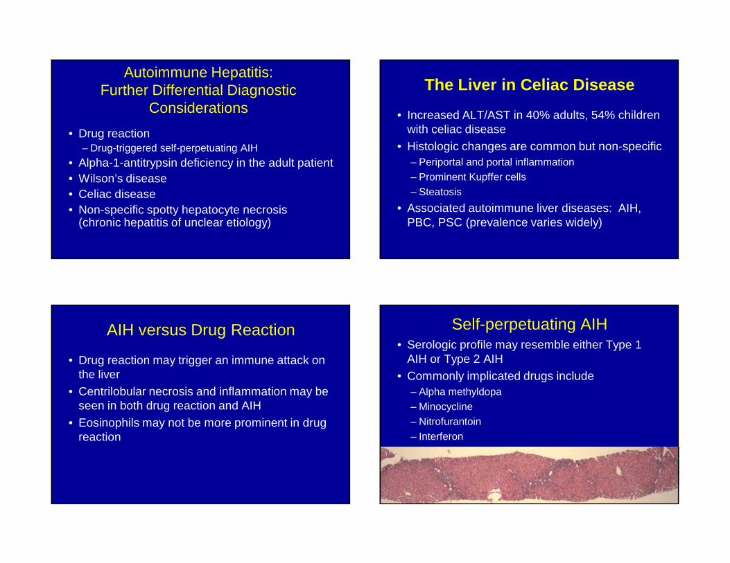
Autoimmune Hepatitis: Further Differential Diagnostic
Considerations
• Drug reaction– Drug-triggered self-perpetuating AIH
• Alpha-1-antitrypsin deficiency in the adult patient• Wilson’s disease• Celiac disease• Non-specific spotty hepatocyte necrosis
(chronic hepatitis of unclear etiology)
The Liver in Celiac Disease
• Increased ALT/AST in 40% adults, 54% children with celiac disease
• Histologic changes are common but non-specific– Periportal and portal inflammation– Prominent Kupffer cells– Steatosis
• Associated autoimmune liver diseases: AIH, PBC, PSC (prevalence varies widely)
AIH versus Drug Reaction
• Drug reaction may trigger an immune attack on the liver
• Centrilobular necrosis and inflammation may be seen in both drug reaction and AIH
• Eosinophils may not be more prominent in drug reaction
Self-perpetuating AIH• Serologic profile may resemble either Type 1
AIH or Type 2 AIH• Commonly implicated drugs include
– Alpha methyldopa– Minocycline– Nitrofurantoin– Interferon

Recently reported Associations with AIH
• Hepatitis A infection• Hepatitis vaccine• Twinrix (HAV + HBV
vaccine)• Interferon tx for HCV, MS• Terbinafine in HBV• Atomoxetine• Phenylpropyluracil
• Black cohosh• Imatinib• Infliximab• Methylphenidate• Statins• Kava kava & St Johns
wort• Minocycline• Respineridone
Primary Biliary Cirrhosis: Definition
• Chronic cholestatic liver disease, considered autoimmune in etiology
• Morphologic hallmark is inflammatory destruction of intrahepatic bile ducts
• Serologic hallmark is circulating AMA
Epidemiology of Primary Biliary Cirrhosis
• Affects women (male-female ratio 1:9)• Median age of onset 50 years (range 21-91)
• Geographical variation: increased prevalence in areas of England, low in developing countries
• More common near “Superfund” sites
• Accounts for up to 2% of deaths from cirrhosis worldwide
Pathogenesis of Primary Biliary Cirrhosis
Considered an autoimmune disorder– association with Sjogren’s disease, RA,
autoimmune thyroiditis– ? Multiple hit mechanism triggered by
mimicry– AMA directed against M2 antigen (E2
component of the pyruvate dehydrogenase complex)
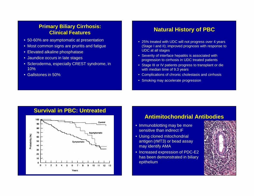
Primary Biliary Cirrhosis: Clinical Features
• 50-60% are asymptomatic at presentation• Most common signs are pruritis and fatigue
• Elevated alkaline phosphatase• Jaundice occurs in late stages• Scleroderma, especially CREST syndrome, in
10%• Gallstones in 50%
Natural History of PBC
• 25% treated with UDC will not progress over 4 years (Stage I and II); improved prognosis with response to UDC at all stages
• Severity of interface hepatitis is associated with progression to cirrhosis in UDC treated patients
• Stage III or IV patients progress to transplant or die with median time of 9.3 years
• Complications of chronic cholestasis and cirrhosis
• Smoking may accelerate progression
Survival in PBC: UntreatedAntimitochondrial Antibodies
• Immunoblotting may be more sensitive than indirect IF
• Using cloned mitochondrial antigen (rMT3) or bead assay may identify AMA
• Increased expression of PDC-E2 has been demonstrated in biliary epithelium
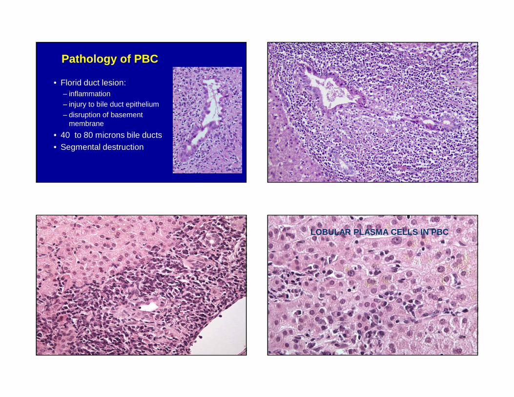
Pathology of PBC
• Florid duct lesion: – inflammation– injury to bile duct epithelium– disruption of basement
membrane
• 40 to 80 microns bile ducts• Segmental destruction
LOBULAR PLASMA CELLS IN PBC
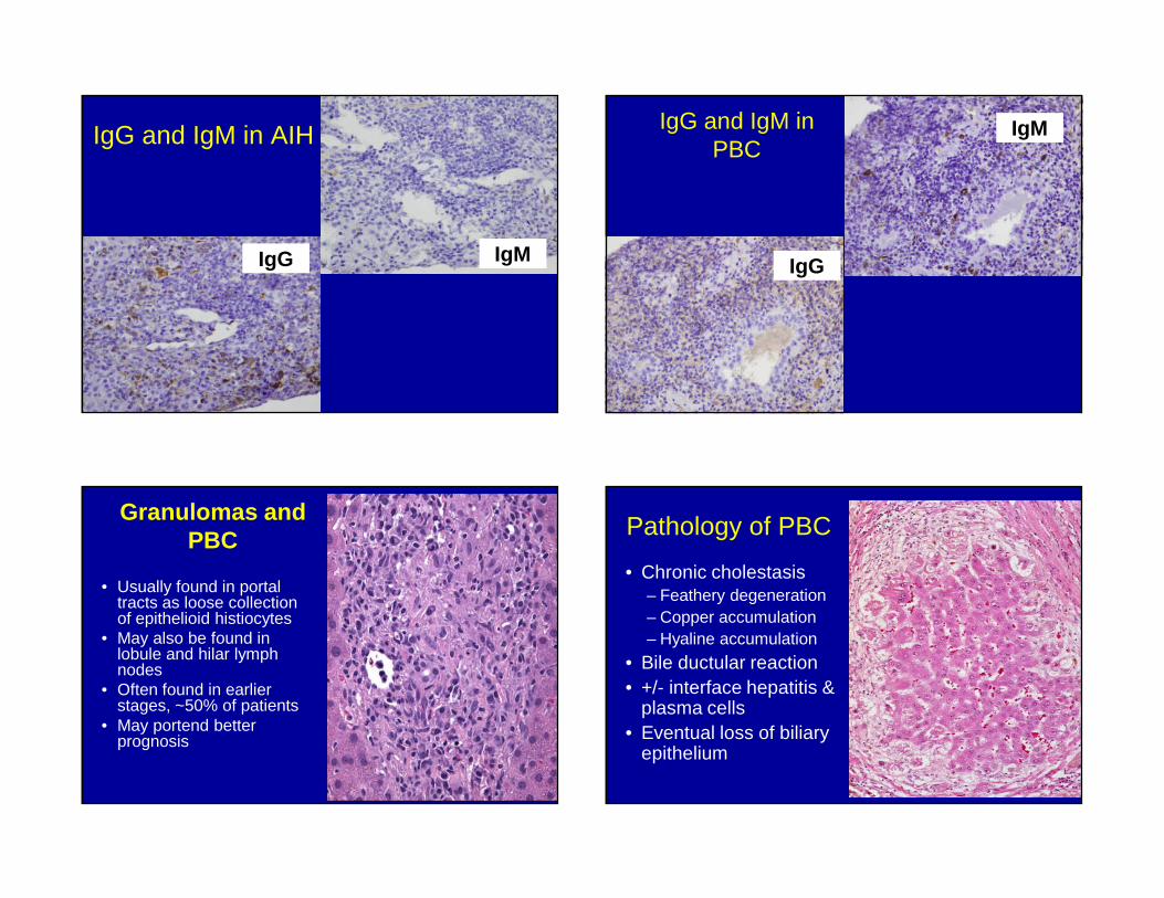
IgG and IgM in AIH
IgMIgG
IgG and IgM in PBC
IgM
IgG
Granulomas and PBC
• Usually found in portal tracts as loose collection of epithelioid histiocytes
• May also be found in lobule and hilar lymph nodes
• Often found in earlier stages, ~50% of patients
• May portend better prognosis
Pathology of PBC
• Chronic cholestasis– Feathery degeneration– Copper accumulation– Hyaline accumulation
• Bile ductular reaction• +/- interface hepatitis &
plasma cells• Eventual loss of biliary
epithelium
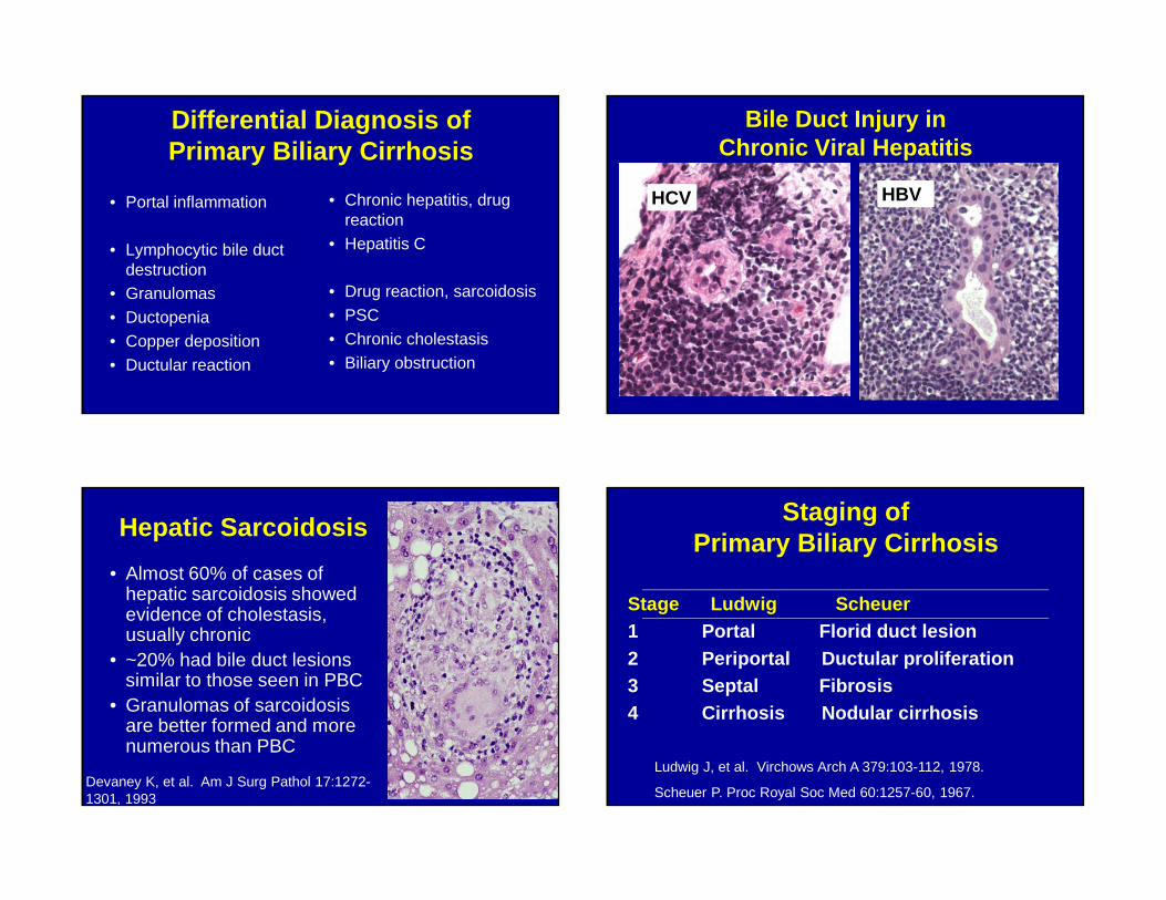
Differential Diagnosis of Primary Biliary Cirrhosis
• Portal inflammation
• Lymphocytic bile duct destruction
• Granulomas• Ductopenia• Copper deposition• Ductular reaction
• Chronic hepatitis, drug reaction
• Hepatitis C
• Drug reaction, sarcoidosis• PSC• Chronic cholestasis• Biliary obstruction
Bile Duct Injury in Chronic Viral Hepatitis
HCV HBV
Hepatic Sarcoidosis
• Almost 60% of cases of hepatic sarcoidosis showed evidence of cholestasis, usually chronic
• ~20% had bile duct lesions similar to those seen in PBC
• Granulomas of sarcoidosis are better formed and more numerous than PBC
Devaney K, et al. Am J Surg Pathol 17:1272-1301, 1993
Staging of Primary Biliary Cirrhosis
Stage Ludwig Scheuer1 Portal Florid duct lesion2 Periportal Ductular proliferation3 Septal Fibrosis4 Cirrhosis Nodular cirrhosis
Ludwig J, et al. Virchows Arch A 379:103-112, 1978.
Scheuer P. Proc Royal Soc Med 60:1257-60, 1967.
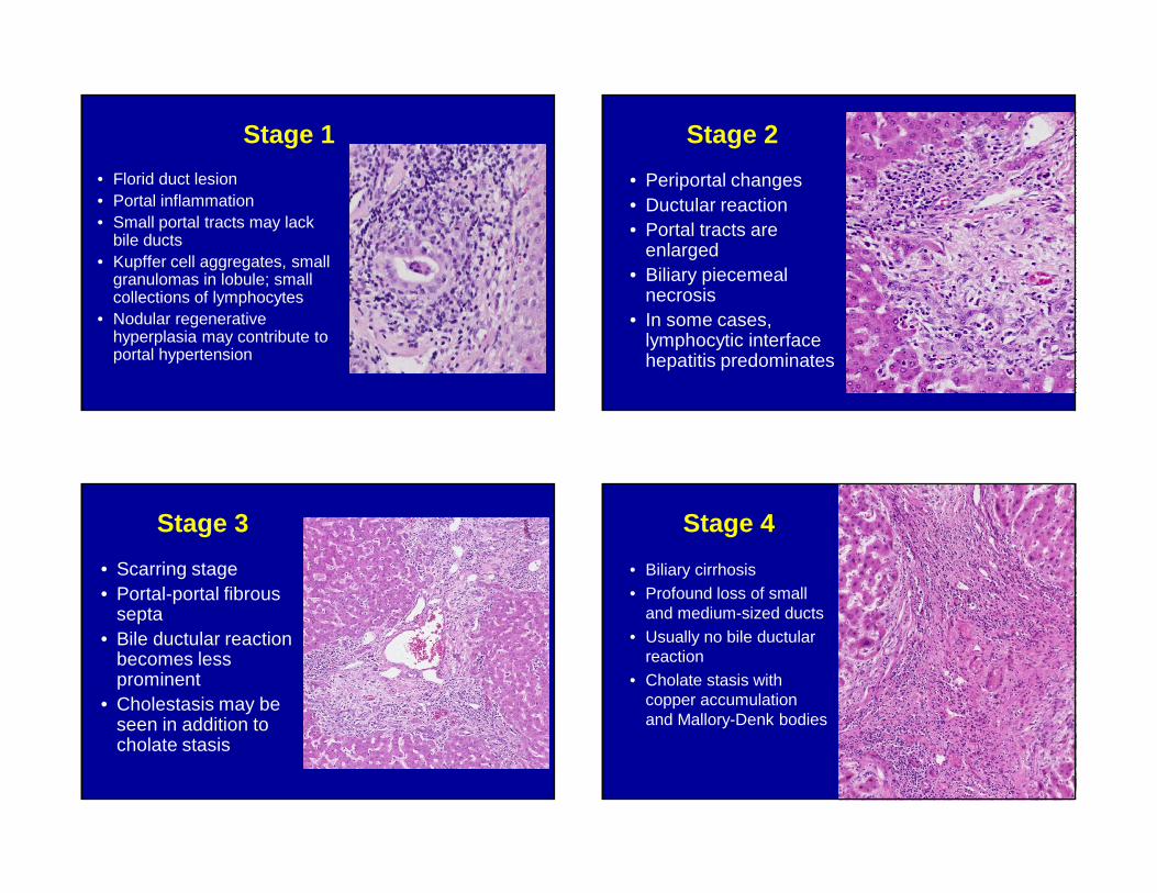
Stage 1
• Florid duct lesion• Portal inflammation• Small portal tracts may lack
bile ducts• Kupffer cell aggregates, small
granulomas in lobule; small collections of lymphocytes
• Nodular regenerative hyperplasia may contribute to portal hypertension
Stage 2
• Periportal changes• Ductular reaction• Portal tracts are
enlarged• Biliary piecemeal
necrosis• In some cases,
lymphocytic interface hepatitis predominates
Stage 3
• Scarring stage• Portal-portal fibrous
septa• Bile ductular reaction
becomes less prominent
• Cholestasis may be seen in addition to cholate stasis
Stage 4
• Biliary cirrhosis• Profound loss of small
and medium-sized ducts• Usually no bile ductular
reaction• Cholate stasis with
copper accumulation and Mallory-Denk bodies
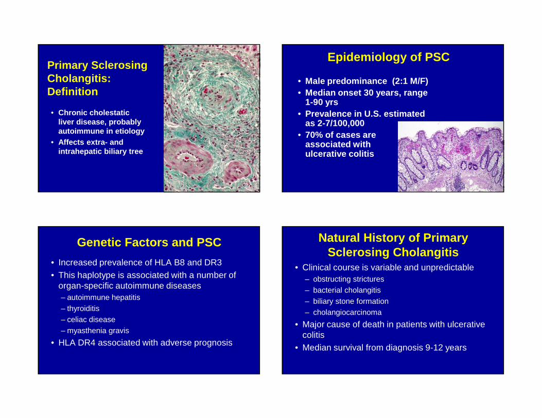
Primary Sclerosing Cholangitis:Definition
• Chronic cholestatic liver disease, probably autoimmune in etiology
• Affects extra- and intrahepatic biliary tree
Epidemiology of PSC
• Male predominance (2:1 M/F)• Median onset 30 years, range
1-90 yrs• Prevalence in U.S. estimated
as 2-7/100,000• 70% of cases are
associated with ulcerative colitis
Genetic Factors and PSC
• Increased prevalence of HLA B8 and DR3• This haplotype is associated with a number of
organ-specific autoimmune diseases– autoimmune hepatitis– thyroiditis– celiac disease– myasthenia gravis
• HLA DR4 associated with adverse prognosis
Natural History of Primary Sclerosing Cholangitis
• Clinical course is variable and unpredictable– obstructing strictures– bacterial cholangitis– biliary stone formation– cholangiocarcinoma
• Major cause of death in patients with ulcerative colitis
• Median survival from diagnosis 9-12 years
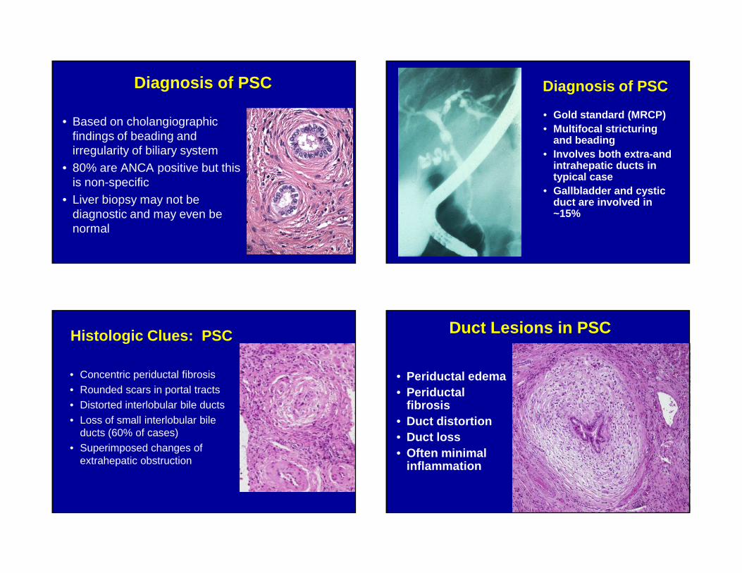
Diagnosis of PSC
• Based on cholangiographic findings of beading and irregularity of biliary system
• 80% are ANCA positive but this is non-specific
• Liver biopsy may not be diagnostic and may even be normal
Diagnosis of PSC
• Gold standard (MRCP)• Multifocal stricturing
and beading• Involves both extra-and
intrahepatic ducts in typical case
• Gallbladder and cystic duct are involved in ~15%
Histologic Clues: PSC
• Concentric periductal fibrosis• Rounded scars in portal tracts• Distorted interlobular bile ducts• Loss of small interlobular bile
ducts (60% of cases)• Superimposed changes of
extrahepatic obstruction
Duct Lesions in PSC
• Periductal edema• Periductal
fibrosis• Duct distortion• Duct loss• Often minimal
inflammation
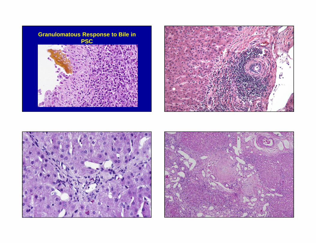
Granulomatous Response to Bile in PSC
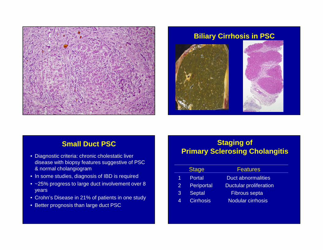
Biliary Cirrhosis in PSC
Small Duct PSC
• Diagnostic criteria: chronic cholestatic liver disease with biopsy features suggestive of PSC & normal cholangiogram
• In some studies, diagnosis of IBD is required
• ~25% progress to large duct involvement over 8 years
• Crohn’s Disease in 21% of patients in one study
• Better prognosis than large duct PSC
Staging of Primary Sclerosing Cholangitis
Stage Features1 Portal Duct abnormalities2 Periportal Ductular proliferation3 Septal Fibrous septa
4 Cirrhosis Nodular cirrhosis
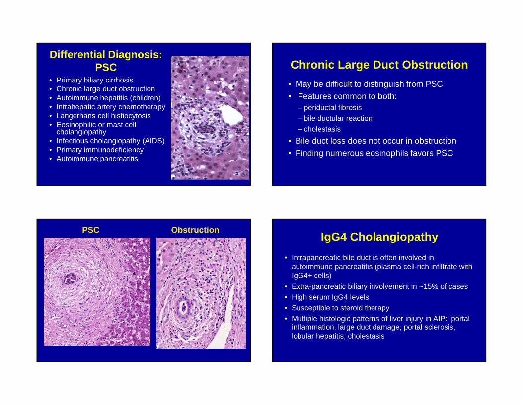
Differential Diagnosis: PSC
• Primary biliary cirrhosis• Chronic large duct obstruction• Autoimmune hepatitis (children)• Intrahepatic artery chemotherapy• Langerhans cell histiocytosis• Eosinophilic or mast cell
cholangiopathy• Infectious cholangiopathy (AIDS)• Primary immunodeficiency• Autoimmune pancreatitis
Chronic Large Duct Obstruction
• May be difficult to distinguish from PSC• Features common to both:
– periductal fibrosis – bile ductular reaction– cholestasis
• Bile duct loss does not occur in obstruction• Finding numerous eosinophils favors PSC
PSC ObstructionIgG4 Cholangiopathy
• Intrapancreatic bile duct is often involved in autoimmune pancreatitis (plasma cell-rich infiltrate with IgG4+ cells)
• Extra-pancreatic biliary involvement in ~15% of cases• High serum IgG4 levels• Susceptible to steroid therapy• Multiple histologic patterns of liver injury in AIP: portal
inflammation, large duct damage, portal sclerosis, lobular hepatitis, cholestasis
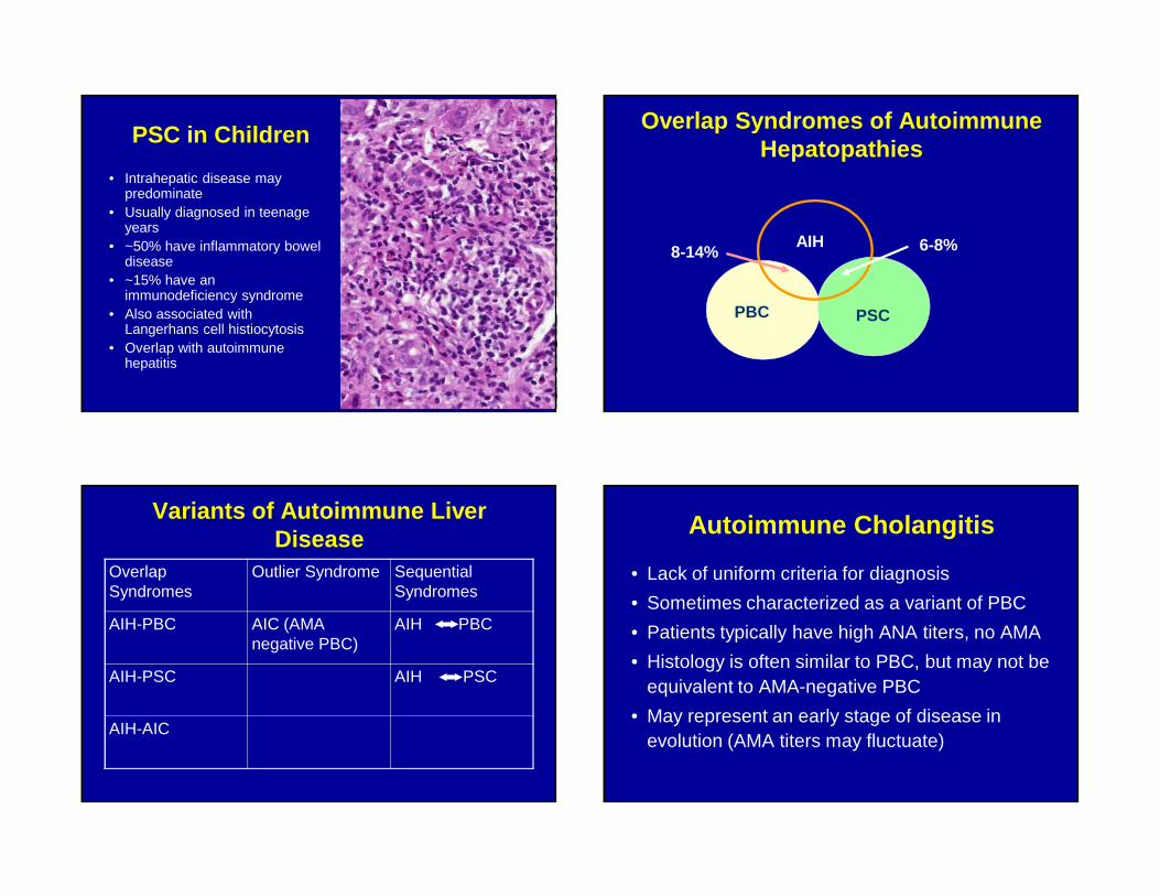
PSC in Children
• Intrahepatic disease may predominate
• Usually diagnosed in teenage years
• ~50% have inflammatory bowel disease
• ~15% have an immunodeficiency syndrome
• Also associated with Langerhans cell histiocytosis
• Overlap with autoimmune hepatitis
Overlap Syndromes of Autoimmune Hepatopathies
AIH
PBC PSC
6-8%8-14%
Variants of Autoimmune Liver Disease
Overlap Syndromes
Outlier Syndrome Sequential Syndromes
AIH-PBC AIC (AMA negative PBC)
AIH PBC
AIH-PSC AIH PSC
AIH-AIC
Autoimmune Cholangitis
• Lack of uniform criteria for diagnosis
• Sometimes characterized as a variant of PBC
• Patients typically have high ANA titers, no AMA
• Histology is often similar to PBC, but may not be equivalent to AMA-negative PBC
• May represent an early stage of disease in evolution (AMA titers may fluctuate)
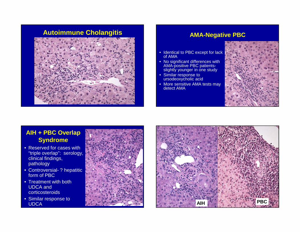
Autoimmune Cholangitis AMA-Negative PBC
• Identical to PBC except for lack of AMA
• No significant differences with AMA-positive PBC patients-slightly younger in one study
• Similar response to ursodeoxycholic acid
• More sensitive AMA tests may detect AMA
AIH + PBC Overlap Syndrome
• Reserved for cases with “triple overlap”: serology, clinical findings, pathology
• Controversial- ? hepatitic form of PBC
• Treatment with both UDCA and corticosteroids
• Similar response to UDCA PBCAIH
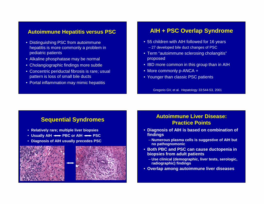
Autoimmune Hepatitis versus PSC
• Distinguishing PSC from autoimmune hepatitis is more commonly a problem in pediatric patients
• Alkaline phosphatase may be normal
• Cholangiographic findings more subtle• Concentric periductal fibrosis is rare; usual
pattern is loss of small bile ducts
• Portal inflammation may mimic hepatitis
AIH + PSC Overlap Syndrome
• 55 children with AIH followed for 16 years– 27 developed bile duct changes of PSC
• Term “autoimmune sclerosing cholangitis” proposed
• IBD more common in this group than in AIH• More commonly p-ANCA +
• Younger than classic PSC patients
Gregorio GV, et al. Hepatology 33:544-53, 2001
Sequential Syndromes
• Relatively rare; multiple liver biopsies• Usually AIH PBC or AIH PSC• Diagnosis of AIH usually precedes PSC
Autoimmune Liver Disease: Practice Points
• Diagnosis of AIH is based on combination of findings– Numerous plasma cells is suggestive of AIH but
no pathognomonic• Both PBC and PSC can cause ductopenia in
biopsies from adult patients– Use clinical (demographic, liver tests, serologic,
radiographic) findings • Overlap among autoimmune liver diseases