association respiratory infection, recurrent hematuria ... · the association of respiratory...
TRANSCRIPT
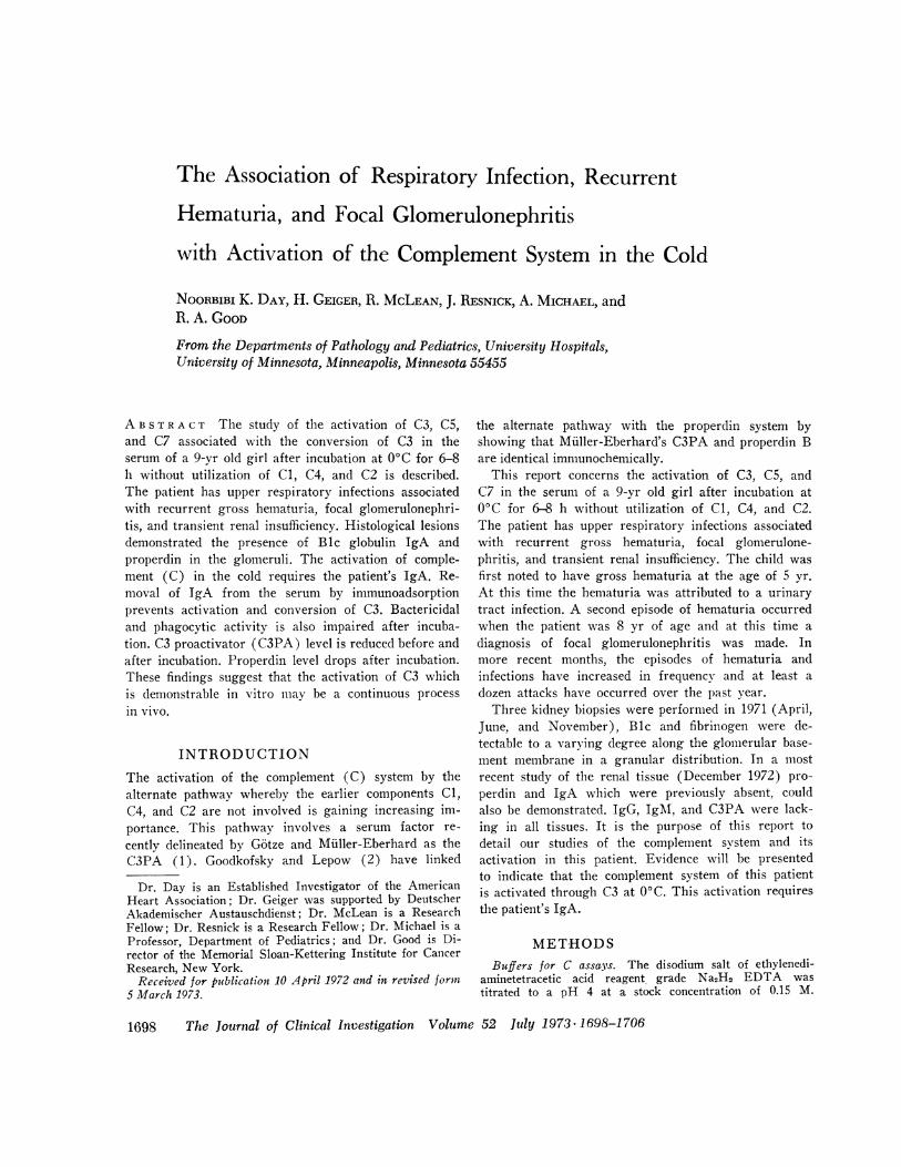
The Association of Respiratory Infection, Recurrent
Hematuria, and Focal Glomerulonephritis
with Activation of the Complement System in the Cold
NOORBIBI K. DAY, H. GEIGER, R. MCLEAN, J. RESNICK, A. MICHAEL, and
R. A. GOODFrom the Departments of Pathology and Pediatrics, University Hospitals,University of Minnesota, Minneapolis, Minnesota 55455
A B S T R A C T The study of the activation of C3, C5,and C7 associated with the conversion of C3 in theserum of a 9-yr old girl after incubation at 0WC for 6-8h without utilization of C1, C4, and C2 is described.The patient has upper respiratory infections associatedwith recurrent gross hematuria, focal glomerulonephri-tis, and transient renal insufficiency. Histological lesionsdemonstrated the presence of Blc globulin IgA andproperdin in the glomeruli. The activation of comple-ment (C) in the cold requires the patient's IgA. Re-moval of IgA from the serum by immunoadsorptionprevents activation and conversion of C3. Bactericidaland phagocytic activity is also impaired after incuba-tion. C3 proactivator (C3PA) level is reduced before andafter incubation. Properdin level drops after incubation.These findings suggest that the activation of C3 whichis demonstrable in vitro may be a continuous processin vivo.
INTRODUCTION
The activation of the complement (C) system by thealternate pathway whereby the earlier components C1,C4, and C2 are not involved is gaining increasing im-portance. This pathway involves a serum factor re-
cently delineated by Gbtze and Miller-Eberhard as theC3PA (1). Goodkofsky and Lepow (2) have linked
Dr. Day is an Established Investigator of the AmericanHeart Association; Dr. Geiger was supported by DeutscherAkademischer Austauschdienst; Dr. McLean is a ResearchFellow; Dr. Resnick is a Research Fellow; Dr. Michael is aProfessor, Department of Pediatrics; and Dr. Good is Di-rector of the Memorial Sloan-Kettering Institute for CancerResearch, NewYork.
Received for publication 10 April 1972 and in revised form5 March 1973.
the alternate pathway with the properdin system byshowing that Miiller-Eberhard's C3PA and properdin Bare identical immunochemically.
This report concerns the activation of C3, C5, andC7 in the serum of a 9-yr old girl after incubation at0C for 6-8 h without utilization of C1, C4, and C2.The patient has upper respiratory infections associatedwith recurrent gross hematuria, focal glomerulone-phritis, and transient renal insufficiency. The child wasfirst noted to have gross hematuria at the age of 5 yr.At this time the hematuria was attributed to a urinarytract infection. A second episode of hematuria occurredwhen the patient was 8 yr of age and at this time adiagnosis of focal glomerulonephritis was made. Inmore recent months, the episodes of hematuria andinfections have increased in frequency and at least adozen attacks have occurred over the past year.
Three kidney biopsies were performed in 1971 (April,June, and November), BMc and fibrinogen were de-tectable to a varying degree along the glomerular base-ment membrane in a granular distribution. In a mostrecent study of the renal tissue (December 1972) pro-perdin and IgA which were previously absent, couldalso be demonstrated. IgG, IgM, and C3PA were lack-ing in all tissues. It is the purpose of this report to
detail our studies of the complement system and itsactivation in this patient. Evidence will be presentedto indicate that the complement system of this patientis activated through C3 at 0C. This activation requiresthe patient's IgA.
MET-HODSBuffers for C assays. The disodium salt of ethylenedi-
aminetetracetic acid reagent grade NaH2 EDTA was
titrated to a pH 4 at a stock concentration of 0.15 M.
1698 The Journal of Clinical Investigation Volume 52 July 1973 1698-1706
Na2 Mg2EDTA (Geigy Chemical Corp., Ardsley, N. Y.)was also titrated to pH 4 at a stock concentration of 0.15M. Gelatin veronal buffer and glucose gelatin veronal bufferwith and without Ca`* and Mg-+ (GGV++, GGV--) wereprepared as described previously (3).
Serum. Blood was allowed to clot for 1 h at room tem-perature. The serum was removed after centrifugation at4VC, aliquoted, and stored at -70'C until used.
Guinea pig C2. Partially purified C2 was prepared fromguinea pig serum (Texas Biological Laboratory, Inc., Ft.Worth, Tex.) according to the method described by Nelson,Jensen, Gigli, and Tamura (4).
EAC1, EAC1,4, and EAC4. Cell intermediates with C1were prepared according to methods described previously(5, 6).
Assays of total complement (CH50) and the C compo-nents. Sensitation of erythrocytes from sheep (3) andthe measurement of total complement in 50% hemolyticunits (CH50) was carried out as described previously (7).Assays of human Cl, C4, and C2 were determined accord-ing to published methods (5).
Assays of human C3, C5, C6, C7, C8, and C9. Func-tionally pure C components for the assays of these com-ponents and EAClgp4-7hu for use in assay of C8 and C9were obtained from Cordis Laboratories (UMiami, Fla.)and the assays were carried out according to the methoddescribed by Nelson et al. (4). The experimental errorof the C components 1-9 ranged between 5 and 10%.Clq, Cls, and C3PA were carried out by the Mancini tech-nique' (8).
Conversion of C3 was determined by immunoelectrophore-sis using antisera against purified C3.
Conversion of C3PA was determined by immunoelectro-phoresis according to the method of Gdtze and Mufller-Eberhard (1) after treatment with inulin and with antiseraagainst purified C3PA.'
Bactericidal activity was measured according to themethod of Muschel and Treffers (9) using an Eschericluiacoli rough strain.
Phagocytic activity was measured according to the stan-dard methods used in this laboratory adapted to usingPseiidomonas aeruginosa type I (10) 2
Alethod of isolation of active factor. 3 ml of the pa-tient's plasma was applied to a 45 X 30 cm block of PevikonC-870 (11) (Mercer Consolidated Corp., New York) pre-pared with barbital buffer, pH 8.6, ionic strength 0.05. Thesample was applied at the cathodal region, electrophoresiswas carried out at 350 V for approximately 18 h at 4VC. Theblocks were cut into 11-cm segments and were eluted withbarbital buffer. The eluates were analyzed for protein byFolin's method.
Assay of active fraction. Approximately 0.7 mg/ml ofeach fraction was reacted with 100 Mul of normal serumfor 2 h at 370C. The mixture was serially diluted in GGV++.0.1 ml of EA1 X 108/ml was then added to each tube andtotal hemolytic C was determined. The active factor ap-peared at the cathodal area and this fraction was concen-trated in an Amicon ultrafiltration device (Amicon Corp.,Lexington, Mass.) using the membrane filter UM-10. Theconcentrated fraction was analyzed by immunoelectrophore-sis with monospecific antisera against IgA, IgM, and IgG.
'Antisera and Mancini plates were kindly supplied by Dr.Hans Muller-Eberhard.
'Biggar, W. D., N. K. Day, D. Windhorst, and R. A.Good. Impaired phagocytosis in Clr-deficient sera. Manu-script in preparation.
Purification by, imninunoadsorptioni. Monospecific anti-serum to IgA prepared according to methods described byLitman and Good (12) was coupled to activated, washedSepharose according to previously described method (13).The antisera wdere shown to be specific f or IgA by themethods outlined in the presentation of Litman and Good(12). 2 ml of plasma (70 mg/ml) was added to the Sepha-rose column. The f ractions were eluted at a rate of 10ml/h. Proteins were eluted over approximately 30 ml. Pro-teins eluted below OD 280 nm= 0.05 were not used forassay. The proteins bound to the immunoadsorbent wererecovered at 20C with 0.2 M glycine-HCl, pH 2.3.
RESULTS
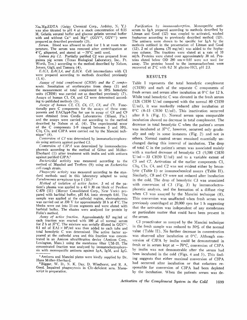
Table I represents the total hemolytic complement(CH50) and each of the separate C components offresh serum and serum after incubation at 0C for 12 h.While total hemolvtic C was elevated in the fresh serum(126 CH50 U/ml compared with the normal 80 CH50U/ml), it was markedly reduced after incubation at0'C (8-15 CH50 U/ml). This inactivation occurredafter 8 h (Fig. 1). Normal serum upon comparableincubation showed no decrease in total complement. Thedecrease in total hemolytic C when the patient's serumwas incubated at 370C, however, occurred only gradu-ally and only in some instances (Fig. 2) and not inothers. Normal control serum remained essentially un-changed during this interval of incubation. The dropof total C in the patient's serum was associated mainlywith a marked decrease of hemolytic C3 (9,500 CH50U/ml - 33 CH50 U/ml) and to a variable extent ofC5 and C7. Activation of the earlier components C1,Clq, Cls, C4, and C2 was not evident either by hemo-lvtic (Table I) or immunochemical assays (Table II).Similarly, C8 and C9 were not reduced after incubationin the cold. The drop of hemolytic C3 was associatedwith conversion of C3 (Fig. 3) by inimunoelectro-phoretic analysis, and the formation of a diffuse ringwhen C3 was assayed by the MA~ancini technique (8).This conversion was unaffected when fresh serum waspreviously centrifuged at 20,000 rpm for 1 h suggestingthat the activation was independent of any membranesor particulate matter that could have been present inthe serum.
C3 proactivator as assayed by the MIancini techniquein the fresh sample was reduced to 50% of the normalvalue (Table II). No further decrease in concentrationwas observed after incubation at 0°C. Although con-version of C3PA by inulin could be demonstrated infresh or in serum kept at - 700C, conversion of C3PAby intulin was not demonstrable after the serum hadbeen incubated in the cold (Figs. 4 and 5). This find-ing suggests that either maximal conversion of C3PAhad occurred after incubation or that cofactors re-sponsible for conversion of C3PA had been depletedby the incubation. When the patients serum was de-
Activation of the Complement System in the Cold 1699
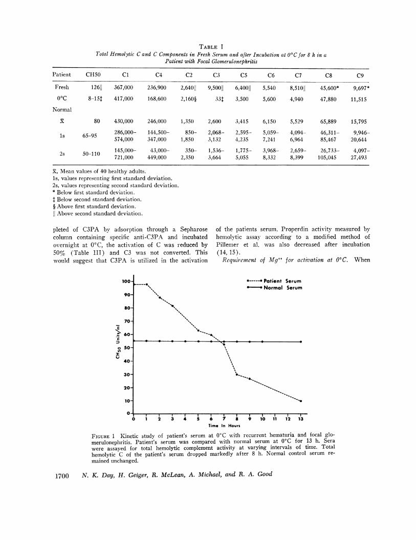
TABLE ITotal Hemolytic C and C Components in Fresh Serum and after Incubation at O'C for 8 h in a
Patient with Focal Glomerulonephritis
Patient CHSO C1 C4 C2 C3 C5 C6 C7 C8 C9
Fresh 12611 367,000 236,900 2,64011 9,50011 6,40011 5,540 8,51011 45,600* 9,697*00C 8-15t 417,000 168,600 2,160§ 33t 3,500 5,600 4,940 47,880 11,515
Normal
X 80 430,000 246,000 1,350 2,600 3,415 6,150 5,529 65,889 15,795
ls 65-95 286,000- 144,500- 850- 2,068- 2,595- 5,059- 4,094- 46,311- 9,946-574,000 347,000 1,850 3,132 4,235 7,241 6,964 85,467 20,644
2s 50-110 145,000- 43,000- 350- 1,536- 1,775- 3,968- 2,659- 26,733- 4,097-721,000 449,000 2,350 3,664 5,055 8,332 8,399 105,045 27,493
X, Mean values of 40 healthy adults.1s, values representing first standard deviation.2s, values representing second standard deviation.* Below first standard deviation.t Below second standard deviation.§ Above first standard deviation.11 Above second standard deviation.
pleted of C3PA by adsorption through a Sepharosecolumn containing specific anti-C3PA and incubatedovernight at 0C, the activation of C was reduced by50% (Table III) and C3 was not converted. Thiswould suggest that C3PA is utilized in the activation
100-
90-
80-
70-
60-.
o S0-
40-
30-
20-
10-
of the patients serum. Properdin activity measured byhemolytic assay according to a modified method ofPillemer et al. was also decreased after incubation(14, 15).
Requirement of Mg++ for activation at 0C. When
*- Patient SerumNormal Serum
FIGURE 1 Kinetic study of patient's serum at 00C with recurrent hematuria and focal glo-merulonephritis. Patient's serum was compared with normal serum at 0'C for 13 h. Serawere assayed for total hemolytic complement activity at varying intervals of time. Totalhemolytic C of the patient's serum dropped markedly after 8 h. Normal control serum re-mained unchanged.
1700 N. K. Day, H. Geiger, R. McLean, A. Michael, and R. A. Good
- - - - 0\
N1""-*'
1 2 3 4 5 6 7 8 9 10 11 12 13Time In Hours
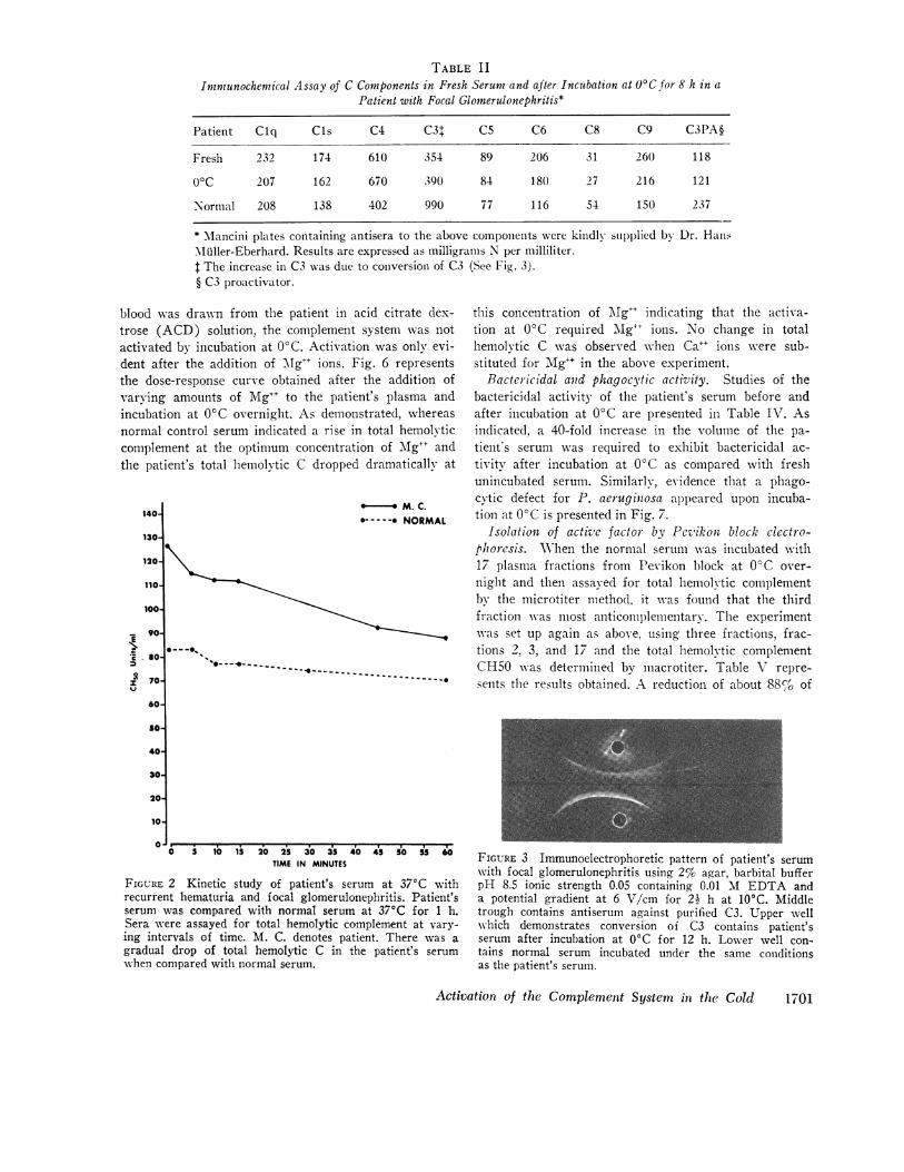
TABLE IIImmunochemical Assay of C Components in Fresh Serum and after Incubation at 0Cfor 8 h in a
Patient with Focal Glomerulonephritis*
Patient Clq CIs C4 C3t C5 C6 C8 C9 C3PA§
Fresh 232 174 610 354 89 206 31 260 118
0C 207 162 670 390 84 180 27 216 121
Normal 208 138 402 990 77 116 54 150 237
* lancini plates containing antisera to the above components were kindly supplied by Dr. HanlsMiuller-Eberhard. Results are expressed as milligrams N per milliliter.t The increase in C3 was due to conversion of C3 (See Fig. 3).§ C3 proactivator.
blood was drawn from the patient in acid citrate dex-trose (ACD) solution, the complement system was notactivated by incubation at 0C. Activation was only evi-dent after the addition of MIg++ ions. Fig. 6 representsthe dose-response curve obtained after the addition ofvarying amounts of Mg`+ to the patient's plasma andincubation at 00C overnight. As demonstrated, whereasnormal control serum indicated a rise in total hemolvticcomplement at the optimum concentration of Mg++ andthe patient's total hemolytic C dropped dramatically at
140
130
120-
110-
100~
E1
z;
90-
so
70-
60
5o-
40-
30J
20-
10-
-* M. C.*-- NORMAL
0---O. ~ ~ .0-- - -- -
0---4-0~~~~~-- --
this concentration of MAg`+ indicating that the activa-tion at 0°C required Mg++ ions. No change in totalhemolytic C was observed when Ca`+ ions were sub-stituted for Mg`+ in the above experiment.
Bactericidal and phagocvtic activity. Studies of thebactericidal activity of the patient's serum before andafter incubation at 00C are presented in Table IV. Asindicated, a 40-fold increase in the volume of the pa-tient's serum was required to exhibit bactericidal ac-tivitv after incubation at 0C as compared with freshunincubated serum. Similarly, evidence that a phago-cytic defect for P. aeruginosa appeared upon incuba-tion at 00C is presented in Fig. 7.
Isolation of active factor by Pevikon block clectro-phoresis. WNhen the normal serum was incubated with17 plasma fractions from Pevikon block at 0°C over-night and then assayed for total hemolytic complementby the microtiter method, it was found that the thirdfraction was most anticonmplementary. The experimentwas set up again as above, using three fractions, frac-tions 2, 3, and 17 and the total hemolytic complementCH50 was determined by macrotiter. Table V repre-sents the results obtained. A reduction of about 88%,o of
0 5 10 15 20 25 30 35 40 45 50550TIME IN MINUTES
FIGURE 2 Kinetic study of patient's serum at 370C withrecurrent hematuria and focal glomerulonephritis. Patient'sserum was compared with normal serum at 370C for 1 h.Sera were assayed for total hemolytic complerment at vary-ing intervals of time. M. C. denotes patient. There wvas agradual drop of total hemolytic C in the patient's serumwhen compared with normal serum.
FIGURE 3 Immunoelectrophoretic pattern of patient's serumwith focal glomerulonephritis using 2% agar, barbital bufferpH 8.5 ionic strength 0.05 containing 0.01 MI EDTA anda potential gradient at 6 V/cm for 2i h at 10'C. Middletrough contains antiserum against purified C3. Upper wellwhich demonstrates conversion of C3 contains patient'sserum after incubation at 0C for 12 h. Lower well con-tains normal serum incubated under the same conditionsas the patient's serum.
Activation of the Complement System in the Cold
I-
.
I
4
i.
4
1701

TABLE I IIPercent Inhibition of Total Hemolytic Complement CH50 and
Conversion of C3 of Patient's Serum Depleted of C3PA andafter Incubation at 0WCOvernight
FIGURE 4 Immunoelectrophoretic pattern of patient's serumafter treatment with inulin according to the method ofGotze and Miiller-Eberhard (1). 0.1 ml of the patient'sfreshly frozen serum (lower well) and normal human serumalso freshly frozen (upper well) was incubated with 20 ulof a 10 mg/ml inulin suspension. The conditions for elec-trophoresis are described in Fig. 3. Conversion of C3 pro-activator occurs in both the patient's serum and in normalserum.
CH50 with fraction 3 occurred as compared with noreduction protluced by fraction 17. Some inhibition wasalso obtained with fraction 2. The three fractions werethen concentrated separately in an Amicon ultrafiltra-tion device (Amicon Corp.) using the membrane filterUM-10. The active fractions were concentrated andwere analyzed by immunoelectrophoresis and by Ouch-terlony with antisera against IgG, IgM, and IgA. Aband appeared with antisera against IgA in fraction 3which was the most active fraction.
Purification by! imimunoadsorption. When the pa-tient's plasma which had been adsorbed by a Sepharosecolumn containing antiserum specific for IgA was in-cubated wvith MIgf+ (2 mg/ml) overnight at 0°C, theCH50 activity dropped only by 4% as compared withunadsorbed control patient's plasma which was incu-bated with Mg++ at 0C overnight. The C activity inthe control plasma dropped by greater than 95%.(Table VI).
Similarly, when the above specific IgA adsorptionwas repeated with the patient's serum and the adsorbedserum then incubated overnight at 0°C, the drop inhemolytic C was only 20% and no conversion of C3could be demonstrated (Fig. 8). When IgM was speci-fically removed from the patient's serum by immuno-adsorption, the percent inhibition of complement was
CHSO %Inhibition C3 conversion
Serum control 90 >90 Yes
Serum depletedof C3PA 66 50 No
61% (Table VII). These experiments indicated thatIgA is responsible for activation of the C system inthis patient's serum. In order to determine whetherthe Sepharose alone was absorbing nonspecifically oneor more proteins involved in the activation of C3 inthe cold, patient's plasma was applied to a Sepharosecolumn not charged with any antiserum. The fractionscontaining the highest activity of hemolytic C wereleft overnight at 0°C with Mg". As shown in Table
E
"I,._
s
* M. C.0-*- - NORMAL
FIGURE 5 Immunoelectrophoretic pattern of patient's serum(lower well) and normal serum (upper well) after in-cubation at 0'C for 12 h. Middle trough contains antiserumto C3PA. The conditions of immunoelectrophoresis wereidentical as described in Fig. 4. No conversion of C3PA isdemonstrable in the patient's serum.
Mg mg/ml
FIGURE 6 Effect of Mg++ ions on patient's plasma andincubation at 0'C for 12 h. As demonstrated inhibition oftotal complement occurred in the patient's plasma at theconcentration necessary for maximum lysis. In contrast,control plasma showed maximum lysis at the same con-centration. M. C. represents patient's plasma.
1702 N. K. Day, H. Geiger, R. McLean, A. Michael, and R. A. Good
I
III
II
:IIIII
IIII
1:0

TABLE IRBactericidal Activity of Fresh Serum and after Incubation at
0C for 8 h in a Patient with Focal Glomerulonephritis
Bactericidal activity*
E. coli+
Fresh Incubated
Patient 0.009 0.035Normal 0.0074 0.0065
* Bactericidal activity performed according to the method ofMuschel and Treffers (9). Results are expressed as the amountof serum necessary to kill 50'-% bacteria.t E. coli rotIgh strain.
VIII, the CH5O activity dropped by 80%cG. The activa-tion of the control patient's serum was 94%. The aboveimnmunoadsorption experiments were performed on thesame day and the columns used were identical in size.The hemolvtic assays were carried out the followingday. In addition, when the fractions passed over such acolumn were analyzed by immunoelectrophoresis with
TIME IN MINUTES
FIGURE 7 Defect of phagocytic function of the patient'sserum after incubation at 00C. ( ----- ), Patient's serumafter incubation at 00C for 8 h. (I- -- ), Pool normalhuman serum after incubation at 0C for 8 h. ( ),Pool normal human serum. ( - --- ), Patient's serumat - 700. The patient's serum was defective in the phago-cytic function for P. aeruginosa type I after incubation atQOC.
TABLE \Anticomplementary Activity of Patient's Plasma Fractions
Obtained by Electrophoresis on Pevikon Blockon Normal Human Serum
Percent inhibitionFraction no. CH50 Urml
2 45
3 88
17 <5
3 ml of the patient's plasma was applied to a 45 cm X 30 cmblock of Pevikon prepared with barbital buffer, pH 8.6, ionicstrength 0.05. The sample was applied at the cathodal region.Electrophoresis was carried out at 350 V for approximately18 h at 40C. The blocks were cut into 1 4-cm segments and wereeluted with barbital buffer. The eluates were analyzed forprotein by Folin's method.Approximately 0.7 mg/ml of each fraction was reacted with100 Ml of normal serum for 2 h at 370C. The mixture wasdiluted in GGX++. 0.1 ml of EA1 X 108nml was then addedto each tube and total hemolvtic C was determined.The results represent percent inhibition of total CH50 whencompared with the control fraction-fraction 17.
antisera against IgG, IgM\, IgA, and JgD, none of theimmunoglobulins were removed nor was C3PA removed.
A control experiment to determine whether IgG wasinvolved in the activation of C3 was then executed.Since it was difficult to remove IgG completely fromthe patients serum by a single immunoadsorption andsince subsequent adsorptions led to a high dilution ofthe serum, the following experiment was carried out.
The patient's plasma was applied to a Pevikon blockand those fractions containing a mixture of IgG andIgA were added to normal serum and incubated at 00Covernight. Inhibition of CH50 activity in the normalserum occurred. The IgA from these fractions wasremoved on Sepharose column containing anti-IgA.The purity of these adsorbed fractions was establishedby immunoelectrophoresis and Ouchterlony (Fig. 9).These fractions were then added to normal human
TABLE VIPercent Inhibition of Total Hemolytic Complement CH50 and
Conversion of C3 of Patient's Fresh Plasma, of PlasmaDepleted of IgA and after Incubation at
0C Overnight
CH5O %7c Inhibition C3 conversion
Placsma control 130 >95 yes
Plasma depletedof IgA 61 4 no
Activation of the Complement System in the Cold 1703
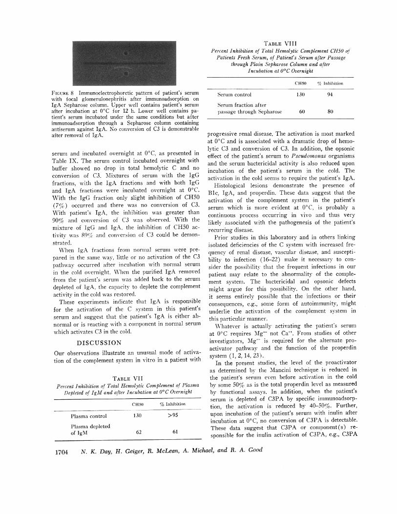
TABLE VI IIPercent Inhibition of Total Hemolytic Complement CH50 of
Patients Fresh Serum, of Patient's Serum after Passagethrough Plain Sepharose Column and after
Incubation at 0C Overnight
FIGURE 8 Immunoelectrophoretic pattern of patient's serumwith focal glomerulonephritis after immunoadsorption onIgA Sepharose column. Upper well contains patient's serumafter incubation at 0'C for 12 h. Lower well contains pa-tient's serum incubated under the same conditions but afterimmunoadsorption through a Sepharose column containingantiserum against IgA. No conversion of C3 is demonstrableafter removal of IgA.
serum and incubated overnight at 00C, as presented inTable IX. The serum control incubated overnight withbuffer showed no drop in total hemolytic C and noconversion of C3. Mixtures of serum with the IgGfractions, with the IgA fractions and with both IgGand IgA fractions were incubated overnight at 0°C.With the IgG fraction only slight inhibition of CH50(7%) occurred and there was no conversion of C3.With patient's IgA, the inhibition was greater than90% and conversion of C3 was observed. With themixture of JgG and IgA. the inhibition of CH50 ac-tivitv was 89(%fi and conversion of C3 could be demon-strated.
When IgA fractions from normal serum were pre-pared in the same way, little or no activation of the C3pathway occurred after incubation with normal serumin the cold overnight. WThen the purified IgA removedfrom the patient's serum was added back to the serumdepleted of IgA, the capacity to deplete the complementactivity in the cold was restored.
These experiments indicate that IgA is responsiblefor the activation of the C system in this patient'sserum and suggest that the patient's IgA is either ab-normal or is reacting with a component in normal serum
which activates C3 in the cold.
DISCUSSIONOur observations illustrate an unusual mode of activa-tion of the complement system in vitro in a patient with
TABLE VIIPercent Inhibition of Total Hemolytic Complement of Plasma
Depleted of IgM and after Incubation at 00C Overnight
CH50 % Inhibition
Plasma control 130 >95
Plasma depletedof IgM 62 61
CH50 %1c Inhibition
Serum control 130 94
Serum fraction afterpassage through Sepharose 60 80
progressive renal disease. The activation is most markedat 0C and is associated with a dramatic drop of hemo-lytic C3 and conversion of C3. In addition, the opsoniceffect of the patient's serum to Pseudomonas organismsand the serum bactericidal activity is also reduced uponincubation of the patient's serum in the cold. Theactivation in the cold seems to require the patient's IgA.
Histological lesions demonstrate the presence ofBic, IgA, and properdin. These data suggest that theactivation of the complement system in the patient'sserum which is more evident at 0WC, is probably acontinuous process occurring in vivo and thus verylikely associated with the pathogenesis of the patient'srecurring disease.
Prior studies in this laboratory and in others linkingisolated deficiencies of the C system with increased fre-quency of renal disease, vascular dise~ase, and suscepti-bility to infection (16-22) make it necessary to con-sider the possibility that the frequent infections in ourpatient may relate to the abnormality of the comple-ment system. The bactericidal and opsonic defectsmight argue for this possibility. On the other hand,it seems entirely possible that the infections or theirconsequences, e.g., some form of autoimmunity, mightunderlie the activation of the complement system inthis particular manner.
XVhatever is actually activating the patient's serumat 0C requires Mg"+ not Ca"+. From studies of otherinvestigators, Mlg+ is required for the alternate pro-activator pathway and the function of the properdinsystem (1,2, 14, 23).
In the present studies, the level of the proactivatoras determined by the Mancini technique is reduced inthe patient's serum even before activation in the coldby some 50% as is the total properdin level as measuredby functional assays. In addition, when the patient'sserum is depleted of C3PA by specific immunoadsorp-tion, the activation is reduced by 40-50%. Further,upon incubation of the patient's serum with inulin afterincubation at 0WC, no conversion of C3PA is detectable.These data suggest that C3PA or component(s) re-
sponsible for the inulin activation of C3PA, e.g., C3PA
1704 N. K. Day, H. Geiger, R. McLean, A. Michael, and R. A. Good

convertase are utilized during the spontaneous activa-tion of C3PA in -vitro.
Although C3PA is not demonstrable in the kidney bi-opsies by immunofluorescent techniques, IgA, and pro-perdin are present. Direct evidence that this patienthas continuous activation in vivo of the C3PA as wellis not vet available and will require studies of utiliza-tion of these components which have not yet beencarried out. Recent studies by G6tze and Mifller-Eber-hard (1) and by Goodkofsky and Lepow suggestthat C3PA and properdin B are identical (2). In ourstudies we have not measured properdin A, but it mightbe desirable to make such studies in light of ourobservations.
The conversion of C3 at 00C is indeed a provocativefinding. Whether an as yet unknowvn enzyme is re-sponsible for this activation or whether the patienthas an activator such as an antibody or autoantibody inher serum which operates preferentially by the alter-nate pathway in the cold must be studied further.Already, however, it is clear that IgA is required forthis strange form of complement activation. Evidencefrom Pevikon block fractionation indicates that the re-sponsible serum component is patient's IgA. MWhen IgAis remooved front either the patient's plasnma or serumby imnmnunoadsorption and then the serum is kept over-night at 00C, no conversion of C3 or drop in hemnolytictiter occurs. \When IgA obtained from Pevikon blockfractions of patient's plasmia is added to the patient'sserum del)letedl of IgA, a drop in hemiolvtic comple-ment and a conversion of C3 occurs in the cold. WNlhenIgG or IgA or mixtureIs of IgG and IgA obtained byPevikon block fractionation front the patient's serumare added to normal serum, and the mixture kept over-night at 0°C, the hemiolytic titer of serum drops bygreater than 80% and a conversion of C3 occurs withinthose mixtures containing IgA, whereas with IgGalone, there is less than 10%,, drop in hemiolytic titerand no conversion of C3. Application of the patient'sserum on Sepharose without antiserum or with Sephla-rose activated w\vith Tg-M, does not remove nonspecifi-callv any of the proteins required for the activation of
FIGURE 9 Ouchterlony pattern of patients plasma afterPevikon block fractionation and after passage throughsepharose column containing anti-IgG antiserum. Upper wellcontains fraction after absorption of IgG absorption. Centerwell contains anti-IgA antiserum.
TABLE IXPercent Inhibition of Total Hemolytic Complement CH5(0 and
Conversion of C3 after Incubation at O'C of Normal Serumwith Pevikon Block Fractions of Patient's Plasma
Containing IgG, IgA, and a Mixture ofJgG plus Ig.l
CH5O % Inhibition C3 conver-ion
Serum control 110 <5.0 No
Serum + lgG 102 7.3 No
Serum+(IgG + IgA)Mixture <1( >90.0 Yes
Serum + IgA 12 89.0 Yes
the C systenm in the cold from either the patient's ornormal sera.
Although present understanding of the complementsy stem, the so-called alternate pathway, and the rela-tionships of these to the properdin system are not yetsufficiently definitive to permit us to explain completelythe association of recurrent renal disease with the un-usual cold activation of the complement system that wehave observed in the serum of our patient, it seemscertain that these phenomena are intimately related inan important way. To our knowledge, this patient'sserum is unique in that its IgA activates complementin the cold without utilizing Cl, C4, and C2. Theassociation of this unique serological process w-ith theclinical picture of recurrent hematuria in unexplainedrenal disease makes it necessary to look at other pa-tients Awho have recurrent unexplained hematuria forsimilar serological perturbations and to look more regu-larlv especially in patients with renal disease and re-current infection for evidence of spontaneous activationsof the alternate pathway by the methodology that wasused in the study of this patient. Since the discoveryof cold activation of the complement system was acci-dental in this instance, it seems likely that directedsearch for this activation mechanism will reveal othercases that can shed light on a most provocative asso-ciation.
ACKNOWLEDGNIENTSWVe thank Dr. Hans Miiller-Eberhard for generously pro-viding us with antisera to C3 proactivator and C3 andMancini plates for immunochemical assays of complementcomponents. We also thank Mrs. Linda Schuveiller, Mrs.Soo Young Yang, Mrs. Linda Campbell, and Mrs. SusanBuron for excellent technical assistance. We thank Dr.Gary Litman for supplying us with antisera against IgA.
This work was supported in part by grants from TheNational Foundation-March of Dimes, U. S. Public HealthService grants AI-08677 and AI-10,704; Special Virus
Activation of the Complement System in the Cold 1 705

Cancer Program (Contract 71-2261); the Kidney Founda-tion of the Upper Midwest; the American Heart Asso-ciation; The American Cancer Society; and the BrownHazen Fund.
REFERENCES1. Gotze, O., and H. J. Mfiller-Eberhard. 1971. The C3-
activator system: an alternate pathway of complementactivation. J. Exp. Med. 134(Suppl.) 905.
2. Goodkofsky, I., and I. H. Lepox. 1971. Functional re-lationship of factor B in the properdin system to C3proactivator of human serum. J. Ihnunitol. 107: 1200.
3. Mayer, M. M. 1961. Procedure for titration of com-plement components with R1, R2, R3 and R4. In Experi-mental Immunochemistry. E. A. Kabat and M. M.Mayer, editors. Charles C Thomas, Publisher, Spring-field, Ill. 2nd edition. 163.
4. Nelson, R. A., Jr., J. Jensen, I. Gigli, and N. Tamura.1966. Methods for the separation, purification andmeasurement of nine components of hemolytic comple-ment in guinea pig serum. Immnniochenmistry. 3: 111.
5. Gewurz, H., A. R. Page, R. J. Pickering, and R. A.Good. 1967. Complement activity and inflammatory neu-trophil exudation in man: studies in patients withglomerulonephritis, essential hypocomplementemia andagammaglobulinemia. Int. Arc/i. Allergy Appl. Ivi-mnunol. 32: 64.
6. Borsos, T., and H. Rapp. 1967. Immune hemolysis: asimplified method for the preparation of EAC4 withguinea pig or with human complement. J. Immnozol.99: 263.
7. Day, N. K., R. J. Pickering, H. Gewurz, and R. A.Good. 1969. Ontogenetic development of the comple-ment system. Immunology. 16: 319.
8. Mancini, G., A. 0. Carbonara, and J. F. Heremans.1965. Immunochemical quantitation of antigens bysingle radial immunodiffusion. Imimunochemistry. 2: 235.
9. Muschel, L. M., and H. B. Treffers. 1956. Quantitativestudies on the bactericidal actions of serum and ofcomplement. I. A rapid photometric growth assay forbactericidal activity. J. Inimmniol. 76: 1.
10. Maaloe, 0. 1946. Doctoral Thesis. Munksgaard, Copen-hagen.
11. Mflller-Eberhard, H. J. 1960. A new supporting mediumfor preparative electrophoresis. Scauzd. J. Clin. Lab. In-vest. 12: 33.
12. Litman, G. WV., and R. A. Good. 1972. Rapid purifica-tion of IgA from normal human serum. Biochim. Bio-phys. Acta. 263: 89.
13. Frommel, D., J. M. Dupuy, G. W. Litman, and R. A.Good. 1970. Use of immunoadsorbent techniques in thepreparation of chemical agammaglobulinemia. J. Im-muotol. 105: 1292.
14. Pillemer, L., L. Blum, I. H. Lepow, 0. A. Ross, E. WV.Todd, and A. C. Wardlaw. 1954. The properdin systemand immunity. I. Demonstration and isolation of anew serum protein, properdin and its role in immunephenomenon. Scienice (Wash. D. C.). 120: 279.
15. McLean, R. H., and A. F. Michael. 1972. The im-munoelectrophoretic pattern of properdin in fresh andaged human serum. Proc. Soc. Exp. Biol. Med. 14:403.
16. Kohler, P. F., and H. J. Mfiller-Eberhard. 1969. Com-plement-immunoglobulin relation: deficiency of C'lqassociated with impaired immunoglobulin synthesis. Sci-ence (Wash. D. C.). 163: 474.
17. Gewurz, H., R. J. Pickering, and R. A. Good. 1968.Complement and complement component activities indiseases associated with repeated infections and malig-nancy. Int. Arch. Allergy Appl. Immunol. 33: 368.
18. Pickering, R. J., G. B. Naff, R. M. Stroud, R. A.Good, and H. Gewurz. 1970. Deficiency of Clr in humanserum. Effects of the structure and function of macro-molecular Cl. J. Exp. Med. 131: 803.
19. Moncada, B., N. K. B. Day, R. A. Good, and D. Wind-horst. 1972. Lupus-erythematosus-like syndrome with afamilial defect of complement. N. Engl. J. Med. 286:689.
20. Day, N. K., H. Geiger, R. Stroud, M. de Bracco, B.Moncada, D. \Vindhorst, and R. A. Good. 1972. Clrdeficiency-an inborn error associated with clinical andrenal disease. J. Clin. Invest. 51: 1102.
21. Agnello, V., M. M. E. deBracco, and H. G. Kunkel.1972. Hereditary C2 deficiency with some manifesta-tions of systemic lupus erythematosus. J. Immunol.108: 837.
22. Day, N. K., H. Geiger, R. McLean, A. Michael, andR. A. Good. 1973. C2 deficiency. Development of lupuserythematosus. J. Clin. Invest. 52: 1601.
23. Lepow, H. J. 1972. Biologically active fragments ofcomplement. In Progress in Immunology. BernardAmos, editor. Academic Press, Inc., New York. 589.
1706 N. K. Day, H. Geiger, R. McLean, A. Michael, and R. A. Good