articulations = joints - master anatomymasteranatomy.org/wp-content/uploads/2015/07/dims05.pdf ·...
TRANSCRIPT

Articulations = JointsPoint of contact bt/n boneand bone or bt/n cartilage and bone.

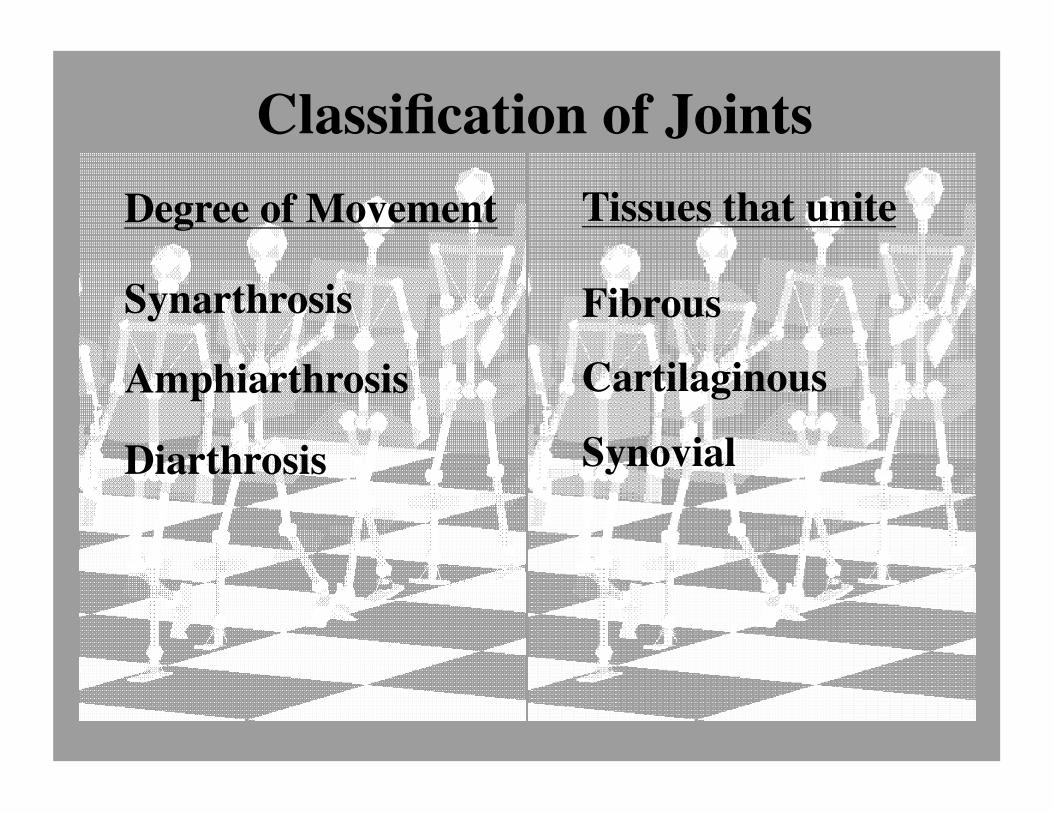
Classification of JointsDegree of Movement
Synarthrosis
Amphiarthrosis
Diarthrosis
Tissues that unite
Fibrous
Cartilaginous
Synovial

Fibrous Joints: Synarthroses ���Sutures ���
The calvaria or brain case is made up of the frontal, parietal, temporal and occipital bones. The joints between these bones are called the sutures.
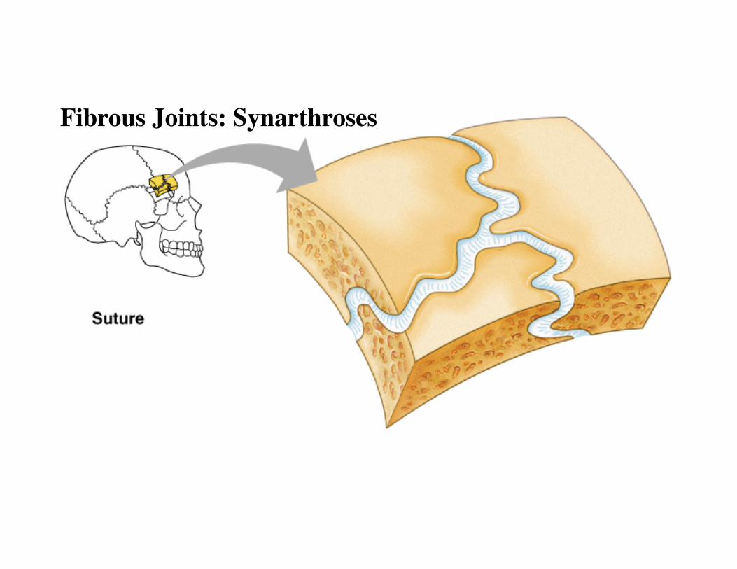
Fibrous Joints: Synarthroses ���

Fibrous Joints – Amphiarthroses��� - Interosseous membranes ���

Fibrous Joints – Amphiarthroses��� - Interosseous membranes ���

Fibrous Joints – Amphiarthroses��� - Interosseous membranes ���

Fibrous Joints – Amphiarthroses��� - Interosseous membranes ���

Interosseus membranes – Fibrous/amphiarthroses

Cartilaginous Joints: AmphiarthrosesIntervertebral discs ���
Pubic symphysis ���

Intervertebral discs – Cartilaginous joints/amphiarthroses

Synchondrosis – - cartilaginous/synarthrosis - joint bt/n 1st rib and sternum

Synchondrosis – - cartilaginous/synarthrosis - epiphyseal plate

Epiphyseal Plate

Diarthrosis/synovial – Hip Joint

Diarthrosis/synovial– Hip Joint

Diarthrosis/synovial– Knee Joint
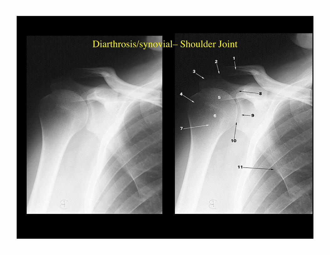
Diarthrosis/synovial– Shoulder Joint

Diarthrosis/synovial – Elbow Joint
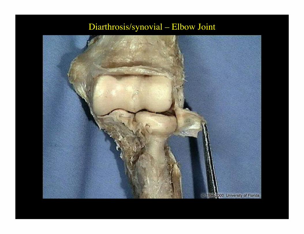
Diarthrosis/synovial – Elbow Joint

Diarthrosis/synovial– Knee Joint

Generic Synovial Joint
Accessory Ligament
SynovialMembrane
Fibrous Capsule
Articular Cartilage
Joint Cavity

Bursae
Shoulder Joint

I. abductor pollicis longus and extensor pollicis brevis II. extensor carpi radialis longus and brevis III. extensor pollicis longus IV. extensor digitorum comunis (four tendons) and extensor indicis V. extensor digiti minimi VI. extensor carpi ulnaris
Tendon Sheaths of the Posterior Wrist

Tendon Sheaths of the Ankle

Synovial Joint – Hinge Type


Elbow JointHinge type

Knee JointHinge type

Synovial Joint – Pivot Type
Radioulnar joint

Atlantoaxial Jointpivot

Pivot

Synovial Joint – Ball-and-Socket Type
Glenohumeral joint


Glenohumeral jointBall-and-socket type

Hip jointBall-and-socket type


Knee – Modified hinge type
Quadriceps tendon
Medial collateral ligament
Patellar ligament
Lateral collateral ligament

Posterior cruciate ligamentAnterior cruciate ligament
Medial condyleLateral condyle
Quadriceps tendon
Patella
Patellar ligament
Medial condyleLateral condyle
Medial collateral ligamentLateral collateral ligament
Medial meniscusLateral meniscus

Quadriceps tendon
Patella
Patellar ligament
Posterior cruciate ligament
Anterior cruciate ligament
Suprapatellar bursa
Patellar bursa
Infrapatellar bursa


Femoral CondylesCruciate Ligaments


ACL
MCL
Medial meniscus

http://faculty.washington.edu/mtuggy/kneeinfo.htm
Telemark Skiing• The combination of torsional
stress and valgus force (distal segment deviates laterally) on the knee results in the common MCL/ACL injury shown in the animated sequence here on the right.
• This injury is commonly found when the skier catches the inner edge of a lightly weighted or non-weighted ski.
• This can occur when the uphill ski, early in the initiation of a turn, in not weighted properly or is flat on the snow instead of being used to carve the early part of the turn.

Tissue-engineered scaffolding may help the body repair weakened knees and joints.
www.nibib1.nih.gov/ eAdvances/80204.htm
• In only two weeks, cartilage—one of the most difficult tissues to repair—was regrown in a rabbit knee joint. The newly restored cartilage, stained dark purple on the right, was rebuilt using a new biomaterial that fosters cartilage growth. With further study, this biomaterial may one day help to repair cartilage damage due to injury.

Frostbite

