arthroscopic treatment of femoroacetabular impingement...arthroscopic treatment of femoroacetabular...
TRANSCRIPT
Tsnptarcwhyotlm
C
GNs
Current Concepts
Arthroscopic Treatment of Femoroacetabular Impingement
Carlos A. Guanche, M.D., and Aaron A. Bare, M.D.
Abstract: The etiology of degenerative joint disease of the hip remains unsolved. A precursor forsome patients, especially younger ones, may be hip impingement. Repetitive microtrauma at maximalflexion can cause chronic pain from the abutment at the femoral head-neck junction caused by anabnormal offset. Chronic impingement from an aspherical head can lead to degenerative labral tearsand acetabular chondral degeneration, which may contribute to the degenerative cascade. Arthro-scopic treatment of hip impingement caused by an abnormal head-neck offset improves symptoms,restores hip morphology, and ultimately may halt the progression toward degenerative joint diseasein certain patients. Early results show that if debridement of the impinging lesion and injured labrumis performed in the setting of normal femoral and acetabular articular surfaces, the results arepromising. Key Words: Hip arthroscopy—Femoroacetabular impingement—Labral tear.
or
pwoctvsaln
jsltita
he causes of hip pain range from intra-articularloose bodies and labral injuries to extra-articular
ources originating from musculotendinous injuries,erve entrapment, bursal inflammation, or referredain. The acetabular labrum has gained increased in-erest because its degeneration frequently is found inssociation with early osteoarthritis of the hip.1-3 Mostecently, femoroacetabular impingement (FAI) has re-eived attention as a structural abnormality associatedith hip pain.4-6 An altered femoral neck morphologyas been implicated as a cause of chronic hip pain inounger patients without obvious degeneration. The-retically, repetitive microtrauma from the neck abut-ing against the acetabular rim can lead to labralesions and acetabular cartilage delamination. Thisechanical impingement of the hip is believed to
From the Southern California Orthopedic Institute, Van Nuys,alifornia, U.S.A.Address correspondence and reprint requests to Carlos A.uanche, M.D., Southern California Orthopedic Institute, 6815oble Ave, Van Nuys, CA 91405, U.S.A. E-mail: cguanche@
coi.com© 2006 by the Arthroscopy Association of North America
w0749-8063/06/2201-05-310$32.00/0doi:10.1016/j.arthro.2005.10.018
Arthroscopy: The Journal of Arthroscopic and Related S
riginate from either a “pistol grip” femoral neck or aetroverted acetabulum4 (Fig 1).
Previously, open exposures and removal of the im-ingement areas have been described.5 Some patientsith FAI, those with decreased femoral head-neckffset on the anterolateral neck (pistol grip deformity),an be successfully treated arthroscopically. In addi-ion, patients with lesser degrees of acetabular retro-ersion can also be treated arthroscopically. Arthro-copic debridement of the anterolateral femoral necklong with treatment of concomitant intra-articularabral lesions attempts to re-establish the normal head-eck relationship and restore hip biomechanics.The causative link between FAI and degenerative
oint disease remains unknown. On the other hand,everal studies have shown an association betweenabral tears and osteoarthritis3 as well as labral tears inhe setting of impingement.4,7 It is possible that treat-ng the structural abnormality can lead to the resolu-ion of hip pain and potentially halt the progression ofhip at risk for future degenerative changes.
BACKGROUND
A nonspherical femoral head has been associated
ith early development of osteoarthritis of the hip.8,995urgery, Vol 22, No 1 (January), 2006: pp 95-106

Ttamseb
cdsnat
dofms
mdwDaaT
Fpw terior
96 C. A. GUANCHE AND A. A. BARE
he effect of asphericity on the acetabular labrum andhe consequent degeneration that ensues has receivedttention in the literature because labral degenerationay lead to early osteoarthritis.1,3,10 McCarthy et al.3
howed that the relative risk of significant chondralrosion approximately doubles in the presence of la-ral lesions.Whereas the asphericity seen in dysplastic heads
an lead to arthritis as a result of the contact of aysplastic head directly with the acetabular chondralurface, aspherical heads stemming from altered head-eck junction morphology can contribute to degener-tive joint disease by a different mechanism. Repeti-
IGURE 1. (A) Normal AP radiograph indicating a central positioatient with offset. (C) Frog lateral view of the patient in Fig 1B. Noith significant acetabular retroversion. Note the overlap of the an
ive abutment of a neck with increased radii, or a
ecreased femoral head-neck offset at extreme rangesf motion, can lead to labral and chondral injuriesrom shearing stresses. Labral lesions from impinge-ent have been shown to be located almost exclu-
ively in the anterosuperior region of the acetabulum.1
Murray11 first introduced the theory of hip impinge-ent as the underlying cause of degenerative joint
isease. The terms head tilt or pistol grip deformityere developed to describe the radiographic findings.ecreased femoral head-neck offset contributes to
butment of the proximal neck with flexion, often withvariable degree of adduction and internal rotation.he repetitive contact can damage the adjacent labrum
e head on the femoral neck. (B) Femoral head-neck junction in asignificant offset of the head center. (D) AP radiograph of a patientand posterior acetabular walls.
n of thte the
nd create chondral injuries. Leunig et al.1 showed
tiptll
cbcpem
haGpwlcbasiaptsfotp
H
dsadoSsool
cs
hpss
P
fiflnfltpcar(iaswoossq
I
tnAcastcp2
tiwftcccw
97FEMOROACETABULAR IMPINGEMENT
hat acetabular rim degeneration is a constant findingn the aged hip and they believed that FAI begins therocess. Beck and his colleagues5 reported that allheir patients treated operatively for impingement hadabral lesions in the anterosuperior quadrant and thatesions correlated with an absent anterolateral offset.
The etiology of a decreased head-neck offset is notompletely understood. The pistol grip deformity haseen attributed to either a form of subclinical slippedapital epiphysis8 or to a growth disturbance of theroximal femur.12,13 Overall, several theories exist toxplain the FAI phenomenon, but there is no agree-ent on the inciting event.In patients with altered morphology of the femoral
ead or acetabulum, less motion is required beforebutment between the neck and acetabulum occurs.anz has et al.4 described 2 distinctive types of im-ingement, cam and pincer. Cam impingement occurshen a nonspherical head abuts against the acetabu-
um, usually with hip flexion. This creates shear forcesausing an outside-in abrasion of the acetabular la-rum in the anterosuperior quadrant. The acetabularrticular surface experiences increased shear and sub-equent chondral delamination, while the labral tear-ng is relatively superficial and localized only to therea of the impingement lesion (Fig 2). Pincer im-ingement occurs as a result of linear contact betweenhe acetabular rim and the head-neck junction. Theource of pincer impingement is the acetabulum, oftenrom anterior overcoverage (acetabular retroversion)r, in some cases, an anterior osteophyte (Fig 3). Inhese cases, the labral tearing occurs from direct com-ression of the neck and causes significant fraying.
CLINICAL PRESENTATION
istory
Although degenerative arthritis is the most commoniagnosis for hip pain of intra-articular origin, FAIhould be considered, especially in younger individu-ls. The typical patient is a middle-aged, athletic in-ividual complaining of groin pain with activity. Thisften occurs during activities requiring hip flexion.imple activities such as walking may aggravateymptoms in some, and sports may aggravate them inthers. Symptoms range from mild to severe and areften intermittent. The groin pain can become activityimiting, especially in athletes.
Patients often have been seen by multiple physi-ians and have been given a wide range of diagnoses,
uch as a sports hernia, tendonitis, and synovitis. Most fave failed conservative treatment because this mor-hologic abnormality is structural. Modalities such astretching often fail and frequently exacerbate theymptoms.
hysical Examination
For anterosuperior impingement, the most commonorm of FAI, patients often exhibit a decrease in bothnternal rotation and adduction while the hip is in aexed position. These maneuvers are often accompa-ied by pain. The impingement test, done by passivelyexing the adducted hip and gradually internally ro-
ating, will often elicit groin pain. This moves theroximal and anterior part of the femoral neck intoontact with the rim of the acetabulum.14 Leunig etl.15 have shown that positive impingement tests cor-elate with labral tears on magnetic resonance imagingMRI) arthrograms, corresponding to the location ofmpingement. A complete examination of the limbnd lumbar spine to rule out other sources of hip pain,uch as bursitis, nerve entrapments, and referred pain,ill help ascertain the diagnosis. In addition, a greatverlap exists in the presentation of hernias. A thor-ugh examination of the lower abdominal musculaturehould be undertaken, or referral to a general surgeonhould be considered in cases where the diagnosis isuestionable.
maging Studies
Although many patients will have been previouslyold that their hip radiographs are normal, subtle ab-ormalities may be present and should be suspected.n anteroposterior (AP) pelvis view allows a gross
omparison of both proximal femora, with particularttention given to the head neck offset. The radiographhould be analyzed with respect to symmetry to assurehat a true AP view of the pelvis is obtained. Theritical assessment includes that the coccyx and sym-hysis pubis be directly overlying with no greater thancm of separation between the 2 structures.There are several measurements that can be ob-
ained for evaluation of subtle hip dysplasia. The firsts the lateral center edge angle (CE angle of Wiberg),hich is determined with a line from the center of the
emoral head vertically, and a line from the lateral rimo the center of the head. An angle less than 20° isonsistent with dysplasia; an angle greater than 25° isonsidered normal. Angles between 20° and 25° areonsidered borderline. The acetabular index of theeight-bearing surface (acetabular index) is the angle
ormed by the acetabular roof “eyebrow or sorcil.”16
FlaNbotch
98 C. A. GUANCHE AND A. A. BARE
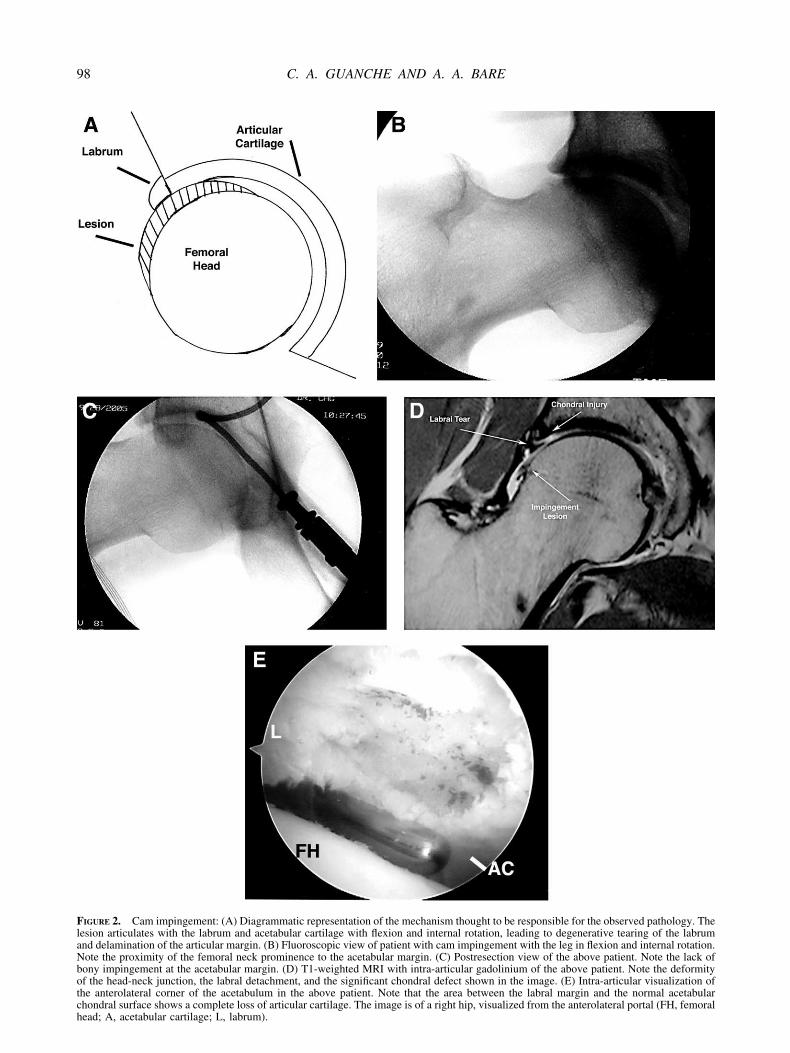
IGURE 2. Cam impingement: (A) Diagrammatic representation of the mechanism thought to be responsible for the observed pathology. Theesion articulates with the labrum and acetabular cartilage with flexion and internal rotation, leading to degenerative tearing of the labrumnd delamination of the articular margin. (B) Fluoroscopic view of patient with cam impingement with the leg in flexion and internal rotation.ote the proximity of the femoral neck prominence to the acetabular margin. (C) Postresection view of the above patient. Note the lack ofony impingement at the acetabular margin. (D) T1-weighted MRI with intra-articular gadolinium of the above patient. Note the deformityf the head-neck junction, the labral detachment, and the significant chondral defect shown in the image. (E) Intra-articular visualization ofhe anterolateral corner of the acetabulum in the above patient. Note that the area between the labral margin and the normal acetabular
hondral surface shows a complete loss of articular cartilage. The image is of a right hip, visualized from the anterolateral portal (FH, femoralead; A, acetabular cartilage; L, labrum).
Nccs
alrtmpopesh
sjj
ttacndpafigpa
pcsali
FovM tabular
99FEMOROACETABULAR IMPINGEMENT
ormal values are from 4° to 10°. Greater than 10° isonsistent with dysplasia. Finally, the neck-shaft anglean be measured. Angles greater than 140° are con-idered to be consistent with dysplasia (Fig 4).
Acetabular version can also be measured.16 Linesre traced from the anterolateral edge of the acetabu-um along the anterior and posterior projections of theim. If the posterior line is traced more laterally thanhe anterior wall, the acetabulum is anteverted (nor-al). If the anterior wall is more lateral than the
osterior wall, the acetabulum is retroverted. A figure-f-8 sign appears when the anterior wall crosses theosterior wall on a tracing. This is consistent with anxcess anterior bony rim (causing a relative retrover-ion), which may cause impingement at the femoralead and neck junction17 (Fig 3).Beyond the measurements discussed above, other
ubtle abnormalities may be present. Obviously theoint should be assessed for any significant decrease in
IGURE 3. Pincer impingement: (A) Diagrammatic representationf the labrum that occurs. There is minimal damage to the chondraersion (left diagram) and retroversion. The figure-of-8 sign as a rRI view of normal acetabular anteversion. (D) MRI view of ace
oint space or degenerative changes. The contour of p
he anterolateral neck should be compared with that ofhe unaffected side. A normal superior neck will have
distinctive concave appearance, with the concaveontour takeoff at the head-neck junction through theeck-greater trochanter region. Lack of this curvatureecreases head-neck offset, which can lead to im-ingement. Flattening and increasing radius of thenterosuperior or anterior portion of the head identi-es a nonspherical head. A cross-table lateral radio-raph is essential in the workup for impingement. Aroperly taken image will show the femoral necklong its anterolateral aspect (Fig 5).16
In some cases, the morphology of the acetabulum orroximal femur requires further delineation by MRI oromputed tomography scanning (Fig 6). Attentionhould be given in these studies to acetabular versions well as soft-tissue impingement along the antero-ateral femoral neck. Specifically, acetabular versions best evaluated on sagittal or coronal views in the
mechanism responsible for pincer impingement. Note the crushingce of the acetabulum. (B) Diagrammatic representation of normalf the overlap of the anterior and posterior surfaces is marked. (C)
retroversion in a symptomatic athlete.
of thel surfaesult o
lane of the hip (Fig 7). A computed tomography scan

weFth
N
tAtimfsm
S
icwfpfstpcmc
FFi
Fttfwie
Ftt
100 C. A. GUANCHE AND A. A. BARE
ith 3-dimensional reconstructions should be consid-red in patients with significant bony deformities.urthermore, MRI arthrography can detect labral pa-
hology in addition to fully assessing the femoralead, neck, and acetabulum.18
IGURE 4. Typical angles that are measured in the assessment ofAI: angle AB, femoral neck-shaft angle; angle CD, acetabular
ndex; angle EF, center-edge angle.
IGURE 5. Cross-table lateral radiograph. White lines are alonghe femoral neck and parallel to the neck at the anterior margin ofhe femoral head. The black arrow indicates the total width of theemoral head. The ratio of the measured distance between the 2hite lines and the total head width is typically greater than 0.152
on anatomically normal hips. Numbers less than 0.152 indicatexcessive posterior offset.16
TREATMENT
onsurgical
Conservative treatment modalities should be at-empted for patients diagnosed with hip impingement.nti-inflammatory medications and activity modifica-
ion may improve or alleviate symptoms. Because hipmpingement is a mechanical problem, conservativeeasures will not eliminate the source. Patients who
ail conservative treatment are candidates for arthro-copic debridement of the femoral neck and the treat-ent of any significant labral lesions.
urgical
A complete arthroscopic examination of the hipncludes visualization of the central and peripheralompartments. The central compartment includes theeight-bearing head, articular cartilage, acetabular
ossa, and the ligamentum teres. The peripheral com-artment contains the non–weight bearing head, theemoral neck, and the hip capsule, as well as theynovial folds and the orbicular ligament. Access tohe anterolateral neck is through the peripheral com-artment and visualization of the labrum though theentral compartment.19 Therefore, arthroscopic treat-ent of hip impingement requires entry into both
ompartments.The setup for this procedure is similar to that for
IGURE 6. MRI (T1-weighted study without intra-articular con-rast) showing both a tear involving the superior labrum as well ashe anatomy of the superior neck.
ther hip arthroscopy procedures.19 Although hip ar-

tb(cfrttita8bp
cptfptiaalpawaa
cfrTaiasn
sccob
FN (B) Tf
Fo
101FEMOROACETABULAR IMPINGEMENT
hroscopy can be performed in either the lateral decu-itus or the supine position, the senior authorC.A.G.), as well as others, believes that peripheralompartment arthroscopy without traction is best per-ormed in the supine position.19,20 The traction appa-atus should be available for examination of the cen-ral compartment and the leg draped so that theraction can be attached and removed without contam-nating the surgical field. Hip and knee flexion withoutraction will relax the anterior capsule and allow easierccess into the anterior peripheral compartment (Fig). There are 30° and 70° arthroscopes are available,ut the 30° device is often the only one needed foreripheral arthroscopy.
IGURE 7. (A) Degenerative labral tear seen from the peripheral coote the reactive synovial tissue laterally (arrow) (L, labrum).
ragmentation of labral tissue.
wIGURE 8. Peripheral compartment arthroscopy with the hip outf traction and in 45° of hip flexion to relax the anterior capsule.
First, assessment of the labrum and other centralompartment structures is performed. The traction ap-aratus is attached in the supine position and 50 lb ofraction is added. A systematic visualization of theemoral head, acetabulum, labrum, and ligamentum iserformed with the anticipation of labral pathology inhe anterosuperior compartment. Labral and chondralnjuries are addressed with the shaver through thenterior portal. The majority of labral lesions associ-ted with hip impingement are degenerative lesionsocated in the anterosuperior quadrant. In the typicalatient, the labrum has some peripheral fraying (in thevascular zone). These lesions are typically treatedith debridement and are unlikely to warrant repair,
lthough their treatment should certainly be individu-lized.
Acetabular chondral injuries may be addressed byhondroplasty, drilling, or microfracture of the af-ected areas in order to stimulate a fibrocartilaginousesponse, similar to the process used in the knee.hese lesions are not uncommon and tend to extendbout 5 to 7 mm in width along the length of thempingement lesion. In most cases of FAI, the femoralrticular surface is intact. Nonetheless, the articularurface should be examined and probed to assureormal surfaces.Areas of chondral delamination of the acetabular
urface should be addressed at this time.16 In someases, a concomitant acetabular rim excision is indi-ated where a significant anteversion or peripheralsteophyte impinges with the femoral neck. The de-ridement of this portion should be undertaken first,
ent. View is of a left hip and approximately the 3 o’clock position.1-weighted MRI of lateral degenerative tear. Arrow indicates
mpartm
ith the peripheral arthroscopy performed later. In

terp
ttat4stcscpwmaac
teptpspbmlp
cepstpcvod
slsciia
pnplibwTdlcmnmr4tfliftipt
patflWtpta
vtaiiinntiu
snph
102 C. A. GUANCHE AND A. A. BARE
hese situations, consideration should be given to ac-tabular takedown in a sharp fashion, followed byeattachment once the debridement has been com-leted, as has been documented in open approaches.5
Peripheral compartment visualization is performedhrough a standard anterolateral portal. Access intohis compartment is gained by use of a guidewire andcannulated trocar system after removing either all of
he traction and placing the leg in a position of about5° of flexion or simply placing the leg in balanceduspension with about 20 lb of traction. Documenta-ion of appropriate guidewire placement with fluoros-opy is recommended. Dienst et al.20 described aystematic diagnostic examination of the peripheralompartment beginning anteriorly. The examinationroceeds in a counterclockwise direction beginningith the anterior neck and proceeding through theedial neck, medial head, anterior head, lateral head,
nd the posterior area. Use of the anterolateral portalllows visualization of nearly the entire peripheralompartment.
In cases where the more lateral and posterior por-ions are not well visualized, an accessory anterolat-ral portal can be considered. The standard portal islaced about 2 cm proximal to the greater trochanter athe anterior margin of the prominence in the coronallane. The accessory portal is 2 to 3 cm distal to thetandard anterolateral portal on the same coronallane. In some cases this portal can be used for looseody removal in the more distal peripheral compart-ent. It can also be used for visualization of the more
ateral aspects of the neck while using the anterolateralortal as the working portal.In addition, in severe cases, a posterolateral portal
an be established (in those cases where a posterolat-ral portal has not already been established) to com-lete the more posterior and lateral impingement le-ions. This portal is located about 2 cm proximal tohe greater trochanter at the posterior margin of therominence in the coronal plane. The most importantonsideration in these cases is to fluoroscopically andisually assess the entire femoral neck from the 12’clock to 6 o’clock position to assure a completeecompression.After completing the diagnostic examination, out-
ide-in development of the working portal is estab-ished. This portal corresponds to the same skin inci-ion for the anterior working portal in the centralompartment. The landmarks for this portal are thentersection of a line drawn from the anterior, superiorliac spine down the femoral shaft, and a line drawn
cross the level of the greater trochanter. The working iortal is best established while viewing the anterioreck. A shaver is introduced through the workingortal and both the arthroscope and shaver are movedaterally. The capsule is excised over the area of thempingement osteophyte for complete visualizationefore bony resection. Capsular excision is performedith a combination of a shaver and a tissue ablator.he area of resection is typically about 2 � 2 cm inimension. The resection is limited to the area over-ying the bony prominence and does not add signifi-ant morbidity because there is still continuity of theajority of the capsule. Gaining access to the lateral
eck is often more difficult than the anterior andedial neck because of a tight zona orbicularis. Dienst
ecommends keeping the hip flexed to approximately5°, abduction of 20° to 40°, and slight external rota-ion of the hip.20 Once in the area of the lateral neck,uoroscopic confirmation of the location on the neck
s recommended. A burr (4.5 mm round) is now usedor the bony resection (Fig 9). Fluoroscopic visualiza-ion is important before beginning the neck contour-ng. The most predictable technique is to outline theroposed area of bony resection and then resect be-ween the margins.17
The goal of the debridement is to restore the appro-riate contour of the anterolateral neck, proceeding insystematic fashion low on the neck toward the head
o restore the normal concavity. The ultimate range ofexion should be between 110° and 115° of flexion.e recommend real-time fluoroscopic examination of
he hip through a range of motion to confirm a com-lete debridement. In addition, arthroscopic visualiza-ion should also be undertaken to document full clear-nce in flexion and rotation.
One concern with femoral neck debridement is theasculature responsible for nutrition of the majority ofhe femoral head.21 The medial femoral circumflexrtery, the main supply, originates from distal to prox-mal and emerges from the obturator externus, pierc-ng the capsule along the posterior superior neck. Thentracapsular branches then divide into 2 to 4 subsy-ovial retinacular vessels and track into the femoraleck. To avoid damage to this area, the distal extent ofhe debridement should be limited to that area thatmpinges only, typically about 10 mm from the artic-lar margin.Finally, the guidelines for the depth of resection are
parse. One study analyzed the percentage of femoraleck resection as a fraction of the total neck size anderformed compressive loading across the femoralead. The amount of resection that predictably ended
n a fracture was greater than 30% of the femoral
Ftda
103FEMOROACETABULAR IMPINGEMENT
IGURE 9. (A) Fluoroscopic view of the femoral neck, the arthroscope and burr in position before debridement. (B) Arthroscopic view ofhe femoral neck lesion. (C) Final fluoroscopic view of the neck following burring. (D) Final arthroscopic view after femoral neck
ebridement. (E) Central compartment arthroscopy shows a degenerative tear of the anterosuperior labrum (FH, femoral head). Note thecetabular cartilage (arrow) and the exposed subchondral bone between the remaining cartilage and the labrum.
n2Pnr
nwanopnte
P
mfwi6atoct
O
tlaaTpcgcd
pptvdhn
hfdet
A
opue2TuisaTa
tnshnsa
npphpadtasruy
ical
104 C. A. GUANCHE AND A. A. BARE
eck.22 For this reason, no more than approximately0% of the width of the neck should be resected.reoperative measurement of the overall width of theeck allows the surgeon to plan for as conservative aesection as is possible.
In addition to the complications germane to femoraleck resection, the common complications associatedith hip arthroscopy should be noted. Most common
re neural injuries, with lateral femoral cutaneouserve lesion the most frequent. These occur as a resultf the proximity of this nerve to the anterior portalosition. In addition, traction injuries to the sciaticerve may occur with excessive traction the extremi-y.23 Finally, there have been cases of excessive fluidxtravasation causing significant neural injuries.24
ostoperative Protocol
Weight bearing restrictions following the debride-ent take into consideration the risk of postoperative
emoral neck fracture. Toe-touch weight bearing for 4eeks is maintained and followed by full weight bear-
ng. No high-impact activities are allowed for at leastweeks. Most patients resume high-impact athletic
ctivities by 12 weeks postoperatively. The impor-ance of early protected weight bearing cannot beveremphasized—there have been 2 documentedases of femoral neck fractures following neck resec-ion.17
RESULTS
pen Debridement
Initial reports on the treatment of FAI have involvedhe use of open surgical dislocation techniques. In theargest series to date,25 the open surgical dislocationpproach was performed on 19 patients with a meange of 36 years and an average follow-up of 4.7 years.he authors25 documented good results in 14 of 19atients with no cases of osteonecrosis. They con-luded that the surgical dislocation approach yieldedood results in patients with early degenerativehanges, but was not beneficial to those with advancedegenerative changes or extensive cartilage damage.Another study assessed a group of 23 hips in 23
atients treated by open surgical debridement for im-ingement.26 Follow-up ranged from 2 to 12 years. Athe most recent evaluation, 7 patients had been con-erted to total hip arthroplasty, 1 had arthroscopicebridement of a recurrent labral tear, and 15 patientsad had no further surgery. No hips developed osteo-
ecrosis. Of the 7 patients who were converted to total Aip arthroplasty, 3 failed early and 4 recovered andunctioned well for between 6.4 and 9.5 years afterebridement. The authors proposed that the procedureffectively treats hips with impingement and main-ained their normal hip in most cases.
rthroscopic Debridement
There are no series to date that have analyzed theutcome of the arthroscopic procedure in a group ofatients. In the senior author’s experience, 10 consec-tive patients treated arthroscopically for FAI werevaluated. Follow-up averaged 16 months (range, 9 to4 months). The average patient age was 34 years.he McCarthy scoring scale for the nonarthritic wassed.27 Eight patients with evidence of FAI and nontra-articular cartilage degenerative disease did sub-tantially better than the 2 patients who had degener-tive disease diagnosed at the time of arthroscopy.he nonarthritic scores averaged 75 preoperativelynd 95 at follow-up (Fig 10).
Sampson17 has reported on a series of 90 patientsreated arthroscopically for FAI. In his experience,early all patients had elimination of the impingementign (pain on flexion and internal rotation) and wereappy with their results. One patient did sustain aondisplaced femoral neck fracture and requiredcrew fixation. The early results, in his estimation,pproached those of the open procedure.
DISCUSSION
Hip impingement can be a difficult entity to diag-ose and treat. The current literature suggests that FAIlays a role in the cascade of hip osteoarthritis in someatients—those with structural proximal femoralead-neck abnormalities.3-7 This entity usually ap-ears in younger and more physically active adultsnd can be debilitating. Subsequent labral and chon-ral lesions have been linked to the repetitive micro-rauma caused by the deformity of the femoral neck orcetabulum.14 Because labral and chondral lesions areeen more frequently in early osteoarthritic hips, earlyecognition and treatment of this entity may halt thenfortunate progression toward osteoarthritis inounger patients.Once the diagnosis of impingement is made, isolat-
ng the source will help determine the appropriateourse of treatment. Conservative treatment should bettempted for all patients. If this treatment fails, theocation of pathology will dictate the surgical plan.
cetabular overhang and other acetabular pathology
mrsrdcamrptam
siaar
shctds
Fjh t the h
105FEMOROACETABULAR IMPINGEMENT
ay require simple debridement and partial acetabularesection, or in some cases a labral takedown andubsequent repair of the affected labrum after the bonyesection is completed. Arthroscopic assessment andebridement of nonspherical femoral heads with de-reased head-neck offset causing impingement offersminimally invasive treatment option to restore hipechanics at the extremes of motion, eliminate the
epetitive microtrauma to the acetabular labrum, andotentially curb the progression of osteoarthritis. Con-ouring of the femoral head and neck to a more normalnatomy removes the offending source of impinge-
IGURE 10. Typical case of FAI in a 19-year-old collegiate basketunction prominence. (B) Arthroscopic view of the typical lesionead-neck junction. (D) Arthroscopic view of the final resection a
ent. The debridement, although performed arthro- s
copically, is fraught with significant complicationsncluding the possibility of femoral neck fracture andvascular necrosis of the femoral head. The surgicalpproach should be well planned and the amount ofesection gauged carefully.
We currently do not fully understand the progres-ion of nontraumatic degenerative joint disease of theip. Although FAI, leading to subsequent labral andhondral injuries, has been associated with early os-eoarthritis of the hip,1,3,4 this cascade has yet to beefinitely proven. While the early results of arthro-copic debridement of FAI are promising, additional
yer. (A) AP radiograph showing the significant femoral head-necketabular cartilage). (C) AP radiograph following resection of the
ead-neck junction (AC, acetabular cartilage).
ball pla(AC, ac
tudies analyzing the clinical and long-term benefit of

ass
1
1
1
1
1
1
1
1
1
1
2
2
2
2
2
2
2
2
106 C. A. GUANCHE AND A. A. BARE
rthroscopic debridement of the femoral neck to re-tore head-neck offset and hip clearance are neces-ary.
REFERENCES
1. Leunig M, Beck M, Woo A, et al. Acetabular rim degenera-tion: A constant finding in the aged hip. Clin Orthop 2003;413:201-207.
2. Seldeg R, Tan V, Hunt J, et al. Anatomy, histologic features,and vascularity of the adult acetabular labrum. Clin Orthop2001;382:232-240.
3. McCarthy JC, Noble PC, Schuck MR, et al. The role of labrallesions to development of early hip disease. Clin Orthop2001;393:25-37.
4. Ganz R, Parvizi J, Beck M, et al. Femoroacetabular impinge-ment: A cause for early osteoarthritis of the hip. Clin Orthop2003;417:112-120.
5. Beck M, Leunig M, Parvizi J, et al. Anterior femoroacetabularimpingement: Part II: Midterm results of surgical treatment.Clin Orthop 2004;418:67-73.
6. Lavigne M, Parvizi J, Beck M, et al. Anterior femoroacetabu-lar impingement: Part I: Technique of joint preserving surgery.Clin Orthop 2004;413:61-66.
7. Ito K, Minka M, Leunig ??, et al. Femoroacetabular impinge-ment and the cam-effect. J Bone Joint Surg Br 2001;83:171-176.
8. Goodman DA, Feighan JE, Smith A, et al. Subclinical slippedcapital femoral epiphysis. J Bone Joint Surg Am 1997;79:1489-1497.
9. Harris WH. Etiology of osteoarthritis of the hip. Clin Orthop1986;213:20-33.
0. Santori N, Villar R. Arthroscopic findings in the initial stagesof hip osteoarthritis. Orthopedics 1999;22:405-409.
1. Murray RO. The aetiology of primary osteoarthritis of the hip.Br J Radiol 1965;38:810-824.
2. Morgan JD, Sommerville EW. Normal and abnormal growthat the upper end of the femur. J Bone Joint Surg Br 1960;42:
810-824.3. Siedenrock KA, Wahab KHA, Werlen S, et al. Abnormal
extension of the femoral head epiphysis as a cause of camimpingement. Clin Orthop 2004;418:54-60.
4. Klaue K, Durnin C, Ganz R. The acetabular rim syndrome.J Bone Joint Surg Br 1991;73:423-429.
5. Leunig M, Werlen S, Ungersbock A, et al. Evaluation of theacetabular labrum by MR arthrography. J Bone Joint Surg Br1997;79:230-234.
6. Mast JW, Brunner RL, Zebrack J. Recognizing acetabularversion in the radiographic presentation of hip dysplasia. ClinOrthop 2004;418:48-53.
7. Sampson TG. Hip morphology and its relationship to pathol-ogy: Dysplasia to impingement. Oper Tech Sports Med 2005;13:37-45.
8. Czerny C, Hofmann S, Urban M, et al. MR arthrography of theadult acetabular capsular-labral complex: Correlation with sur-gery and anatomy. Am J Radiol 1999;173:345-349.
9. Byrd TJW. Hip arthroscopy utilizing the supine position. Ar-throscopy 1994;10:275-280.
0. Dienst M, Godde S, Seil R, et al. Hip arthroscopy withouttraction: In vivo anatomy of the peripheral hip joint cavity.Arthroscopy 2001;17:924-931.
1. Gautier E, Ganz K, Krügel N, et al. Anatomy of the medialfemoral circumflex artery and its surgical implications. J BoneJoint Surg Br 2000;82:679-683.
2. Mardones RM, Gonzalez C, Chen Q, et al. Surgical treatmentof femoroacetabular impingement: Evaluation of the effect ofthe size of the resection. J Bone Joint Surg Am 2005;87:273-279.
3. Clarke MT, Arora A, Villar RN. Hip arthroscopy: Complica-tions in 1054 cases. Clin Orthop 2003;406:84-88.
4. Funke EL, Munzinger U. Complications in hip arthroscopy.Arthroscopy 1996;12:156-159.
5. Beck M, Leunig M, Parvizi J, et al. Anterior femoroacetabularimpingement: Part II. Midterm results of surgical treatment.Clin Orthop 2004;418:67-73.
6. Murphy S, Tannast M, Kim YJ, et al. Debridement of the adulthip for femoroacetabular impingement: Indications and pre-liminary clinical results. Clin Orthop 2004;429:178-181.
7. Christensen C, McCarthy J, Mittleman M. Outcomes. In: Mc-Carthy JC ed. Early hip disorders. New York: Springer, 2003;
195-200.