appendicitis as the presenting manifestation of kawasaki disease
TRANSCRIPT

CASE REPORT
Appendicitis as the presenting manifestation of Kawasaki disease
Gwendolyn M. Garnett • Sarah Kimball • Marian E. Melish •
Karen S. Thompson • Devin P. Puapong • Sidney M. Johnson •
Russell K. Woo
Accepted: 27 November 2013 / Published online: 7 December 2013
� Springer-Verlag Berlin Heidelberg 2013
Abstract In cases of Kawasaki’s disease (KD) presenting
as acute surgical abdomen, rarely has the presence of acute
appendicitis been found. We report two cases of histolog-
ically confirmed acute appendicitis in the presence of KD
and a review of the literature as it pertains to acute abdo-
men and atypical presentations of KD.
Keywords Kawasaki disease � Acute appendicitis �Acute abdomen � Vasculitis � Coronary artery aneurysm
Introduction
While the pathogenesis of Kawasaki’s disease (KD)
remains unclear, the prevalence of serious and potentially
life-threatening sequelae has been reduced with early rec-
ognition of classic clinical features of the disease and
prompt initiation of therapy. A diagnostic challenge is
posed when the disease presents atypically, as is frequently
seen in patients with severe gastrointestinal complications,
and can result in detrimental therapeutic delays. While it is
known that KD can present as an acute abdomen, rarely has
acute appendicitis been found as a source of peritonitis at
the time of diagnosis. We report two such cases of histo-
logically confirmed acute appendicitis in the presence of
KD. In both cases, Kawasaki’s disease was diagnosed
postoperatively. Evaluation for Kawasaki’s disease was
initiated because of unusual clinical features during the
postoperative period. Describing the critical manifestations
seen in this unique presentation has important diagnostic
and treatment implications, as special consideration must
be given to the timing and appropriateness of certain
therapies. While KD is not considered a surgical disease by
convention, surgeons may be the first to recognize this
atypical presentation. Favorable outcomes for these atypi-
cal KD patients may depend upon the keen judgment of
surgeons familiar with the disease’s clinical presentation
and course.
Case histories
Case 1
A 3-year-old previously healthy male presented with a 72-h
history of periumbilical abdominal pain, nausea, vomiting,
diarrhea, and fever. Laboratory data were significant for
leukocytosis with bandemia, acidosis, and elevated
C-reactive protein (CRP). Abdominal ultrasound was
inconclusive. Computed tomography (CT) abdomen/pelvis
showed free fluid in the peritoneal cavity, and while por-
tions of the appendix appeared normal, the entire appendix
was not visualized. Tachycardia combined with the
development of peritoneal signs prompted laparoscopic
exploration. Intraoperative findings included free pelvic
fluid, and an injected appendix with serosal changes but
minimal thickening. No other sources of pathology were
identified and appendectomy was performed without
complication. Pathology showed an intact appendix with
focal intramuscular inflammation with neutrophil invasion.
G. M. Garnett (&) � S. Kimball
Department of Surgery, University of Hawai’i,
John A. Burns School of Medicine, Honolulu,
HI 96813, USA
e-mail: [email protected]
M. E. Melish � K. S. Thompson � D. P. Puapong �S. M. Johnson � R. K. Woo
Kapi’olani Medical Center for Women and Children,
Honolulu, HI 96826, USA
123
Pediatr Surg Int (2014) 30:549–552
DOI 10.1007/s00383-013-3439-9
On final report this was felt to be consistent with acute
appendicitis. The patient’s postoperative course was char-
acterized by anorexia, persistent abdominal pain, diarrhea,
transaminitis, and high fevers. Physical exam was notable
for peripheral edema involving the hands, fingers, and feet,
conjunctival injection, and dry, cracked lips. Suspecting
KD, an echocardiogram was obtained which revealed
pericardial effusion, and dilatation of the coronary arteries.
Intravenous gammaglobulin (IVIG) and aspirin were star-
ted on hospital day 7. One additional IVIG dose was given
for recurrent fever. The patient was discharged 3 days later
upon complete resolution of symptoms. At 2-year follow-
up, echocardiogram revealed normal caliber coronary
arteries.
Case 2
A previously healthy 7-year-old female presented with a
4-day history of abdominal pain, emesis, diarrhea, and
fever. Laboratory data showed a markedly elevated CRP
level and white blood cell count. CT abdomen/pelvis with
rectal contrast demonstrated dilated loops of small bowel,
free fluid in the pelvis, but poor visualization of the
appendix. Perforated appendicitis was suspected and the
patient was taken for laparoscopic exploration. Intraoper-
ative findings included suppurative appendicitis, purulent
pelvic fluid, and uniform dilatation of the entire small
bowel. Pathology was consistent with mild neutrophilic
and eosinophilic appendicitis, with acute appendicitis being
given as the final diagnosis. The postoperative course was
complicated by persistent abdominal pain, fever, the
development of a raised rash on the arms, trunk and but-
tocks, and dry, cracked lips. Bilateral non-exudative con-
junctivitis was present since admission. A repeat CT
abdomen/pelvis showed colitis, though no evidence of
abscess formation. Again suspecting KD, an echocardio-
gram was obtained and revealed a medium-sized aneurysm
of the left anterior descending coronary artery. IVIG and
antiplatelet therapy was initiated. All symptoms of KD
resolved and the patient was discharged on hospital day 11.
The patient required one additional hospitalization after
follow-up echocardiogram showed enlargement of the
coronary artery aneurysm. Recent echocardiograms have
shown aneurysm regression.
Discussion
KD is an acute, self-limited vasculitis predominately
occurring in early childhood [1]. While there are clinical
and epidemiological features suggestive of an infectious
cause and a possible role for genetic predisposition, the
etiology of KD remains unknown [1, 2]. The major
sequelae of KD are cardiovascular in nature with coronary
artery involvement found in all fatal cases. While coronary
artery aneurysm formation is the most dreaded complica-
tion, it is important to recognize KD is a systemic disease
involving blood vessels throughout the entire body, and can
manifest in several organ systems. KD thus has the
capacity to present in a number of ways often times mas-
querading as other illnesses particularly in the early stages
of disease onset [1].
While there are many clinical and laboratory findings
associated with KD, diagnosis is based on the presence of
persistent fever for five or more days duration, accompa-
nied by at least four of five principle clinical findings.
These findings include bilateral non-exudative conjuncti-
vitis, changes of the extremities, rash, cervical lymphade-
nopathy, and erythema of the lips and oral mucosa [1–3].
Atypical or incomplete Kawasaki’s disease may be diag-
nosed in patients who have some clinical features of the
disease but fail to meet the classic criteria listed above.
These patients carry a higher risk of coronary artery
involvement [4–6]. Because delay in treatment raises
morbidity and mortality of KD particularly in infants and
older children, KD should be considered in any child with
persistent fever, laboratory evidence of systemic inflam-
mation, and no other explanation of febrile illness. Echo-
cardiographic abnormalities, chiefly coronary artery
involvement, often provide the basis for diagnosis in
patients with persistent fever and two or three of the
principle KD clinical features [1].
Owing to its propensity to involve vessels throughout
the entire body, KD often mimics other disease processes
including those that are surgical in nature [1, 7]. Gastro-
intestinal symptoms including abdominal pain with dis-
tention, vomiting, and diarrhea are common and occur in
approximately two-thirds of patients [3]. On rare occasion,
KD has presented as acute surgical abdomen thus landing
these unusual patients on the surgical service. Two tertiary
care centers with a large referral base reported acute sur-
gical abdomen as the presenting manifestation in 4.6 % of
their Kawasaki patients. Notable clinical features of these
patients distinguishing them from patient’s with classic KD
include older age ([5 years old), elevated transaminases,
drastically elevated white blood cell count ([20,000 units)
and incomplete presentation. These patients also had a high
incidence of coronary artery aneurysm development at
50 %. Operative findings in these patients most commonly
revealed gallbladder involvement such as gallbladder
hydrops and cholestasis, though paralytic ileus, and hem-
orrhagic duodenitis were other pertinent findings. Just one
case of acute appendicitis was identified in this series [7].
Overall, there are five reported cases of Kawasaki dis-
ease with appendicular involvement [6–9]. Most of these
cases were limited to appendicular vasculitis (arteritis) with
550 Pediatr Surg Int (2014) 30:549–552
123
only two cases providing histological confirmation of acute
appendicitis [6, 7]. Both our patients were taken for sur-
gical exploration for peritonitis and the presumed diagnosis
of appendicitis. During surgical exploration, inflammatory
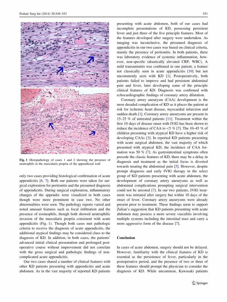
changes of the appendix were visualized in both cases
though were more prominent in case two. No other
abnormalities were seen. The pathology reports varied and
noted unusual features such as focal infiltration and the
presence of eosinophils, though both showed neutrophilic
invasion of the muscularis propria consistent with acute
appendicitis (Fig. 1). Though both cases met pathologic
criteria to receive the diagnosis of acute appendicitis, the
additional atypical findings may be considered clues to the
diagnosis of KD. In addition, in both cases, the patients’
advanced initial clinical presentation and prolonged post-
operative course without improvement did not correlate
with the gross surgical and pathologic findings of non-
complicated acute appendicitis.
Our two cases shared a number of clinical features with
other KD patients presenting with appendicitis and acute
abdomen. As in the vast majority of reported KD patients
presenting with acute abdomen, both of our cases had
incomplete presentations of KD, possessing persistent
fever and just three of the five principle features. Most of
the features developed after surgery were undertaken. As
imaging was inconclusive, the presumed diagnosis of
appendicitis in our two cases was based on clinical criteria,
mainly the presence of peritonitis. In both patients, there
was laboratory evidence of systemic inflammation, how-
ever, non-specific (drastically elevated CRP, WBC). A
mild transaminitis was confirmed in one patient, a feature
not classically seen in acute appendicitis [10] but not
uncommonly seen with KD [1]. Postoperatively, both
patients failed to improve and had persistent abdominal
pain and fever, later developing some of the principle
clinical features of KD. Diagnosis was confirmed with
echocardiographic findings of coronary artery dilatation.
Coronary artery aneurysm (CAA) development is the
most dreaded complication of KD as it places the patient at
risk for ischemic heart disease, myocardial infarction and
sudden death [1]. Coronary artery aneurysms are present in
15–25 % of untreated patients [11]. Treatment within the
first 10 days of disease onset with IVIG has been shown to
reduce the incidence of CAA to\5 % [7]. The 10–45 % of
children presenting with atypical KD have a higher risk of
developing CAAs [3]. In reported KD patients presenting
with acute surgical abdomen, the vast majority of which
presented with atypical KD, the incidence of CAA for-
mation was 50 % [7]. As gastrointestinal symptoms often
precede the classic features of KD, there may be a delay in
diagnosis and treatment as the initial focus is diverted
towards treating the abdominal pain [5]. However, despite
prompt diagnosis and early IVIG therapy in the select
group of KD patients presenting with acute abdomen, the
development of coronary artery aneurysms as well as
abdominal complications prompting surgical intervention
could not be arrested [7]. In our two patients, IVIG treat-
ment was initiated after surgery but within 10 days of the
onset of fever. Coronary artery aneurysms were already
present prior to treatment. These findings seem to support
Zulian’s suggestion that KD patients presenting with acute
abdomen may possess a more severe vasculitis involving
multiple systems including the intestinal tract and carry a
more aggressive form of the disease [7].
Conclusion
In cases of acute abdomen, surgery should not be delayed.
However, familiarity with the clinical features of KD is
essential as the persistence of fever, particularly in the
postoperative period, and the presence of two or three of
these features should prompt the physician to consider the
diagnosis of KD. While uncommon, Kawasaki patients
Fig. 1 Histopathology of cases 1 and 2 showing the presence of
neutrophils in the muscularis propria of the appendiceal wall
Pediatr Surg Int (2014) 30:549–552 551
123
initially presenting with acute abdomen will likely be
admitted to the surgical service. As there is evidence to
suggest that these patients carry a more aggressive form of
the disease, positive outcomes in this unique group will
rely on a surgeon’s ability to promptly diagnosis and ini-
tiate treatment of this unusual disease.
References
1. Newburger JW et al (2004) Diagnosis, treatment, and long-term
management of Kawasaki disease: a statement for professionals
from the committee on rheumatic fever, endocarditis and kawa-
saki disease, council on cardiovascular disease in the young,
American Heart Association. Circulation 110:2747–2771
2. Melish ME, Hick RV (1990) Kawasaki syndrome: clinical fea-
tures, pathophysiology, etiology and therapy. J Rheumatol Suppl
24:2–10
3. Baker AL et al (2009) Associated symptoms in the ten days
before diagnosis of Kawasaki disease. J Pediatr 154:592–595
4. Rowley AH et al (1987) Incomplete Kawasaki disease with
coronary artery involvement. J Pediatr 110:409–413
5. Minich LL et al (2007) Delayed diagnosis of Kawasaki disease:
what are the risk factors? Pediatrics 120:1434–1440
6. Bartlet AH, Dishop MK, Baker CJ (2006) An unusual case of
appendicitis in a child. Semin Pediatr Infect Dis 17(3):111–2,
177–9
7. Zulian F et al (2003) Acute surgical abdomen as presenting
manifestation of Kawasaki disease. J Pediatr 142:731–735
8. Chiba T (1998) Two cases of appendicitis in Kawasaki disease.
Arch Jpn Chir 67(3–4):69–71
9. Maggio M, Liotta A et al (2007) A case of Kawasaki disease
mimicking acute appendicitis. Med J Aust 187(5):316–317
10. Anderson KD, Parry RL (1998) Appendicitis. In: O’Neill JA Jr,
Rowe MI, Grosfeld JL, Fonkalsrud EW, Coran AG (eds) Pedi-
atric Surgery, 5th edn. St Louis (MO), Mosby, p 1369
11. Kato H, Sugimura T, Akagi T et al (1996) Long-term conse-
quences of Kawasaki disease: a 10- to 21-year follow-up study of
594 patients. Circulation 94:1379–1385
552 Pediatr Surg Int (2014) 30:549–552
123