anticancer potentials of secondary metabolites from ... · anticancer potentials of secondary...
TRANSCRIPT
Int.J.Curr.Microbiol.App.Sci (2013) 2(2):44-45
44
Original Research Article
Anticancer potentials of secondary metabolites from endophytes of Barringtonia acutangula and its molecular characterization
P. Jayashree Lakshmi and *K. Vanmathi Selvi
Department of Microbiology, Sri Akilandeswari Women s College, Vandavasi, T.V.Malai, Tamil Nadu, India.
*Corresponding author: [email protected]
A B S T R A C T
Introduction
Fungi are plant-like organisms that lack chlorophyll. An endophytic fungi is a fungal microorganism, which spends the whole or part of its life cycle colonizing inter and /or intra-cellularly inside the healthy tissues of the host plants, typically causing no apparent symptoms of diseases. Endophytes are now considered as an important component of biodiversity. The distribution of endophytic mycoflora differs with the host. Some Endophytes are widespread and others are highly specific to single host in single environment. These endophytes
protect their hosts from infectious agents and adverse conditions by secreting bioactive secondary metabolites (Azevedo et al., 2000; Carroll and Carroll, 1978; Strobel, 2003). The endophytic fungi play important physiological and ecological roles in their host life. In recent years, there has been evidence that the production of secondary metabolites by an endophyte is not random, but seems to be correlated with its ecological niche. The metabolic interactions of endophytes with its host may favor the synthesis of biologically active secondary metabolites.
ISSN: 2319-7706 Volume 2 Number (2013) pp.44 55. http://www.ijcmas.com
K E Y W O R D S
Endophytic; Barringtonia acutangula; anticancer assay, Genomic DNA; PCR Standardization; BLAST of the endophytic fungus.
Two species of Endophytic fungi (EFB01 and EFB02) were isolated from leaves of Barringtonia acutangula, a medicinal plant and secondary metabolite extraction was performed using organic solvents. The extracts were screened for anticancer assay against Human Colon Cancer cell lines HT29. Extract obtained from endophytic fungus, EFB01 showed highest cytotoxicity. Endophytic fungus, EFB01 was selected for molecular identification. Genomic DNA was extracted using CTAB (Cetyl trimethyl ammonium bromide) buffer. The total genomic DNA concentration was estimated by absorbance at 260 nm. PCR standardization was carried out using ITS (Internal transcribed spacer) primers for endophytic fungus, EFB01.Using sequence analysis of ITS region and BLAST (Basic local alignment search tool) the endophytic fungus, EFB01 was identified as Colletotrichum gloeosporioides. .
Int.J.Curr.Microbiol.App.Sci (2013) 2(2):44-45
45
Plant endophytic fungi are novel source of bioactive secondary metabolites with potential application in agriculture, medicine and food industry. Some of the interesting compounds produced by endophytic microbes are taxol, cryptocin, cryptocandin, jesterone, oocydin, isopestacin, the psuedomycins and ambuic acid.
Endophytes can be identified in several ways, usually through amplifying and sequencing a small piece of DNA. Some endophytes can be cultured from a small piece of their host plant in an appropriate growth medium. Colletotrichum is a genus of fungi that are obligate symbionts to plants in the form of endophytes. The amount of taxol produced by this endophytic fungus, Colletotrichum gloeosporioides (strain JGC-9) was about 163.4 g/L (Gangadevi and Muthumary, 2008). This taxol has Strong cytotoxic activity towards BT 220, H116, Int 407, HL 251 and HLK 210 human cancer cells in vitro, tested by Apoptotic assay. Medicinal plants are known to harbour endophytic fungi that are believed to be associated with the production of pharmaceutical products (Zhang et al., 2006). In the present study, Barringtonia acutangula, an important medicinal plant, was investigated for the presence of endophytic community.
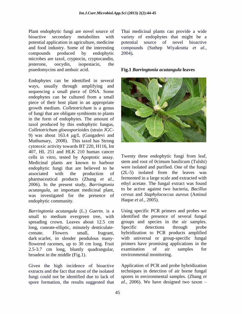
Barringtonia acutangula (L.) Gaertn. is a small to medium evergreen tree, with spreading crown. Leaves about 12.5 cm long, cuneate-elliptic, minutely denticulate-crenate. Flowers small, fragrant, dark scarlet, in slender pendulous many-flowered racemes, up to 30 cm long. Fruit 2.5-3.7 cm long, bluntly quadrangular, broadest in the middle (Fig.1).
Given the high incidence of bioactive extracts and the fact that most of the isolated fungi could not be identified due to lack of spore formation, the results suggested that
Thai medicinal plants can provide a wide variety of endophytes that might be a potential source of novel bioactive compounds (Suthep Wiyakrutta et al., 2004).
Fig.1 Barringtonia acutangula leaves
Twenty three endophytic fungi from leaf, stem and root of 0cimum basilicum (Tulshi) were isolated and purified. One of the fungi (2L-5) isolated from the leaves was fermented in a large scale and extracted with ethyl acetate. The fungal extract was found to be active against two bacteria, Bacillus cereus and Staphylococcus aureus (Aminul Haque et al., 2005).
Using specific PCR primers and probes we identified the presence of several fungal groups and species in the air samples. Specific detections through probe hybridization to PCR products amplified with universal or group-specific fungal primers have promising applications in the examination of air samples for environmental monitoring.
Application of PCR and probe hybridization techniques in detection of air borne fungal spores in environmental samples. (Zhang et al., 2006). We have designed two taxon
Int.J.Curr.Microbiol.App.Sci (2013) 2(2):44-45
46
selective primers for the internal transcribed spacer (ITS) region in the nuclear ribosomal repeat unit.
The pimers, ITS1-F and ITS4-B,were intended to be specific to fungi and basidiomycetes, the universal primers preferentially amplify the fungal ITS region. The Present study was initiated to isolate endophytic fungus from Barringtonia acutangula and their medicinal and molecular studies performed.
Materials and Methods
Collection of barringtonia leaves
Healthy leaves from Barringtonia acutangula tree were collected from the river bed in Kovilanchery, Chennai. Rinse the leaves with distilled water until the surface is cleaned thoroughly. Keep the leaves over the tissue paper for drying. After the leaves are dried, the leaves are cut over the midrib region. The alternative midrib pieces are taken to avoid colonization of same organism.
Isolation of endophytic fungi
The surface sterilized leaves were trimmed using sterile blade and inoculated into plates containing sterile Potato Dextrose Agar medium. The plates were incubated at 28°C for 7 days and observed for growth of fungus from leaves.
Extraction of secondary metabolites
Minimal media was prepared and autoclaved. Endophytic Fungus EFB01, EFB02 was inoculated into minimal media. Flasks were then incubated at 28°C for 10
14 days in shaker. After incubation, extraction was done with different solvents
(Chloroform, Ethyl acetate). First the fungus grown in the media is removed by filtering the medium using filter paper. Then the solvents are added to the media in the ratio 1:1in separating funnel and left for 15 minutes. The organic phase was collected and kept for drying at 37°C. The dry weight of the extract was determined.
Cell line study using animal cell culture
Culturing of animal cells in in vitro condition is called animal cell culture. This is done to know the drug response, gene expression, antibody production (MOAbs), cytotoxic analysis, propagation of viruses, recombinant proteins, antitumour evaluation/screening. Media is the major constituent factor in the culturing of animal cells. For the growth of the cell in invitro condition, media is required. Media is now commercially available with all the nutrients present in the required volume. Widely used media are Dulbecco s Minimal Essential Medium (DMEM) and Rosewell Park Memorial Institute (RPMI 1640) medium.
RPMI 1640 Media Preparation
16.4gms of the RPMI 1640 medium was suspended in 900ml tissue culture grade water with constant, gentle stirring until the powder is completely dissolved. It may be necessary to lower the pH to 4.0 with 1N HCl to completely dissolve the powder, after it has dissolved completely; the pH can be raised to 7.2 with 1N NaOH prior to the addition of sodium bicarbonate. 2.0gms of sodium bicarbonate powder were added for 26.67ml, therefore 7.5% sodium bicarbonate for 1 litre of medium and stir until dissolved. The pH was adjusted to 0.2-0.3 pH units below the desired pH 1N
Int.J.Curr.Microbiol.App.Sci (2013) 2(2):44-45
47
HCl or 1N NaOH since the pH tends to rise during filtration. The final volume was made up to 1000ml with tissue culture grade water. The medium was sterilised immediately by filtering through a sterile membrane filter with a porosity of 0.22 micron or less using positive pressure rather than vacuum to minimize the loss of carbon dioxide. The sterile supplements were aseptically added as required and the desired amount of sterile medium was dispensed into sterile containers. The liquid medium is stored at 2-8ºC and in dark till use.
Maintenance of cell line
For maintaining the cell line, sub culturing was done. This was done to facilitate the cell growth by removing the cells from the medium and introducing them into a new fresh medium. Mostly, enzymatic methods were used for cell maintenance. The enzyme used here was TPVG.
Cell culture flasks were selected by confluency by observing under microscope. Growth medium was removed from the flasks and the cells were incubated at 37ºC after adding the enzyme. Initially all cells get detached from the surface. The cells were suspended in 5ml of the (RPMI 1640 with 10% FBS) medium. The suspension was aspired few times to break cell clumps. Cell line, date of seeding and passage number was marked on the bottom. The cell suspension was transferred to fresh T-Flasks. 5ml of cell suspension was added to each well.
Seeding of HT 29 cell line
The cell lines were seeded in 2 kinds of plate, 24titre plate and 96titre plate. 24 titre plate consists of 420 l of RPMI 1640 and 80 l of cell suspension and 96 titre plate
consists of 80 l of medium
and 20 l of cell
suspension.
Both the plates are allowed to grow till the formation of monolayer. Then these plates are used for DNA fragmentation and MTT assay (Methyl thiazol tetrazolium assay).
MTT assay
This method is based on the ability of live but not dead cells to reduce a yellow tetrazolium dye to a purple formazan product. In brief, approximately 5x10³cells/well (cell line) were seeded into 96 well plate, 100µl of RPMI 1640 medium was added and incubated at 37ºC. After 24hours, the medium was discarded and fresh medium was added with different concentration of extract (Chloroform, Ethyl acetate) such as 125, 250, 500 (100-2000µg/ml). The plates were incubated for 1-3 days at 37ºC in a CO2 incubator. After respective incubation period, medium was discarded and 100µl fresh medium was added with 10µl of MTT (5mg/ml). After incubation at 37ºC in a CO2 incubator for 4 hours, the medium was discarded and 200µl of DMSO (Dimethyl sulfoxide) was added to dissolve the formazon crystals. Then the absorbance was read in a spectrophotometer at 630nm and cell survival was calculated by the following formula.
Viability % = Test OD / Control OD X 100 Cytotoxocity % = 100 viability%
Molecular characterization of endophytic fungus by genomic DNA isolation
Add 10 ml (for a 0.5 g pad) of CTAB extraction buffer gently mix to wet the entire powdered pad. Place in 65º C water bath for 30 min. Cool, add equal volume of CHCl3: IAA (24:1). Mix, centrifuge for 10 min at full speed. Transfer aqueous
Int.J.Curr.Microbiol.App.Sci (2013) 2(2):44-45
48
supernatant to a new tube. Add an equal volume of isopropanol. High molecular weight DNA should spool out upon mixing. spool out the DNA with a glass rod or hook, pour out the remaining supernatant. Rinse the spooled DNA with 70% ethanol. Air dry, add 1- 5 ml TE containing 20 µg/ ml RNase suspend the samples, place in 65º C bath, or allow pellets to re suspend overnight at 4oC .
Analysis of DNA by Agarose gel electrophoresis
Agarose was weighed and transferred to a conical flask. 50 ml of 1X TAE was added and Agarose was melted to a clear solution by heating. It was allowed to cool until it reached bearable temperature. 2.5µl of ethidium bromide stock solution was added. A gel casting tray was placed in a leveling table and the melted agarose was poured. After the gel solidified, the comb was taken out carefully. The casted gel was placed in an electrophoresis tank and 1X TAE buffer was added until the gel was completely submerged. DNA sample was mixed with the gel loading buffer and loaded into the well. The samples were then electrophoresed at 50V until the gel loading buffer reached 2/3rd of the gel. This gel was then viewed under UV Transilluminator.
Quantification of DNA by spectrophotomeric method
The spectrophotometer was calibrated and the UV lamp was turned on. The wavelength was set at 260nm and 280nm. Absorbance at 260 and 280 nm was set at zero with TE buffer or sterile water as blank. 3µl of the DNA was taken in a quartz cuvette and made up to 3ml with TE buffer or sterile water. Absorbance of the sample at 260 and 280 nm was noted.
Polymerase chain reaction
50- 100ng of DNA is used for molecular identification. The PCR reaction is performed for 20µl . The PCR tubes were placed in thermocycler and the samples were amplified in thermocycler. Amplified samples were then electrophoresed on 1.5% agarose gel. EFB01 Fungus was taken for molecular identification as it showed very high significant anticancer activity (Table.1,2).
Table.1 PCR reaction conditions
Initial denaturation 94 C 3min Denaturation 94 C 1min Annealing 51 C 1min Extension 72 C 1min 20sec Final extension 72 C 7min Hold 4 C Total number of cycles 32
Analysis of PCR product by agarose gel electrophoresis
Agarose was weighed and transferred to a conical flask. 50 ml of 1X TAE was added and Agarose was melted to a clear solution by heating. It was allowed to cool until it reached bearable temperature. 2.5µl of ethidium bromide stock solution was added. A gel casting tray was placed in a leveling table and the melted agarose was poured. After the gel solidified, the comb was taken out carefully. The casted gel was placed in an electrophoresis tank and 1X TAE buffer was added until the gel was completely submerged. DNA sample was mixed with the gel loading buffer and loaded into the well. The samples were then electrophoresed at 50V until the gel loading buffer reached 2/3rd of the gel. This gel was then viewed under UV Transilluminator. The PCR product was subjected to DNA sequencing and the results were analysed using BLAST tool.
Int.J.Curr.Microbiol.App.Sci (2013) 2(2):44-45
49
Table.2 Reaction set up for ITS amplification - standardization
Contents Stock Conc Final Conc Final Volume
Mili Q Water - - 10.8µl Taq Buffer (with Mgcl2)
10x (20mM MgCl2)
1x (2.0mM MgCl2) 2µl
dNTPs 2mM 0.2mM 2µl ITS-F 3µM 0.3µM 2µl ITS-R 3µM 0.3µM 2µl Template DNA - 100 ng/µl 1µl Taq Polymerase 5U/µl 1 U 0.2µl
Results
In the present study, Barringtonia acutangula, an important medicinal plant, was investigated for the presence of endophytic fungi. Healthy leaves from Barringtonia acutangula tree were collected from the river bed in Kovilanchery, Chennai and the leaves are washed with distilled water. The leaves are surface sterilized using chemicals aseptically. The midrib pieces of leaves are selected for inoculation.
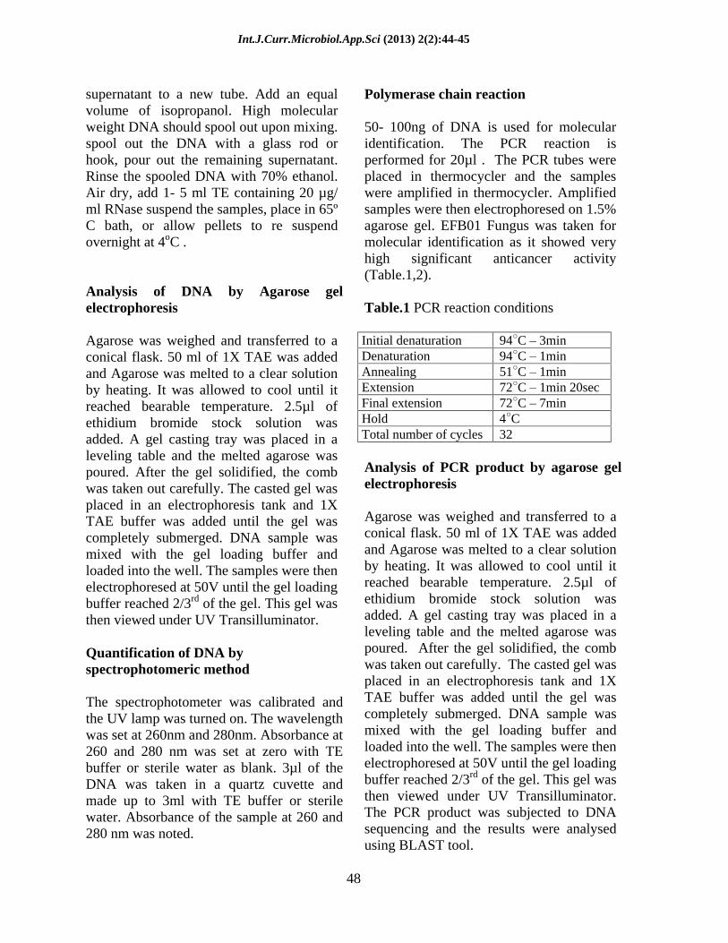
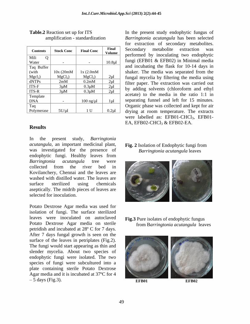
Potato Dextrose Agar media was used for isolation of fungi. The surface sterilized leaves were inoculated on autoclaved Potato Dextrose Agar media on sterile petridish and incubated at 28º C for 7 days. After 7 days fungal growth is seen on the surface of the leaves in petriplates (Fig.2). The fungi would start appearing as thin and slender mycelia. About two species of endophytic fungi were isolated. The two species of fungi were subcultured into a plate containing sterile Potato Dextrose Agar media and it is incubated at 37ºC for 4 5 days (Fig.3).
In the present study endophytic fungus of Barringtonia acutangula has been selected for extraction of secondary metabolites. Secondary metabolite extraction was performed by inoculating two endophytic fungi (EFB01 & EFB02) in Minimal media and incubating the flask for 10-14 days in shaker. The media was separated from the fungal mycelia by filtering the media using filter paper. The extraction was carried out by adding solvents (chloroform and ethyl acetate) to the media in the ratio 1:1 in separating funnel and left for 15 minutes. Organic phase was collected and kept for air drying at room temperature. The extracts were labelled as: EFB01-CHCl3, EFB01-EA, EFB02-CHCl3 & EFB02-EA.
Fig. 2 Isolation of Endophytic fungi from Barringtonia acutangula leaves
Fig.3 Pure isolates of endophytic fungus from Barringtonia acutangula leaves
EFB01 EFB02

Int.J.Curr.Microbiol.App.Sci (2013) 2(2):44-45
50
Anti-cancer assay of secondary metabolic product of fungi
The cytotoxic effect of fungal endophyte isolated from Barringtonia acutangula was tested by the MTT assay, which showed the effect of its secondary metabolites on the cell viability in HT29, human colon cancer cell line. The cells
treated with the fungal extracts of concentration ranging between 125µg/ml and 500µg/ml showed a significant decrease in the cell viability (Table.3,4). The fungal extract from EFB01 showed 52% cyto-toxicity while that of EFB02 exhibited 40% cyto-toxicity which is nominal when compared to the metabolite from EFB01.
Table. 3 Anti-cancer assay of secondary metabolic product of fungi (EFB01)
Activity EFB01 Control PC
Concentration 500µg 250µg 125µg Viability 47.16024 52.28495 49.27239 100 25.7644
Cytotoxicity 52.8398 47.7151 50.7276 0 74.2356
Table. 4 Anti-cancer assay of secondary metabolic product of fungi (EFB02)
Activity EFB02 Control PC Concentration 500µg 250µg 125µg
Viability 59.16156 60.41667 62.3265 100 25.7644 Cytotoxicity 40.8384 39.5833 37.6735 0 74.2356
Table.5 DNA quantification by UV spectrophotometry Method
Sample A260 A280 Concentration of
DNA (ng/µl) Purity ratio =A260/A280
EFB01 0.40 0.23 20000 1.739
EFB02 0.34 0.21 17000 1.619
Int.J.Curr.Microbiol.App.Sci (2013) 2(2):44-45
51
Molecular characterization of endophytic fungi
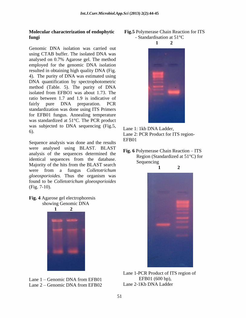
Genomic DNA isolation was carried out using CTAB buffer. The isolated DNA was analysed on 0.7% Agarose gel. The method employed for the genomic DNA isolation resulted in obtaining high quality DNA (Fig. 4). The purity of DNA was estimated using DNA quantification by spectrophotometric method (Table. 5). The purity of DNA isolated from EFBO1 was about 1.73. The ratio between 1.7 and 1.9 is indicative of fairly pure DNA preparation. PCR standardization was done using ITS Primers for EFB01 fungus. Annealing temperature was standardized at 51°C. The PCR product was subjected to DNA sequencing (Fig.5, 6).
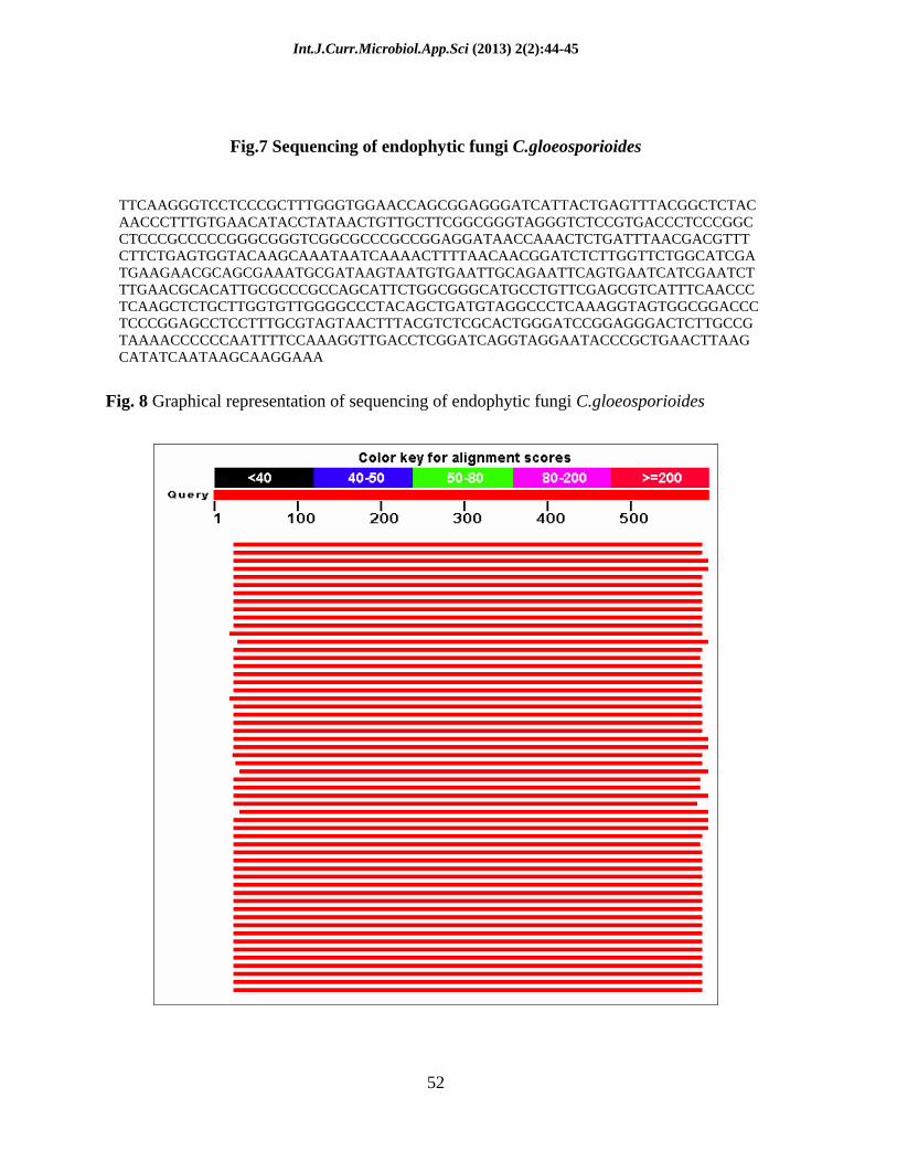
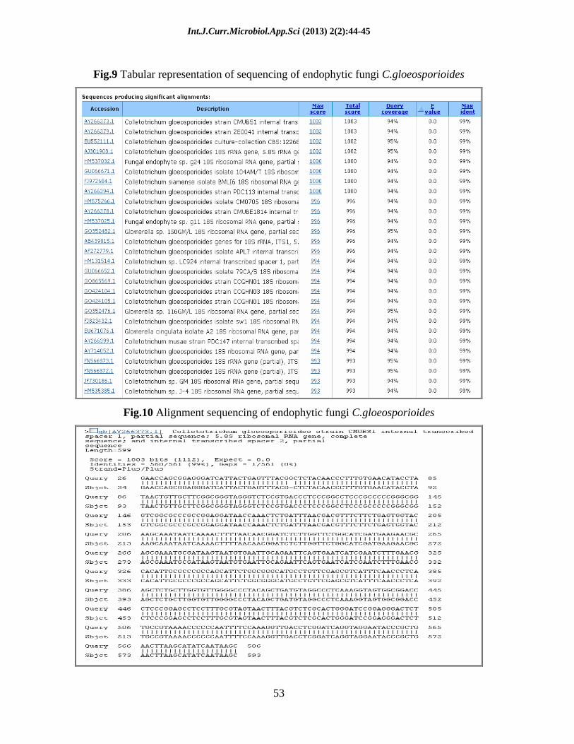
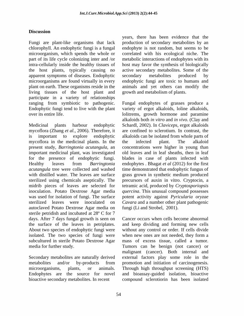
Sequence analysis was done and the results were analysed using BLAST. BLAST analysis of the sequences determined the identical sequences from the database. Majority of the hits from the BLAST search were from a fungus Colletotrichum gloeosporioides. Thus the organism was found to be Colletotrichum gloeosporioides (Fig. 7-10).
Fig. 4 Agarose gel electrophoresis showing Genomic DNA 1 2
Lane 1 Genomic DNA from EFB01 Lane 2 Genomic DNA from EFB02
Fig.5 Polymerase Chain Reaction for ITS - Standardisation at 51°C
1 2
Lane 1: 1kb DNA Ladder, Lane 2: PCR Product for ITS region- EFB01
Fig. 6 Polymerase Chain Reaction ITS Region (Standardized at 51°C) for Sequencing
1 2
Lane 1-PCR Product of ITS region of EFB01 (600 bp), Lane 2-1Kb DNA Ladder
Int.J.Curr.Microbiol.App.Sci (2013) 2(2):44-45
52
Fig.7 Sequencing of endophytic fungi C.gloeosporioides
TTCAAGGGTCCTCCCGCTTTGGGTGGAACCAGCGGAGGGATCATTACTGAGTTTACGGCTCTACAACCCTTTGTGAACATACCTATAACTGTTGCTTCGGCGGGTAGGGTCTCCGTGACCCTCCCGGCCTCCCGCCCCCGGGCGGGTCGGCGCCCGCCGGAGGATAACCAAACTCTGATTTAACGACGTTTCTTCTGAGTGGTACAAGCAAATAATCAAAACTTTTAACAACGGATCTCTTGGTTCTGGCATCGATGAAGAACGCAGCGAAATGCGATAAGTAATGTGAATTGCAGAATTCAGTGAATCATCGAATCTTTGAACGCACATTGCGCCCGCCAGCATTCTGGCGGGCATGCCTGTTCGAGCGTCATTTCAACCCTCAAGCTCTGCTTGGTGTTGGGGCCCTACAGCTGATGTAGGCCCTCAAAGGTAGTGGCGGACCCTCCCGGAGCCTCCTTTGCGTAGTAACTTTACGTCTCGCACTGGGATCCGGAGGGACTCTTGCCGTAAAACCCCCCAATTTTCCAAAGGTTGACCTCGGATCAGGTAGGAATACCCGCTGAACTTAAGCATATCAATAAGCAAGGAAA
Fig. 8 Graphical representation of sequencing of endophytic fungi C.gloeosporioides
Int.J.Curr.Microbiol.App.Sci (2013) 2(2):44-45
53
Fig.9 Tabular representation of sequencing of endophytic fungi C.gloeosporioides
Fig.10 Alignment sequencing of endophytic fungi C.gloeosporioides
Int.J.Curr.Microbiol.App.Sci (2013) 2(2):44-45
54
Discussion
Fungi are plant-like organisms that lack chlorophyll. An endophytic fungi is a fungal microorganism, which spends the whole or part of its life cycle colonizing inter and /or intra-cellularly inside the healthy tissues of the host plants, typically causing no apparent symptoms of diseases. Endophytic microorganisms are found virtually in every plant on earth. These organisms reside in the living tissues of the host plant and participate in a variety of relationships ranging from symbiotic to pathogenic. Endophytic fungi tend to live with the plant over its entire life.
Medicinal plants harbour endophytic mycoflora (Zhang et al., 2006). Therefore, it is important to explore endophytic mycoflora in the medicinal plants. In the present study, Barringtonia acutangula, an important medicinal plant, was investigated for the presence of endophytic fungi. Healthy leaves from Barringtonia acutangula tree were collected and washed with distilled water. The leaves are surface sterilized using chemicals aseptically. The midrib pieces of leaves are selected for inoculation. Potato Dextrose Agar media was used for isolation of fungi. The surface sterilized leaves were inoculated on autoclaved Potato Dextrose Agar media on sterile petridish and incubated at 28º C for 7 days. After 7 days fungal growth is seen on the surface of the leaves in petriplates. About two species of endophytic fungi were isolated. The two species of fungi were subcultured in sterile Potato Dextrose Agar media for further study.
Secondary metabolites are naturally derived metabolites and/or by-products from microorganisms, plants, or animals. Endophytes are the source for novel bioactive secondary metabolites. In recent
years, there has been evidence that the production of secondary metabolites by an endophyte is not random, but seems to be correlated with his ecological niche. The metabolic interactions of endophytes with its host may favor the synthesis of biologically active secondary metabolites. Some of the secondary metabolites produced by endophytic fungi are toxic to humans and animals and yet others can modify the growth and metabolism of plants.
Fungal endophytes of grasses produce a variety of ergot alkaloids, loline alkaloids, lolitrems, growth hormone and paramine alkaloids both in vitro and in vivo. (Clay and Schardl, 2002). In Claviceps, ergot alkaloids are confined to sclerotium. In contrast, the alkaloids can be isolated from whole parts of the infected plant. The alkaloid concentrations were higher in young than old leaves and in leaf sheaths, then in leaf blades in case of plants infected with endophytes . Bhagat et al (2012) for the first time demonstrated that endophytic fungus of grass grown in synthetic medium produced precursors of auxin in vitro. Cryptocin, a tetramic acid, produced by Cryptosporiopsis quercina. This unusual compound possesses potent activity against Pyricularia oryzae Cavara and a number other plant pathogenic fungi (Li and Strobel, 2001).
Cancer occurs when cells become abnormal and keep dividing and forming new cells without any control or order. If cells divide when new ones are not needed, they form a mass of excess tissue, called a tumor. Tumors can be benign (not cancer) or malignant (cancer). Both internal and external factors play some role in the promotion and initiation of carcinogenesis. Through high throughput screening (HTS) and bioassay-guided isolation, bioactive compound sclerotiorin has been isolated

Int.J.Curr.Microbiol.App.Sci (2013) 2(2):44-45
55
from an endophytic fungus Cephalotheca faveolata (Periyasamy Giridharan et al., 2012). Sclerotiorin was found to be potent anti-proliferative against different cancer cells. An endophytic fungus Colletotrichum gloeosporioides (strain JGC-9) was isolated from Justicia gendarussa, a medicinal plant and screened for taxol production (Gangadevi and Muthumary, 2008). This fungal taxol isolated from the organic extract of this fungal culture also had strong cytotoxic activity towards BT 220, H116, Int 407, HL 251 and HLK 210 human cancer cells in vitro, tested by Apoptotic assay.
In the present study the anticancer activity of extracts against human colon cancer cell lines, HT29 was tested by using MTT assay. The extracts showed a high significant activity against the cancer cells. The fungal extract from EFB01 showed 52% cytotoxicity and that of EFB02 exhibited 40% cytotoxicity. EFB01 showed the highest cytotoxicity, hence it was chosen for molecular characterization. Therefore, any research on endophyte-plant symbiosis, such as in this study is of value, especially taking into account the positive biological activity as anti-tumour agents. Effective extracts could provide potential leads towards the development of novel and eco friendly biologically agents. Thus, biological controls to prevent diseases offer an attractive alternative to disease management without the negative impact of chemical control. This study reinforced the assumption that endophytes of medicinal plants play an important role in the search for anti-tumoral compounds .The observations from our study encourage further investigation on these plants.
References
Aminul Haque, M.D., Shawkat Hossain, M., Rahman, M.Z., and Rezaur Rahman, M. 2005. Isolation of Bioactive secondary
metabolites from the endophytic fungus of Ocimum basilicum. J Pharm Sci. 4(2): 127-130.
Azevedo, J.L., Pereira, J.O., and Araújo, W.L. 2000. Endophytic microorganisms: a review on insect control and recent advances on tropical plants. Electronic J Biotechnol. 3(1): 40-65.
Carroll, G.C., and Carroll, F.E. 1978. Studies on the incidence of coniferous needle endophytes in the Pacific Northwest. Can J Bot. 56: 3032- 43.
Clay, K., and Schardl, C. 2002. Evolutionary origins and ecological consequences of endophyte symbiosis with grasses. American Nat. 160: 599 5127.
Gangadevi, V., and Muthumary, J. 2008. Isolation of Colletotrichum gloeosporioides, a novel endophytic taxol producing fungus from the leaves of a medicinal plant, Justicia gendarussa. Mycologia Balcanica. 5: 1 4.
Li, J.Y., and Strobel, G.A. 2001. Jesterone and Hydroxy
jesterone antioomycete Cyclohexanone epoxides from the endophytic fungi Pestalotiopsis jesteri. Phytochem. 57: 261 265.
Periyasamy G., Shilpa, A.V., Amit, K., Mishra, L., and Deshmukh, S.K. 2012. Anticancer activity of sclerotiorin, isolated from an endophytic fungus Cephalotheca faveolata. Indian J Experi biolo. 50(7): 464 468.
Strobel, G.A., 2003. Endophytes as source of bioactive products. Microbe Infect 5(6) : 535 44.
Suthep, W., Nongluksna, S., and Strobel, G.A. 2004. Endophytic fungi with anti-microbial, anticancer and anti-malarial activities isolated from Thai medicinal plants World J. Microbiol. Biotechnol. 20(3) : 265 72.
Zhang, H.W., Song, Y.C., and Tan, R.X. 2006. Biology and chemistry of endophytes. Nat. Pro. Repc. 23: 753
71.