antibodies: indispensable tools for biomedical research
TRANSCRIPT

TIBS 25 – DECEMBER 2000
5930968 – 0004/00/$ – See front matter © 2000, Elsevier Science Ltd. All rights reserved. PII: S0968-0004(00)01725-4
REVIEWS
this issue of TiBS). Burridge was one ofthe first to demonstrate that specificantigens could be detected directly inSDS-gels using polyclonal antibodies fol-lowed by incubation with 125I-labeledgoat anti-rabbit IgG and autoradiogra-phy4. The disadvantages of this tech-nique were related to the long incu-bation and washing times (up to50–60 h) that were necessary to obtainsatisfactory results. In addition, thistechnique required the use of relativelylarge amounts of antibodies, and themultiple steps involved frequently led tothe breakage of delicate gels.
Other laboratories adopted similarmethods, but it wasn’t until 1979 thatseveral laboratories began experiment-ing with the transfer of proteins fromSDS-gels to solid matrices. Initially,these techniques involved transfer bydiffusion of proteins from gels to immo-bilizing matrices such as diazotizedpaper5. This procedure required about12 h for the transfer, long incubationtimes for primary and secondary anti-bodies, and was inefficient, leavingmuch of the protein in the gel. In thesame year, Towbin and co-workers came up with theextremely clever idea of elec-trophoretically transferringproteins from one- and two-dimensional gels to nitro-cellulose sheets. I remembervividly the impact of thistechnique. It made antibodydetection simple and fast (a few hours of work), andprovided a much more effi-cient transfer of the majorityof proteins to produce anidentical replica of thebanding pattern seen on our Coomassie-stained gels.Once in the nitrocellulosepaper, the proteins could bestained using a variety ofantigen-detection methods,including 125I-, fluorescein- or horseradish peroxidase-labeled secondary antibod-ies6. This simple but elegantmethod, now called Westernblotting, transformed the
detection of antigen–antibody interac-tions into a fast, efficient and high-reso-lution technique readily carried out inany laboratory.
Antibody-based blotting procedureswere subsequently exploited in manyways. In our laboratory, one of the mostuseful of these was the introduction ofantibodies to clone genes. This methodused antibodies to screen blots of bac-terial colonies expressing hybrid pro-teins derived from cDNA libraries7.Other techniques involved the micro-affinity purification of antibodies. Theytook advantage of the complexesformed between antigens and antibod-ies in the matrices used for blotting. For example, following transfer fromSDS-gels, protein bands could be cutfrom diazophenylthioether (DPT) paperand incubated in complex antiseracontaining numerous antibodies.Elution of the bound antibodies pro-duced highly purified preparations,which reacted only with the protein ofinterest in immunofluorescence andWestern blotting assays8.
Similarly, immunoaffinity techniqueswere developed for the efficient andlarge-scale purification of proteins orother antigens for biochemical pur-poses. For example, one method tookadvantage of strains of Staphylococcusaureus that are Protein A positive9.Protein A is present at the bacterial cellsurface and binds to the Fc regions of many IgG subclasses. Therefore,
Antibodies: indispensable toolsfor biomedical research
Today, every student of biology recog-nizes that biomedical research is inex-tricably linked to the use of antibodies.However, it is only over the past 25years that antibody production, charac-terization and detection have comewithin easy reach of almost every re-searcher. In this short article, I discusssome of the most important techniquesthat have brought about this revolutionin antibody usage. For a comprehensiveoverview and an excellent technicalmanual, interested readers should con-sult Harlow and Lane1.
Antibodies are targeted reagents thatrecognize and bind to specific antigens.Antigens, especially proteins, usuallypossess numerous epitopes, therebyproducing polyclonal antibodies follow-ing injection into an animal. Each anti-body in the resulting antiserum is spe-cific for a given epitope. A majorbreakthrough in the production of largeamounts of antibodies directed againsta single epitope came with the develop-ment of the monoclonal antibody tech-nique2. It is the application and use ofboth polyclonal and monoclonal anti-bodies that has revolutionized manyareas of basic research.
My introduction to antibodies camewhen we immunized our first rabbit. I will never forget the analysis of ourfirst ‘bleed’. The most accessible tech-nique available at that time was theOuchterlony double-immunodiffusionassay. This was a slow anxiety-buildingprocedure, taking days or even weeks insome cases to obtain the ultimate re-ward of a precipitin line, demonstratingthat our animal was responding to theprotein of interest. This remained thenorm for antibody detection until the mid-1970s when faster, more effi-cient and higher-resolution methods fordetermining antigen–antibody interac-tions started to become available. Inour laboratory, these techniquesproved to be enormously helpful in ex-panding the utility of antibodies. Fromthe beginning, these methods took ad-vantage of the separation of complexmixtures of cellular proteins by sodiumdodecyl sulfate (SDS) polyacrylamidegel electrophoresis (Ref. 3; see p. 590 in
Figure 1A PtK2 epithelial cell fixed and processed for double-labelimmunofluorescence using a rabbit polyclonal antibodydirected against bovine hoof prekeratin (fluorescein,green) and a mouse monoclonal antibody directedagainst nuclear lamin A (rhodamine, red). Conventionalepifluorescence was used to obtain this image.
REVIEWS TIBS 25 – DECEMBER 2000
594
heat-inactivated, formalin-fixed bacteriawere used to adsorb antigen–antibodycomplexes from cell lysates. Once ad-sorbed, the bacteria were centrifugedand washed and highly purified antigenswere released. This procedure has beenimproved by employing Protein A linkedto inert beads. Purified preparations ofan antibody can be crosslinked to thebeads for use as an affinity column. Upto 10 000-fold purifications of proteinscan be achieved using these tech-niques1. An extension of the Protein Amethod, which provides insights intoprotein–protein interactions, involvesthe analysis of immunoprecipitates ob-tained from gently lysed cells. Underthese conditions, antibodies bind totheir primary antigens, which in turn re-main associated with their normal inter-acting partners. We, and many others,have found this to be one of the mostimportant biochemical tools for the ini-tial characterization of multicomponentprotein complexes and their potentialfunctions in cells1.
At the same time as improved assaysfor antibody–antigen interactions andpurification were being developed, newand very useful techniques becameavailable for antibody production. Oneparticularly useful approach, to which Iwas introduced by Klaus Weber andElias Lazarides while I was on sabbaticalat the Cold Spring Harbor Laboratory in 1973, involved the purification of
proteins by slicing them from SDS-poly-acrylamide gels. Importantly, it wasfound that the resulting denatured pro-teins, such as actin, are antigenic andcan produce antibodies that react withtheir native forms10. Other techniqueshave involved the use of synthetic pep-tides derived from the known sequencesof a protein. Remarkably, antibodiesdirected against very short peptides(approximately six amino acids) are ca-pable of reacting with the full-lengthnative protein11.
Antibodies prepared in this fashionhave had an enormous impact on struc-ture–function studies. For example, syn-thetic peptides were used to make spe-cific tyrosinated and non-tyrosinateda-tubulin antibodies, which yieldedcritical insights into the structure andfunction of microtubules12. Antibodieshave also been prepared against spe-cific phosphorylated variants of pro-teins using the same approach. This hasbeen especially useful in gaining in-sights into the regulation of cytoskeletalcomponents such as intermediate fila-ments13. With the ever-increasing avail-ability of protein sequences and theever-expanding insights into the regula-tion of proteins by post-translationalmodifications, the use of synthetic pep-tides as antigens is gaining more andmore popularity. Another related andimportant finding that has been used inantibody production is related to the
presentation or display ofantigens in the immune re-sponse. For example, it hasbeen found that the highlyordered arrays of antigensdisplayed by virus coat pro-teins elicit a greater andmore specific immune re-sponse than the sameproteins in solution1,24,25.
The use of antibodies forthe subcellular localization of antibody–antigen inter-actions by immunofluores-cence microscopy has led toimportant new insights intostructure–function relation-ships and protein–proteininteractions within both thenuclear and cytoplasmiccompartments. This can beattributed not only to theready availability of a widerange of antibodies directedagainst virtually every subcel-lular organelle or structuralentity but also to remarkableadvances in light microscopy.
Immunofluorescence was originallydeveloped in the 1940s by Coons usingantibodies directly conjugated to fluo-rescein14. Subsequently, Coons’ groupintroduced the technique of indirect im-munofluorescence, an extremely impor-tant modification of their originalmethod1,15. This employed the use of un-conjugated primary antibodies and sec-ondary antibodies conjugated withfluorochromes such as fluorescein andrhodamine. This was a vast improve-ment, as it obviated the need for directconjugation of antibodies and the likeli-hood of decreasing their activity andspecificity. However, numerous obsta-cles inherent in labeling cells with anti-bodies and then visualizing them in thefluorescence microscope had to beovercome before the method becamewidespread in the 1970s.
Until then, a conventional transmittedlight optical system equipped with adarkfield condenser was used to visual-ize cultured fibroblasts that had beenfixed and stained with fluorescein-tagged antibodies. This set-up providedthe contrast required for visualizing thelight emitted by the fluorochrome with-out the background excitatory light. Theresult was a very weak signal requiringprolonged exposure times for 35-mmfilm recording. I can remember waiting15–30 min to capture one mediocreimage. The introduction of the first com-mercially available epifluorescence sys-tems between the late 1960s and themid-1970s permitted researchers to takefull advantage of the highest numericalaperture (NA) oil immersion objectives.In this optical set-up, the objective lensserved as both the condenser and theobjective and the emitted light was seenon a black background, optimizing bothspecimen contrast and the chances ofdetecting weaker signals. Epifluor-escence also required the developmentof efficient dichroic mirrors that re-flected shorter wavelengths and trans-mitted longer wavelengths, optimizinglight intensities. You can imagine the ex-citement upon trying out our first ZeissIII RS epifluorescence system, which al-lowed us, for the first time, to capture animage in 30 s or less (for an extensive re-view of fluorescence microscopy, seeRef. 15).
One of the earliest demonstrations ofthe utility of these new microscopicmethods, which drew a large amount ofattention, was the localization of actin10.The striking images of stress fibers incultured non-muscle cells achieved byepifluorescence triggered an avalanche
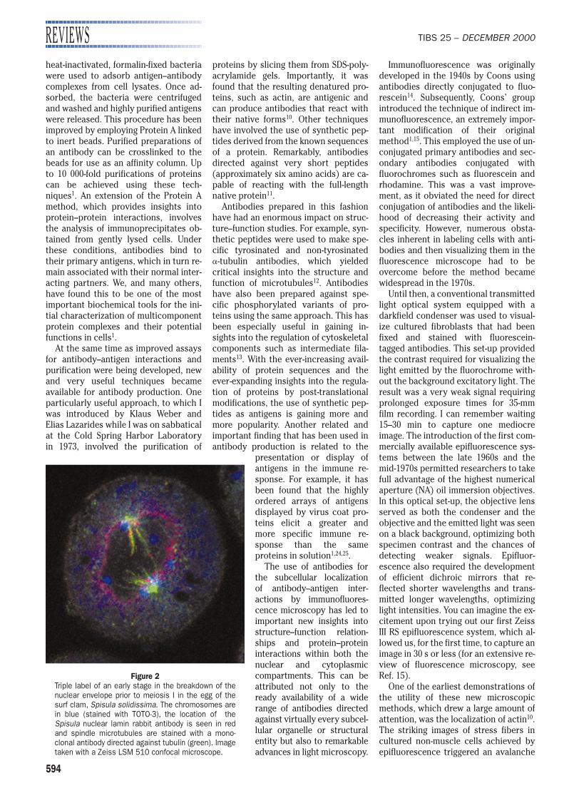
Figure 2Triple label of an early stage in the breakdown of thenuclear envelope prior to meiosis I in the egg of thesurf clam, Spisula solidissima. The chromosomes arein blue (stained with TOTO-3), the location of theSpisula nuclear lamin rabbit antibody is seen in redand spindle microtubules are stained with a mono-clonal antibody directed against tubulin (green). Imagetaken with a Zeiss LSM 510 confocal microscope.
TIBS 25 – DECEMBER 2000
595
of remarkable images of subcellularcompartments that continues to thisday. Fluorescence image capturing hasalso been significantly advanced overthe last two decades through the intro-duction of video cameras, and low-light-sensitive silicon intensified and intensified charge-coupled device (CCD)cameras15–17. Recent innovations inhigh-resolution fluorescent antibodyimaging include the use of confocal, de-convolution and multiphoton micro-scopes18,19. The major advantage ofthese optical systems is that they re-move out-of-focus light, thereby provid-ing optimal contrast and resolution. Inall three types of instruments, three-di-mensional images of the distribution ofantigens within cells or tissues can bereadily obtained15.
The technological advances in fluor-escence microscopy, including the useof narrow-band filter systems and theready availability of a wide range offluorochrome-conjugated second anti-bodies, have now made double andtriple label immunofluorescence com-monplace15 (see Figs 1,2). In the early1980s, Allison Adams and John Pringlefrom the University of Michigan visitedmy laboratory, during the PhysiologyCourse at the Woods Hole MarineBiological Laboratory, to learn immuno-fluorescence. They wanted to localizeactin and tubulin in Saccharomyces cere-visiae. I was quite skeptical at the timethat any useful information could be de-rived from cells that were so tiny com-pared with fibroblasts. But I was wrong!Once fixation protocols had been opti-mized, remarkable details of cytoskel-etal organization were revealed in yeastcells20. Major advances have also takenplace in other important model geneticsystems including Drosophila21 andCaenorhabditis elegans22. The develop-ment of reliable epifluorescence obser-vations of a large number of antigens inthese important systems has providedimportant new insights into the regu-lation of the early developmental stagesof wild-type and mutant organisms15.Antibody localization has also shed newlight on the structure and function of nu-merous proteins within cells at the ultra-structural level. This has entailed eitherpre-embedding or post-embeddinglabeling with a variety of electron-densereporters linked to secondary antibod-ies. These include colloidal gold andnano-gold. These techniques have beenextremely useful in localizing cell sur-face and nuclear components at highresolution15.
Antibodies have also proved to bevery useful in in vivo investigations.These involve either the microinjectionof antibodies into cells, or the exposureof live cells to antibodies directedagainst cell-surface antigens, followedby the analysis of their effects on cellmorphology and/or behavior. For exam-ple, in a key paper, Thomas Kreis wasable to demonstrate that the accessibil-ity of nine amino acids at the C-terminalend of the vesicular stomatitis virusglycoprotein was essential for transportto the cell surface23.
Another important application ofantibodies lies in the recognition of epi-tope tags. Epitope tags are short aminoacid sequences that are added to pro-teins expressed by modified cDNAs.These short peptide sequences are ex-tremely useful, as in most cases theyhave no apparent effects on either thestructure or function of their expressedfusion products. When expressed incells, their products are readily distin-guished from the same or similar pro-teins expressed endogenously by veryspecific antibodies generated againstthe tags. This approach also allows theisolation and purification of expressedproteins containing a tag. Immuno-precipitation can be used to identifyother proteins bound to the antigen.Among the most widely used tags arethe myc tag derived from the c-myc pro-tein, the HA tag derived from flu hemag-glutinin and FLAG, a synthetic peptidetag (see Ref. 1 for a review).
In conclusion, I can only marvel at theenormous impact that antibodies havehad in all types of biomedical investiga-tions. This brief review has touched ononly a few of the major methods thathave influenced my own research groupover the past 25 years or so. Many use-ful applications have not been dis-cussed, but the techniques describedare representative of virtually all theantibody-based methods now in wideuse in the life sciences.
AcknowledgementsI thank Ying-Hao Chou and Anne
Goldman for reading this manuscript.Research in my laboratory is supportedby a MERIT Award from the NIGMS andgrants from NCI and NIDR.
References1 Harlow, E. and Lane, D. (1999) Using Antibodies: A
Laboratory Manual, Cold Spring Harbor Press2 Kohler, G. and Milstein, C. (1975) Continuous cultures
of fused cells secreting antibody of a predefinedspecificity. Nature 256, 495–497
3 Studier, F.W. (2000) Slab-gel electrophoresis. Trends Biochem. Sci. 25, 588–590
4 Burridge, K. (1976) Changes in cellular glycoproteinsafter transformation: identification of specificglycoproteins and antigens in sodium dodecyl sulfategels. Proc. Natl. Acad. Sci. U. S. A. 73,4457–4461
5 Renart, J. et al. (1979) Transfer of proteins from gelsto diazobenzyloxymethyl-paper and detection withantisera: a method for studying antibody specificity andantigen structure. Proc. Natl. Acad. Sci. U. S. A. 76,3116–3120
6 Towbin, H. et al. (1979) Electrophoretic transfer ofproteins from polyacrylamide gels to nitrocellulosesheets: procedures and some applications. Proc. Natl. Acad. Sci. U. S. A. 76, 4350–4354
7 Young, R.A. and Davis, R.W. (1983) Efficient isolationof genes using antibody probes. Proc. Natl. Acad. Sci.U. S. A. 80, 1194–1198
8 Olmsted, J.B. (1981) Affinity purification of antibodiesfrom diazotized paper blots of heterogeneous proteinsamples. J. Biol. Chem. 256, 11955–11957
9 Kessler, S.W. (1975) Rapid isolation of antigens fromcells with a staphylococcal protein A–antibodyadsorbent: parameters of the interaction ofantibody–antigen complexes with protein A. J. Immunol. 115, 1617–1624
10 Lazarides, E. and Weber, K. (1974) Actin antibody: thespecific visualization of actin filaments in non-musclecells. Proc. Natl. Acad. Sci. U. S. A. 71,2268–2272
11 Lerner, R.A. (1982) Tapping the immunologicalrepertoire to produce antibodies of predeterminedspecificity. Nature 299, 592–596
12 Gundersen, G.G. et al. (1984) Distinct populations ofmicrotubules: tyrosinated and non-tyrosinated a tubulinare distributed differently in vivo. Cell 38,779–789
13 Inagaki, M. et al. (1996) Dynamic properties ofintermediate filaments: regulation by phosphorylation.Bioessays 18, 481–487
14 Coons, A.H. et al. (1942) The demonstration ofpneumococcal antigen in tissues by the use offluorescent antibody. J. Immunol. 45,159–170
15 Spector, D. et al. (1998) Cells: A Laboratory Manual(Vol. 2), Cold Spring Harbor Laboratory Press
16 Allen, R.D. and Allen, N.S. (1983) Video-enhancedmicroscopy with a computer frame memory. J. Microsc. 129, 3–17
17 Inoue, S. (1986) Video Microscopy, Plenum18 White, J.G. et al. (1987) An evaluation of confocal
versus conventional imaging of biological structures byfluorescence light microscopy. J. Cell Biol. 105, 41–48
19 Agard, D.A. et al. (1989) Fluorescence microscopy inthree dimensions. Methods Cell Biol. 30,353–377
20 Adams, A.E.M. and Pringle, J.R. (1984) Relationship ofactin and tubulin distribution to bud growth in wild-typeand morphogenetic-mutant Saccharomyces cerevisiae.J. Cell Biol. 98, 934–945
21 Mitchison, T.J. and Sedat, J. (1983) Localization ofantigenic determinants in whole Drosophila embryos.Dev. Biol. 99, 261–264
22 Strome, S. and Wood, W.B. (1982)Immunofluorescence visualization of germ-line-specificcytoplasmic granules in embryos, larvae and adults of Caerorhabditis elegans. Proc. Natl. Acad. U. S. A.79, 1558–1562
23 Kreis, T.E. (1986) Microinjected antibodies against thecytoplasmic domain of vesicular stomatitis virusglycoprotein block its transport to the cell surface.EMBO J. 5, 931–941
24 Aebi, U. et al. (1977) New method for localizingproteins in periodic structures: Fab fragment labelingcombined with image processing of electronmicrographs. Proc. Natl. Acad. Sci. U. S. A. 74,5514–5518
25 Bachmann, M.F. and Zinkernagel, R.M. (1996) Theinfluence of virus structure on antibody response andvirus serotype formation. Immunol. Today 17, 553
ROBERT D. GOLDMAN
Dept of Cell and Molecular Biology,Northwestern University Medical School,303 E. Chicago Avenue, W11-145, Chicago,IL 60611, USA.Email: [email protected]
REVIEWS