anti-inflammatory actions of the anticoagulant, activated ...cdn.intechweb.org/pdfs/25192.pdf ·...
TRANSCRIPT
4
Anti-Inflammatory Actions of the Anticoagulant, Activated Protein C
Christopher John Jackson and Meilang Xue Sutton Arthritis Research Laboratories, Department of Rheumatology,
Institute of Bone and Joint Research, Kolling Institute, University of Sydney at Royal North Shore Hospital, St Leonards
Australia
1. Introduction
Protein C is a vitamin K–dependent zymogen, discovered in 1976 in bovine plasma (Stenflo,
1976). It is derived from the human PROC gene on chromosome 2 (2q13-q14) which contains
9 exons (Rezaie, 1993). Post-translational modifications include -hydroxylation at Asp71,
N-linked glycosylation at residues 97, 248, 313 and 329 and ┛-carboxylation of 9 glutamic
acid residues which forms the Gla domain at the amino terminus. Human protein C is 62kD
protein and consists of 419 amino acids. The four major moieties that make up the protein C
molecule are a Gla domain, two epidermal growth factor (EGF)- like regions, a small
activation peptide, and an active serine protease domain (Griffin, 2005). Mature 62 000 Da
human protein C is cleaved by a furin-like endoprotease that releases Lys156–Arg157 before
secretion from liver cells. Protein C is activated on the endothelial surface when thrombin
binds to thrombomodulin and cleaves protein C’s activation peptide. This conversion to
activated protein C (APC) is augmented by endothelial cell protein C receptor (EPCR)
(Fukudome & Esmon, 1994). Protein C circulates in plasma at 70 nM whereas APC is present
in much lower concentrations (40 pM or ~ 2.3 ng/mL) (Gruber & Griffin, 1992).
APC was first recognized as an anticoagulant. In the presence of its cofactor, protein S, APC
degrades the coagulation factors Va and VIIIa and inhibits thrombin generation. The light
chain provides anticoagulant activity by having highly specific protein–protein interactions
with factors Va and VIIIa followed by proteolytic inactivation of factor Va by cleavage at
Arg (506) and Arg (306) and of factor VIIIa by cleavage at Arg (336) and Arg (562) (Zlokovic
& Griffin, 2011). In addition, APC promotes fibrinolysis by binding to plasminogen activator
inhibitor which prevents inhibition of plasminogen conversion to plasmin. The significance
of APC as an anticoagulant is reflected by the findings that deficiencies in protein C result in
severe familial disorders of thrombosis (Baker & Bick, 1999). Replenishment of protein
C/APC in patients with systemic or local hypercoagulation can reverse the abnormality.
2. Anti-inflammatory and cytoprotective functions of APC
In addition to its anticoagulant activity, APC exerts a broad range of cytoprotective and
anti-inflammatory actions described below.
www.intechopen.com
Inflammatory Diseases – A Modern Perspective 46
2.1 Inflammation
Independent of its effect on coagulation, APC has potent anti-inflammatory properties (Joyce, 2001; Mosnier & Griffin, 2003) associated with a decrease in pro-inflammatory cytokines and a reduction of leukocyte recruitment. Joyce et al (Joyce, 2001) have shown that
APC directly suppresses expression of p50 and p52 nuclear factor (NF)-B subunits in
human umbilical vein endothelial cells. The NF-B pathway is important for the expression of a wide variety of inflammatory genes including tumor necrosis factor (TNF)-┙ and cell adhesion molecules that are associated with diseases ranging from inflammation to cancer
(Li & Verma, 2002). Direct inhibition of NF-B is sufficient to block symptoms of many inflammatory diseases so these inhibitors have potential therapeutic value (Bell, 1915; Li & Verma, 2002; Calzado, 1914; Calzado, 2007). APC inhibits the expression and activation of
NF-B in unstimulated and stimulated monocytes (White, 2000; Xue, 2007; Yuksel, 2002), keratinocytes (Xue, 2004), endothelial cells (Franscini, 2004). APC also suppresses
inflammation in vivo by inhibition of NF-B (Cheng, 2006). In addition, APC has the ability to upregulate and activate matrix metalloproteinase (MMP)-2 (Nguyen, 2000; Xue, 2004), a MMP with anti-inflammatory properties (Itoh, 2002; McQuibban, 2002) and to suppress gelatinase B (Cheng, 2006; Xue, 2007), a MMP associated with many inflammatory conditions (Itoh, 2002; Ram, 2006). During acute inflammation, plasma APC levels are diminished (Liaw, 2004) and inflammatory cytokines such as interleukin (IL)-1┚ and TNF-┙, as well as endotoxin, can attenuate thrombomodulin and EPCR expression which further reduces the ability of endothelial cells to generate APC. Acute inflammation is exacerbated in mice genetically predisposed to a severe protein C deficiency (Lay, 2007). APC also regulates the immuno/inflammatory response. Monocytes treated with APC
decrease the release of tissue factor (Toltl, 2008), the pro-inflammatory cytokines TNF-┙
(Grey, 1994), IL-1┚, IL-6, and & IL-8 (Stephenson, 2006). Additionally, APC induces the
release of the anti-inflammatory cytokine IL-10 from monocytes (Toltl, 2008).
APC targets CD8+ dendritic cells to reduce the mortality of endotoxemia in mice (Kerschen,
2010). Expression of EPCR in mature murine immune cells is limited to a subset of CD8+
conventional dendritic cells. Adoptive transfer of splenic CD11chiPDCA-1- dendritic cells
from wild-type mice into animals with hematopoietic EPCR deficiency restored the
therapeutic efficacy of APC, whereas transfer of EPCR-deficient CD11chi dendritic cells or
wild-type CD11chi dendritic cells depleted of EPCR+ cells did not. These data reveal an
essential role for EPCR and PAR1 on hematopoietic cells, identify EPCR-expressing
dendritic immune cells as a critical target of APC therapy, and document EPCR-
independent anti-inflammatory effects of APC on innate immune cells.
2.2 Cell proliferation and apoptosis
APC induces growth of cultured human umbilical vein endothelial cells (HUVEC) (Uchiba,
2004). In smooth muscle cells, APC elicits an increase in [(3)H]-thymidine incorporation
(Bretschneider, 2007) and enhances proliferation and migration of human skin keratinocytes
(Xue, 2005). Consistent with the stimulatory effects on cell growth, APC displays strong
anti-apoptotic properties. APC decreases sepsis-induced apoptosis resulting from increased
p21 and p53 proteins in mice (Sakar, 2007) and modulates Bcl-2 and Bax and inhibits
caspase-3 and -8 activity which results in inhibition of apoptosis in a number of cell types
(Joyce, 2001). During hypoxic stress of brain endothelial cells, APC inhibits p53, reduces pro-
apoptotic Bax and maintains levels of protective Bcl-2 protein, thereby preventing the
www.intechopen.com
Anti-Inflammatory Actions of the Anticoagulant, Activated Protein C 47
stimulation of the intrinsic apoptotic pathway (Cheng, 2003). In human skin keratinocytes,
APC prevents cell apoptosis via inhibition of caspase-3 activation (Xue, 2004) and in
podocytes, APC protects against glucose-induced apoptosis both in vitro and in vivo
(Isermann, 2007). APC inhibits bisphosphonate-induced endothelial cell death via EPCR-
induced inactivation of caspase-3 and NF-κB, and also suggests that APC has the potential
to be a therapeutic drug in various vascular diseases induced by endothelial cell damage
(Seol, 2011).
2.3 Barrier stabilization
Endothelial cells normally form a dynamically regulated stable barrier at the blood-tissue
interface, and breakdown of this barrier is a key pathogenic factor in inflammatory
disorders, such as sepsis. APC boosts the barrier via at least two different mechanisms.
First, APC enhances sphingosine-1-phosphate (S-1-P) production, which signals through its
G-protein coupled receptor to stabilize the cytoskeleton and reduce endothelial permeability
(Feistritzer & Riewald, 2005; Finigan, 2005). Second, APC utilizes the angiopoietin
(Ang)/Tie2 axis to promote endothelial barrier function (Minhas, 2010). APC significantly
up-regulates gene and protein expression of Tie2 and Ang1 in a dose (0.01-10 µg/ml) and
time (0.5 h – 24 h) dependent manner in HUVEC, whilst it markedly inhibits Ang2 with an
IC50 of ~ 0.1 µg/ml. HUVEC permeability, measured using Evans blue dye transfer, is
significantly reduced in the presence of APC and, in concordance, the tight junction
associated protein, zona occludens (ZO)-1, is up regulated and localized peripherally
around cells, compared to control. Smooth muscle cell migration towards APC-stimulated
HUVEC is elevated compared to unstimulated cells. Blocking antibodies and small
interfering (si) RNA treatment, compared to isotype or scrambled siRNA controls, show that
APC requires three receptors, endothelial protein C receptor (EPCR), protease activated
receptor (PAR)-1 and Tie2 to perform all these barrier stabilization functions (Minhas, 2010).
We have shown that HUVEC produce protein C that acts through novel mediators to
enhance their own functional integrity (Xue, 2010). When endogenous protein C or its
receptor, EPCR, is suppressed by si RNA, HUVEC proliferation is decreased and apoptosis
elevated. Interestingly, protein C or EPCR siRNA significantly increases HUVEC
permeability, which occurs via a reduction of the Ang1/Ang2 ratio and inhibition of the
peripheral localization of the tight junction protein, ZO-1. In addition, protein C or EPCR
siRNA inhibits type IV collagen and MMP-2, providing the first evidence that protein C
contributes to vascular basement membrane formation (Xue, 2010). Barrier stabilization is
more effective when APC is derived endogenously and functions in an autocrine manner,
than when the source of APC is exogenous (Feistritzer, 2006).
The barrier protective effect of APC is also relevant to epidermal keratinocytes (Xue, 2011).
In response to APC, Tie2, a tyrosine kinase receptor, is rapidly activated within 30 minutes,
and relocates to cell-cell contacts. APC also increases junction proteins ZO-1, claudin-1 and
VE-cadherin. Inhibition of Tie2 by its peptide inhibitor or small interfering RNA abolished
the barrier protective effect of APC (Xue, 2011).
3. APC cellular signalling
APC exerts its anti-inflammatory, cyto-protective and barrier stabilization effects by acting on receptors which initiates cellular signaling, as described below.
www.intechopen.com
Inflammatory Diseases – A Modern Perspective 48
3.1 EPCR and PARs
Many of the cyto-protective actions of APC are mediated through EPCR, which itself is anti-inflammatory (Esmon, 2004). This receptor binds protein C and APC with similar affinity (Fukudome & Esmon, 1994), and protein C can be converted to APC whilst remaining bound to EPCR. EPCR is a type I transmembrane protein which shares homology with the major histocompatibility class 1/CD1 family of proteins involved in the immune response. EPCR was discovered on endothelial cells, however EPCR was subsequently found on some leukocytes (Esmon, 2004) and is strongly expressed by the basal layer of keratinocytes in skin epidermis as well as in cultured keratinocytes (Xue, 2005). Recent studies show that EPCR has important physiological functions. For example, over-expression of EPCR protects transgenic mice from endotoxin-induced injury (Li, 2005) and EPCR is essential for normal embryonic development as deletion of the EPCR gene in mice is lethal by embryonic day 10 (Gu, 2002). Recently, EPCR has been identified as a marker of certain stem cells in mice (Balazs, 2006;
Kent, 2009). EPCR is expressed at high levels within the bone marrow in hematopoietic
stem cells (HSCs). Mouse bone marrow cells isolated on the basis of EPCR expression alone
are highly enriched HSCs, showing levels of engraftment in vivo comparable to that of stem
cells purified using the most effective conventional methods (Balazs, 2006). Moreover, they
showed that hematopoietic stem cell activity is always associated with EPCR-expressing
cells (Balazs, 2006). In addition, high EPCR-expressing cells are observed in basal-like
tumours in breast cancer (Park, 2010).
EPCR does not mediate cell signalling, but acts as a homing receptor to allow APC to cleave
PAR-1 (Riewald, 2002). The PARs are G-protein coupled receptors found on most cells. The
four known PARs are activated via proteolytic cleavage by various proteases that results in
an intra-molecular tethered ligand that triggers activation of a G protein and subsequent
intracellular signalling (Coughlin, 2000). Thrombin activates PAR-1, PAR-3, and PAR-4,
whereas other serine proteases, including APC (Riewald, 2002). but not thrombin, activate
PAR-2. Subsequent functional activity of APC cleavage of PAR-2 is yet to be fully
elucidated, although our experiments indicate that APC acts through PAR-2 to promote
wound healing in mice (Julovi et al, personal communication) . While the majority of
reports cast PAR-2 as pro-inflammatory, others show that PAR-2 agonists are beneficial in
several mouse models that involve inflammation or ischemia (Milia, 2002). Among the
PARs, PAR-1 is most widely expressed and has been most extensively studied. APC
bound to EPCR can activate PAR-1 and promote the anti-inflammatory and anti-apoptotic
actions of APC (Riewald, 2002).
Both thrombin and APC can cleave PAR-1 at identical locations. Thrombin cleavage causes
platelet activation, increases vascular permeability, activates NF-B and elevates
inflammatory cytokines, all of which promote an inflammatory response. Unexpectedly,
when APC cleaves PAR-1, its actions are directly opposite to that of thrombin. APC strongly
inhibits vascular permeability, activation of NF-B, endothelial adhesion molecule
expression, cytokine production and monocyte migration (Riewald, 2002). Studies using
endothelial cells and other cell types show that when APC activates PAR-1 in an EPCR-
dependent manner, it causes alterations in gene expression profiles and exerts direct anti-
apoptotic effects (Guo, 2004; Joyce, 2001). However, compared to thrombin, APC is
relatively inefficient and requires ~104 higher concentration (Kuliopulos, 1999) to cleave
PAR-1. This has raised doubts about the possibility of APC having a physiological effect by
www.intechopen.com
Anti-Inflammatory Actions of the Anticoagulant, Activated Protein C 49
acting through PAR-1. Bae et al (Bae, 2007) have partially solved this issue by showing that
APC cleaves PAR-1 on lipid rafts in endothelial cells. They subsequently identified a novel
pathway whereby EPCR is associated with caveolin-1 in lipid rafts in endothelial cells (Bae,
2007). These discrete, cholesterol and sphingolipid enriched microdomains of the cell
membrane provide a protective compartment for APC to act in isolation. When APC
binds to EPCR in the lipid raft, caveolin-1 is replaced with PAR-1 which couples with the
pertussis toxin sensitive Gi-protein to initiate a protective signalling pathway. In contrast,
when thrombin cleaves PAR-1 outside the lipid raft signalling occurs via Gq and/or
G12/13 which exert inflammatory effects. Interestingly, if EPCR is occupied on the lipid
raft, even thrombin, can induce activation of the Gi protein and mimic the protective
effects of APC (Bae, 2007).
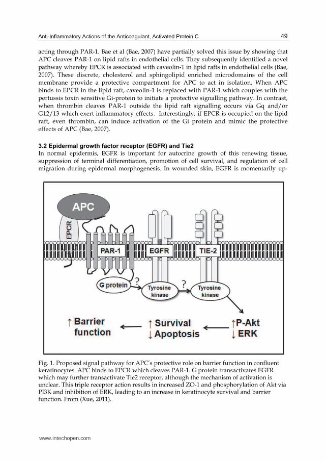
3.2 Epidermal growth factor receptor (EGFR) and Tie2
In normal epidermis, EGFR is important for autocrine growth of this renewing tissue, suppression of terminal differentiation, promotion of cell survival, and regulation of cell migration during epidermal morphogenesis. In wounded skin, EGFR is momentarily up-
Fig. 1. Proposed signal pathway for APC’s protective role on barrier function in confluent keratinocytes. APC binds to EPCR which cleaves PAR-1. G protein transactivates EGFR which may further transactivate Tie2 receptor, although the mechanism of activation is unclear. This triple receptor action results in increased ZO-1 and phosphorylation of Akt via PI3K and inhibition of ERK, leading to an increase in keratinocyte survival and barrier function. From (Xue, 2011).
www.intechopen.com
Inflammatory Diseases – A Modern Perspective 50
regulated and is a major contributor to the proliferative and migratory aspects of wound re-epithelialization. EGFR is able to regulate cell adhesion, expression of matrix degrading proteinases, and cell migration to provide a vital contribution to the migratory and invasive potential of keratinocytes (Hudson & McCawley, 1998). APC appears to act through EGFR to regulate lymphocyte migration (Feistritzer, 2006) and wound healing (Xue, 2007). When keratinocytes are stimulated with APC, the expression and phosphorylation of EGFR is markedly increased and conversely when cells are treated with protein C siRNA, the phosphorylated form of EGFR in cell lysates is inhibited by more than 50% (Xue, 2007). Using dual immunofluorescent staining, we found that both EPCR and activated EGFR are co-localized in basal and suprabasal keratinocytes in the epidermis, which is identical to protein C localization in skin epidermis (Xue, 2007). Furthermore, APC does not activate Tie2 through its major ligand, Ang-1, in keratinocytes, but instead acts by binding to EPCR, cleaving PAR-1 and trans-activating EGFR followed by transactivation of Tie2 (Figure 1). When activation of Akt, but not ERK, is inhibited, the barrier protective effect of APC on keratinocytes is abolished. Another report has indicated that, extracellularly, APC engages EPCR, PAR-1, and EGFR in order to increase the invasiveness of MDA-MB-231 cells (Gramling, 2010).
3.3 Other receptors
EPCR-independent signaling components of the APC pathway have been identified in monocytes. Apolipoprotein E receptor 2 (ApoER2) binding of APC results in phosphorylation of Dab1 and activation of the PI3K and Akt pathway resulting in decreased tissue factor release from monocytes (Yang, 2009). The efficacy of APC in murine endotoxemia is dependent on integrin CD11b. Genetic inactivation of CD11b, PAR1, or sphingosine kinase-1, but not EPCR, abolished the ability of APC to suppress the macrophage inflammatory response in vitro. Using a LPS-induced mouse model of lethal endotoxemia, Cao et al (Cao, 2010) showed that APC administration reduced the mortality of wild-type mice, but not CD11b-deficient mice.
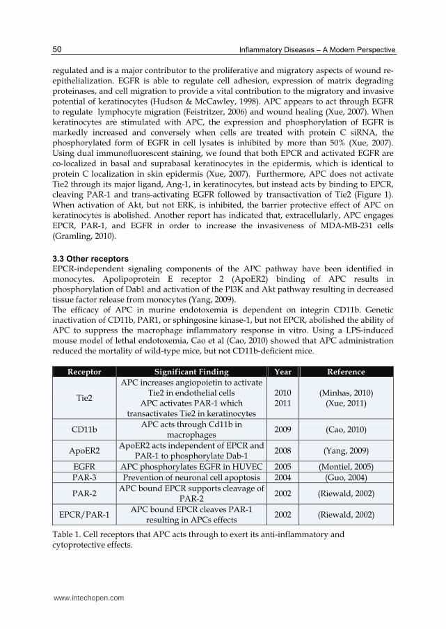
Receptor Significant Finding Year Reference
Tie2 APC increases angiopoietin to activate
Tie2 in endothelial cells APC activates PAR-1 which
transactivates Tie2 in keratinocytes
2010 2011
(Minhas, 2010) (Xue, 2011)
CD11b APC acts through Cd11b in macrophages
2009 (Cao, 2010)
ApoER2 ApoER2 acts independent of EPCR and PAR-1 to phosphorylate Dab-1
2008 (Yang, 2009) EGFR APC phosphorylates EGFR in HUVEC 2005 (Montiel, 2005)
PAR-3 Prevention of neuronal cell apoptosis 2004 (Guo, 2004) PAR-2 APC bound EPCR supports cleavage of
PAR-22002 (Riewald, 2002)
EPCR/PAR-1 APC bound EPCR cleaves PAR-1 resulting in APCs effects
2002 (Riewald, 2002)
Table 1. Cell receptors that APC acts through to exert its anti-inflammatory and cytoprotective effects.
www.intechopen.com
Anti-Inflammatory Actions of the Anticoagulant, Activated Protein C 51
3.4 Intracellular signaling
The transcription factors, NF-B and the AP-1 complex, a transcriptionally active heterodimer of Fos and Jun proteins, regulate the expression of genes involved in immune and inflammatory responses. They play a pivotal role in the regulation of inflammation
(Carmi & Razin, 2007). APC prevents activation of NF-B and AP-1 stimulated by LPS and endotoxin in human monocytes. The MAP kinase pathway is a prerequisite for growth factor stimulated mitogenesis in many cell types. Three major downstream MAP kinase cascades are mitogen- activated ERK1/2 and stress/cytokine-activated p38 and c-Jun N-terminal kinases. APC induces cell proliferation via activation of the ERK1/2 pathway in endothelial cells (Uchiba, 2004). Similarly, stimulation of smooth muscle cells with APC induces a synergistic effect on ERK-1/2 phosphorylation and DNA synthesis (Bretschneider, 2007). In human keratinocytes, blocking protein C expression or inhibiting its binding to EPCR/EGFR decreases the phosphorylation of ERK1/2 but increases p38 activation. Furthermore, inhibition of ERK completely abolishes APC’s stimulatory effect on proliferation. These results indicate that keratinocyte-derived protein C promotes cell growth in an autocrine manner via EPCR, EGFR and activation of ERK1/2 (Xue, 2007). Furthermore, when activation of Akt, but not ERK, is inhibited, the barrier protective effect of APC on keratinocytes is abolished. Thus, APC activates Tie2, which selectively enhances the PI3K/Akt signalling to stimulate junctional complexes and reduce keratinocyte permeability (Xue, 2011).
4. APC in inflammatory disease
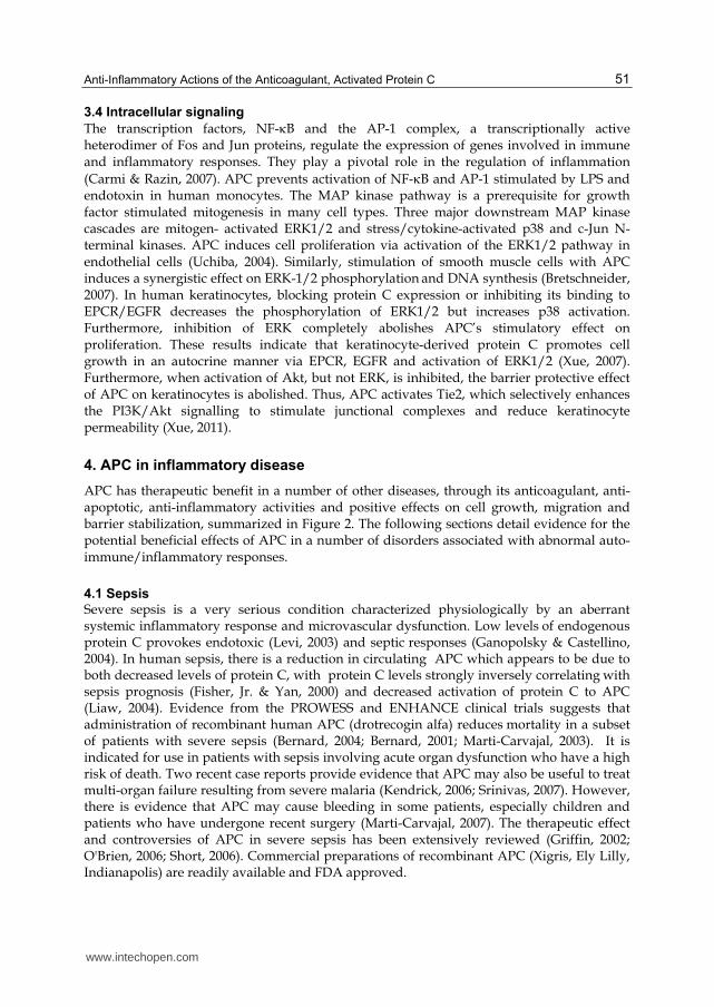
APC has therapeutic benefit in a number of other diseases, through its anticoagulant, anti-apoptotic, anti-inflammatory activities and positive effects on cell growth, migration and barrier stabilization, summarized in Figure 2. The following sections detail evidence for the potential beneficial effects of APC in a number of disorders associated with abnormal auto-immune/inflammatory responses.
4.1 Sepsis
Severe sepsis is a very serious condition characterized physiologically by an aberrant systemic inflammatory response and microvascular dysfunction. Low levels of endogenous protein C provokes endotoxic (Levi, 2003) and septic responses (Ganopolsky & Castellino, 2004). In human sepsis, there is a reduction in circulating APC which appears to be due to both decreased levels of protein C, with protein C levels strongly inversely correlating with sepsis prognosis (Fisher, Jr. & Yan, 2000) and decreased activation of protein C to APC (Liaw, 2004). Evidence from the PROWESS and ENHANCE clinical trials suggests that administration of recombinant human APC (drotrecogin alfa) reduces mortality in a subset of patients with severe sepsis (Bernard, 2004; Bernard, 2001; Marti-Carvajal, 2003). It is indicated for use in patients with sepsis involving acute organ dysfunction who have a high risk of death. Two recent case reports provide evidence that APC may also be useful to treat multi-organ failure resulting from severe malaria (Kendrick, 2006; Srinivas, 2007). However, there is evidence that APC may cause bleeding in some patients, especially children and patients who have undergone recent surgery (Marti-Carvajal, 2007). The therapeutic effect and controversies of APC in severe sepsis has been extensively reviewed (Griffin, 2002; O'Brien, 2006; Short, 2006). Commercial preparations of recombinant APC (Xigris, Ely Lilly, Indianapolis) are readily available and FDA approved.
www.intechopen.com
Inflammatory Diseases – A Modern Perspective 52
Endogenous APC signaling is critical to protection of mice from LPS-induced septic shock
(Xu, 2009). Many mechanisms have been described, but the exact manner in which APC
affects sepsis patients is unclear. Therapeutic APC can regulate neutrophil migration and
extravasation by direct engagement of ┚1 and ┚3 integrins (Elphick, 2009), suppress
macrophage activation dependent on integrin CD11b/CD18 (Kerschen, 2010), control
maturation and activation of CD8+ DCs dependent on EPCR (Kerschen, 2010), and
neutralize late-stage inflammatory mediators by degrading nuclear histones from apoptotic
cells (Xu, 2009).
A recent Cochrane review suggested that APC should not be used for treating patients with
severe sepsis or septic shock and that APC is associated with a higher risk of bleeding
(Marti-Carvajal, 2011). Unless additional RCTs provide evidence of a treatment effect,
policy-makers, clinicians and academics have been advised not promote the use of APC
(Marti-Carvajal, 2011). Nonetheless, new reports continue to show that patients with septic
shock who were treated with APC had a reduced in-hospital mortality compared with those
not treated with APC (Sadaka, 2011).
Fig. 2. The diverse biological effects of APC. APC exerts numerous molecular effects which
lead to cellular responses which cause protective effects in a number of diseases. Ang=
angiopoietin; EGFR= epidermal growth factor receptor; IL=interleukin; MMP=matrix
metalloproteinase; TGF=transforming growth factor; TNF=tumour necrosis factor-┙;
ZO=zona occludens; Bcl=B-cell lymphoma; BAX=Bcl-2–associated X protein.
www.intechopen.com
Anti-Inflammatory Actions of the Anticoagulant, Activated Protein C 53
4.2 Spinal cord injury (SCI)
SCI can be induced by a physical insult resulting in an inflammatory response that leads to tissue destruction and life-long disabilities. Therapeutic intervention in SCI is largely being directed at reducing or alleviating inflammation (Okajima, 2004). In rat models, the
inflammatory response occurs due to infiltration by neutrophils and release of TNF- (Taoka, 2000). Taoka et al (Taoka, 1997) demonstrated that a P-selectin mediated interaction between activated neutrophils and endothelial cells may be a critical step in endothelial cell damage leading to spinal cord injury in rats. These authors (Taoka, 1998) originally showed that rats subjected to compression-trauma induced SCI and treated with APC have a marked reduction in motor disturbances (Taoka, 1998), however, 12 years later this paper was retracted by the authors (2011). APC significantly reduces motor disturbances and micro-infarctions of the spinal cord in rats subjected to ischemia/reperfusion-induced SCI
(Hirose, 2000). The increased tissue levels of TNF- and neutrophils in the injured part of the spinal cord were significantly reduced in animals that received APC. APC has direct neuroprotective effects, independent of its anticoagulant activity (Zlokovic, 2005). In a rabbit model of ischemic spinal cord injury, APC eases the functional deficits and increases the number of motor neurons (Yamauchi, 2006). Interestingly, APC induces insulin-like growth factor (IGF)-1, IGF-1 receptor and the downstream p-Akt which might partially explain the neuroprotective effects of APC after transient spinal cord ischemia in rabbit.
4.3 Brain injury and stroke
Elevated plasma protein C levels are linked to a lower incidence of ischemic stroke in humans (Folsom, 1999) and conversely, lower circulating APC levels are found in patients with post-infection ischemic stroke compared to the control subjects (Macko, 1996). These data imply that the protein C pathway may protect against stroke. In a murine model of focal cerebral ischemia, APC significantly improved cerebral blood flow in the ischemic hemisphere and markedly reduced the volume of brain injury caused by middle cerebral vein occlusion. These effects were dependent on EPCR and PAR-1 and seemingly independent of APC’s anticoagulant effects (Fernandez, 2003; Guo, 2004; Shibata, 2001). APC directly prevents apoptosis in hypoxic human brain endothelium through inhibition of p53, normalization of the pro-apoptotic Bax/Bcl-2 ratio, and reduction of caspase-3 signalling (Guo, 2004). APC's cytoprotection of endothelial cells in vitro requires EPCR and PAR-1(Guo, 2004; Mosnier & Griffin, 2003). APC can also directly protect perturbed neurons from cell injury and apoptosis. In vitro, APC reduces apoptosis in mouse cortical neurons treated with N-methyl-D-aspartate (NMDA) and staurosporine (Guo, 2004). Interestingly, PAR-1 and PAR-3 are required for this effect. Intra-cerebral APC infusion dose-dependently reduces NMDA excitotoxic injury in mice (Guo, 2004). Overall, APC maintains the patency of ischemic vasculature by inhibiting hypoxia-induced endothelial cell apoptosis and also directly protects the integrity and functionality of the neuronal network. Periventricular leukomalacia is the dominant form of brain injury in premature infants, characterized by white matter injury. The underlying pathogenic mechanisms include hypoperfusion, procoagulant activity, apoptotic cell death, microglial activation and inflammation associated with maternal and/or fetal infection (Genc, 2006). They hypothesized that APC, which modulates many of these processes, is a promising therapeutic agent for periventricular leukomalacia.
www.intechopen.com
Inflammatory Diseases – A Modern Perspective 54
4.4 Alzheimer’s disease
Alzheimer’s disease is characterized by elevated levels of amyloid ┚ peptide (A┚) in the brain that are associated with neuronal and vascular toxicity and is the major cause of dementia with advancing age. Inflammation, A┚ deposition in the brain parenchyma and vessels, blood–brain barrier dysfunction, oxidative stress, formation of advanced end glycation products and endothelial and neural cell death have all been implicated in the pathogenesis of Alzheimer’s disease (Zlokovic, 2005). It has been proposed that APC could be useful in treating Alzheimer’s disease, since it has anti-inflammatory, anti-oxidant, profibrinolytic, neurovascular protectant and pro-angiogenic properties (Genc, 2007). Furthermore, APC exerts anti-inflammatory and anti-apoptotic activities directly on endothelial and neural cells (Griffin, 2006). APC protects against neurovascular injury in experimental stroke models, protects endothelial barrier integrity and decreases fibrin deposition (Griffin, 2006). Similar to its anti-inflammatory effect on other cells, APC may inhibit the activation status of the microglia that plays a key role in neuroinflammation.
4.5 Acute kidney injury
In a rat model of endotoxemia, rat APC significantly improved peritubular capillary flow and reduced leukocyte adhesion and rolling, 3 hours after treatment with LPS (Gupta, 2007). After 24 hr, APC treatment significantly improved renal blood flow. In addition, APC modulated the renin-angiotensin system by reducing mRNA expression levels of angiotensin converting enzyme-1, angiotensinogen in the kidney (Gupta, 2007). Thus, APC can suppress LPS-induced acute renal failure by modulating factors involved in vascular inflammation. Ischemia/reperfusion-induced renal injury is an important pathologic mechanism leading
to acute renal failure (Thadhani, 1996). In a rat model, intravenous administration of APC
markedly reduces ischemia/reperfusion -induced renal dysfunction and tubular necrosis,
whereas heparin or inactive APC has no effect (Mizutani, 2000). Furthermore, APC
significantly inhibited the ischemia/reperfusion -induced decrease in renal tissue blood
flow, the increase in the vascular permeability, and renal levels of TNF-, IL-8, and
myeloperoxidase. Leukocytopenia produced effects similar to those of APC. These findings
strongly suggested that APC has a protective effect against ischemia/reperfusion-induced
renal injury by inhibiting activation of leukocytes rather than inhibiting coagulation
(Okajima, 2004). Recently, Isemann et al (Isermann, 2007) have shown that APC protects
against diabetic nephropathy by inhibiting endothelial and podocyte apoptosis. In a rat
model of sepsis by cecal ligation and puncture (CLP), treatment with APC significantly
inhibited sepsis-induced elevations in creatinine, LDH levels, and improved renal
architecture. Furthermore, sepsis-induced inhibition of interferon (INF)-┛ and increase in IL-
1┚ and IL-10 were attenuated by APC treatment. The authors suggested that APC confers a
survival advantage by reducing systemic inflammation and, in doing so, preserves organ
function (Keller, 2011).
4.6 Lung disorders
In a mouse model of acute lung injury, APC inhalation attenuates LPS-induced amplification of neutrophils and macrophages in bronchoalveolar lavage fluid as well as VCAM-1 protein levels in lung tissue (Kotanidou, 2006). In a murine model of asthma, inhalation of APC significantly inhibits the expression of T helper 2 (Th2) cytokines, IgE,
www.intechopen.com
Anti-Inflammatory Actions of the Anticoagulant, Activated Protein C 55
eosinophilic inflammation, and hyper-responsiveness in bronchiolar lavage fluid (Yuda, 2004). Electromobility shift assays show that translation of signal transducer and activator of
transcription 6 (STAT6) and NF-B to the nucleus is reduced in lung samples from mice treated with inhaled APC. Although APC is thought to have a short half-life of ~ 20 mins, it can be detected in mice bronchoalveolar lavage fluid 24 h after inhalation. Thus, APC inhalation might offer a long-acting alternative route of administration to the lungs to attenuate pulmonary inflammation in acute lung injury. It is unclear whether APC has any beneficial effect in humans with inflammatory lung diseases. Nick et al (Nick, 2004) performed a double-blinded, placebo-controlled study of APC in a human model of endotoxin-induced pulmonary inflammation and showed that APC significantly reduced leukocyte accumulation to the airspaces, independent of pulmonary cytokine or chemokine release. Bronchoalveolar lavage fluid neutrophils of patients receiving APC demonstrated decreased chemotaxis ex vivo but no change in cytokine release, cell survival, or apoptosis. The major components of the protein C pathway, including thrombomodulin, protein C and EPCR are all expressed by human airway epithelial cells (Hataji, 2002). Activation of protein C is reduced in sputum of patients with bronchial asthma compared to control subjects, which may contribute to exacerbation of the inflammatory response in the airway of asthmatic patients (Hataji, 2002). However, Schouten et al (Schouten, 2011) showed that endogenous protein C has strong effects on the host response to lethal influenza A infection, on the one hand inhibiting pulmonary coagulopathy and inflammation, but on the other hand facilitating neutrophil influx and protein leak and accelerating mortality.
4.7 Acute pancreatitis
Acute pancreatitis is a local inflammatory process that leads to a systemic inflammatory
response and multiple organ failure (Kirschenbaum & Astiz, 2005). Disseminated
intravascular coagulation and thromboembolism are related to overall morbidity of this
disease which provides a setting in which APC could play a therapeutic role. Ottesen et al
(Ottesen, 1999) found a decrease in levels of protein C in animals with acute pancreatitis. In
a rodent model of pancreatitis (Alsfasser, 2006), treatment with APC reduces inflammation
in the pancreas and lungs and significantly improves survival compared to controls (86% vs
38%; P=.05). This animal model exhibits severe consumptive coagulopathy, however APC’s
anti-coagulation properties did not worsen this condition. In another study, when APC was
given 6 hours after the induction of pancreatitis, it significantly reduced acinar necrosis,
tissue edema, fat necrosis, IL-6 and TNF-┙ and inflammatory infiltration compared to
controls. Inhibition of expression of pancreatic p38 MAPK and JNK and upregulation of
ERK1/2 expression by APC treatment protects against pancreatic injury, thus ameliorating
severity of the disease (Chen, 2007).
4.8 Type 1 Diabetes (T1D)
T1D is a chronic progressive autoimmune disease that affects genetically prone individuals.
The physiological destruction of -cells is a crucial event for disease onset (Mathis, 2001). Inflammation and auto-immunity play an important role in the destruction of pancreatic
islet -cells in T1D, with apoptosis being the dominant form of -cell death in both animal
models of diabetes and humans. Replacement of the -cells by transplantation of islet cells is a radical therapy for human T1D. A major problem with this therapy is the large loss of
www.intechopen.com
Inflammatory Diseases – A Modern Perspective 56
viability of islet cells during the procedure. APC’s strong anti-inflammatory and anti-apoptotic properties appear to be beneficial in preventing destruction of islet cells. Exogenous administration of APC significantly reduces loss of functional islet mass after intraportal transplantation in diabetic mice (Contreras, 2004). Animals given APC exhibit better glucose control, higher glucose disposal rates and higher arginine-stimulated acute insulin release (Contreras, 2004). These effects are associated with a reduction in plasma proinsulin, intrahepatic fibrin deposition, and islet apoptosis early after the transplant. APC treatment is also associated with a significant reduction of proinflammatory cytokine release and prevents endothelial cell activation and dysfunction. This study suggests that APC therapy will improve the take-rate of the transplanted islet cells and thus decrease the number of the cells required for human pancreatic islet transplantation (Contreras, 2004). Interestingly, plasma levels of protein C/APC are reduced in humans with T1D (Gruden, 1997; Vukovich & Schernthaner, 1986). In addition, soluble EPCR, which binds to APC and inhibits its activity, is increased in T1D (Wu, 2000). These data together indicate that circulating APC activity is markedly reduced in T1D. Whether replenishment of APC levels will benefit patients with T1D is yet to be resolved. However, APC exhibits great potential in preventing glucose-induced apoptosis in endothelial cells and podocytes (Isermann, 2007).
4.9 Rheumatoid Arthritis (RA)
RA is a chronic autoimmune disease characterized by persistent inflammation of multiple
synovial joints which results in progressive tissue destruction of bone and cartilage. Protein
C/APC is present in RA synovial tissues and co-localizes with MMP-2 in endothelial and
synovial lining cells (Buisson-Legendre, 2004). EPCR is also strongly expressed by synovial
tissue in patients with RA and co-localizes with CD68 positive staining cells, indicating that
these cells are largely macrophage/monocytes (Xue, 2007). Inhibiting the activation of
monocytes/macrophages reduces the severity of arthritis in patients with RA and in animal
models of RA (Bondeson, 1999; Kwasny-Krochin, 2002). APC inhibits activation of normal
monocytes by preventing their migration and production of proinflammatory
cytokines/chemokines, such as TNF- and macrophage migration inhibitory factor levels
(Schmidt-Supprian, 2000). When monocytes from RA patients are pre-treated with APC,
their migration towards monocyte chemoattractant protein-1 (MCP-1) is inhibited in a dose-
dependent manner (Xue, 2007). Pre-incubation of these cells with RCR252, an antibody
which blocks APC binding to EPCR, abolishes this inhibitory effect of APC, indicating that
APC acts through EPCR to inhibit the chemotactic response of RA monocytes. MMP-9
regulation may be at least one downstream effector of APC’s inhibition of monocyte
migration. When added to purified monocytes from RA patients, APC dose-dependently
inhibits the production of MMP-9 (Xue, 2007). MMP-9 not only allows cell migration but
also exerts direct pro-inflammatory effects, such as activation of cytokines which would
enhance the beneficial effect of APC in RA.
NF-B is activated in synovium from RA patients (Marok, 1996) and in cultured RA
synovial fibroblasts (Fujisawa, 1996). Inhibition of NF-B activity strongly reduces the severity of disease in animal models of arthritis by inhibiting leukocyte infiltration
(Blackwell, 2004). High levels of NF-B activity are present in RA monocytes under basal
conditions. APC (20 g/ml) dramatically inhibits the active form of NF-B in both control
and LPS stimulated monocytes from RA patients. Downstream of NF-B is one of the most
www.intechopen.com
Anti-Inflammatory Actions of the Anticoagulant, Activated Protein C 57
potent inflammatory cytokines in RA, TNF- (Xue, 2007). Specific inhibition of TNF- using biological agents such as the monoclonal antibody, Adalimumab (marketed as Humira), is proving very successful in treating moderate to severe RA in adults who have had a poor response to other anti-rheumatic drugs. Monocytes/macrophages are the
primary source of TNF- which, after release, promotes the proinflammatory activity of
these and other surrounding cells. APC significantly decreases TNF- both in control and LPS-stimulated RA monocytes (Xue, 2007). Thus, APC may mimic the action of the
“biological” agents, through inhibition of TNF-. However, by acting on multiple targets, APC may be more effective.
4.10 Cancer
APC can activate signaling molecules to promote MDA-MB-231 breast cancer cell and endothelial cell motility (Gramling, 2010). However, accumulating evidence suggests that the APC pathway limits cancer progression. Acquired protein C deficiency is observed in cancer patients, especially in patients using certain types of chemotherapy (Feffer, 1989; Mewhort-Buist, 2008; Rogers, 1988; Woodley-Cook, 2006). The loss of expression of thrombomodulin (or increase in its soluble form), a receptor required to convert protein C to APC, on cancer cells correlates with advanced stage and poor prognosis (Hanly, 2006; Hanly & Winter, 2007; Lindahl, 1993). Low levels of thrombomodulin also increase the invasive ability of cancer cells in vitro (Matsushita, 1998) and is significantly correlated with a high relapse rate in breast cancer (Kim, 1997). It is likely that low thrombomodulin levels induce metastasis due to reduced endogenous APC levels, although several alternative mechanisms have also been proposed (Hanly & Winter, 2007). Additionally, EPCR, the specific receptor for APC is detected in several cell lines derived from various types of cancer (Van Sluis, 2010). For example, EPCR is expressed on human breast cancer cells, with an extremely high frequency (Tsuneyoshi, 2001). EPCR on the vascular wall inhibits cancer cell adhesion and transmigration (Bezuhly, 2009; Lindahl, 1993). Finally, significant resistance to APC was found in women with a lymph-node-positive breast carcinoma (Bezuhly, 2009; Lindahl, 1993; Nijziel, 2003). In a mouse model, endogenous APC has been found to limit cancer cell extravasation via sphingosine-1-phosphate receptor-1 and VE-cadherin-dependent vascular barrier enhancement. Van Sluis et al (Van Sluis, 2011) showed that in the absence of endogenous APC, fibrinogen depletion does not prevent cancer cell dissemination and secondary tumor formation in immune-competent mice. Overall, they show that endogenous APC is essential for immune-mediated cancer cell elimination (Van Sluis, 2011). The exact mechanisms on how about prevents cancer are not clear. However, recently, a number of studies have shown that activation of PAR1 are involved in limiting of tumour cell migration and invasion/metastasis in vitro and in vivo (Kamath, 2001; Nierodzik, 1998; Villares, 2011) and intact barrier function can effectively prevent tumour cell migration and metastasis. APC can enhance both endothelial and epithelial barrier functions via activation of PAR1 which may partly explain the underlying mechanisms(Minhas, 2010; Xue, 2011).
4.11 Skin injuries
Chronic wounds are a common health problem. There are many different factors which lead to a chronic wound, including advancing age, persistent inflammatory stimuli, tissue hypoxia, diabetes mellitus, immunodeficiency, a smoking history or the use of certain medications, but they can affect patients at any race or economic background. The common
www.intechopen.com

Inflammatory Diseases – A Modern Perspective 58
types of chronic wounds include: peripheral ulcers, which are the most frequent cause of lower limb amputation in patients with type I and type II diabetes (Levin, 1993); decubitus ulcers, a result of prolonged, unrelieved pressure over a bony prominence and venous stasis ulcers, where venous congestion of the lower extremities results in local hypoxia (Trent, 2005). With the current diabetes epidemic and increasing aging population, chronic wounds are a serious concern to the health system. They require dedicated care including regular dressings, frequent clinic appointments and when complications arise, hospital admission potentially requiring surgery or even amputation. The “state-of-the-art” treatments for chronic wounds are expensive and have limited success. Cutaneous wound repair can be divided into a series of overlapping phases including formation of fibrin clot, inflammatory response, granulation tissue formation, which includes re-epithelialization and angiogenesis, and matrix remodeling. Re-epithelialization is an important component of wound repair as it serves to restore the barrier function of skin. Newly formed blood vessels provide nutrition and oxygen to the growing tissue and allow leukocytes to enter the site of injury. A chronic wound or ulcer occurs when the co-coordinated cellular and biochemical response to injury are disrupted.
Fig. 3. APC treatment used in conjunction with topical negative pressure (TNP) to treat a
recalcitrant orthopaedic wound present for 2 years. APC and TNP were applied on day 0
and twice a week until day 19. Day 0 shows exposed bone (arrow) and day 7 shows healthy
granulation tissue covering bone. Wound is fully healed by day 28 and remained healed at 8
months follow-up. Taken from Wijewardana et al, Int J Lower Extrem Wounds in press,
2011.
The role of inflammation in wound healing is under debate. Many researchers believe that
the inflammatory phase is vital for wound healing to proceed, however, there is evidence to
suggest otherwise. Whereas normal adult healing results in a fibrous scar, early fetal
wounds which have very little, if any, inflammatory response, exhibit scarless healing with
complete restoration of the normal skin architecture. Scar formation exacerbates when
inflammation is provoked in fetal wounds, suggesting that the absence of inflammation
www.intechopen.com

Anti-Inflammatory Actions of the Anticoagulant, Activated Protein C 59
contributes to the rapid and flawless repair of these wounds (Szpaderska & DiPietro, 2005).
Compared to dermal wounds, oral wounds have substantially lower levels of macrophage,
neutrophil, and T-cell infiltration and heal rapidly with minimal inflammation and often
with minimal scar formation (Szpaderska, 2003). Redd et al (Redd, 2004) studied wound
healing in the PU.1 null mouse, which is genetically incapable of raising an inflammatory
response because several haematopoietic lineages, including macrophages and
neutrophils, are absent or severely delayed in their differentiation. Wounds in these
animals rapidly repair with increased vascularity at the wound site and faster
reepithelialisation of the wound surface, as well as being scarless (Redd, 2004). They
hypothesised that the lower the level of some growth factors in adult wounds the closer it
would mimic scar-free healing in the embryo. This is relevant to many chronic wounds
which are often associated with excess inflammation and become locked in this
inflammatory phase. Thus, APC’s anti-inflammatory effects may actually benefit, rather
than hinder, the healing of chronic wounds.
Keratinocytes and endothelial cells are two major cell types in skin and play critical roles in
the healing of skin injury. Keratinocytes of the epidermis provide the major cellular
component of the outermost barrier to the environment. When the skin is broken, a critical
response is triggered to restore its protective function. Within 24 hours of wounding,
keratinocytes from the wound margins begin to migrate and invade the wound bed, where
they proliferate to form the new epithelium. We have shown that protein C is produced by
skin keratinocytes, especially those in the basal layer (Xue, 2007). This endogenous protein C
is activated on the cell surface, with the resulting APC stimulating a wound healing
phenotype in keratinocytes (Xue, 2005; Xue, 2007). Furthermore, the autocrine actions of
APC are necessary for normal keratinocyte growth and function (Xue, 2007). APC
stimulates proliferation, MMP-2 activity, migration and prevents apoptosis in skin
keratinocytes, all vital processes of re-epithelialization (Xue, 2005; Xue, 2004). Endothelial
migration and proliferation are vital to generate new blood vessels for wound healing. APC
stimulates endothelial cell proliferation and induces tube-like structure formation in vitro
(Uchiba, 2004) and cell migration (Brueckmann, 2003). In the chick chorioallantoic
membrane (CAM) assay, APC stimulates angiogenesis and re-epithelialization. In vivo, APC
induces corneal angiogenesis in a mouse model (Uchiba, 2004). APC also stimulates the
proliferation of smooth muscle cells (Bretschneider, 2007), which would contribute to the
formation of mature blood vessels. In a full-thickness rat skin-healing model, a single
topical application of APC enhances wound healing compared to saline control at least
partly via stimulating angiogenesis and re-epithelialisation (Jackson, 2005).
Recent evidence suggests that APC will be effective in humans with chronic wounds. An
open label pilot study was conducted on 4 patients whose wounds were not improving,
despite standard wound treatment for 4 months or greater (Whitmont, 2008). APC was
applied topically to wounds once weekly for 4 weeks. All 4 patients showed rapid positive
response to treatment which was maintained during a 4 month follow-up period. Overall,
there was more than 80% reduction in wound size. The treatment was well tolerated with no
significant side effects or complications experienced. In another recent study, APC treatment
was used in conjunction with topical negative pressure (TNP), to treat recalcitrant long-
standing orthopaedic wounds (Wijewardana et al, Combination of activated protein C and
topical negative pressure vacuum therapy rapidly regenerates granulation tissue over
www.intechopen.com
Inflammatory Diseases – A Modern Perspective 60
exposed bone to heal recalcitrant orthopaedic wounds, in press, 2011, Int J Lower Extrem
Wounds). One example is a 47 year old male with no significant co-morbidities who had
tibial and fibula fractures following an assault and being hit in the leg by a baseball bat, 2
years prior to presentation. The fractures required open reduction and fixation with plates
and screws. Two months post-operatively he developed an infection and breakdown of the
wound. He was started on antibiotic treatment and had wash out of the wound and
scraping of the bone which showed osteomyelitis. Antibiotic treatment was continued but
failed to control the infection so all metal-ware was removed and the wound was debrided.
There was malunion of the bone and after 3 months the plates and screws were replaced
and the wound was covered with a fasciocutaneous flap. After one month, he again
developed infection and breakdown of the flap. For the following year he had continued
treatment with antibiotics, conventional and TNP dressings, however the wound remained
non-healing. He was then treated with APC plus topical negative pressure and after 7 days a
layer of healthy granulation tissue covered the bone and almost filled the entire wound
space (Figure 3). By day 10 re-epithelialization had occurred on top of the dermis around the
perimeter of the wound and by day 19 the wound had completely healed. Follow up at 8
month showed the wound had continued to remain intact
By dampening inflammation and accelerating angiogenesis and re-epithellialisation, APC
is also likely to minimize scar formation, which holds great potential for burn victims and
those susceptible to keloid scarring. Patients with burn injuries and inhalation trauma
have a significant increase in thrombin generation in the airways compared with control
patients, as reflected by increased lavage fluid levels of thrombin-antithrombin complexes
and fibrin degradation products, and decreased lavage fluid levels of APC and
antithrombin (Hofstra, 2011).
5. Engineered protein C/APC
The stereospecific interactions of APC with factors Va and VIIIa involve both the APC
enzymatic active-site region and residues that are not part of the immediate APC active site.
These residues are termed ‘exosites’ on the APC active enzyme surface and can be mutated
to diminish the anticoagulant activity of APC without altering the cell-signaling activity of
the molecule (Bae, 2007; Gale, 2002; Kerschen, 2007; Mosnier, 2004). Replacement of a cluster
of five positively charged residues by alanine residues (ie. 5A-APC) on the top surface of the
APC heavy-chain protease domain restructures this crucial positively charged exosite,
causing >98% loss of the anticoagulant activity of human APC while leaving intact APC cell-
signaling activities on different cell types within the neurovascular unit (Mosnier, 2007;
Mosnier, 2009). Replacement of three of these five residues (i.e. lysine residues 191–193) by
three alanine residues produces the 3K3A-APC variant (Deane, 2009; Guo, 2009; Wang,
2009) which has a similar effect to the 5A-APC variant, causing loss of >92% of APC
anticoagulant activity. These engineered variants provide APC for therapeutic purposes in
which the risk of serious bleeding caused by the anticoagulant activity of APC is diminished
while the cytoprotective effects of APC are preserved. In preclinical animal models of ALS
(Zhong, 2009), stroke (Guo, 2009; Wang, 2009), brain injury (Walker, 2010) and sepsis
{Kerschen, 2007 KERSCHEN2007 /id} and cytoprotective function (Ni, 2011) these APC
variants show beneficial effects that were equivalent to, or sometimes greater than, the wild-
type recombinant APC. Bir et al (Bir, 2011) demonstrated that 5A-APC could attenuate lung
www.intechopen.com
Anti-Inflammatory Actions of the Anticoagulant, Activated Protein C 61
damage caused by P. aeruginosa in critically ill patients. In addition, 5A-APC inhibits the
inflammatory response of conventional dendritic cells independent of EPCR and suppresses
IFN-gamma production by natural killer-like dendritic cells (Kerschen, 2010). Recently, an
APC variant (APC-L38D/N329Q) was generated with minimal anticoagulant activity, but 5-
fold enhanced endothelial barrier protective function and 30-fold improved anti-apoptotic
function when compared with wild type APC (Ni, 2011).
An APC variant with minimal cell-signaling activity but with substantially increased anticoagulant activity, E149A-APC, has been useful for proof of concept studies and for antithrombotic indications (Mosnier, 2009). E149A-APC has superior antithrombotic activity in a mouse model of arterial thrombosis compared to wt-APC, but has no benefit in an endotoxemia sepsis model where wild type (wt)-APC or the 5A-APC variant reduced mortality {Kerschen, 2007 KERSCHEN2007 /id} (Mosnier, 2009).
6. Conclusion
APC, an anticoagulant with anti-inflammatory, anti-apoptotic, proliferative and barrier
stabilization properties, has not only emerged as a therapeutic agent for use in selected
patients with severe sepsis, but also appears to have considerable benefit in chronic wounds
and a number of other autoimmune/inflammatory diseases. The in vitro, preclinical and
limited clinical data for these diseases indicate that APC holds a remarkable promise.
Further insights into the mechanisms of action of APC will be required for the translation of
preclinical study results to the bedside.
More than three decades since the discovery of APC, we are only beginning to learn of its
biological diversity. The clinical evidence for APC as a treatment for severe sepsis is
perplexing (Marti-Carvajal, 2007; Marti-Carvajal, 2011). Strong evidence indicates that APC
will benefit other disorders involving various organ injuries including kidney injury, spinal
cord injury, respiratory function and stroke. Blinded and controlled clinical trials will
elucidate its clinical protective effects in these disorders. With the recent discovery that the
largest organ of the body, the skin, positively responds to APC, it is likely that APC will
accelerate healing of skin injuries and disorders.
7. References
Retraction for yuji taoka et Al., (2011). "activated protein C reduces the severity of compression-induced spinal cord injury in rats by inhibiting activation of leukocytes". J.Neurosci. Vol.31, No.23, pp. 8697
Alsfasser, G., Warshaw, A.L., Thayer, S.P., Antoniu, B., Laposata, M., Lewandrowski, K.B., & Fernandez-del, C.C. (2006). Decreased inflammation and improved survival with recombinant human activated protein C treatment in experimental acute pancreatitis. Arch.Surg. Vol.141, No.7, pp. 670-676
Bae, J.S., Yang, L., Manithody, C., & Rezaie, A.R. (2007). Engineering a disulfide bond to stabilize the calcium binding loop of activated protein C eliminates its anticoagulant but not protective signaling properties. J.Biol.Chem. Vol.282, No.35, pp.25493-25500
Bae, J.S., Yang, L., Manithody, C., & Rezaie, A.R. (2007). The ligand occupancy of endothelial protein C receptor switches the protease-activated receptor 1-dependent signaling
www.intechopen.com
Inflammatory Diseases – A Modern Perspective 62
specificity of thrombin from a permeability-enhancing to a barrier-protective response in endothelial cells. Blood. Vol.110, No.12, pp. 3909-3916
Bae, J.S., Yang, L., & Rezaie, A.R. (2007). Receptors of the protein C activation and activated protein C signaling pathways are colocalized in lipid rafts of endothelial cells. Proc.Natl.Acad.Sci.U.S.A. Vol.104, No.8, pp. 2867-2872
Baker, W.F. & Bick, R.L. (1999). Treatment of hereditary and acquired thrombophilic disorders. Semin.Thromb.Hemostasis. Vol.25, No.4, pp. 387-405
Balazs, A.B., Fabian, A.J., Esmon, C.T., & Mulligan, R.C. (2006). Endothelial protein C receptor (CD201) explicitly identifies hematopoietic stem cells in murine bone marrow. Blood. Vol.107, No.6, pp. 2317-2321
Bernard, G.R., Margolis, B.D., Shanies, H.M., Ely, E.W., Wheeler, A.P., Levy, H., Wong, K., & Wright, T.J. (2004). Extended Evaluation of Recombinant Human Activated Protein C United States Trial (ENHANCE US): A Single-Arm, Phase 3B, Multicenter Study of Drotrecogin Alfa (Activated) in Severe Sepsis. Chest. Vol.125, No.6, pp. 2206-2216
Bernard, G.R., Vincent, J.L., Laterre, P.F., LaRosa, S.P., Dhainaut, J.F., Lopez-Rodriguez, A., Steingrub, J.S., Garber, G.E., Helterbrand, J.D., Ely, E.W., & Fisher, C.J. (2001). Efficacy and Safety of Recombinant Human Activated Protein C for Severe Sepsis. New Engl. J Med. Vol.344, No.10, pp. 699-709
Bezuhly, M., Cullen, R., Esmon, C.T., Morris, S.F., West, K.A., Johnston, B., & Liwski, R.S. (2009). Role of activated protein C and its receptor in inhibition of tumor metastasis. Blood. Vol.113, No.14, pp. 3371-3374
Bir, N., Lafargue, M., Howard, M., Goolaerts, A., Roux, J., Carles, M., Cohen, M.J., Iles, K.E., Fernandez, J.A., Griffin, J.H., & Pittet, J.F. (2011). Cytoprotective-selective Activated Protein C Attenuates P. aeruginosa-induced Lung Injury in Mice. Am.J.Respir.Cell Mol.Biol. Epub ahead of print
Blackwell, N.M., Sembi, P., Newson, J.S., Lawrence, T., Gilroy, D.W., & Kabouridis, P.S. (2004). Reduced infiltration and increased apoptosis of leukocytes at sites of inflammation by systemic administration of a membrane-permeable IkappaBalpha repressor. Arthritis Rheum. Vol.50, No.8, pp. 2675-2684
Bondeson, J., Browne, K.A., Brennan, F.M., Foxwell, B.M.J., & Feldmann, M. (1999). Selective Regulation of Cytokine Induction by Adenoviral Gene Transfer of I{kappa}B{alpha} into Human Macrophages: Lipopolysaccharide-Induced, But Not Zymosan-Induced, Proinflammatory Cytokines Are Inhibited, But IL-10 Is Nuclear Factor-{kappa}B Independent. J. Immunol. Vol.162, No.5, pp. 2939-2945
Bretschneider, E., Uzonyi, B., Weber, A.A., Fischer, J.W., Pape, R., Lotzer, K., & Schror, K. (2007). Human vascular smooth muscle cells express functionally active endothelial cell protein C receptor. Circ.Res. Vol.100, No.2, pp. 255-262
Brueckmann, M., Marx, A., Martin, W.H., Liebe, V., Lang, S., Kaden, J.J., Zieger, W., Borggrefe, M., Huhle, G., & Konstantin, H.K. (2003). Stabilization of monocyte chemoattractant protein-1-mRNA by activated protein C. Thromb.Haemost. Vol.89, No.1, pp. 149-160
Buisson-Legendre, N., Smith, S., March, L., & Jackson, C. (2004). Elevation of activated protein C in synovial joints in rheumatoid arthritis and its correlation with matrix metalloproteinase 2. Arthritis Rheum. Vol.50, No.7, pp. 2151-2156
Calzado, M.A., Bacher, S., & Schmitz, M.L. (2007). NF-kappaB inhibitors for the treatment of inflammatory diseases and cancer. Curr.Med.Chem. Vol.14, No.3, pp.367-376
www.intechopen.com
Anti-Inflammatory Actions of the Anticoagulant, Activated Protein C 63
Calzado, M.A., Bacher, S., & Schmitz, M.L. (2007). NF-kappaB inhibitors for the treatment of inflammatory diseases and cancer. Curr.Med.Chem. Vol.14, No.3, pp. 367-376
Cao, C., Gao, Y., Li, Y., Antalis, T.M., Castellino, F.J., & Zhang, L. (2010). The efficacy of activated protein C in murine endotoxemia is dependent on integrin CD11b. J. Clin Invest. Vol.120, No.6, pp. 1971-1980
Carmi, I. & Razin, E. (2007). The role played by key transcription factors in activated mast cells. Immunol.Rev. Vol.217:280-91., No.280-291
Chen, P., Zhang, Y., Qiao, M., & Yuan, Y. (2007). Activated protein C, an anticoagulant polypeptide, ameliorates severe acute pancreatitis via regulation of mitogen-activated protein kinases. J.Gastroenterol. Vol.42, No.11, pp. 887-896
Cheng, T., Liu, D., Griffin, J.H., Fernandez, J.A., Castellino, F., Rosen, E.D., Fukudome, K., & Zlokovic, B.V. (2003). Activated protein C blocks p53-mediated apoptosis in ischemic human brain endothelium and is neuroprotective. Nat.Med. Vol.9, No.3, pp. 338-342
Cheng, T., Petraglia, A.L., Li, Z., Thiyagarajan, M., Zhong, Z., Wu, Z., Liu, D., Maggirwar, S.B., Deane, R., Fernandez, J.A., LaRue, B., Griffin, J.H., Chopp, M., & Zlokovic, B.V. (2006). Activated protein C inhibits tissue plasminogen activator-induced brain hemorrhage. Nat. Med. Vol.12, No.11, pp. 1278-1285
Contreras, J.L., Eckstein, C., Smyth, C.A., Bilbao, G., Vilatoba, M., Ringland, S.E., Young, C., Thompson, J.A., Fernandez, J.A., Griffin, J.H., & Eckhoff, D.E. (2004). Activated protein C preserves functional islet mass after intraportal transplantation: a novel link between endothelial cell activation, thrombosis, inflammation, and islet cell death. Diabetes. Vol.53, No.11, pp. 2804-2814
Coughlin, S.R. (2000). Thrombin signalling and protease-activated receptors [In Process Citation]. Nature. Vol.407, No.6801, pp. 258-264
Deane, R., LaRue, B., Sagare, A.P., Castellino, F.J., Zhong, Z., & Zlokovic, B.V. (2009). Endothelial protein C receptor-assisted transport of activated protein C across the mouse blood-brain barrier. J.Cereb.Blood Flow Metab. Vol.29, No.1, pp. 25-33
Elphick, G.F., Sarangi, P.P., Hyun, Y.M., Hollenbaugh, J.A., Ayala, A., Biffl, W.L., Chung, H.L., Rezaie, A.R., McGrath, J.L., Topham, D.J., Reichner, J.S., & Kim, M. (2009). Recombinant human activated protein C inhibits integrin-mediated neutrophil migration. Blood. Vol.113, No.17, pp. 4078-4085
Esmon, C.T. (2004). Crosstalk between inflammation and thrombosis. Maturitas. Vol.47, No.4, pp. 305-314
Esmon, C.T. (2004). Structure and functions of the endothelial cell protein C receptor. Crit Care Med. Vol.32, No.5 Suppl, pp. S298-S301
Feffer, S.E., Carmosino, L.S., & Fox, R.L. (989). Acquired protein C deficiency in patients with breast cancer receiving cyclophosphamide, methotrexate, and 5-fluorouracil. Cancer. Vol.63, No.7, pp. 1303-1307
Feistritzer, C., Mosheimer, B.A., Sturn, D.H., Riewald, M., Patsch, J.R., & Wiedermann, C.J. (2006). Endothelial protein C receptor-dependent inhibition of migration of human lymphocytes by protein C involves epidermal growth factor receptor. J.Immunol. Vol.176, No.2, pp. 1019-1025
Feistritzer, C. & Riewald, M. (2005). Endothelial barrier protection by activated protein C through PAR1-dependent sphingosine 1-phosphate receptor-1 crossactivation. Blood. Vol.105, No.8, pp. 3178-3184
www.intechopen.com
Inflammatory Diseases – A Modern Perspective 64
Feistritzer, C., Schuepbach, R.A., Mosnier, L.O., Bush, L.A., Di, C.E., Griffin, J.H., & Riewald, M. (2006). Protective signaling by activated protein C is mechanistically linked to protein C activation on endothelial cells. J. Biol. Chem. Vol.281, No.29, pp. 20077-20084
Fernandez, J.A., Xu, X., Liu, D., Zlokovic, B.V., & Griffin, J.H. (2003). Recombinant murine-activated protein C is neuroprotective in a murine ischemic stroke model. Blood Cells Mol.Dis. Vol.30, No.3, pp. 271-276
Finigan, J.H., Dudek, S.M., Singleton, P.A., Chiang, E.T., Jacobson, J.R., Camp, S.M., Ye, S.Q., & Garcia, J.G. (2005). Activated protein C mediates novel lung endothelial barrier enhancement: Role of sphingosine 1-phosphate receptor transactivation. J.Biol.Chem. Vol.280, No.17, pp.17286-17293
Fisher, C.J., Jr. & Yan, S.B. (2000). Protein C levels as a prognostic indicator of outcome in sepsis and related diseases. Crit Care Med. Vol.28, No.9 Suppl, pp. S49-S56
Folsom, A.R., Rosamond, W.D., Shahar, E., Cooper, L.S., Aleksic, N., Nieto, F.J., Rasmussen, M.L., & Wu, K.K. (1999). Prospective study of markers of hemostatic function with risk of ischemic stroke. The Atherosclerosis Risk in Communities (ARIC) Study Investigators. Circulation. Vol.100, No.7, pp. 736-742
Franscini, N., Bachli, E.B., Blau, N., Leikauf, M.S., Schaffner, A., & Schoedon, G. (2004). Gene expression profiling of inflamed human endothelial cells and influence of activated protein C. Circulation. Vol.110, No.18, pp. 2903-2909
Fujisawa, K., Aono, H., Hasunuma, T., Yamamoto, K., Mita, S., & Nishioka, K. (1996). Activation of transcription factor NF-kappa B in human synovial cells in response to tumor necrosis factor alpha. Arthritis Rheum. Vol.39, No.2, pp. 197-203
Fukudome, K. & Esmon, C.T. (1994). Identification, cloning, and regulation of a novel endothelial cell protein c activated protein c receptor. J. Biol. Chem. Vol.269, No.42, pp. 26486-26491
Gale, A.J., Tsavaler, A., & Griffin, J.H. (2002). Molecular characterization of an extended binding site for coagulation factor Va in the positive exosite of activated protein C. J. Biol. Chem. Vol.277, No.32, pp. 28836-28840
Ganopolsky, J.G. & Castellino, F.J. (2004). A Protein C Deficiency Exacerbates Inflammatory and Hypotensive Responses in Mice During Polymicrobial Sepsis in a Cecal Ligation and Puncture Model. Am. J. Pathol. Vol.165, No.4, pp. 1433-1446
Genc, K. (2007). The rationale for activated protein C treatment in perinatal white matter injury. Med.Hypotheses. Vol.68, No.6, pp. 1418-1419
Genc, K. (2007). Activated protein C: therapeutic implications for Alzheimer's disease. Med Hypotheses. Vol.69, No.3, pp. 701-702
Gramling, M.W., Beaulieu, L.M., & Church, F.C. (2010). Activated protein C enhances cell motility of endothelial cells and MDA-MB-231 breast cancer cells by intracellular signal transduction. Exp.Cell Res. Vol.316, No.3, pp. 314-328
Grey, S.T., Tsuchida, A., Hau, H., Orthner, C.L., Salem, H.H., & Hancock, W.W. (1994). Selective inhibitory effects of the anticoagulant activated protein C on the responses of human mononuclear phagocytes to LPS, IFN-gamma, or phorbol ester. J. Immunol. Vol.153, No.8, pp. 3664-3672
Griffin, J.H., Fernandez, J.A., Gale, A.J., & Mosnier, L.O. (2005). Activated protein C. J.Thromb.Haemost.2007.Vol.5,No.suppl 1, pp.73-80.
Griffin, J.H., Fernandez, J.A., Mosnier, L.O., Liu, D., Cheng, T., Guo, H., & Zlokovic, B.V. (2006). The promise of protein C. Blood Cells Mol.Dis. Vol.36, No.2, pp. 211-216
www.intechopen.com
Anti-Inflammatory Actions of the Anticoagulant, Activated Protein C 65
Griffin, J.H., Zlokovic, B., & Fernandez, J.A. (2002). Activated protein C: potential therapy for severe sepsis, thrombosis, and stroke. Semin.Hematol. Vol.39, No.3, pp. 197-205
Gruber, A. & Griffin, J.H. (1992). Direct detection of activated protein C in blood from human subjects. Blood. Vol.79, No.9, pp. 2340-2348
Gruden, G., Olivetti, C., Cavallo-Perin, P., Bazzan, M., Stella, S., Tamponi, G., & Pagano, G. (1997). Activated protein C resistance in type I diabetes. Diabetes Care. Vol.20, No.3, pp. 424-425
Gu, J.M., Crawley, J.T.B., Ferrell, G., Zhang, F., Li, W., Esmon, N.L., & Esmon, C.T. (2002). Disruption of the Endothelial Cell Protein C Receptor Gene in Mice Causes Placental Thrombosis and Early Embryonic Lethality. J. Biol. Chem. Vol.277, No.45, pp. 43335-43343
Guo, H., Gu, F., Li, W., Zhang, B., Niu, R., Fu, L., Zhang, N., & Ma, Y. (2009). Reduction of protein kinase C zeta inhibits migration and invasion of human glioblastoma cells. J.Neurochem. Vol.109, No.1, pp. 203-213
Guo, H., Liu, D., Gelbard, H., Cheng, T., Insalaco, R., Fernandez, J.A., Griffin, J.H., & Zlokovic, B.V. (2004). Activated protein C prevents neuronal apoptosis via protease activated receptors 1 and 3. Neuron. Vol.41, No.4, pp. 563-572
Guo, H., Singh, I., Wang, Y., Deane, R., Barrett, T., Fernandez, J.A., Chow, N., Griffin, J.H., & Zlokovic, B.V. (2009). Neuroprotective activities of activated protein C mutant with reduced anticoagulant activity. Eur J Neurosci. Vol.29, No.6, pp. 1119-1130
Guo, H., Liu, D., Gelbard, H., Cheng, T., Insalaco, R., Fernandez, J.A., Griffin, J.H., & Zlokovic, B.V. (2004). Activated Protein C Prevents Neuronal Apoptosis via Protease Activated Receptors 1 and 3. Neuron. Vol.41, No.4, pp. 563-572
Gupta, A., Rhodes, G.J., Berg, D.T., Gerlitz, B., Molitoris, B.A., & Grinnell, B.W. (2007). Activated protein C ameliorates LPS-induced acute kidney injury and downregulates renal INOS and angiotensin 2. Am.J.Physiol. Renal Physiol. Vol.293, No.1, pp. F245-F254
Hanly, A.M., Redmond, M., Winter, D.C., Brophy, S., Deasy, J.M., Bouchier-Hayes, D.J., & Kay, E.W. (2006). Thrombomodulin expression in colorectal carcinoma is protective and correlates with survival. Br J Cancer. Vol.94, No.9, pp. 1320-1325
Hanly, A.M. & Winter, D.C. (2007). The role of thrombomodulin in malignancy. Semin Thromb Hemost. Vol.33, No.7, pp. 673-679
Hataji, O., Taguchi, O., Gabazza, E.C., Yuda, H., Fujimoto, H., Suzuki, K., & Adachi, Y. (2002). Activation of protein C pathway in the airways. Lung. Vol.180, No.1, pp. 47-59
Hirose, K., Okajima, K., Taoka, Y., Uchiba, M., Tagami, H., Nakano, K., Utoh, J., Okabe, H., & Kitamura, N. (2000). Activated protein C reduces the ischemia/reperfusion-induced spinal cord injury in rats by inhibiting neutrophil activation. Ann Surg. Vol.232, No.2, pp. 272-280
Hofstra, J.J., Vlaar, A.P., Knape, P., Mackie, D.P., Determann, R.M., Choi, G., van Der, P.T., Levi, M., & Schultz, M.J. (2011). Pulmonary Activation of Coagulation and Inhibition of Fibrinolysis After Burn Injuries and Inhalation Trauma. J Trauma. Epub ahead of print
Hudson, L.G. & McCawley, L.J. (1998). Contributions of the epidermal growth factor receptor to keratinocyte motility. Microsc.Res.Tech. Vol.43, No.5, pp. 444-455
Isermann, B., Vinnikov, I.A., Madhusudhan, T., Herzog, S., Kashif, M., Blautzik, J., Corat, M.A.F., Zeier, M., Blessing, E., Oh, J., Gerlitz, B., Berg, D.T., Grinnell, B.W.,
www.intechopen.com
Inflammatory Diseases – A Modern Perspective 66
Chavakis, T., Esmon, C.T., Weiler, H., Bierhaus, A., & Nawroth, P.P. (2007). Activated protein C protects against diabetic nephropathy by inhibiting endothelial and podocyte apoptosis. Nat Med. Vol.13, No.11, pp. 1349-1358
Itoh, T., Matsuda, H., Tanioka, M., Kuwabara, K., Itohara, S., & Suzuki, R. (2002). The role of matrix metalloproteinase-2 and matrix metalloproteinase-9 in antibody-induced arthritis. J.Immunol. Vol.169, No.5, pp. 2643-2647
Jackson, C.J., Xue, M., Thompson, P., Davey, R.A., Whitmont, K., Smith, S., Buisson-Legendre, N., Sztynda, T., Furphy, L.J., Cooper, A., Sambrook, P., & March, L. (2005). Activated protein C prevents inflammation yet stimulates angiogenesis to promote cutaneous wound healing. Wound.Repair Regen. Vol.13, No.3, pp. 284-294
Joyce, D.E., Gelbert, L., Ciaccia, A., DeHoff, B., & Grinnell, B.W. (2001). Gene expression profile of antithrombotic protein c defines new mechanisms modulating inflammation and apoptosis. J.Biol.Chem. Vol.276, No.14, pp. 11199-11203
Kamath, L., Meydani, A., Foss, F., & Kuliopulos, A. (2001). Signaling from protease-activated receptor-1 inhibits migration and invasion of breast cancer cells. Cancer Res. Vol.61, No.15, pp. 5933-5940
Keller, S.A., Moore, C.C., Evans, S.L., McKillop, I.H., & Huynh, T. (2011). Activated protein C alters inflammation and protects renal function in sepsis. J Surg.Res. Vol.168, No.1, pp. e103-e109
Kendrick, B.J., Gray, A.G., Pickworth, A., & Watters, M.P. (2006). Drotrecogin alfa (activated) in severe falciparum malaria. Anaesthesia. Vol.61, No.9, pp. 899-902
Kent, D.G., Copley, M.R., Benz, C., Wohrer, S., Dykstra, B.J., Ma, E., Cheyne, J., Zhao, Y., Bowie, M.B., Zhao, Y., Gasparetto, M., Delaney, A., Smith, C., Marra, M., & Eaves, C.J. (2009). Prospective isolation and molecular characterization of hematopoietic stem cells with durable self-renewal potential. Blood. Vol.113, No.25, pp. 6342-6350
Kerschen, E., Hernandez, I., Zogg, M., Jia, S., Hessner, M.J., Fernandez, J.A., Griffin, J.H., Huettner, C.S., Castellino, F.J., & Weiler, H. (2010). Activated protein C targets CD8+ dendritic cells to reduce the mortality of endotoxemia in mice. J Clin Invest. Vol.120, No.9, pp. 3167-3178
Kerschen, E.J., Fernandez, J.A., Cooley, B.C., Yang, X.V., Sood, R., Mosnier, L.O., Castellino, F.J., Mackman, N., Griffin, J.H., & Weiler, H. (2007). Endotoxemia and sepsis mortality reduction by non-anticoagulant activated protein C. J Exp Med. Vol.204, No.10, pp. 2439-2448
Kim, S.J., Shiba, E., Ishii, H., Inoue, T., Taguchi, T., Tanji, Y., Kimoto, Y., Izukura, M., & Takai, S. (1997). Thrombomodulin is a new biological and prognostic marker for breast cancer: an immunohistochemical study. Anticancer Res. Vol.17, No.3C, pp. 2319-2323
Kirschenbaum, L. & Astiz, M. (2005). Acute pancreatitis: a possible role for activated protein C? Crit Care. Vol.9, No.3, pp. 243-244
Kotanidou, A., Loutrari, H., Papadomichelakis, E., Glynos, C., Magkou, C., Armaganidis, A., Papapetropoulos, A., Roussos, C., & Orfanos, S.E. (2006). Inhaled activated protein C attenuates lung injury induced by aerosolized endotoxin in mice. Vascul.Pharmacol. Vol.45, No.2, pp. 134-140
Kuliopulos, A., Covic, L., Seeley, S.K., Sheridan, P.J., Helin, J., & Costello, C.E. (1999). Plasmin desensitization of the PAR1 thrombin receptor: kinetics, sites of truncation, and implications for thrombolytic therapy. Biochemistry. Vol.38, No.14, pp. 4572-4585
www.intechopen.com
Anti-Inflammatory Actions of the Anticoagulant, Activated Protein C 67
Kwasny-Krochin, B., Bobek, M., Kontny, E., Gluszko, P., Biedron, R., Chain, B.M., Maslinski, W., & Marcinkiewicz, J. (2002). Effect of taurine chloramine, the product of activated neutrophils, on the development of collagen-induced arthritis in DBA 1/J mice. Amino.Acids. Vol.23, No.4, pp. 419-426
Lay, A.J., Donahue, D., Tsai, M.J., & Castellino, F.J. (2007). Acute inflammation is exacerbated in mice genetically predisposed to a severe protein C deficiency. Blood. Vol.109, No.5, pp. 1984-1991
Levi, M., Dorffler-Melly, J., Reitsma, P., Buller, H., Florquin, S., van der Poll, T., & Carmeliet, P. (2003). Aggravation of endotoxin-induced disseminated intravascular coagulation and cytokine activation in heterozygous protein-C-deficient mice. Blood. Vol.101, No.12, pp. 4823-4827
Levin, M.E. (1993). Diabetic foot ulcers: pathogenesis and management. J.ET Nurs. Vol.20, No.5, pp. 191-198
Li, Q. & Verma, I.M. (2002). NF-kappaB regulation in the immune system. Nat.Rev.Immunol. Vol.2, No.10, pp. 725-734
Li, W., Zheng, X., Gu, J., Hunter, J., Ferrell, G.L., Lupu, F., Esmon, N.L., & Esmon, C.T. (2005). Overexpressing endothelial cell protein C receptor alters the hemostatic balance and protects mice from endotoxin. J Thromb.Haemost. Vol.3, No.7, pp. 1351-1359
Liaw, P.C., Esmon, C.T., Kahnamoui, K., Schmidt, S., Kahnamoui, S., Ferrell, G., Beaudin, S., Julian, J.A., Weitz, J.I., Crowther, M., Loeb, M., & Cook, D.J. (2004). Patients with severe sepsis vary markedly in their ability to generate activate protein C. Blood. Vol.104, No,13, pp 3958-3964
Lindahl, A.K., Boffa, M.C., & Abildgaard, U. (1993). Increased plasma thrombomodulin in cancer patients. Thromb Haemost. Vol.69, No.2, pp. 112-114
Macko, R.F., Ameriso, S.F., Gruber, A., Griffin, J.H., Fernandez, J.A., Barndt, R., Quismorio, F.P., Jr., Weiner, J.M., & Fisher, M. (1996). Impairments of the protein C system and fibrinolysis in infection-associated stroke. Stroke. Vol.27, No.11, pp. 2005-2011
Marok, R., Winyard, P.G., Coumbe, A., Kus, M.L., Gaffney, K., Blades, S., Mapp, P.I., Morris, C.J., Blake, D.R., Kaltschmidt, C., & Baeuerle, P.A. (1996). Activation of the transcription factor nuclear factor-kappa-b in human inflamed synovial tissue. Arthritis Rheum. Vol.39, No.4, pp. 583-591
Marti-Carvajal, A., Salanti, G., & Cardona, A.F. (2003). Human recombinant activated protein C for severe sepsis. Cochrane.Database.Syst.Rev. Vol.5,CD004388
Marti-Carvajal, A., Salanti, G., & Cardona, A.F. (2007). Human recombinant activated protein C for severe sepsis. Cochrane.Database.Syst.Rev. Vol.3, CD004388
Marti-Carvajal, A.J., Sola, I., Lathyris, D., & Cardona, A.F. (2011). Human recombinant activated protein C for severe sepsis. Cochrane.Database.Syst.Rev. Vol.4,.CD004388
Mathis, D., Vence, L., & Benoist, C. (2001). [beta]-Cell death during progression to diabetes. Nature. Vol.414, No.6865, pp. 792-798
Matsushita, Y., Yoshiie, K., Imamura, Y., Ogawa, H., Imamura, H., Takao, S., Yonezawa, S., Aikou, T., Maruyama, I., & Sato, E. (1998). A subcloned human esophageal squamous cell carcinoma cell line with low thrombomodulin expression showed increased invasiveness compared with a high thrombomodulin-expressing clone--thrombomodulin as a possible candidate for an adhesion molecule of squamous cell carcinoma. Cancer Lett. Vol.127, No.1-2, pp. 195-201
www.intechopen.com
Inflammatory Diseases – A Modern Perspective 68
McQuibban, G.A., Gong, J.H., Wong, J.P., Wallace, J.L., Clark-Lewis, I., & Overall, C.M. (2002). Matrix metalloproteinase processing of monocyte chemoattractant proteins generates CC chemokine receptor antagonists with anti-inflammatory properties in vivo. Blood. Vol.100, No.4, pp. 1160-1167
Mewhort-Buist, T.A., Liaw, P.C., Patel, S., Atkinson, H.M., Berry, L.R., & Chan, A.K. (2008). Treatment of endothelium with the chemotherapy agent vincristine affects activated protein C generation to a greater degree in newborn plasma than in adult plasma. Thromb Res. Vol.122, No.3, pp. 418-426
Milia, A.F., Salis, M.B., Stacca, T., Pinna, A., Madeddu, P., Trevisani, M., Geppetti, P., & Emanueli, C. (2002). Protease-activated receptor-2 stimulates angiogenesis and accelerates hemodynamic recovery in a mouse model of hindlimb ischemia. Circ.Res. Vol.91, No.4, pp. 346-352
Minhas, N., Xue, M., Fukudome, K., & Jackson, C.J. (2010). Activated protein C utilizes the angiopoietin/Tie2 axis to promote endothelial barrier function. FASEB J. Vol.24, No.3, pp. 873-881
Mizutani, A., Okajima, K., Uchiba, M., & Noguchi, T. (2000). Activated protein C reduces ischemia/reperfusion-induced renal injury in rats by inhibiting leukocyte activation. Blood. Vol.95, No.12, pp. 3781-3787
Montiel, M., de la Blanca, E.P., & Jimenez, E. (2005). Angiotensin II induces focal adhesion kinase/paxillin phosphorylation and cell migration in human umbilical vein endothelial cells. Biochem.Biophys.Res.Commun. Vol.327, No.4, pp. 971-978
Mosnier, L.O., Gale, A.J., Yegneswaran, S., & Griffin, J.H. (2004). Activated protein C variants with normal cytoprotective but reduced anticoagulant activity. Blood. Vol.104, No.6, pp 1740-1744
Mosnier, L.O. & Griffin, J.H. (2003). Inhibition of staurosporine-induced apoptosis of endothelial cells by activated protein C requires protease-activated receptor-1 and endothelial cell protein C receptor. Biochem.J. Vol.373, No.Pt 1, pp. 65-70
Mosnier, L.O., Yang, X.V., & Griffin, J.H. (2007). Activated protein C mutant with minimal anticoagulant activity, normal cytoprotective activity, and preservation of thrombin activable fibrinolysis inhibitor-dependent cytoprotective functions. J.Biol.Chem. Vol.282, No.45, pp. 33022-33033
Mosnier, L.O., Zampolli, A., Kerschen, E.J., Schuepbach, R.A., Banerjee, Y., Fernandez, J.A., Yang, X.V., Riewald, M., Weiler, H., Ruggeri, Z.M., & Griffin, J.H. (2009). Hyperantithrombotic, noncytoprotective Glu149Ala-activated protein C mutant. Blood. Vol.113, No.23, pp. 5970-5978
Nguyen, M., Arkell, J., & Jackson, C.J. (3000). Activated protein C directly activates human endothelial gelatinase A. J. Biol. Chem.Vol.275, No.13, pp. 9095-9098
Ni, A.F., O'Donnell, J.S., Johnson, J.A., Brown, L., Gleeson, E.M., Smith, O.P., & Preston, R.J. (2011). Activated protein C N-linked glycans modulate cytoprotective signaling function on endothelial cells. J. Biol. Chem. Vol.286, No.2, pp. 1323-1330
Nick, J.A., Coldren, C.D., Geraci, M.W., Poch, K.R., Fouty, B.W., O'Brien, J., Gruber, M., Zarini, S., Murphy, R.C., Kuhn, K., Richter, D., Kast, K.R., & Abraham, E. (2004). Recombinant human activated protein C reduces human endotoxin-induced pulmonary inflammation via inhibition of neutrophil chemotaxis. Blood. Vol.104, No.13, pp 3878-3885
Nierodzik, M.L., Chen, K., Takeshita, K., Li, J.J., Huang, Y.Q., Feng, X.S., D'Andrea, M.R., ndrade-Gordon, P., & Karpatkin, S. (1998). Protease-activated receptor 1 (PAR-1) is
www.intechopen.com
Anti-Inflammatory Actions of the Anticoagulant, Activated Protein C 69
required and rate-limiting for thrombin-enhanced experimental pulmonary metastasis. Blood. Vol.92, No.10, pp. 3694-3700
Nijziel, M.R., van, O.R., Christella, M., Thomassen, L.G., van Pampus, E.C., Hamulyak, K., Tans, G., & Rosing, J. (2003). Acquired resistance to activated protein C in breast cancer patients. Br.J Haematol. Vol.120, No.1, pp. 117-122
O'Brien, L.A., Gupta, A., & Grinnell, B.W. (2006). Activated protein C and sepsis. Front Biosci. Vol.11, No.1, pp. 676-698
Okajima, K. (2004). Regulation of inflammatory responses by activated protein C: the molecular mechanism(s) and therapeutic implications. Clin.Chem.Lab Med. Vol.42, No.2, pp. 132-141
Ottesen, L.H., Bladbjerg, E.M., Osman, M., Lausten, S.B., Jacobsen, N.O., Gram, J., & Jensen, S.L. (1999). Protein C activation during the initial phase of experimental acute pancreatitis in the rabbit. Dig.Surg. Vol.16, No.6, pp. 486-495
Park, S.Y., Lee, H.E., Li, H., Shipitsin, M., Gelman, R., & Polyak, K. (2010). Heterogeneity for Stem CellGÇôRelated Markers According to Tumor Subtype and Histologic Stage in Breast Cancer. Clin. Cancer Res. Vol.16, No.3, pp. 876-887
Ram, M., Sherer, Y., & Shoenfeld, Y. (2006). Matrix metalloproteinase-9 and autoimmune diseases. J Clin Immunol. Vol.26, No.4, pp. 299-307
Redd, M.J., Cooper, L., Wood, W., Stramer, B., & Martin, P. (2004). Wound healing and inflammation: embryos reveal the way to perfect repair. Philos.Trans.R.Soc.Lond B Biol.Sci. Vol.359, No.1445, pp. 777-784
Rezaie, A.R., Neuenschwander, P.F., Morrissey, J.H., & Esmon, C.T. (1993). Analysis of the functions of the first epidermal growth factor-like domain of factor X. J.Biol.Chem. Vol.268, No.11, pp. 8176-8180
Riewald, M., Petrovan, R.J., Donner, A., Mueller, B.M., & Ruf, W. (2002). Activation of endothelial cell protease activated receptor 1 by the protein C pathway. Science. Vol.296, No.5574, pp. 1880-1882
Rogers, J.S., Murgo, A.J., Fontana, J.A., & Raich, P.C. (1988). Chemotherapy for breast cancer decreases plasma protein C and protein S. J Clin Oncol. Vol.6, No.2, pp. 276-281
Sadaka, F., O'Brien, J., Migneron, M., Stortz, J., Vanston, A., & Taylor, R.W. (2011). Activated protein C in septic shock: a propensity-matched analysis. Crit Care. Vol.15, No.2, pp. R89-
Sakar, A., Vatansever, S., Sepit, L., Ozbilgin, K., & Yorgancioglu, A. (2007). Effect of recombinant human activated protein C on apoptosis-related proteins. Eur.J Histochem. Vol.51, No.2, pp. 103-109
Schmidt-Supprian, M., Murphy, C., While, B., Lawler, M., Kapurniotu, A., Voelter, W., Smith, O., & Bernhagen, J. (2000). Activated protein C inhibits tumor necrosis factor and macrophage migration inhibitory factor production in monocytes. Eur Cytokine Netw. Vol.11, No.3, pp. 407-413
Schouten, M., de Boer, J.D., van der Sluijs, K.F., Roelofs, J.J., van, '., V, Levi, M., Esmon, C.T., & van Der, P.T. (2011). Impact of Endogenous Protein C on Pulmonary Coagulation and Injury During Lethal H1N1 Influenza in Mice. Am J Respir.Cell Mol Biol. Vol.44, No.3, pp. 377-383
Seol, J.W., Lee, Y.J., Jackson, C.J., Sambrook, P.N., & Park, S.Y. (2011). Activated protein C inhibits bisphosphonate-induced endothelial cell death via the endothelial protein C receptor and nuclear factor-kappaB pathways. Int J Mol Med. Vol.27, No.6, pp. 835-840
www.intechopen.com
Inflammatory Diseases – A Modern Perspective 70
Shibata, M., Kumar, S.R., Amar, A., Fernandez, J.A., Hofman, F., Griffin, J.H., & Zlokovic, B.V. (2001). Anti-inflammatory, antithrombotic, and neuroprotective effects of activated protein C in a murine model of focal ischemic stroke. Circulation. Vol.103, No.13, pp. 1799-1805
Short, M.A., Schlichting, D., & Qualy, R.L. (2006). From bench to bedside: a review of the clinical trial development plan of drotrecogin alfa (activated). Curr.Med.Res.Opin. Vol.22, No.12, pp. 2525-2540
Srinivas, R., Agarwal, R., & Gupta, D. (2007). Severe sepsis due to severe falciparum malaria and leptospirosis co-infection treated with activated protein C. Malar.J. Vol.6, No.6, pp.42
Stenflo, J. (1976). A new vitamin K-dependent protein. Purification from bovine plasma and preliminary characterization. J.Biol.Chem. Vol.251, No.2, pp. 355-363
Stephenson, D.A., Toltl, L.J., Beaudin, S., & Liaw, P.C. (2006). Modulation of monocyte function by activated protein C, a natural anticoagulant. J.Immunol. Vol.177, No.4, pp. 2115-2122
Szpaderska, A.M. & DiPietro, L.A. (2005). Inflammation in surgical wound healing: friend or foe? Surgery. Vol.137, No.5, pp. 571-573
Szpaderska, A.M., Zuckerman, J.D., & DiPietro, L.A. (2003). Differential injury responses in oral mucosal and cutaneous wounds. J.Dent.Res. Vol.82, No.8, pp. 621-626
Taoka, Y., Okajima, K., Uchiba, M., & Johno, M. (2000). Neuroprotection by recombinant thrombomodulin. Thromb Haemost. Vol.83, No.3, pp. 462-468
Taoka, Y., Okajima, K., Uchiba, M., Murakami, K., Harada, N., Johno, M., & Naruo, M. (1998). Activated protein C reduces the severity of compression-induced spinal cord injury in rats by inhibiting activation of leukocytes. J Neurosci. Vol.18, No.4, pp. 1393-1398
Taoka, Y., Okajima, K., Uchiba, M., Murakami, K., Kushimoto, S., Johno, M., Naruo, M., Okabe, H., & Takatsuki, K. (1997). Role of neutrophils in spinal cord injury in the rat. Neuroscience. Vol.79, No.4, pp. 1177-1182
Thadhani, R., Pascual, M., & Bonventre, J.V. (1996). Acute renal failure. N.Engl.J.Med. Vol.334, No.22, pp. 1448-1460
Toltl, L.J., Beaudin, S., & Liaw, P.C. (2008). Activated protein C up-regulates IL-10 and inhibits tissue factor in blood monocytes. J.Immunol. Vol.181, No.3, pp. 2165-2173
Trent, J.T., Falabella, A., Eaglstein, W.H., & Kirsner, R.S. (2005). Venous ulcers: pathophysiology and treatment options. Ostomy.Wound Manage. Vol.51, No.5, pp. 38-54
Tsuneyoshi, N., Fukudome, K., Horiguchi, S., Ye, X., Matsuzaki, M., Toi, M., Suzuki, K., & Kimoto, M. (2001). Expression and anticoagulant function of the endothelial cell protein C receptor (EPCR) in cancer cell lines. Thromb Haemost. Vol.85, No.2, pp. 356-361
Uchiba, M., Okajima, K., Oike, Y., Ito, Y., Fukudome, K., Isobe, H., & Suda, T. (2004). Activated protein C induces endothelial cell proliferation by mitogen-activated protein kinase activation in vitro and angiogenesis in vivo. Circ Res. Vol.95, No.1, pp. 34-41
Van Sluis, G.L., Bruggemann, L.W., Esmon, C.T., Kamphuisen, P.W., Richel, D.J., Buller, H.R., Van Noorden, C.J., & Spek, C.A. (2011). Endogenous activated protein C is essential for immune-mediated cancer cell elimination from the circulation. Cancer Lett. Vol.306, No.1, pp. 106-110
www.intechopen.com

Anti-Inflammatory Actions of the Anticoagulant, Activated Protein C 71
Van Sluis, G.L., Buller, H.R., & Spek, C.A. (2010). The role of activated protein C in cancer progression. Thromb Res. Vol.125 Suppl 2, No.S138-S142
Villares, G.J., Zigler, M., Dobroff, A.S., Wang, H., Song, R., Melnikova, V.O., Huang, L., Braeuer, R.R., & Bar-Eli, M. (2011). Protease activated receptor-1 inhibits the Maspin tumor-suppressor gene to determine the melanoma metastatic phenotype. Proc Natl Acad Sci U.S.A. Vol.108, No.2, pp. 626-631
Vukovich, T.C. & Schernthaner, G. (1986). Decreased protein C levels in patients with insulin-dependent type I diabetes mellitus. Diabetes. Vol.35, No.5, pp. 617-619
Walker, C.T., Marky, A.H., Petraglia, A.L., Ali, T., Chow, N., & Zlokovic, B.V. (2010). Activated protein C analog with reduced anticoagulant activity improves functional recovery and reduces bleeding risk following controlled cortical impact. Brain Res. Vol.1347, No.125-131
Wang, Y., Thiyagarajan, M., Chow, N., Singh, I., Guo, H., Davis, T.P., & Zlokovic, B.V. (2009). Differential neuroprotection and risk for bleeding from activated protein C with varying degrees of anticoagulant activity. Stroke. Vol.40, No.5, pp. 1864-1869
White, B., Schmidt, M., Murphy, C., Livingstone, W., O'Toole, D., Lawler, M., O'Neill, L., Kelleher, D., Schwarz, H.P., & Smith, O.P. (2000). Activated protein C inhibits lipopolysaccharide-induced nuclear translocation of nuclear factor kappaB (NF-kappaB) and tumour necrosis factor alpha (TNF-alpha) production in the THP-1 monocytic cell line. Br.J.Haematol. Vol.110, No.1, pp. 130-134
Whitmont, K., Reid, I., Tritton, S., March, L., Xue, M., Lee, M., Fulcher, G., Sambrook, P., Slobedman, E., Cooper, A., & Jackson, C. (2008). Treatment of chronic leg ulcers with topical activated protein C. Arch.Dermatol. Vol.144, No.11, pp. 1479-1483
Woodley-Cook, J., Shin, L.Y., Swystun, L., Caruso, S., Beaudin, S., & Liaw, P.C. (2006). Effects of the chemotherapeutic agent doxorubicin on the protein C anticoagulant pathway. Mol Cancer Ther. Vol.5, No.12, pp. 3303-3311
Wu, J., Zhou, Z., Ye, S., Dai, H., Ma, L., Xu, X., & Li, X. (2000). [Detection of soluble endothelial protein C receptor (sEPCR) in patients with CHD, DM and SLE]. Zhonghua Xue.Ye.Xue.Za Zhi. Vol.21, No.9, pp. 472-474
Xu, J., Ji, Y., Zhang, X., Drake, M., & Esmon, C.T. (2009). Endogenous activated protein C signaling is critical to protection of mice from lipopolysaccaride-induced septic shock. J Thromb Haemost. Vol.7, No.5, pp. 851-856
Xu, J., Zhang, X., Pelayo, R., Monestier, M., Ammollo, C.T., Semeraro, F., Taylor, F.B., Esmon, N.L., Lupu, F., & Esmon, C.T. (2009). Extracellular histones are major mediators of death in sepsis. Nat Med. Vol.15, No.11, pp. 1318-1321
Xue, M., Campbell, D., Sambrook, P.N., Fukudome, K., & Jackson, C.J. (2005). Endothelial protein C receptor and protease-activated receptor-1 mediate induction of a wound-healing phenotype in human keratinocytes by activated protein C. J. Invest. Dermatol. Vol.125, No.6, pp. 1279-1285
Xue, M., Chow, S.O., Dervish, S., Chan, Y.K., Julovi, S.M., & Jackson, C.J. (2011). Activated protein C enhances human keratinocyte barrier integrity via sequential activation of epidermal growth factor receptor and Tie2. J. Biol. Chem. Vol.286, No.8, pp. 6742-6750
Xue, M., March, L., Sambrook, P.N., Fukudome, F., & Jackson, C.J. (2007). Endothelial protein C receptor is over-expressed in rheumatoid arthritic (RA) synovium and mediates the anti-inflammatory effects of activated protein C in RA monocytes. Ann.Rheum.Dis. Vol.66, No.12, pp.1574-1580
www.intechopen.com
Inflammatory Diseases – A Modern Perspective 72
Xue, M., March, L., Sambrook, P.N., & Jackson, C.J. (2007). Differential regulation of matrix metalloproteinase 2 and matrix metalloproteinase 9 by activated protein C: relevance to inflammation in rheumatoid arthritis. Arthritis Rheum. Vol.56, No.9, pp. 2864-2874
Xue, M., Minhas, N., Chow, S.O., Dervish, S., Sambrook, P.N., March, L., & Jackson, C.J. (2010). Endogenous protein C is essential for the functional integrity of human endothelial cells. Cell Mol Life Sci. Vol.67, No.9, pp.1537-1546
Xue, M., Thompson, P., Kelso, I., & Jackson, C. (2004). Activated protein C stimulates proliferation, migration and wound closure, inhibits apoptosis and upregulates MMP-2 activity in cultured human keratinocytes. Exp Cell Res. Vol.299, No.1, pp. 119-127
Xue, M., Campbell, D., & Jackson, C.J. (2007). Protein C is an autocrine growth factor for human skin keratinocytes. J. Biol. Chem. Vol.282, No.18, pp. 13610-13616
Yamauchi, T., Sakurai, M., Abe, K., Takano, H., & Sawa, Y. (2006). Neuroprotective effects of activated protein C through induction of insulin-like growth factor-1 (IGF-1), IGF-1 receptor, and its downstream signal phosphorylated serine-threonine kinase after spinal cord ischemia in rabbits. Stroke. Vol.37, No.4, pp. 1081-1086
Yang, X.V., Banerjee, Y., Fernandez, J.A., Deguchi, H., Xu, X., Mosnier, L.O., Urbanus, R.T., de Groot, P.G., White-Adams, T.C., McCarty, O.J., & Griffin, J.H. (2009). Activated protein C ligation of ApoER2 (LRP8) causes Dab1-dependent signaling in U937 cells. Proc. Nat. Acad. Sci. U.S.A. Vol.106, No.1, pp. 274-279
Yuda, H., Adachi, Y., Taguchi, O., Gabazza, E.C., Hataji, O., Fujimoto, H., Tamaki, S., Nishikubo, K., Fukudome, K., D'Alessandro-Gabazza, C.N., Maruyama, J., Izumizaki, M., Iwase, M., Homma, I., Inoue, R., Kamada, H., Hayashi, T., Kasper, M., Lambrecht, B.N., Barnes, P.J., & Suzuki, K. (2004). Activated protein C inhibits bronchial hyperresponsiveness and Th2 cytokine expression in mice. Blood. Vol.103, No.6, pp. 2196-2204
Yuksel, M., Okajima, K., Uchiba, M., Horiuchi, S., & Okabe, H. (2002). Activated protein C inhibits lipopolysaccharide-induced tumor necrosis factor-alpha production by inhibiting activation of both nuclear factor-kappa B and activator protein-1 in human monocytes. Thromb.Haemost. Vol.88, No.2, pp. 267-273
Zhong, Z., Ilieva, H., Hallagan, L., Bell, R., Singh, I., Paquette, N., Thiyagarajan, M., Deane, R., Fernandez, J.A., Lane, S., Zlokovic, A.B., Liu, T., Griffin, J.H., Chow, N., Castellino, F.J., Stojanovic, K., Cleveland, D.W., & Zlokovic, B.V. (2009). Activated protein C therapy slows ALS-like disease in mice by transcriptionally inhibiting SOD1 in motor neurons and microglia cells. J. Clin. Invest. Vol.119, No.11, pp. 3437-3449
Zlokovic, B.V. (2005). Neurovascular mechanisms of Alzheimer's neurodegeneration. Trends Neurosci. Vol.28, No.4, pp. 202-208
Zlokovic, B.V. & Griffin, J.H. (2011). Cytoprotective protein C pathways and implications for stroke and neurological disorders. Trends Neurosci. Vol.34, No.4, pp. 198-209
www.intechopen.com
Inflammatory Diseases - A Modern PerspectiveEdited by Dr. Amit Nagal
ISBN 978-953-307-444-3Hard cover, 240 pagesPublisher InTechPublished online 16, December, 2011Published in print edition December, 2011
InTech EuropeUniversity Campus STeP Ri Slavka Krautzeka 83/A 51000 Rijeka, Croatia Phone: +385 (51) 770 447 Fax: +385 (51) 686 166www.intechopen.com
InTech ChinaUnit 405, Office Block, Hotel Equatorial Shanghai No.65, Yan An Road (West), Shanghai, 200040, China
Phone: +86-21-62489820 Fax: +86-21-62489821
"Inflammatory Diseases - A Modern Perspective" represents an extended and thoroughly revised collection ofpapers on inflammation. This book explores a wide range of topics relevant to inflammation and inflammatorydiseases while its main objective is to help in understanding the molecular mechanism and a concrete reviewof inflammation. One of the interesting things about this book is its diversity in topics which includepharmacology, medicine, rational drug design, microbiology and biochemistry. Each topic focuses oninflammation and its related disease thus giving a unique platform which integrates all the useful informationregarding inflammation.
How to referenceIn order to correctly reference this scholarly work, feel free to copy and paste the following:
Christopher John Jackson and Meilang Xue (2011). Anti-Inflammatory Actions of the Anticoagulant, ActivatedProtein C, Inflammatory Diseases - A Modern Perspective, Dr. Amit Nagal (Ed.), ISBN: 978-953-307-444-3,InTech, Available from: http://www.intechopen.com/books/inflammatory-diseases-a-modern-perspective/anti-inflammatory-actions-of-the-anticoagulant-activated-protein-c