anti-bioadhesion on hierarchically structured, superhydrophobic surfaces
TRANSCRIPT
This journal is c The Royal Society of Chemistry 2013 Chem. Commun., 2013, 49, 9191--9193 9191
Cite this: Chem. Commun.,2013,49, 9191
Anti-bioadhesion on hierarchically structured,superhydrophobic surfaces†
Jie Zhao,a Lingjie Song,b Jinghua Yinb and Weihua Ming*a
We prepared hierarchically structured, superhydrophobic surfaces, with
single-, dual-, and triple-scale roughness, via a layer-by-layer (LbL)
particle deposition approach. The dual-/triple-scale structured, super-
hydrophobic surfaces exhibited significantly reduced protein adsorption
(up to a 90% decrease). Furthermore, platelet adhesion and activation
was completely suppressed on the triple-scale structured surface.
Undesired protein and platelet adsorption is challenging in variousbiomedical applications such as blood storage bags, urinarycatheters, and biosensors, which may result in functional failure.1–3
Thus, surfaces that can effectively resist bioadhesion such as proteinadsorption and platelet adhesion have attracted considerableattention for years. Hydrophilic surfaces have been used toresist nonspecific protein adsorption and platelet adhesion,primarily due to the hydrated layer formed on the surface.4–6
Alternatively, a superhydrophobic surface may also possessanti-bioadhesion properties. In nature, there are many elegantsuperhydrophobic surfaces, such as the lotus leaf, which aredue to the combination of appropriate surface chemistry andhierarchically structured surface topography,7 and have inspiredpreparation of a wide range of man-made superhydrophobicsurfaces.8–10 For a protein-containing water droplet sitting ona superhydrophobic surface, air becomes entrapped betweenwater and the structured surface in the Cassie–Baxter wettingregime,11 significantly reducing the liquid/solid interfacialarea, thus potentially minimizing protein adsorption.12,13
So far, only limited studies have been performed on super-hydrophobic surfaces to explore their potential resistance toprotein adsorption and platelet adhesion;14–17 the exact roles ofthe surface topography (dimension-wise) and chemistry in anti-bioadhesion have not been fully understood.
Recently, we made superhyrophobic surfaces with raspberry-likesurface topography.18,19 We envisage that these hierarchically struc-tured, superhydrophobic surfaces should possess anti-bioadhesionproperties, especially when the outermost structure is at thenanometer scale. In this work, we designed hierarchicallystructured surfaces with dual- and triple-scale roughness viathe layer-by-layer (LbL) deposition20–22 of silica particles ofdifferent sizes, which would allow us to systematically examinethe role of the structural dimensions, as well as the surfacechemistry, on the bioadhesion properties.
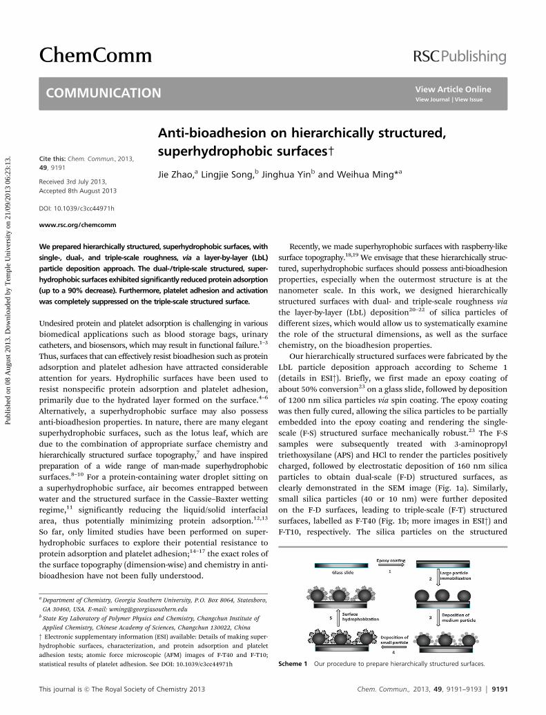
Our hierarchically structured surfaces were fabricated by theLbL particle deposition approach according to Scheme 1(details in ESI†). Briefly, we first made an epoxy coating ofabout 50% conversion23 on a glass slide, followed by depositionof 1200 nm silica particles via spin coating. The epoxy coatingwas then fully cured, allowing the silica particles to be partiallyembedded into the epoxy coating and rendering the single-scale (F-S) structured surface mechanically robust.23 The F-Ssamples were subsequently treated with 3-aminopropyltriethoxysilane (APS) and HCl to render the particles positivelycharged, followed by electrostatic deposition of 160 nm silicaparticles to obtain dual-scale (F-D) structured surfaces, asclearly demonstrated in the SEM image (Fig. 1a). Similarly,small silica particles (40 or 10 nm) were further depositedon the F-D surfaces, leading to triple-scale (F-T) structuredsurfaces, labelled as F-T40 (Fig. 1b; more images in ESI†) andF-T10, respectively. The silica particles on the structured
Scheme 1 Our procedure to prepare hierarchically structured surfaces.
a Department of Chemistry, Georgia Southern University, P.O. Box 8064, Statesboro,
GA 30460, USA. E-mail: [email protected] State Key Laboratory of Polymer Physics and Chemistry, Changchun Institute of
Applied Chemistry, Chinese Academy of Sciences, Changchun 130022, China
† Electronic supplementary information (ESI) available: Details of making super-hydrophobic surfaces, characterization, and protein adsorption and plateletadhesion tests; atomic force microscopic (AFM) images of F-T40 and F-T10;statistical results of platelet adhesion. See DOI: 10.1039/c3cc44971h
Received 3rd July 2013,Accepted 8th August 2013
DOI: 10.1039/c3cc44971h
www.rsc.org/chemcomm
ChemComm
COMMUNICATION
Publ
ishe
d on
08
Aug
ust 2
013.
Dow
nloa
ded
by T
empl
e U
nive
rsity
on
21/0
9/20
13 0
6:23
:13.
View Article OnlineView Journal | View Issue
9192 Chem. Commun., 2013, 49, 9191--9193 This journal is c The Royal Society of Chemistry 2013
surfaces were further consolidated by reacting with SiCl4. Some ofthese surfaces were also treated with 1H,1H,2H,2H-perfluorodecyltrichlorosilane via chemical vapour deposition to hydrophobizethe surfaces (designated as -Rf samples).
We first examined the wettability of the perfluorinatedsurfaces. Before fluorination, all structured surfaces showed,unsurprisingly, high hydrophilicity with water contact angles(CA) lower than 301. The fluorinated single-scale surface (F-S-Rf)was much more hydrophobic (CA: 1451, Fig. 2a), but the waterCAs were significantly lower than those on F-D-Rf, F-T40-Rf, andF-T10-Rf (all above 1651), since the dual-scale and triple-scalestructures would allow more air to be entrapped between waterand the solid surface (the Cassie–Baxter wetting regime11), whichis the key to achieving superhydrophobicity. Roll-off angle datarevealed an even more pronounced difference: while significanttilting (>401) was required for a 10 mL water droplet to roll off theF-S-Rf surface (Fig. 2b), only 41 was needed for a 2 mL waterdroplet on the F-D-Rf surface. The water roll-off angle was evensmaller on the F-T-Rf surface (Fig. 2b); even the slightestvibration would lead to the roll-off of water droplets as smallas 2 mL. Detailed investigation of the structural impact on thesurface super-repellency will be reported elsewhere.
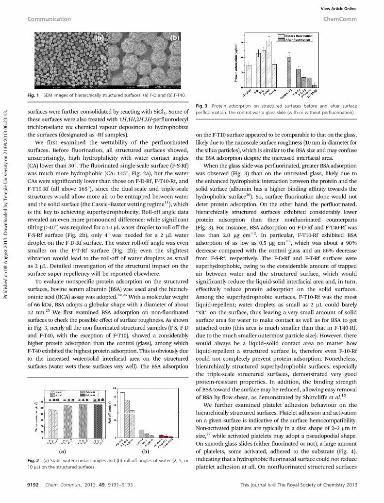
To evaluate nonspecific protein adsorption on the structuredsurfaces, bovine serum albumin (BSA) was used and the bicinch-oninic acid (BCA) assay was adopted.24,25 With a molecular weightof 66 kDa, BSA adopts a globular shape with a diameter of about12 nm.25 We first examined BSA adsorption on non-fluorinatedsurfaces to check the possible effect of surface roughness. As shownin Fig. 3, nearly all the non-fluorinated structured samples (F-S, F-Dand F-T40, with the exception of F-T10), showed a considerablyhigher protein adsorption than the control (glass), among whichF-T40 exhibited the highest protein adsorption. This is obviously dueto the increased water/solid interfacial area on the structuredsurfaces (water wets these surfaces very well). The BSA adsorption
on the F-T10 surface appeared to be comparable to that on the glass,likely due to the nanoscale surface roughness (10 nm in diameter forthe silica particles), which is similar to the BSA size and may confusethe BSA adsorption despite the increased interfacial area.
When the glass slide was perfluorinated, greater BSA adsorptionwas observed (Fig. 3) than on the untreated glass, likely due tothe enhanced hydrophobic interaction between the protein and thesolid surface (albumin has a higher binding affinity towards thehydrophobic surface26). So, surface fluorination alone would notdeter protein adsorption. On the other hand, the perfluorinated,hierarchically structured surfaces exhibited considerably lowerprotein adsorption than their nonfluorinated counterparts(Fig. 3). For instance, BSA adsorption on F-D-Rf and F-T40-Rf wasless than 2.0 mg cm�2. In particular, F-T10-Rf exhibited BSAadsorption of as low as 0.5 mg cm�2, which was about a 90%decrease compared with the control glass and an 86% decreasefrom F-S-Rf, respectively. The F-D-Rf and F-T-Rf surfaces weresuperhydrophobic, owing to the considerable amount of trappedair between water and the structured surface, which wouldsignificantly reduce the liquid/solid interfacial area and, in turn,effectively reduce protein adsorption on the solid surfaces.Among the superhydrophobic surfaces, F-T10-Rf was the mostliquid-repellent; water droplets as small as 2 mL could barely‘‘sit’’ on the surface, thus leaving a very small amount of solidsurface area for water to make contact as well as for BSA to getattached onto (this area is much smaller than that in F-T40-Rf,due to the much smaller outermost particle size). However, therewould always be a liquid–solid contact area no matter howliquid-repellent a structured surface is, therefore even F-10-Rfcould not completely prevent protein adsorption. Nonetheless,hierarchically structured superhydrophobic surfaces, especiallythe triple-scale structured surfaces, demonstrated very goodprotein-resistant properties. In addition, the binding strengthof BSA toward the surface may be reduced, allowing easy removalof BSA by flow shear, as demonstrated by Shirtcliffe et al.15
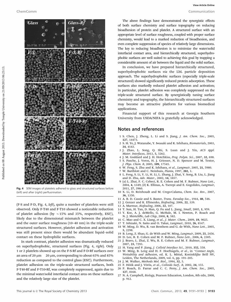
We further examined platelet adhesion behaviour on thehierarchically structured surfaces. Platelet adhesion and activationon a given surface is indicative of the surface hemocompatibility.Non-activated platelets are typically in a disc shape of 2–3 mm insize,27 while activated platelets may adopt a pseudopodial shape.On smooth glass slides (either fluorinated or not), a large amountof platelets, some activated, adhered to the substrate (Fig. 4),indicating that a hydrophobic fluorinated surface could not reduceplatelet adhesion at all. On nonfluorinated structured surfaces
Fig. 1 SEM images of hierarchically structured surfaces: (a) F-D and (b) F-T40.
Fig. 2 (a) Static water contact angles and (b) roll-off angles of water (2, 5, or10 mL) on the structured surfaces.
Fig. 3 Protein adsorption on structured surfaces before and after surfaceperfluorination. The control was a glass slide (with or without perfluorination).
Communication ChemComm
Publ
ishe
d on
08
Aug
ust 2
013.
Dow
nloa
ded
by T
empl
e U
nive
rsity
on
21/0
9/20
13 0
6:23
:13.
View Article Online
This journal is c The Royal Society of Chemistry 2013 Chem. Commun., 2013, 49, 9191--9193 9193
(F-S and F-D, Fig. 4, left), quite a number of platelets were stillobserved. Only F-T40 and F-T10 showed a noticeable reductionof platelet adhesion (by B15% and 35%, respectively, ESI†),likely due to the dimensional mismatch between the plateletand the outer surface roughness (10–40 nm) in the triple-scalestructured surfaces. However, platelet adhesion and activationwas still present since there would be abundant liquid–solidcontact on these hydrophilic surfaces.
In stark contrast, platelet adhesion was dramatically reducedon superhydrophobic, structured surfaces (Fig. 4, right). Only1 or 2 platelets showed up on the F-S-Rf and F-D-Rf surfaces overan area of 20 mm� 20 mm, corresponding to about 65% and 85%reduction as compared to the control glass (ESI†). Furthermore,platelet adhesion on the triple-scale structured surfaces, bothF-T40-Rf and F-T10-Rf, was completely suppressed, again due tothe minimal water/solid interfacial contact area on these surfacesand the relatively large size of the platelet.
The above findings have demonstrated the synergistic effectsof both surface chemistry and surface topography on reducingbioadhesion of protein and platelet. A structured surface with anappropriate level of surface roughness, coupled with proper surfacechemistry, would lead to a marked reduction of bioadhesion, andeven complete suppression of species of relatively large dimensions.The key to reducing bioadhesion is to minimize the water/solidinterfacial contact area, and hierarchically structured, superhydro-phobic surfaces are well suited to achieving this goal by trapping aconsiderable amount of air between the liquid and the solid surface.
In conclusion, we have prepared hierarchically structured,superhydrophobic surfaces via the LbL particle depositionapproach. The superhydrophobic surfaces (especially triple-scalestructured) showed significantly reduced protein adsorption. Thesesurfaces also markedly reduced platelet adhesion and activation;in particular, platelet adhesion was completely suppressed on thetriple-scale structured surface. By synergistically tuning surfacechemistry and topography, the hierarchically structured surfacesmay become an attractive platform for various biomedicalapplications.
Financial support of this research at Georgia SouthernUniversity from USDA/NIFA is gratefully acknowledged.
Notes and references1 S. Chen, J. Zheng, L. Li and S. Jiang, J. Am. Chem. Soc., 2005,
127, 14473.2 S. H. Ye, J. Watanabe, Y. Iwasaki and K. Ishihara, Biomaterials, 2003,
24, 4143.3 J. Zhao, L. Song, Q. Shi, S. Luan and J. Yin, ACS Appl.
Mater. Interfaces, 2013, 5, 5262.4 J. M. Goddard and J. H. Hotchkiss, Prog. Polym. Sci., 2007, 32, 698.5 S. Pasche, J. Voros, H. J. Griesser, N. D. Spencer and M. Textor,
J. Phys. Chem. B, 2005, 109, 17545.6 W. Feng, S. Zhu and K. Ishihara, et al., Langmuir, 2005, 21, 5980.7 W. Barthlott and C. Neinhuis, Planta, 1997, 202, 1.8 L. Feng, S. Li, Y. Li, H. Li, L. Zhang, J. Zhai, Y. Song, B. Liu, L. Jiang
and D. Zhu, Adv. Mater., 2002, 14, 1857.9 (a) L. Zhai, F. C. Cebeci, R. E. Cohen and M. F. Rubner, Nano Lett.,
2004, 4, 1349; (b) K. Ellinas, A. Tserepi and E. Gogolides, Langmuir,2011, 27, 3960.
10 X. Li, D. Reinhoudt and M. Crego-Calama, Chem. Soc. Rev., 2007,36, 1350.
11 A. B. D. Cassie and S. Baxter, Trans. Faraday Soc., 1944, 40, 546.12 J. Genzer and K. Efimenko, Biofouling, 2006, 22, 339.13 A. Marmur, Biofouling, 2006, 22, 107.14 T. Sun, H. Tan, D. Han, Q. Fu and L. Jiang, Small, 2005, 1, 959.15 Y. Koc, A. J. deMello, G. McHale, M. I. Newton, P. Roach and
N. J. Shirtcliffe, Lab Chip, 2008, 8, 582.16 C. Mao and C. X. Liang, et al., J. Mater. Chem., 2009, 19, 9025.17 T. Ishizaki, N. Saito and O. Takai, Langmuir, 2010, 26, 8147.18 W. Ming, D. Wu, R. van Benthem and G. de With, Nano Lett., 2005,
5, 2298.19 B. Leng, Z. Shao, G. de With and W. Ming, Langmuir, 2009, 25, 2456.20 D. Lee, R. E. Cohen and M. F. Rubner, Nano Lett., 2006, 6, 2305.21 J. Bravo, L. Zhai, Z. Wu, R. E. Cohen and M. F. Rubner, Langmuir,
2007, 23, 7293.22 H. Yang and P. Jiang, J. Colloid Interface Sci., 2010, 352, 558.23 W. Ming, B. Leng and H. F. Hoefnagels, et al., in ‘‘Contact Angle,
Wettability and Adhesion, ed. K. L. Mittal, Koninklijke Brill NV,Leiden, The Netherlands, 2009, vol. 6, pp. 191–205.
24 J. M. Walker, Methods Mol. Biol., 1994, 32, 5.25 F. Hook and J. Voros, et al., Colloids Surf., B, 2002, 24, 155.26 P. Roach, D. Farrar and C. C. Perry, J. Am. Chem. Soc., 2005,
127, 8168.27 N. A. Campbell, Biology, Pearson Education, London, 8th edn, 2008,
p. 912.
Fig. 4 SEM images of platelets adhered to glass and structured surfaces before(left) and after (right) perfluorination.
ChemComm Communication
Publ
ishe
d on
08
Aug
ust 2
013.
Dow
nloa
ded
by T
empl
e U
nive
rsity
on
21/0
9/20
13 0
6:23
:13.
View Article Online