anatomy & physiology lesson 9. the cardiovascular system the blood, heart, and blood vessels...
TRANSCRIPT
Anatomy & PhysiologyAnatomy & Physiology
Lesson 9Lesson 9
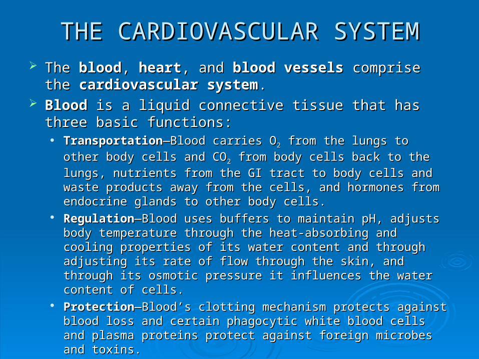
THE CARDIOVASCULAR SYSTEMTHE CARDIOVASCULAR SYSTEM The The bloodblood, , heartheart, and , and blood vesselsblood vessels comprise the comprise the
cardiovascular systemcardiovascular system.. Blood Blood is a liquid connective tissue that has three basic is a liquid connective tissue that has three basic
functions:functions: TransportationTransportation—Blood carries O—Blood carries O22 from the lungs to other body from the lungs to other body
cells and COcells and CO22 from body cells back to the lungs, nutrients from from body cells back to the lungs, nutrients from
the GI tract to body cells and waste products away from the the GI tract to body cells and waste products away from the cells, and hormones from endocrine glands to other body cells.cells, and hormones from endocrine glands to other body cells.
RegulationRegulation—Blood uses buffers to maintain pH, adjusts body —Blood uses buffers to maintain pH, adjusts body temperature through the heat-absorbing and cooling properties temperature through the heat-absorbing and cooling properties of its water content and through adjusting its rate of flow through of its water content and through adjusting its rate of flow through the skin, and through its osmotic pressure it influences the water the skin, and through its osmotic pressure it influences the water content of cells.content of cells.
ProtectionProtection—Blood’s clotting mechanism protects against blood —Blood’s clotting mechanism protects against blood loss and certain phagocytic white blood cells and plasma loss and certain phagocytic white blood cells and plasma proteins protect against foreign microbes and toxins.proteins protect against foreign microbes and toxins.
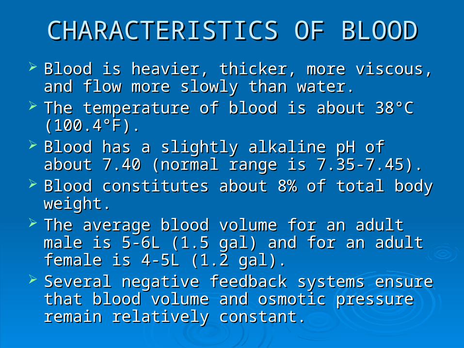
CHARACTERISTICS OF BLOODCHARACTERISTICS OF BLOOD Blood is heavier, thicker, more viscous, and flow Blood is heavier, thicker, more viscous, and flow
more slowly than water.more slowly than water. The temperature of blood is about 38°C The temperature of blood is about 38°C
(100.4°F).(100.4°F). Blood has a slightly alkaline pH of about 7.40 Blood has a slightly alkaline pH of about 7.40
(normal range is 7.35-7.45).(normal range is 7.35-7.45). Blood constitutes about 8% of total body weight.Blood constitutes about 8% of total body weight. The average blood volume for an adult male is The average blood volume for an adult male is
5-6L (1.5 gal) and for an adult female is 4-5L 5-6L (1.5 gal) and for an adult female is 4-5L (1.2 gal).(1.2 gal).
Several negative feedback systems ensure that Several negative feedback systems ensure that blood volume and osmotic pressure remain blood volume and osmotic pressure remain relatively constant.relatively constant.
COMPONENTS OF BLOODCOMPONENTS OF BLOOD
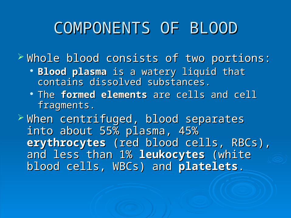
Whole blood consists of two portions:Whole blood consists of two portions: Blood plasma Blood plasma is a watery liquid that contains is a watery liquid that contains
dissolved substances.dissolved substances. The The formed elementsformed elements are cells and cell are cells and cell
fragments.fragments. When centrifuged, blood separates into When centrifuged, blood separates into
about 55% plasma, 45% about 55% plasma, 45% erythrocyteserythrocytes (red blood cells, RBCs), and less than 1% (red blood cells, RBCs), and less than 1% leukocytesleukocytes (white blood cells, WBCs) and (white blood cells, WBCs) and plateletsplatelets..
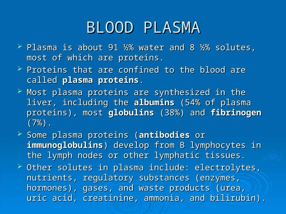
BLOOD PLASMABLOOD PLASMA Plasma is about 91 ½% water and 8 ½% solutes, most Plasma is about 91 ½% water and 8 ½% solutes, most
of which are proteins.of which are proteins. Proteins that are confined to the blood are called Proteins that are confined to the blood are called plasma plasma
proteinsproteins.. Most plasma proteins are synthesized in the liver, Most plasma proteins are synthesized in the liver,
including the including the albuminsalbumins (54% of plasma proteins), most (54% of plasma proteins), most globulinsglobulins (38%) and (38%) and fibrinogen fibrinogen (7%).(7%).
Some plasma proteins (Some plasma proteins (antibodiesantibodies or or immunoglobulinsimmunoglobulins) develop from B lymphocytes in the ) develop from B lymphocytes in the lymph nodes or other lymphatic tissues.lymph nodes or other lymphatic tissues.
Other solutes in plasma include: electrolytes, nutrients, Other solutes in plasma include: electrolytes, nutrients, regulatory substances (enzymes, hormones), gases, and regulatory substances (enzymes, hormones), gases, and waste products (urea, uric acid, creatinine, ammonia, waste products (urea, uric acid, creatinine, ammonia, and bilirubin). and bilirubin).
FORMED ELEMENTSFORMED ELEMENTS
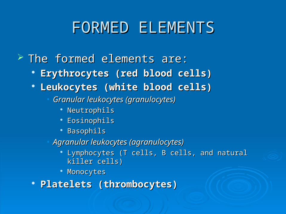
The formed elements are:The formed elements are: Erythrocytes (red blood cells)Erythrocytes (red blood cells) Leukocytes (white blood cells)Leukocytes (white blood cells)
• Granular leukocytes (granulocytes)Granular leukocytes (granulocytes) NeutrophilsNeutrophils EosinophilsEosinophils BasophilsBasophils
• Agranular leukocytes (agranulocytes)Agranular leukocytes (agranulocytes) Lymphocytes (T cells, B cells, and natural killer cells)Lymphocytes (T cells, B cells, and natural killer cells) MonocytesMonocytes
Platelets (thrombocytes)Platelets (thrombocytes)
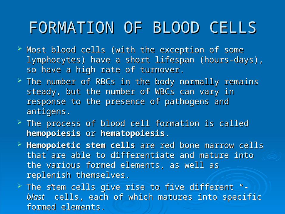
FORMATION OF BLOOD CELLSFORMATION OF BLOOD CELLS Most blood cells (with the exception of some Most blood cells (with the exception of some
lymphocytes) have a short lifespan (hours-days), so lymphocytes) have a short lifespan (hours-days), so have a high rate of turnover.have a high rate of turnover.
The number of RBCs in the body normally remains The number of RBCs in the body normally remains steady, but the number of WBCs can vary in response to steady, but the number of WBCs can vary in response to the presence of pathogens and antigens.the presence of pathogens and antigens.
The process of blood cell formation is called The process of blood cell formation is called hemopoiesishemopoiesis or or hematopoiesis hematopoiesis..
Hemopoietic stem cellsHemopoietic stem cells are red bone marrow cells that are red bone marrow cells that are able to differentiate and mature into the various are able to differentiate and mature into the various formed elements, as well as replenish themselves.formed elements, as well as replenish themselves.
The stem cells give rise to five different “-The stem cells give rise to five different “-blastblast” cells, ” cells, each of which matures into specific formed elements.each of which matures into specific formed elements.
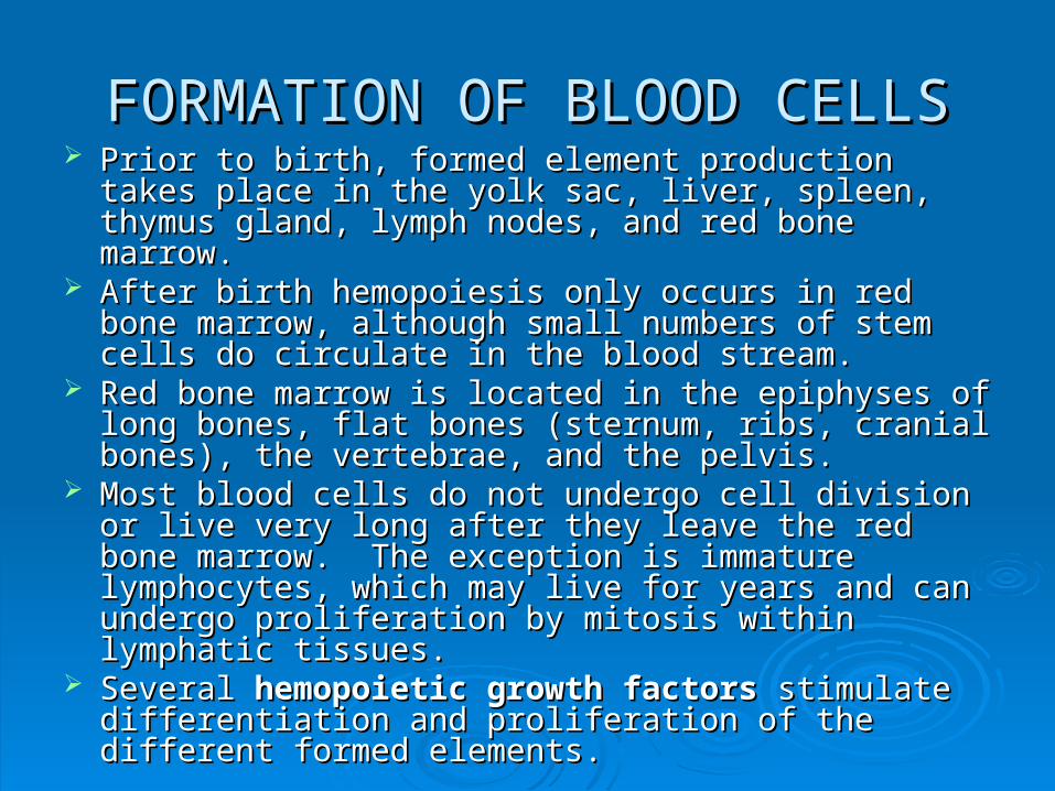
FORMATION OF BLOOD CELLSFORMATION OF BLOOD CELLS Prior to birth, formed element production takes place in Prior to birth, formed element production takes place in
the yolk sac, liver, spleen, thymus gland, lymph nodes, the yolk sac, liver, spleen, thymus gland, lymph nodes, and red bone marrow.and red bone marrow.
After birth hemopoiesis only occurs in red bone marrow, After birth hemopoiesis only occurs in red bone marrow, although small numbers of stem cells do circulate in the although small numbers of stem cells do circulate in the blood stream.blood stream.
Red bone marrow is located in the epiphyses of long Red bone marrow is located in the epiphyses of long bones, flat bones (sternum, ribs, cranial bones), the bones, flat bones (sternum, ribs, cranial bones), the vertebrae, and the pelvis.vertebrae, and the pelvis.
Most blood cells do not undergo cell division or live very Most blood cells do not undergo cell division or live very long after they leave the red bone marrow. The long after they leave the red bone marrow. The exception is immature lymphocytes, which may live for exception is immature lymphocytes, which may live for years and can undergo proliferation by mitosis within years and can undergo proliferation by mitosis within lymphatic tissues.lymphatic tissues.
Several Several hemopoietic growth factorshemopoietic growth factors stimulate stimulate differentiation and proliferation of the different formed differentiation and proliferation of the different formed elements.elements.
RED BLOOD CELLSRED BLOOD CELLS Erythrocytes contain the oxygen-carrying protein Erythrocytes contain the oxygen-carrying protein
hemoglobinhemoglobin.. It is hemoglobin that give whole blood its red It is hemoglobin that give whole blood its red
color.color. Mature RBCs are shaped as biconcave discs, Mature RBCs are shaped as biconcave discs,
lack a nucleus or other organelles, and can lack a nucleus or other organelles, and can neither reproduce or carry on other metabolic neither reproduce or carry on other metabolic functions.functions.
Hemoglobin constitutes about 33% of the cell’s Hemoglobin constitutes about 33% of the cell’s weight.weight.
The primary function of erythrocytes is oxygen The primary function of erythrocytes is oxygen transportation.transportation.
HEMATOCRIT & ANEMIAHEMATOCRIT & ANEMIA HematocritHematocrit or or HctHct refers to the percentage of RBCs in refers to the percentage of RBCs in
blood.blood. The normal hematocrit range in adult women is 38-46% The normal hematocrit range in adult women is 38-46%
(42% average) and in men it is 40-54% (47% average).(42% average) and in men it is 40-54% (47% average). A significant drop in hematocrit indicates A significant drop in hematocrit indicates anemiaanemia.. Some factors that may lead to anemia include an Some factors that may lead to anemia include an
inability of red bone marrow to respond to inability of red bone marrow to respond to erythropoietinerythropoietin (a hormone that stimulate RBC (a hormone that stimulate RBC production), nutritional deficiencies (inadequate iron, production), nutritional deficiencies (inadequate iron, certain amino acids, or vitamin Bcertain amino acids, or vitamin B1212), lack of intrinsic ), lack of intrinsic
factor (which aids with the absorption of vitamin Bfactor (which aids with the absorption of vitamin B1212, ,
which helps red bone marrow to produce erythrocytes), which helps red bone marrow to produce erythrocytes), or leukemia.or leukemia.
WHITE BLOOD CELLSWHITE BLOOD CELLS
Unlike RBCs, leukocytes have a nucleus and do Unlike RBCs, leukocytes have a nucleus and do not contain hemoglobin.not contain hemoglobin.
The two major groups of WBCs are The two major groups of WBCs are granulocytesgranulocytes and and agranulocytesagranulocytes.. Granulocytes have granules in the cytoplasm that can Granulocytes have granules in the cytoplasm that can
be seen under a light microscope.be seen under a light microscope. Agranulocytes do not have cytoplasmic granules that Agranulocytes do not have cytoplasmic granules that
can be seen under a light microscope.can be seen under a light microscope. The various types of WBCs are primarily The various types of WBCs are primarily
responsible for protecting the body against responsible for protecting the body against disease conditions.disease conditions.
WHITE BLOOD CELLS & DISEASEWHITE BLOOD CELLS & DISEASE An increase in the number of WBCs usually indicates the presence An increase in the number of WBCs usually indicates the presence
of inflammation or infection.of inflammation or infection. Since each type of WBC plays a different role, determining the Since each type of WBC plays a different role, determining the
percentage of each type in the blood (a percentage of each type in the blood (a differential white blood differential white blood cell countcell count) helps to diagnose specific conditions.) helps to diagnose specific conditions.
A high neutrophil count may indicate bacterial infections, burns, A high neutrophil count may indicate bacterial infections, burns, stress, or inflammation, while a low count can result from radiation, stress, or inflammation, while a low count can result from radiation, certain drugs, vit. Bcertain drugs, vit. B1212 deficiency, or systemic lupus erythematosus. deficiency, or systemic lupus erythematosus.
A high eosinophil count suggests allergic reaction, parasitic A high eosinophil count suggests allergic reaction, parasitic infection, autoimmune disease, or adrenal insufficiency, while a low infection, autoimmune disease, or adrenal insufficiency, while a low count could be due to certain drugs, stress, or Cushing’s syndrome.count could be due to certain drugs, stress, or Cushing’s syndrome.
Basophils may increase with some allergic responses, leukemias, Basophils may increase with some allergic responses, leukemias, cancers, and hypothyroidism, but may decrease with pregnancy, cancers, and hypothyroidism, but may decrease with pregnancy, ovulation, stress and hyperthyroidism.ovulation, stress and hyperthyroidism.
High lymphocyte counts could indicate viral infections, immune High lymphocyte counts could indicate viral infections, immune diseases, and some leukemias. Low counts might result from diseases, and some leukemias. Low counts might result from prolonged, severe illness, high steroid levels, and prolonged, severe illness, high steroid levels, and immunosuppresion.immunosuppresion.
Excess monocytes may result from certain viral or fungal infections, Excess monocytes may result from certain viral or fungal infections, tuberculosis, some leukemias, and chronic disease. Low monocyte tuberculosis, some leukemias, and chronic disease. Low monocyte levels seldom occur.levels seldom occur.
PLATELETSPLATELETS Hemopoietic stem cells also develop into Hemopoietic stem cells also develop into
megakaryoblastsmegakaryoblasts, as well as erythrocytes and , as well as erythrocytes and leukocytes.leukocytes.
Under the influence of the hormone Under the influence of the hormone thrombopoetinthrombopoetin megakaryoblasts transform into huge megakaryocyte megakaryoblasts transform into huge megakaryocyte cells.cells.
Megakaryocytes splinter into 2000-3000 membrane-Megakaryocytes splinter into 2000-3000 membrane-enclosed pieces each. These fragments are called enclosed pieces each. These fragments are called platelets (thrombocytes)platelets (thrombocytes)..
Platelets are small, disc-shaped, and contain no nucleus, Platelets are small, disc-shaped, and contain no nucleus, but do have many granules.but do have many granules.
Platelets help stop blood loss from damaged blood Platelets help stop blood loss from damaged blood vessels by forming a platelet plug.vessels by forming a platelet plug.
The granules in platelets also contain chemicals that The granules in platelets also contain chemicals that help promote blood clotting.help promote blood clotting.
HEMOSTASISHEMOSTASIS HemostasisHemostasis is the process by which is the process by which
bleeding is stopped.bleeding is stopped. In the presence of damage to blood In the presence of damage to blood
vessels, hemostasis must be quick, vessels, hemostasis must be quick, localized to the area that is damaged, and localized to the area that is damaged, and carefully controlled.carefully controlled.
Three basic mechanisms reduce blood Three basic mechanisms reduce blood loss:loss: Vascular spasmVascular spasm Platelet plug formationPlatelet plug formation Blood clotting (coagulation)Blood clotting (coagulation)
VASCULAR SPASMVASCULAR SPASM When arteries or arterioles are damaged, When arteries or arterioles are damaged,
the smooth muscles in their walls the smooth muscles in their walls immediately contract, reducing the amount immediately contract, reducing the amount of blood loss.of blood loss.
Vascular spasmVascular spasm may last for several may last for several minutes to several hours, while other minutes to several hours, while other hemostatic mechanisms go into effect.hemostatic mechanisms go into effect.
Vascular spasm probably occurs in Vascular spasm probably occurs in response to damage to the smooth muscle response to damage to the smooth muscle and from reflexes initiated by pain and from reflexes initiated by pain receptors.receptors.
PLATELET PLUG FORMATIONPLATELET PLUG FORMATION Although platelets are small, they are packed with an Although platelets are small, they are packed with an
impressive arsenal of chemicals to help achieve impressive arsenal of chemicals to help achieve hemostasis.hemostasis.
Platelet plug formation occurs in three steps:Platelet plug formation occurs in three steps: Platelet adhesionPlatelet adhesion—Platelets contact and stick to parts of the —Platelets contact and stick to parts of the
damaged blood vessel.damaged blood vessel. Platelet release reactionPlatelet release reaction—Adhered platelets become —Adhered platelets become
“activated,” they extend projections which allow them to contact “activated,” they extend projections which allow them to contact each other, and they begin to release their chemicals. The each other, and they begin to release their chemicals. The various chemicals serve to activate nearby platelets and contract various chemicals serve to activate nearby platelets and contract the blood vessel muscles, restricting blood flow through the the blood vessel muscles, restricting blood flow through the injured vessel.injured vessel.
Platelet aggregationPlatelet aggregation—As other platelets in the area become —As other platelets in the area become activated, they stick to the originally activated platelets, forming activated, they stick to the originally activated platelets, forming a mass called a a mass called a platelet plugplatelet plug. .
Although a platelet plug is initially soft, it can become Although a platelet plug is initially soft, it can become quite tight when reinforced by fibrin threads formed quite tight when reinforced by fibrin threads formed during clotting. A platelet plug can be very effective in during clotting. A platelet plug can be very effective in preventing blood loss in a small vessel.preventing blood loss in a small vessel.
CLOTTING (COAGULATION)CLOTTING (COAGULATION) When blood is removed from a blood vessel, it forms a When blood is removed from a blood vessel, it forms a
gel. Eventually, the gel will separate from the liquid. gel. Eventually, the gel will separate from the liquid. The liquid is called The liquid is called serumserum—it is plasma without its —it is plasma without its clotting proteins. The gel is called a clotting proteins. The gel is called a clotclot and is and is composed of insoluble fibrin (protein fibers) trapping the composed of insoluble fibrin (protein fibers) trapping the formed elements of the blood.formed elements of the blood.
The formation of the clot is called The formation of the clot is called clottingclotting or or coagulation.coagulation.
If blood clots too easily, a If blood clots too easily, a thrombosis thrombosis may occur, and if may occur, and if it is too slow to clot, it is too slow to clot, hemorrhagehemorrhage may result. may result.
Clotting involves several substances known as Clotting involves several substances known as clotting clotting factorsfactors—these include Ca—these include Ca2+2+, several inactive enzymes , several inactive enzymes that are produced in the liver, and various molecules that are produced in the liver, and various molecules associated with platelets or released by damaged associated with platelets or released by damaged tissues.tissues.
Clotting involves a complex chain reaction in which the Clotting involves a complex chain reaction in which the different coagulation factors activate each other in a different coagulation factors activate each other in a positive feedback manner to produce a large amount of positive feedback manner to produce a large amount of product.product.
CLOTTING (COAGULATION)CLOTTING (COAGULATION) Clotting occurs in three basic stages:Clotting occurs in three basic stages:
Stage 1) Production of prothrombinaseStage 1) Production of prothrombinase—This is —This is initiated by either the initiated by either the extrinsic pathwayextrinsic pathway or the or the intrinsic pathwayintrinsic pathway. Prothrombinase is a catalytic . Prothrombinase is a catalytic enzyme.enzyme.
• The The extrinsic pathwayextrinsic pathway is simpler and quicker (within is simpler and quicker (within seconds, if trauma is severe). It is so named because the seconds, if trauma is severe). It is so named because the initiating factor is leaked into the blood from cells initiating factor is leaked into the blood from cells outside outside (extrinsic to) (extrinsic to) the blood.the blood.
• The The intrinsic pathwayintrinsic pathway is more complex and may take is more complex and may take several minutes. It receives its name because its activators several minutes. It receives its name because its activators are in direct contact with or contained are in direct contact with or contained within (intrinsic to)within (intrinsic to) the the blood.blood.
Stage 2)Stage 2) Regardless of the pathway utilized, in the Regardless of the pathway utilized, in the second stage prothrombinase and Casecond stage prothrombinase and Ca2+2+ catalyze the catalyze the conversion of conversion of prothrombinprothrombin into into thrombin.thrombin.
Stage 3)Stage 3) Thrombin, with Ca Thrombin, with Ca2+2+, in turn converts the , in turn converts the soluble soluble fibrinogenfibrinogen into insoluble into insoluble fibrinfibrin threads and threads and activates a clotting factor that stabilizes the fibrin activates a clotting factor that stabilizes the fibrin threads into a sturdy clot.threads into a sturdy clot.
INTRAVASCULAR COAGULATIONINTRAVASCULAR COAGULATION Normally, blood remains fluid in undamaged blood Normally, blood remains fluid in undamaged blood
vessels.vessels. Sometimes, however, the endothelial surface of a blood Sometimes, however, the endothelial surface of a blood
vessel may become roughened (from atherosclerosis, vessel may become roughened (from atherosclerosis, trauma, or infection) or blood may flow too slowly. When trauma, or infection) or blood may flow too slowly. When this happens, platelets may aggregate and form a clot.this happens, platelets may aggregate and form a clot.
Clotting in an unbroken blood vessel (usually a vein) is Clotting in an unbroken blood vessel (usually a vein) is calledcalled thrombosis thrombosis. The clot itself is called a. The clot itself is called a thrombus thrombus..
A thrombus may dissolve spontaneously or may break A thrombus may dissolve spontaneously or may break off and be carried away in the blood.off and be carried away in the blood.
Any foreign object in the bloodstream is called anAny foreign object in the bloodstream is called an embolus.embolus.
An embolus which gets lodged in the lungs is called a An embolus which gets lodged in the lungs is called a pulmonary embolismpulmonary embolism..
An embolus that breaks away from an arterial wall may An embolus that breaks away from an arterial wall may become lodged in a smaller artery, blocking bloodflow to become lodged in a smaller artery, blocking bloodflow to a vital organ.a vital organ.
BLOOD GROUPS & BLOOD BLOOD GROUPS & BLOOD TYPESTYPES
The surfaces of erythrocytes contain The surfaces of erythrocytes contain glycoproteins and glycolipids that can act as glycoproteins and glycolipids that can act as antigens. These antigens. These isoantigensisoantigens or or agglutinogensagglutinogens are normal parts of one person’s RBCs, but may are normal parts of one person’s RBCs, but may trigger antigen-antibody responses in another trigger antigen-antibody responses in another person.person.
Based on the presence or absence of Based on the presence or absence of isoantigens, blood is classified intoisoantigens, blood is classified into blood blood groupsgroups..
Each blood group may have two or more Each blood group may have two or more blood blood typestypes..
Two major blood groups areTwo major blood groups are ABO ABO and and RhRh.. There are at least 24 different blood groups and There are at least 24 different blood groups and
more than 100 isoantigens that can be identified more than 100 isoantigens that can be identified on the surfaces of RBCs.on the surfaces of RBCs.
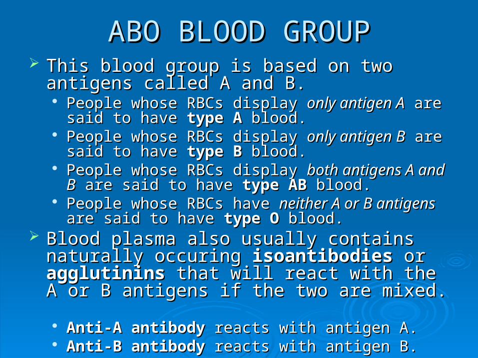
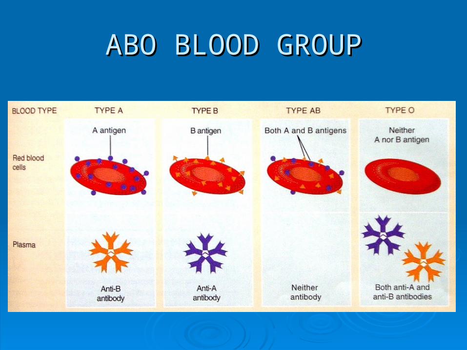
ABO BLOOD GROUPABO BLOOD GROUP This blood group is based on two antigens called This blood group is based on two antigens called
A and B.A and B. People whose RBCs display People whose RBCs display only antigen Aonly antigen A are said are said
to have to have type Atype A blood. blood. People whose RBCs display People whose RBCs display only antigen Bonly antigen B are said are said
to have to have type Btype B blood. blood. People whose RBCs display People whose RBCs display both antigens A and Bboth antigens A and B
are said to have are said to have type ABtype AB blood. blood. People whose RBCs have People whose RBCs have neither A or B antigensneither A or B antigens are are
said to have said to have type O type O blood.blood. Blood plasma also usually contains naturally Blood plasma also usually contains naturally
occuring occuring isoantibodiesisoantibodies or or agglutinins agglutinins that will that will react with the A or B antigens if the two are react with the A or B antigens if the two are mixed. mixed. Anti-A antibodyAnti-A antibody reacts with antigen A. reacts with antigen A. Anti-B antibodyAnti-B antibody reacts with antigen B. reacts with antigen B.
ABO BLOOD GROUPABO BLOOD GROUP
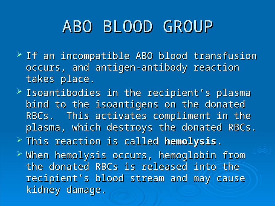
If an incompatible ABO blood transfusion occurs, If an incompatible ABO blood transfusion occurs, and antigen-antibody reaction takes place.and antigen-antibody reaction takes place.
Isoantibodies in the recipient’s plasma bind to Isoantibodies in the recipient’s plasma bind to the isoantigens on the donated RBCs. This the isoantigens on the donated RBCs. This activates compliment in the plasma, which activates compliment in the plasma, which destroys the donated RBCs.destroys the donated RBCs.
This reaction is called This reaction is called hemolysishemolysis.. When hemolysis occurs, hemoglobin from the When hemolysis occurs, hemoglobin from the
donated RBCs is released into the recipient’s donated RBCs is released into the recipient’s blood stream and may cause kidney damage.blood stream and may cause kidney damage.
ABO BLOOD GROUPABO BLOOD GROUP
Rh BLOOD GROUPRh BLOOD GROUP The Rh blood group got its name because it was The Rh blood group got its name because it was
first discovered in the Rhesus monkey.first discovered in the Rhesus monkey. People whose RBCs have Rh antigens are People whose RBCs have Rh antigens are
designated designated RhRh++ while those without Rh antigens while those without Rh antigens are are RhRh--..
Normally, plasma does not contain anti-Rh Normally, plasma does not contain anti-Rh antibodies, but if an Rhantibodies, but if an Rh-- person receives an Rh person receives an Rh++ blood transfusion, the immune system will begin blood transfusion, the immune system will begin to make anti-Rh antibodies.to make anti-Rh antibodies.
If a second transfusion of RhIf a second transfusion of Rh++ blood is given, the blood is given, the anit-Rh antibodies will cause hemolysis of the anit-Rh antibodies will cause hemolysis of the donated blood, potentially resulting in a severe donated blood, potentially resulting in a severe reaction.reaction.
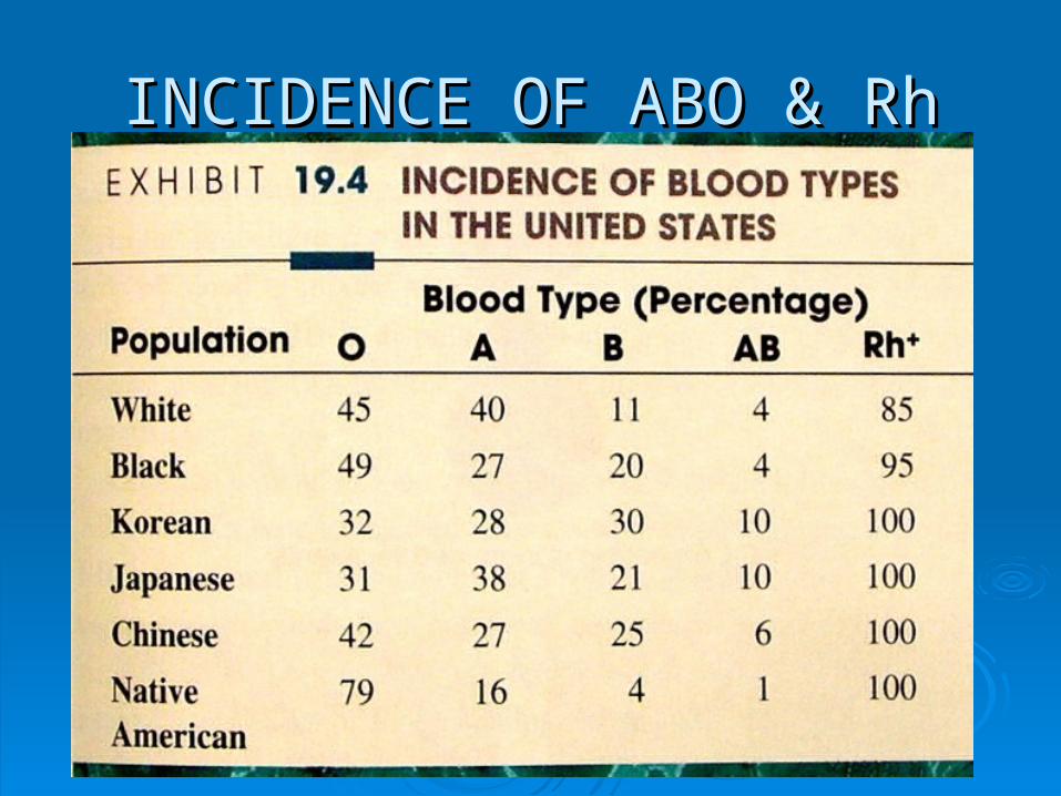
INCIDENCE OF ABO & RhINCIDENCE OF ABO & Rh
THE HEARTTHE HEART The heart is a hollow, cone-shaped organ, about The heart is a hollow, cone-shaped organ, about
the size of a person’s closed fist, and averaging the size of a person’s closed fist, and averaging 300 g (10 oz) in an adult.300 g (10 oz) in an adult.
It is about 12 cm (5 in) long, 9 cm (3 ½ in) wide, It is about 12 cm (5 in) long, 9 cm (3 ½ in) wide, and 6 cm (2 ½ in) thick.and 6 cm (2 ½ in) thick.
It contains four chambers: 2 It contains four chambers: 2 atriaatria and 2 and 2 ventriclesventricles..
It rests on the diaphragm, near the middle of the It rests on the diaphragm, near the middle of the thoracic cavity, in the mediastinum.thoracic cavity, in the mediastinum.
The pointed inferior end of the heart is called the The pointed inferior end of the heart is called the apexapex and the broad superior/posterior portion is and the broad superior/posterior portion is called the called the basebase..
THE HEARTTHE HEART The heart pumps blood into two closed circuits—The heart pumps blood into two closed circuits—
the the systemic circulationsystemic circulation and the and the pulmonary pulmonary circulationcirculation..
The left side of the heart pumps blood into the The left side of the heart pumps blood into the systemic circulation, supplying freshly systemic circulation, supplying freshly oxygenated blood to all body tissues (except the oxygenated blood to all body tissues (except the alveoli of the lungs) and removing carbon alveoli of the lungs) and removing carbon dioxide waste.dioxide waste.
The right side of the heart pumps deoxygenated The right side of the heart pumps deoxygenated blood into the pulmonary circulation, where it blood into the pulmonary circulation, where it picks up oxygen and unloads carbon dioxide in picks up oxygen and unloads carbon dioxide in the lungs.the lungs.
Freshly oxygenated blood from the lungs returns Freshly oxygenated blood from the lungs returns to the left side of the heart, where it begins to the left side of the heart, where it begins another trip through the systemic circulation.another trip through the systemic circulation.
PERICARDIUMPERICARDIUM The The pericardiumpericardium is a triple-layered sac that is a triple-layered sac that
surrounds and protects the heart, anchoring it surrounds and protects the heart, anchoring it within the mediastinum, yet allowing it ample within the mediastinum, yet allowing it ample freedom for contraction.freedom for contraction. The outer The outer fibrous pericardiumfibrous pericardium is composed of is composed of
tough, inelastic, dense irregular CT.tough, inelastic, dense irregular CT. The inner The inner serous pericardiumserous pericardium is thinner, more is thinner, more
delicate, and forms a double layer around the heart.delicate, and forms a double layer around the heart.• The outer The outer parietal layerparietal layer is fused to the fibrous pericardium. is fused to the fibrous pericardium. • The inner The inner visceral layervisceral layer or or epicardiumepicardium adheres tightly to adheres tightly to
the surface of the heart.the surface of the heart.• Between the parietal and visceral layers is the Between the parietal and visceral layers is the pericardial pericardial
cavitycavity, which contains , which contains pericardial fluidpericardial fluid. This fluid reduces . This fluid reduces friction between the membranes as the heart moves.friction between the membranes as the heart moves.
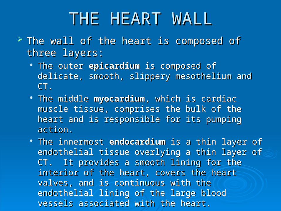
THE HEART WALLTHE HEART WALL The wall of the heart is composed of three The wall of the heart is composed of three
layers:layers: The outer The outer epicardiumepicardium is composed of delicate, is composed of delicate,
smooth, slippery mesothelium and CT.smooth, slippery mesothelium and CT. The middle The middle myocardiummyocardium, which is cardiac muscle , which is cardiac muscle
tissue, comprises the bulk of the heart and is tissue, comprises the bulk of the heart and is responsible for its pumping action.responsible for its pumping action.
The innermost The innermost endocardiumendocardium is a thin layer of is a thin layer of endothelial tissue overlying a thin layer of CT. It endothelial tissue overlying a thin layer of CT. It provides a smooth lining for the interior of the heart, provides a smooth lining for the interior of the heart, covers the heart valves, and is continuous with the covers the heart valves, and is continuous with the endothelial lining of the large blood vessels endothelial lining of the large blood vessels associated with the heart.associated with the heart.
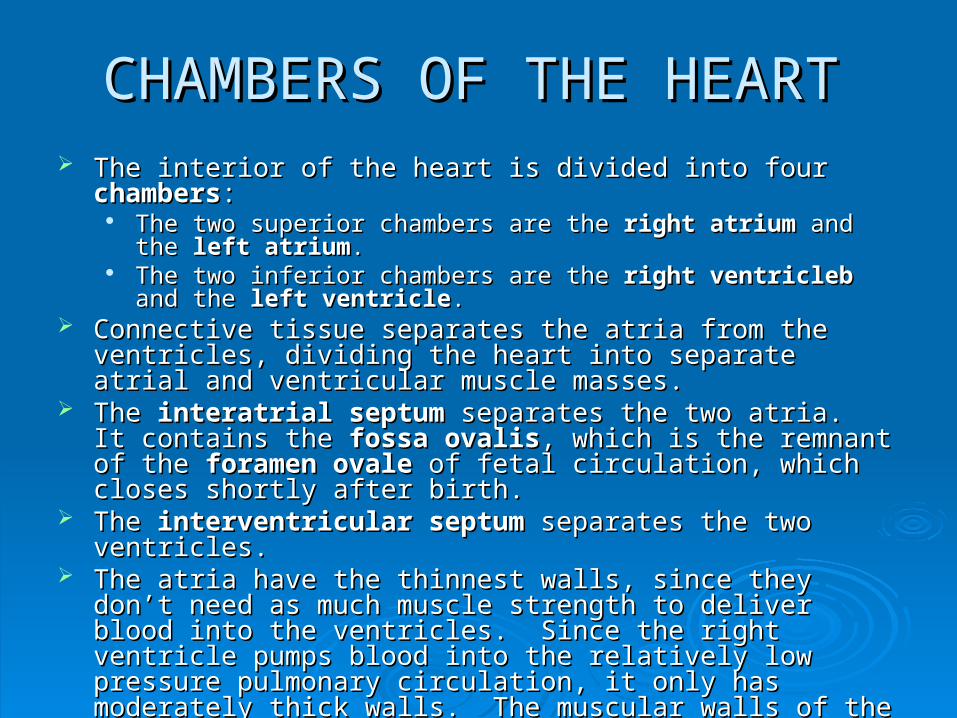
CHAMBERS OF THE HEARTCHAMBERS OF THE HEART The interior of the heart is divided into four The interior of the heart is divided into four chamberschambers::
The two superior chambers are the The two superior chambers are the right atriumright atrium and the and the left atriumleft atrium.. The two inferior chambers are the The two inferior chambers are the right ventriclebright ventricleb and the and the left left
ventricleventricle.. Connective tissue separates the atria from the ventricles, dividing Connective tissue separates the atria from the ventricles, dividing
the heart into separate atrial and ventricular muscle masses.the heart into separate atrial and ventricular muscle masses. The The interatrial septuminteratrial septum separates the two atria. It contains the separates the two atria. It contains the
fossa ovalisfossa ovalis, which is the remnant of the , which is the remnant of the foramen ovaleforamen ovale of fetal of fetal circulation, which closes shortly after birth.circulation, which closes shortly after birth.
The The interventricular septuminterventricular septum separates the two ventricles. separates the two ventricles. The atria have the thinnest walls, since they don’t need as much The atria have the thinnest walls, since they don’t need as much
muscle strength to deliver blood into the ventricles. Since the right muscle strength to deliver blood into the ventricles. Since the right ventricle pumps blood into the relatively low pressure pulmonary ventricle pumps blood into the relatively low pressure pulmonary circulation, it only has moderately thick walls. The muscular walls of circulation, it only has moderately thick walls. The muscular walls of the left ventricle are four times as thick as the right ventricle, since it the left ventricle are four times as thick as the right ventricle, since it requires extra strength to pump blood throughout the systemic requires extra strength to pump blood throughout the systemic circulation.circulation.
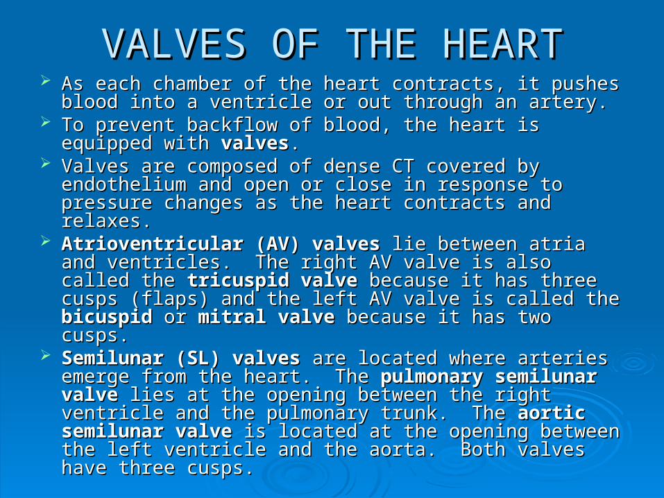
VALVES OF THE HEARTVALVES OF THE HEART As each chamber of the heart contracts, it pushes blood As each chamber of the heart contracts, it pushes blood
into a ventricle or out through an artery.into a ventricle or out through an artery. To prevent backflow of blood, the heart is equipped with To prevent backflow of blood, the heart is equipped with
valvesvalves. . Valves are composed of dense CT covered by Valves are composed of dense CT covered by
endothelium and open or close in response to pressure endothelium and open or close in response to pressure changes as the heart contracts and relaxes.changes as the heart contracts and relaxes.
Atrioventricular (AV) valvesAtrioventricular (AV) valves lie between atria and lie between atria and ventricles. The right AV valve is also called the ventricles. The right AV valve is also called the tricuspid valvetricuspid valve because it has three cusps (flaps) and because it has three cusps (flaps) and the left AV valve is called the the left AV valve is called the bicuspid bicuspid oror mitral valve mitral valve because it has two cusps.because it has two cusps.
Semilunar (SL) valvesSemilunar (SL) valves are located where arteries are located where arteries emerge from the heart. The emerge from the heart. The pulmonary semilunar pulmonary semilunar valvevalve lies at the opening between the right ventricle and lies at the opening between the right ventricle and the pulmonary trunk. The the pulmonary trunk. The aortic semilunar valveaortic semilunar valve is is located at the opening between the left ventricle and the located at the opening between the left ventricle and the aorta. Both valves have three cusps.aorta. Both valves have three cusps.
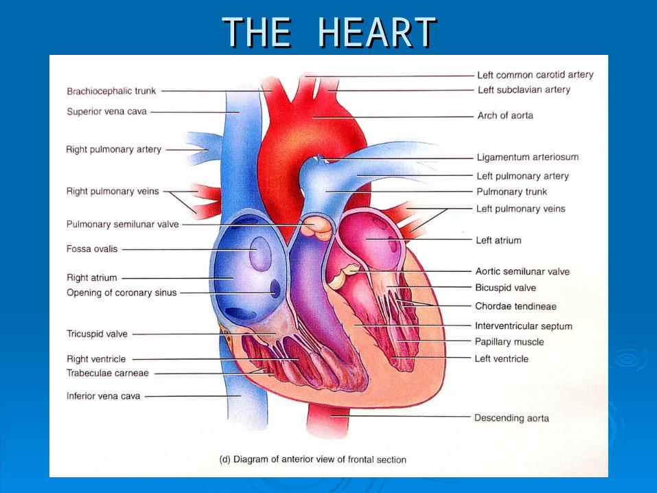
THE HEARTTHE HEART
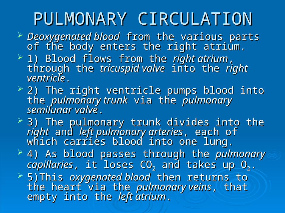
PULMONARY CIRCULATIONPULMONARY CIRCULATION Deoxygenated bloodDeoxygenated blood from the various parts of from the various parts of
the body enters the right atrium.the body enters the right atrium. 1) Blood flows from the 1) Blood flows from the right atriumright atrium, through the , through the
tricuspid valvetricuspid valve into the into the right ventricleright ventricle.. 2) The right ventricle pumps blood into the 2) The right ventricle pumps blood into the
pulmonary trunkpulmonary trunk via the via the pulmonary semilunar pulmonary semilunar valvevalve..
3) The pulmonary trunk divides into the 3) The pulmonary trunk divides into the rightright and and left pulmonary arteriesleft pulmonary arteries, each of which carries , each of which carries blood into one lung.blood into one lung.
4) As blood passes through the 4) As blood passes through the pulmonary pulmonary capillariescapillaries, it loses CO, it loses CO22 and takes up O and takes up O22. .
5)This 5)This oxygenated bloodoxygenated blood then returns to the then returns to the heart via the heart via the pulmonary veinspulmonary veins, that empty into , that empty into the the left atriumleft atrium..
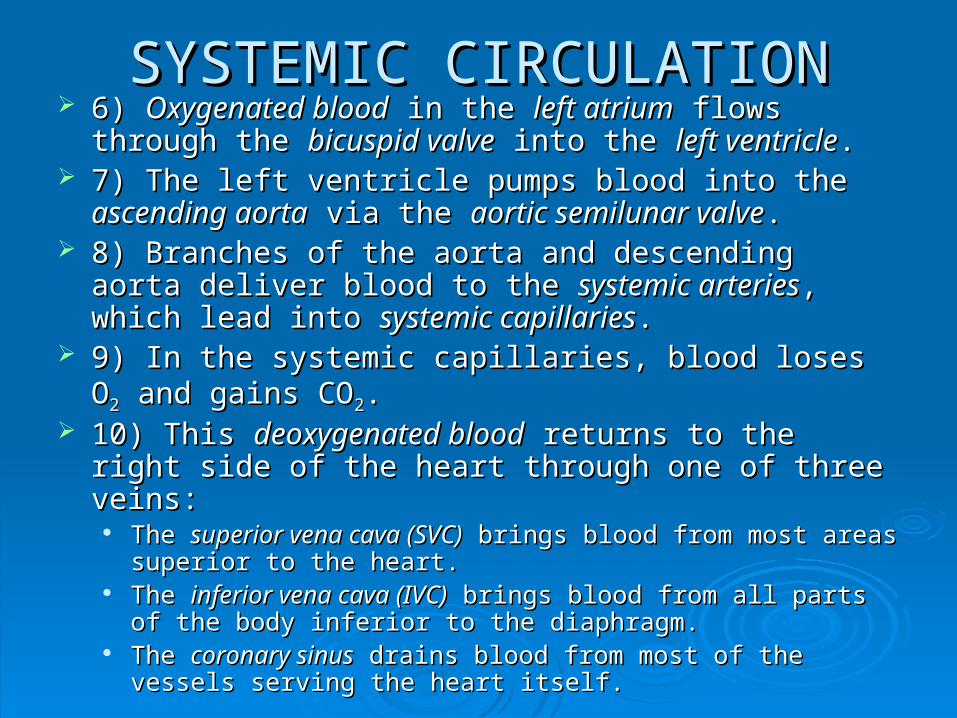
SYSTEMIC CIRCULATIONSYSTEMIC CIRCULATION 6) 6) Oxygenated bloodOxygenated blood in the in the left atriumleft atrium flows through the flows through the
bicuspid valvebicuspid valve into the into the left ventricleleft ventricle.. 7) The left ventricle pumps blood into the 7) The left ventricle pumps blood into the ascending ascending
aortaaorta via the via the aortic semilunar valveaortic semilunar valve.. 8) Branches of the aorta and descending aorta deliver 8) Branches of the aorta and descending aorta deliver
blood to the blood to the systemic arteriessystemic arteries, which lead into , which lead into systemic systemic capillariescapillaries..
9) In the systemic capillaries, blood loses O9) In the systemic capillaries, blood loses O22 and gains and gains COCO22..
10) This 10) This deoxygenated blooddeoxygenated blood returns to the right side of returns to the right side of the heart through one of three veins:the heart through one of three veins:
The The superior vena cava (SVC)superior vena cava (SVC) brings blood from most areas brings blood from most areas superior to the heart.superior to the heart.
The The inferior vena cava (IVC)inferior vena cava (IVC) brings blood from all parts of the brings blood from all parts of the body inferior to the diaphragm.body inferior to the diaphragm.
The The coronary sinuscoronary sinus drains blood from most of the vessels drains blood from most of the vessels serving the heart itself.serving the heart itself.
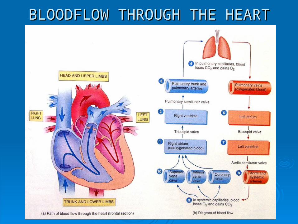
BLOODFLOW THROUGH THE HEARTBLOODFLOW THROUGH THE HEART
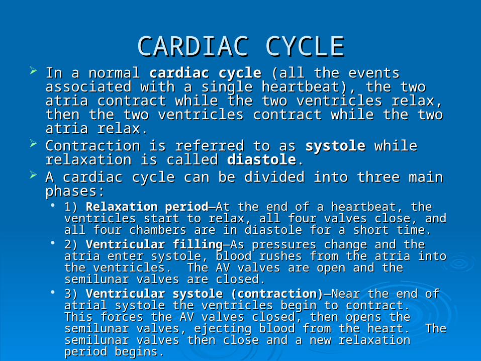
CARDIAC CYCLECARDIAC CYCLE In a normal In a normal cardiac cyclecardiac cycle (all the events associated with (all the events associated with
a single heartbeat), the two atria contract while the two a single heartbeat), the two atria contract while the two ventricles relax, then the two ventricles contract while the ventricles relax, then the two ventricles contract while the two atria relax.two atria relax.
Contraction is referred to as Contraction is referred to as systolesystole while relaxation is while relaxation is called called diastolediastole..
A cardiac cycle can be divided into three main phases:A cardiac cycle can be divided into three main phases: 1) 1) Relaxation periodRelaxation period—At the end of a heartbeat, the ventricles —At the end of a heartbeat, the ventricles
start to relax, all four valves close, and all four chambers are in start to relax, all four valves close, and all four chambers are in diastole for a short time.diastole for a short time.
2) 2) Ventricular fillingVentricular filling—As pressures change and the atria enter —As pressures change and the atria enter systole, blood rushes from the atria into the ventricles. The AV systole, blood rushes from the atria into the ventricles. The AV valves are open and the semilunar valves are closed.valves are open and the semilunar valves are closed.
3) 3) Ventricular systole (contraction)Ventricular systole (contraction)—Near the end of atrial —Near the end of atrial systole the ventricles begin to contract. This forces the AV systole the ventricles begin to contract. This forces the AV valves closed, then opens the semilunar valves, ejecting blood valves closed, then opens the semilunar valves, ejecting blood from the heart. The semilunar valves then close and a new from the heart. The semilunar valves then close and a new relaxation period begins.relaxation period begins.
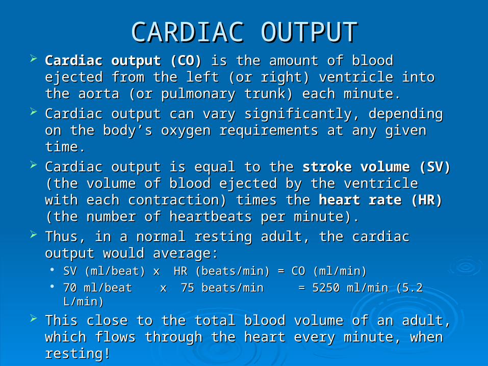
CARDIAC OUTPUTCARDIAC OUTPUT Cardiac output (CO) Cardiac output (CO) is the amount of blood ejected is the amount of blood ejected
from the left (or right) ventricle into the aorta (or from the left (or right) ventricle into the aorta (or pulmonary trunk) each minute.pulmonary trunk) each minute.
Cardiac output can vary significantly, depending on the Cardiac output can vary significantly, depending on the body’s oxygen requirements at any given time.body’s oxygen requirements at any given time.
Cardiac output is equal to the Cardiac output is equal to the stroke volume (SV)stroke volume (SV) (the (the volume of blood ejected by the ventricle with each volume of blood ejected by the ventricle with each contraction) times the contraction) times the heart rate (HR)heart rate (HR) (the number of (the number of heartbeats per minute).heartbeats per minute).
Thus, in a normal resting adult, the cardiac output would Thus, in a normal resting adult, the cardiac output would average:average:
SV (ml/beat) x HR (beats/min) = CO (ml/min) SV (ml/beat) x HR (beats/min) = CO (ml/min) 70 ml/beat x 75 beats/min = 5250 ml/min (5.2 L/min)70 ml/beat x 75 beats/min = 5250 ml/min (5.2 L/min)
This close to the total blood volume of an adult, which This close to the total blood volume of an adult, which flows through the heart every minute, when resting!flows through the heart every minute, when resting!
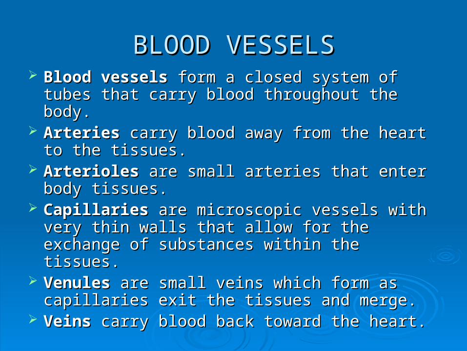
BLOOD VESSELSBLOOD VESSELS Blood vesselsBlood vessels form a closed system of tubes form a closed system of tubes
that carry blood throughout the body.that carry blood throughout the body. Arteries Arteries carry blood away from the heart to the carry blood away from the heart to the
tissues.tissues. ArteriolesArterioles are small arteries that enter body are small arteries that enter body
tissues.tissues. CapillariesCapillaries are microscopic vessels with very are microscopic vessels with very
thin walls that allow for the exchange of thin walls that allow for the exchange of substances within the tissues.substances within the tissues.
VenulesVenules are small veins which form as are small veins which form as capillaries exit the tissues and merge.capillaries exit the tissues and merge.
VeinsVeins carry blood back toward the heart. carry blood back toward the heart.
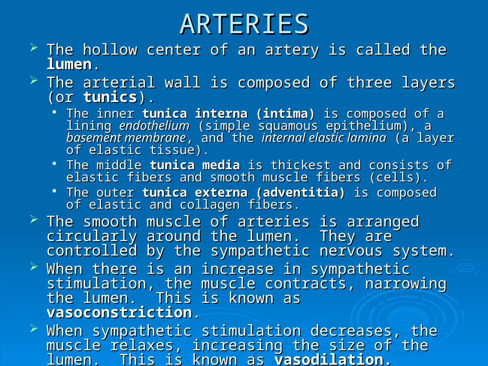
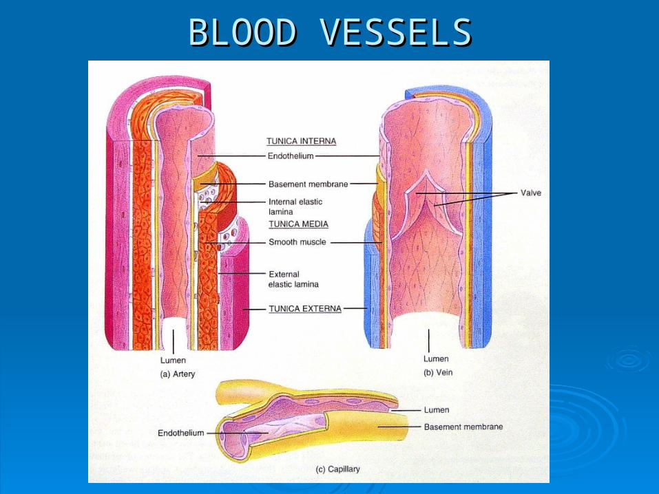
ARTERIESARTERIES The hollow center of an artery is called the The hollow center of an artery is called the lumenlumen.. The arterial wall is composed of three layers (or The arterial wall is composed of three layers (or tunicstunics).).
The inner The inner tunica interna (intima)tunica interna (intima) is composed of a lining is composed of a lining endotheliumendothelium (simple squamous epithelium), a (simple squamous epithelium), a basement basement membranemembrane, and the , and the internal elastic laminainternal elastic lamina (a layer of elastic (a layer of elastic tissue).tissue).
The middle The middle tunica mediatunica media is thickest and consists of elastic is thickest and consists of elastic fibers and smooth muscle fibers (cells).fibers and smooth muscle fibers (cells).
The outer The outer tunica externa (adventitia)tunica externa (adventitia) is composed of elastic is composed of elastic and collagen fibers.and collagen fibers.
The smooth muscle of arteries is arranged circularly The smooth muscle of arteries is arranged circularly around the lumen. They are controlled by the around the lumen. They are controlled by the sympathetic nervous system.sympathetic nervous system.
When there is an increase in sympathetic stimulation, When there is an increase in sympathetic stimulation, the muscle contracts, narrowing the lumen. This is the muscle contracts, narrowing the lumen. This is known as known as vasoconstrictionvasoconstriction..
When sympathetic stimulation decreases, the muscle When sympathetic stimulation decreases, the muscle relaxes, increasing the size of the lumen. This is known relaxes, increasing the size of the lumen. This is known as as vasodilation.vasodilation.
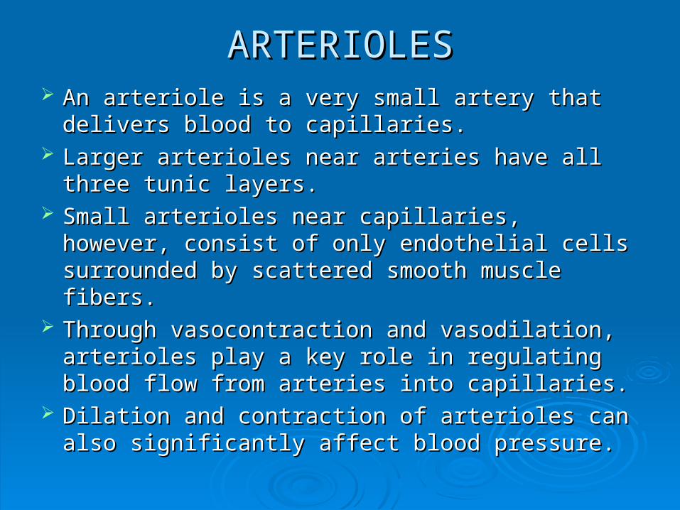
ARTERIOLESARTERIOLES An arteriole is a very small artery that delivers An arteriole is a very small artery that delivers
blood to capillaries.blood to capillaries. Larger arterioles near arteries have all three Larger arterioles near arteries have all three
tunic layers.tunic layers. Small arterioles near capillaries, however, Small arterioles near capillaries, however,
consist of only endothelial cells surrounded by consist of only endothelial cells surrounded by scattered smooth muscle fibers.scattered smooth muscle fibers.
Through vasocontraction and vasodilation, Through vasocontraction and vasodilation, arterioles play a key role in regulating blood flow arterioles play a key role in regulating blood flow from arteries into capillaries.from arteries into capillaries.
Dilation and contraction of arterioles can also Dilation and contraction of arterioles can also significantly affect blood pressure.significantly affect blood pressure.
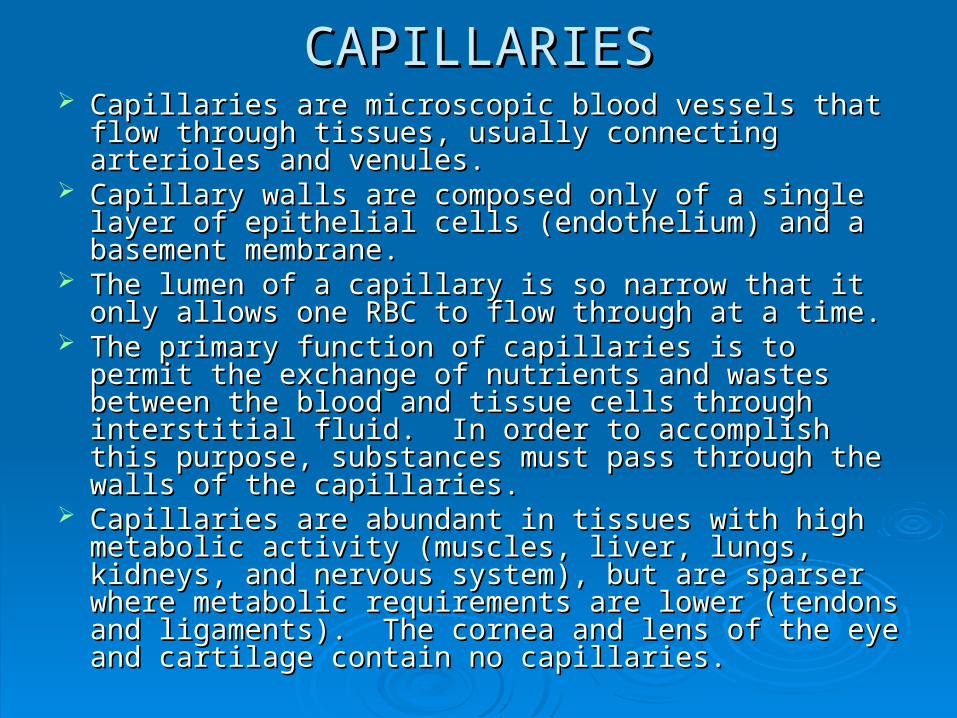
CAPILLARIESCAPILLARIES Capillaries are microscopic blood vessels that flow Capillaries are microscopic blood vessels that flow
through tissues, usually connecting arterioles and through tissues, usually connecting arterioles and venules.venules.
Capillary walls are composed only of a single layer of Capillary walls are composed only of a single layer of epithelial cells (endothelium) and a basement epithelial cells (endothelium) and a basement membrane.membrane.
The lumen of a capillary is so narrow that it only allows The lumen of a capillary is so narrow that it only allows one RBC to flow through at a time.one RBC to flow through at a time.
The primary function of capillaries is to permit the The primary function of capillaries is to permit the exchange of nutrients and wastes between the blood exchange of nutrients and wastes between the blood and tissue cells through interstitial fluid. In order to and tissue cells through interstitial fluid. In order to accomplish this purpose, substances must pass through accomplish this purpose, substances must pass through the walls of the capillaries.the walls of the capillaries.
Capillaries are abundant in tissues with high metabolic Capillaries are abundant in tissues with high metabolic activity (muscles, liver, lungs, kidneys, and nervous activity (muscles, liver, lungs, kidneys, and nervous system), but are sparser where metabolic requirements system), but are sparser where metabolic requirements are lower (tendons and ligaments). The cornea and lens are lower (tendons and ligaments). The cornea and lens of the eye and cartilage contain no capillaries.of the eye and cartilage contain no capillaries.
VENULESVENULES Venules form from several capillaries uniting as Venules form from several capillaries uniting as
they exit body tissues.they exit body tissues. Venules collect blood from capillaries and drain Venules collect blood from capillaries and drain
it into veins. it into veins. Venules close to capillaries of only an Venules close to capillaries of only an
endothelial tunica interna and a tunica media of endothelial tunica interna and a tunica media of a few scattered smooth muscle fibers.a few scattered smooth muscle fibers.
As venules approach veins they become larger As venules approach veins they become larger and acquire the tunica externa that is and acquire the tunica externa that is characteristic of veins.characteristic of veins.
VEINSVEINS The walls of veins are composed of essentially The walls of veins are composed of essentially
the same three tunics as arteries, but there are the same three tunics as arteries, but there are significant differences in their relative significant differences in their relative thicknesses.thicknesses.
The tunica interna is thinner and the tunica The tunica interna is thinner and the tunica media is much thinner, with relatively little media is much thinner, with relatively little smooth muscle and elastic fibers. The tunica smooth muscle and elastic fibers. The tunica externa is the thickest layer of veins, consisting externa is the thickest layer of veins, consisting of collagen and elastic fibers. Veins also do not of collagen and elastic fibers. Veins also do not contain the external or internal elastic laminae contain the external or internal elastic laminae found in arteries.found in arteries.
The lumen of a vein is larger than that of a The lumen of a vein is larger than that of a comparable artery.comparable artery.

VEINSVEINS
The average blood pressure in veins is The average blood pressure in veins is considerably lower than in arteries.considerably lower than in arteries.
Many veins contain one-way valves that prevent Many veins contain one-way valves that prevent blood from flowing backwards, away from the blood from flowing backwards, away from the heart. These valves are important since veins heart. These valves are important since veins have lower blood pressure and weaker, less have lower blood pressure and weaker, less muscular walls, so it is more difficult to keep muscular walls, so it is more difficult to keep blood moving forward through them.blood moving forward through them.
As veins pass through groups of skeletal As veins pass through groups of skeletal muscles, contractions of the muscles also help muscles, contractions of the muscles also help to push blood forward, toward the heart.to push blood forward, toward the heart.
BLOOD VESSELSBLOOD VESSELS