anatomy of the thoracolumbar spine physician name physician institution date
TRANSCRIPT

Anatomyof the Thoracolumbar Spine
Physician Name
Physician Institution
Date

Thoracolumbar Spine Anatomy

Thoracic Spine Anatomy
• Unique characteristics• Rib attachments• Relatively stiff kyphotic curve• Vertebral bodies vary greatly in shape and size
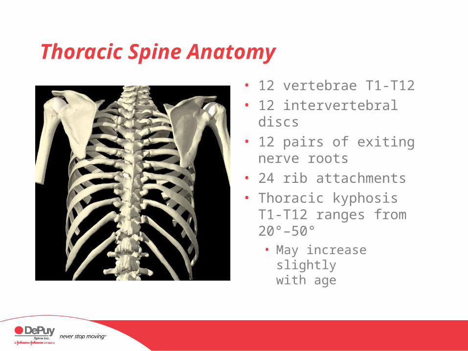
• 12 vertebrae T1-T12• 12 intervertebral discs • 12 pairs of exiting
nerve roots• 24 rib attachments • Thoracic kyphosis T1-
T12 ranges from 20°–50°• May increase slightly
with age
Thoracic Spine Anatomy

Thoracic Spine Anatomy
Costal facets• Rib articulates with
the vertebral bodies
• Ribs articulate with the transverse process (T1-T10)
• Ribs overlie the disc space (T1-T9)
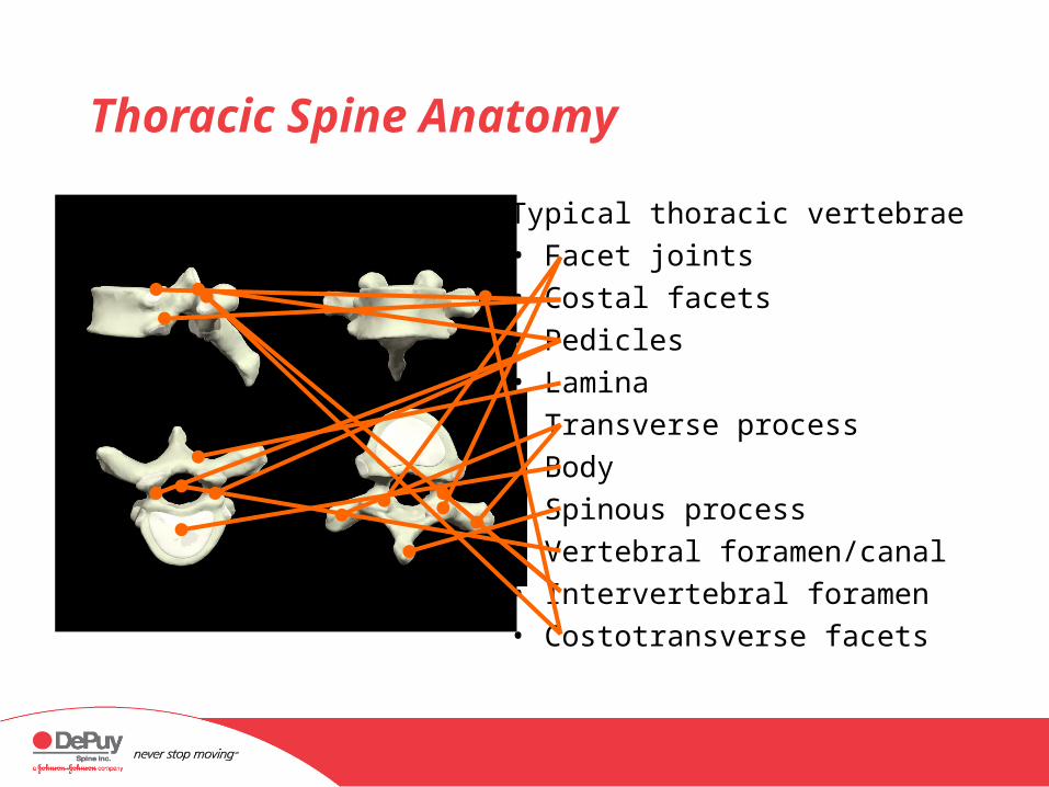
Typical thoracic vertebrae• Facet joints• Costal facets• Pedicles• Lamina• Transverse process• Body• Spinous process• Vertebral foramen/canal• Intervertebral foramen• Costotransverse facets
Thoracic Spine Anatomy
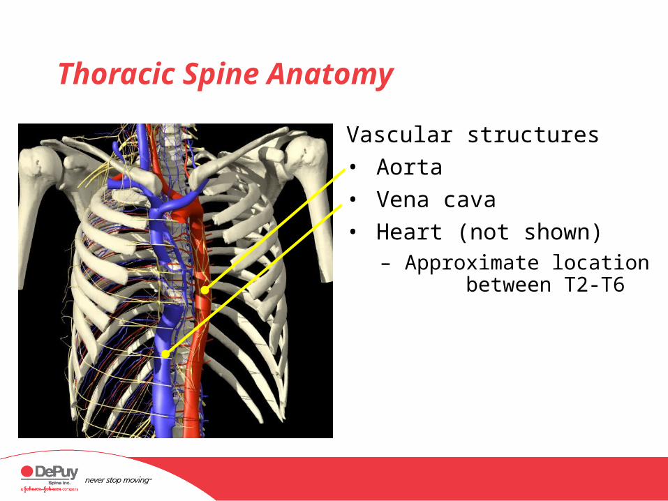
Vascular structures
• Aorta
• Vena cava
• Heart (not shown)– Approximate location
between T2-T6
Thoracic Spine Anatomy

Muscular structures
• Multifidus– Between transverse
and spinous processes
Thoracic Spine Anatomy

Muscular structures
• Longissimus– Between transverse
process and thoracolumbar fascia
Thoracic Spine Anatomy
Lumbar Spine Anatomy

Lumbar Anatomy
• 5 vertebrae L1-L5• 5 intervertebral discs • 5 pair of exiting nerve
roots• Lumbar lordosis L1-S1
ranges from 30°–80°• The apex of lumbar
lordosis L3-L4
11
22
33
44
55

Sacral Anatomy
• The sacrum is a series of 3, 4, or 5 fused coccygeal vertebrae
• The coccyx articulates with the inferior aspect of the sacrum11
223344
CC

Lumbar Spine Anatomy
Typical lumbar vertebra (L2)• Body• Vertebral foramen/canal• Intervertebral foramen• Pedicle• Transverse process• Lamina• Spinous process• Facet joints• Pars interarticularis
inferior
Superior
Anterior (oblique)
A Lateral P
Posterior (oblique)
Superior
Inferiorsuperior
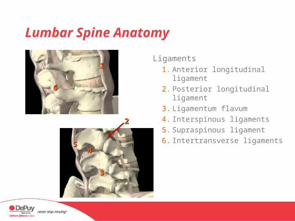
Lumbar Spine Anatomy
Ligaments1. Anterior longitudinal ligament
2. Posterior longitudinal ligament
3. Ligamentum flavum
4. Interspinous ligaments
5. Supraspinous ligament
6. Intertransverse ligaments
11
22
33
4455
66

Lumbar Spine Anatomy: Nerve Structures
• The spinal cord and nerve roots are often affected by skeletal problems
• Discs and bony tissue can interfere with normal nerve function and cause pain

Lumbar Spine Anatomy: Nerve Structures
Conus medularis • The point at which the
thick, single strand of the spinal cord ends
• Typically at T12 or L1
Note: in this illustration, the posterior elements of the spine, along with the dura mater and arachnoid mater, are not shown.

Lumbar Spine Anatomy: Nerve Structures
Cauda equina• The point at which
individual nerve roots continue down through the spinal canal
Note: in this illustration, the posterior elements of the spine, along with the dura mater and arachnoid mater, are not shown.

Lumbar Spine Anatomy: Nerve Structures
Exiting nerve root• Passes medial to the pedicle
of the anatomic segment
Traversing nerve root • Passes across the disc
space and beneath the pedicles of the inferior segment
Note: in this illustration, the posterior elements of the spine, along with the dura mater and arachnoid mater, are not shown.

Lumbar Spine Anatomy: Vascular Structures
The aorta and vena cava bifurcate around the level of the L3/L4 disc space
1. Aorta
2. Vena cava
3. Iliac arteries
4. Iliac veins
5. Midsacral vessels
