anatomy of a muscle from fascia to filament. connective tissue coverings skeletal muscle- organ...
TRANSCRIPT
ANATOMY OF A MUSCLE
FROM FASCIA TO FILAMENT
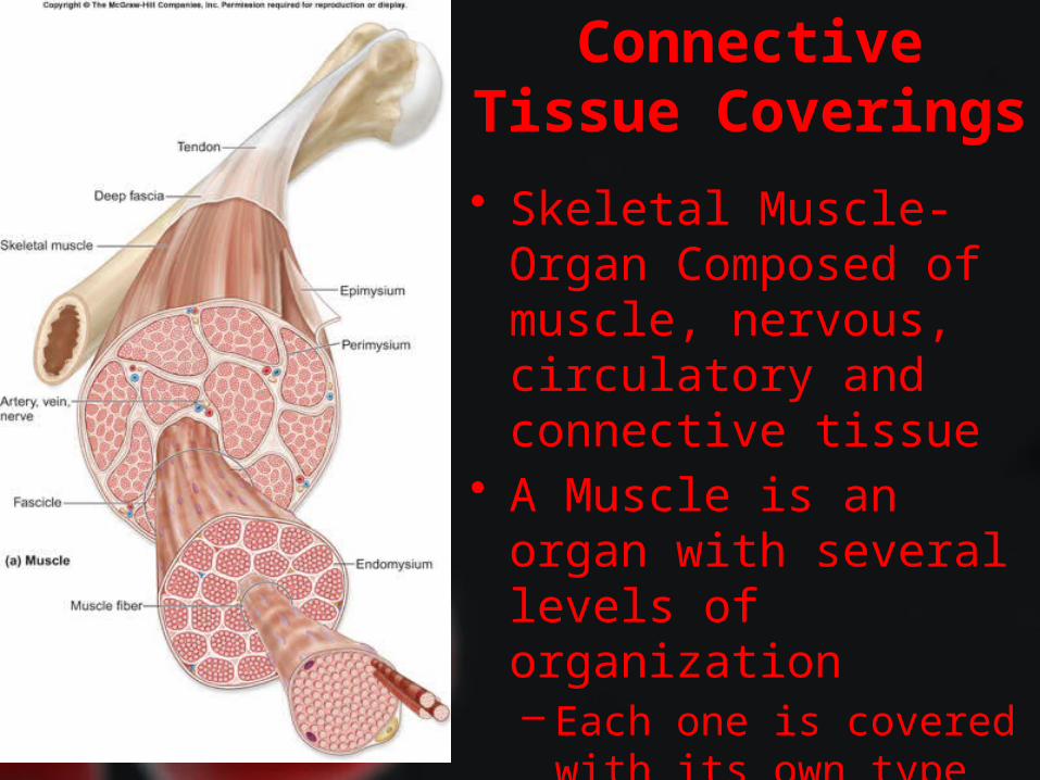
Connective Tissue Coverings
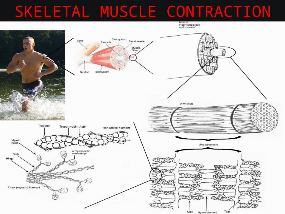
• Skeletal Muscle- Organ Composed of muscle, nervous, circulatory and connective tissue
• A Muscle is an organ with several levels of organization– Each one is covered with
its own type of connective tissue to hold its position
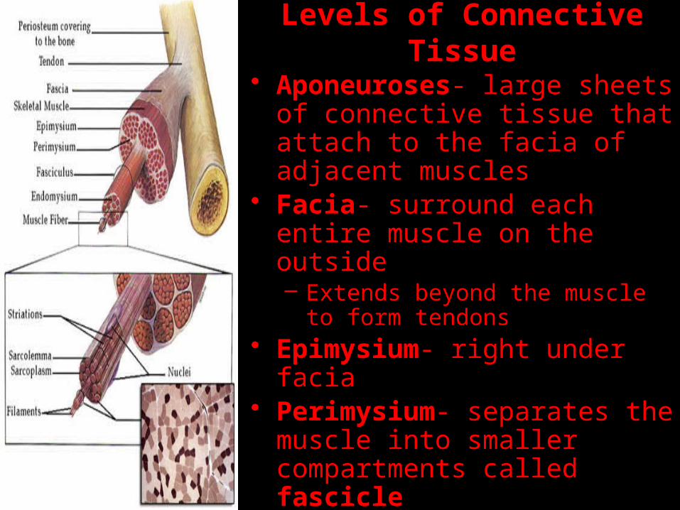
Levels of Connective Tissue
• Aponeuroses- large sheets of connective tissue that attach to the facia of adjacent muscles
• Facia- surround each entire muscle on the outside– Extends beyond the muscle to form
tendons• Epimysium- right under facia• Perimysium- separates the
muscle into smaller compartments called fascicle
• Endomysium- separates each fascicle into individual muscle fibers
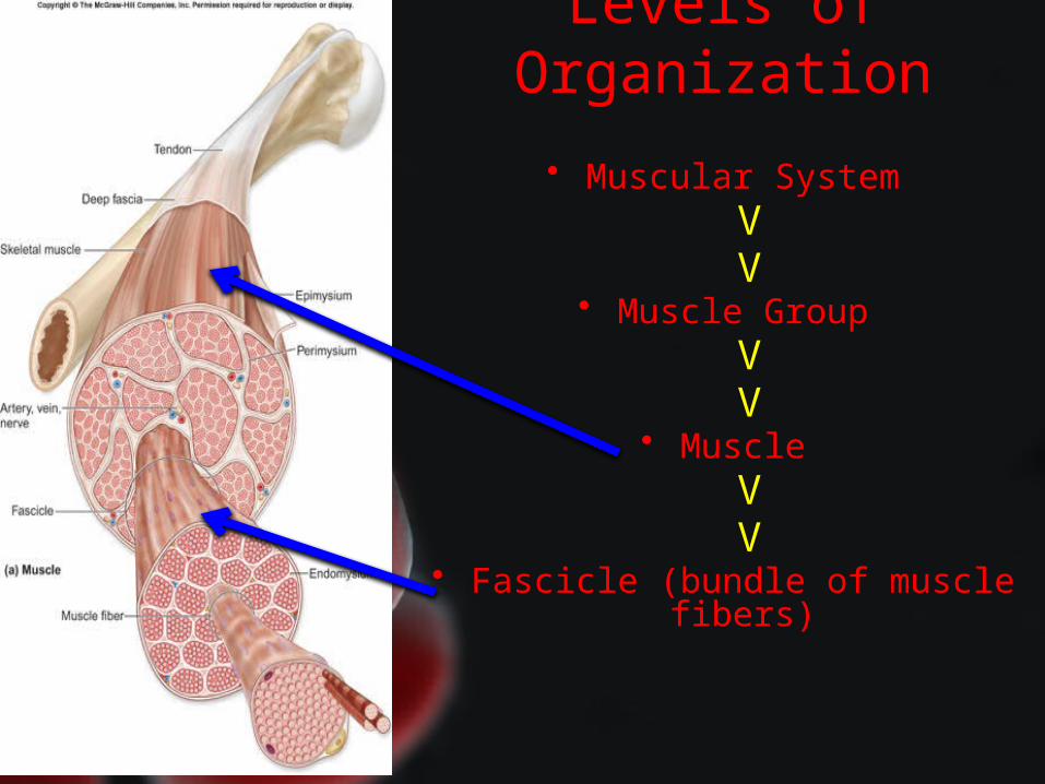
Levels of Organization
• Muscular SystemVV
• Muscle GroupVV
• MuscleVV
• Fascicle (bundle of muscle fibers)
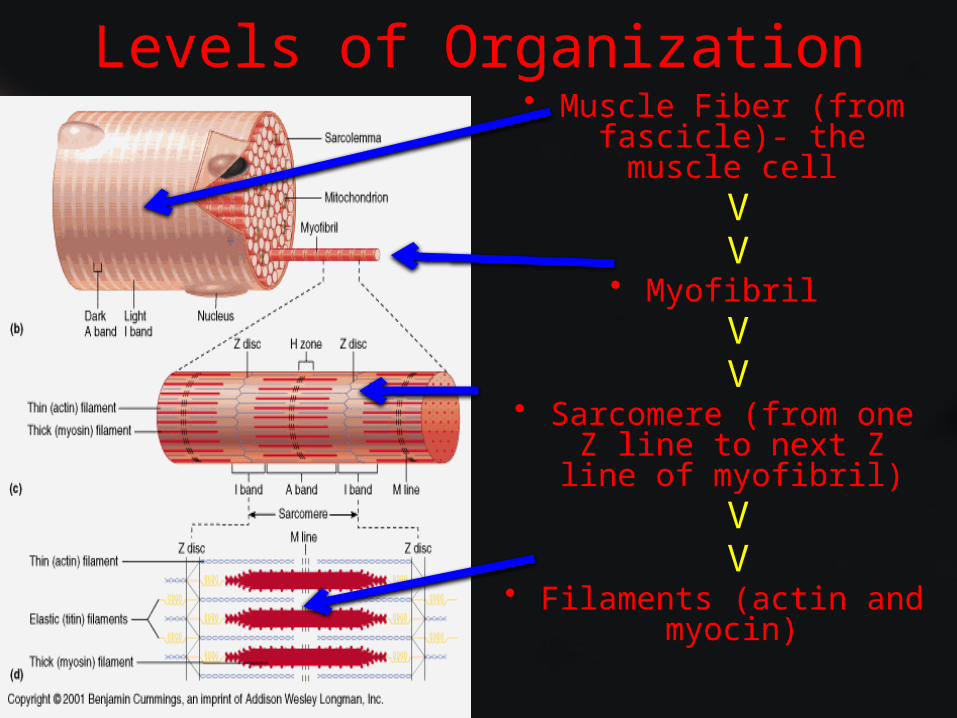
Levels of Organization• Muscle Fiber (from fascicle)- the muscle cell
VV
• MyofibrilVV
• Sarcomere (from one Z line to next Z line of myofibril)
VV
• Filaments (actin and myocin)
Skeletal Muscle Fibers
• Muscle Fiber- a cell that responds to stimuli & relaxes when stimuli ends– Actually the fusion of many cells called a myoblast– Multinucleated due to this fusion– Other cellular structures have special names due to
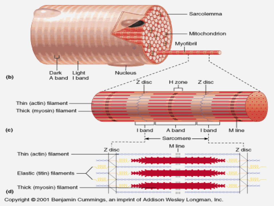
the fiber’s unique structure• Sarcolemma- outer covering• Sarcoplasm- inner fluid• Sarcoplasmic Reticulum- nework of internal channels• Transverse Tubules- channels that lead outside
– Combination of two networks activate muscle contractions
Skeletal Muscle Fibers• Each fiber is very dense with mitochondria
• Myofibrils- parallel threadlike subdivisions in each fiber• Fundamental in muscle contraction
– Actin (thin) & Myosin (thick)- two types of protein filaments• The alternating of these filaments produces muscle striations
– Muscle Striations- Two Main Parts• I Bands- light bands made up of thin actin filaments directly attached to
structures called Z lines• A Bands- dark bands composed of thick myosin filaments overlapping with
thin actin filaments– The H Zone is the middle of the A Band consisting of the M line
(thickening of the myosin filaments)• Sarcomere- segment of the myofibril that extends from Z line to Z line
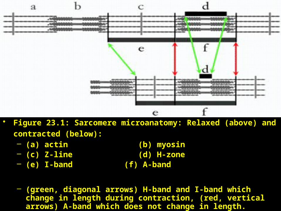
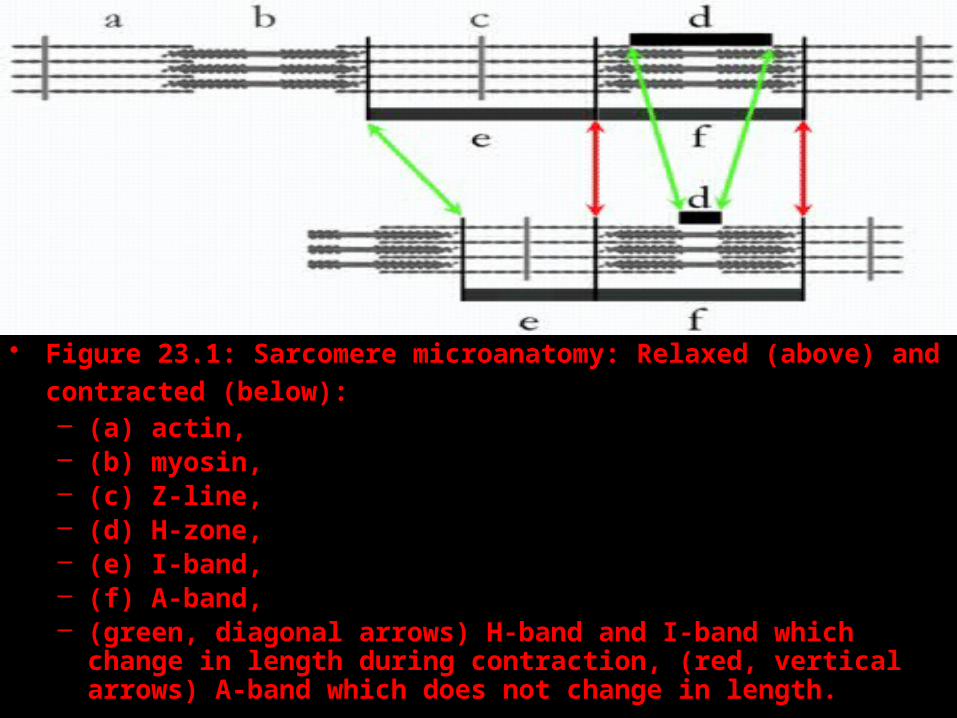
• Figure 23.1: Sarcomere microanatomy: Relaxed (above) and
contracted (below): – (a) actin (b) myosin– (c) Z-line (d) H-zone– (e) I-band (f) A-band
– (green, diagonal arrows) H-band and I-band which change in length during contraction, (red, vertical arrows) A-band which does not change in length.
NEUROMUSCULAR JUNCTION

Motor unit• A motor unit is a single motor
neuron and all of the corresponding muscle fibers it innervates.
• When a motor unit is activated, all of its fibers contract.
• Groups of motor units often work together to coordinate the contractions of a single muscle;
• all of the motor units that subserve a single muscle are considered a motor unit pool.
SKELETAL MUSCLE CONTRACTION
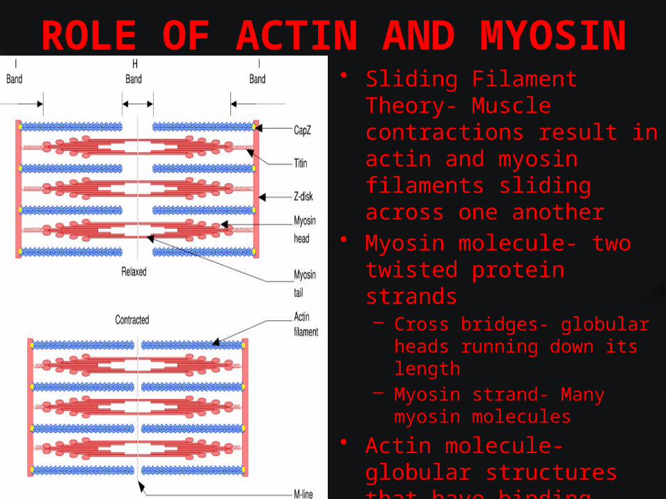
ROLE OF ACTIN AND MYOSIN• Sliding Filament Theory-
Muscle contractions result in actin and myosin filaments sliding across one another
• Myosin molecule- two twisted protein strands – Cross bridges- globular heads
running down its length– Myosin strand- Many myosin
molecules
• Actin molecule- globular structures that have binding sites for the myosin cross bridges– Actin strand- many actin
molecules in a helix
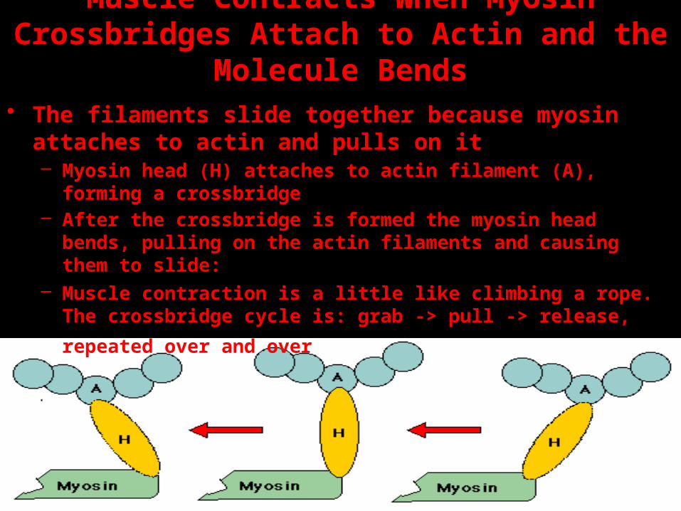
Muscle Contracts When Myosin Crossbridges Attach to Actin and the Molecule Bends
• The filaments slide together because myosin attaches to actin and pulls on it– Myosin head (H) attaches to actin filament (A), forming a
crossbridge– After the crossbridge is formed the myosin head bends, pulling on
the actin filaments and causing them to slide:– Muscle contraction is a little like climbing a rope. The crossbridge
cycle is: grab -> pull -> release, repeated over and over
ATP is Required for Both Contraction and Relaxation of Muscle
• ATP is the energy supply for contraction• It is required for the sliding of the filaments which is
accomplished by a bending movement of the myosin heads
• It is also required for the separation of actin and myosin which relaxes the muscle
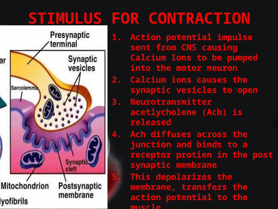
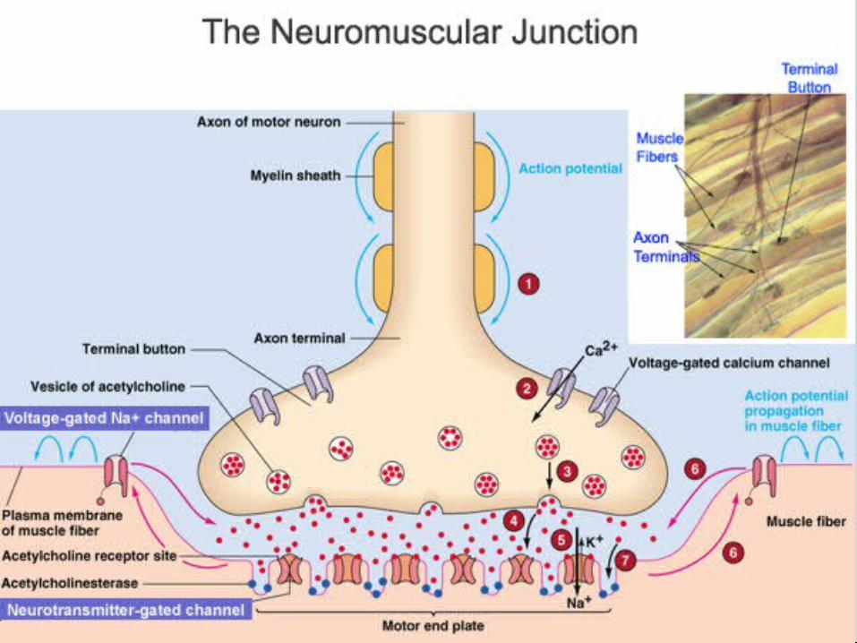
STIMULUS FOR CONTRACTION1. Action potential impulse sent from
CNS causing Calcium Ions to be pumped into the motor neuron
2. Calcium ions causes the synaptic vesicles to open
3. Neurotransmitter acetlycholene (Ach) is released
4. Ach diffuses across the junction and binds to a receptor protien in the post synaptic membrane
5. This depolarizes the membrane, transfers the action potential to the muscle
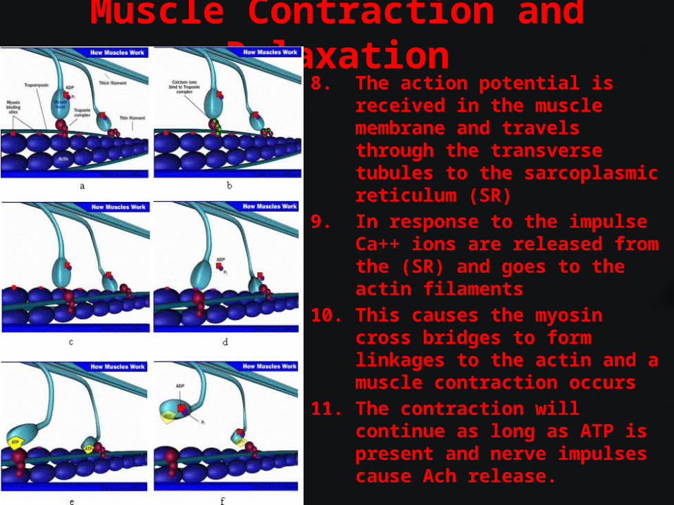
Muscle Contraction and Relaxation8. The action potential is received in
the muscle membrane and travels through the transverse tubules to the sarcoplasmic reticulum (SR)
9. In response to the impulse Ca++ ions are released from the (SR) and goes to the actin filaments
10. This causes the myosin cross bridges to form linkages to the actin and a muscle contraction occurs
11. The contraction will continue as long as ATP is present and nerve impulses cause Ach release.
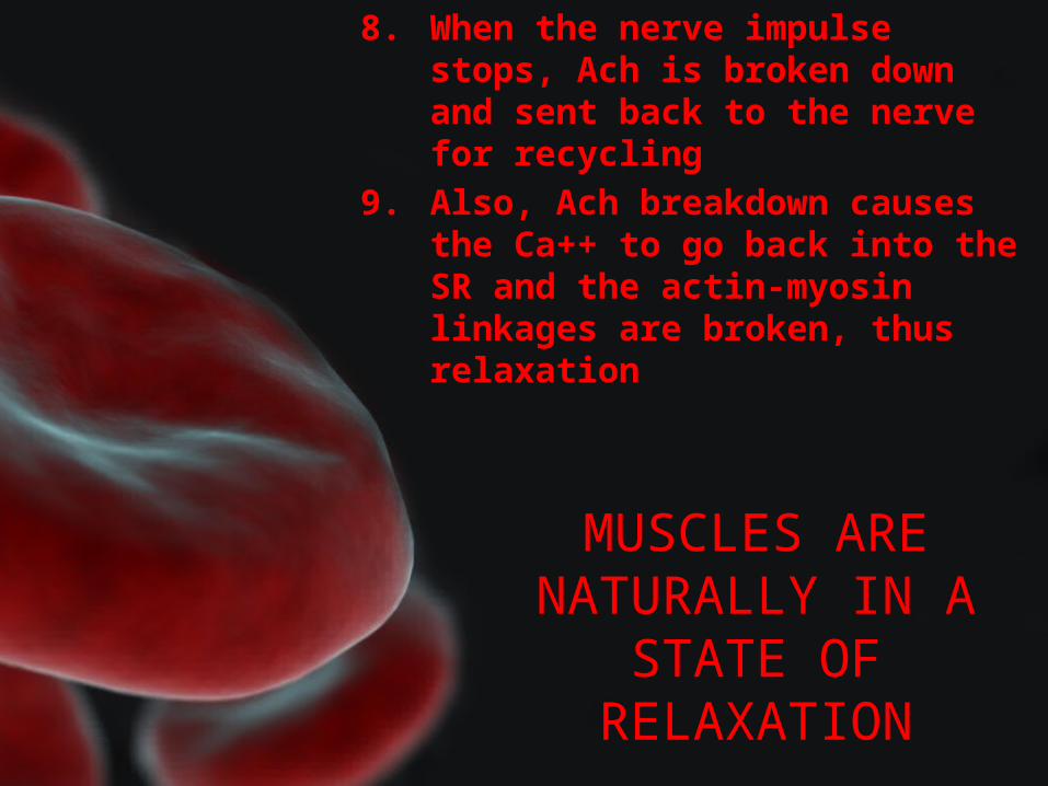
MUSCLES ARE NATURALLY IN A
STATE OF RELAXATION
8. When the nerve impulse stops, Ach is broken down and sent back to the nerve for recycling
9. Also, Ach breakdown causes the Ca++ to go back into the SR and the actin-myosin linkages are broken, thus relaxation
• Figure 23.1: Sarcomere microanatomy: Relaxed (above) and
contracted (below): – (a) actin, – (b) myosin, – (c) Z-line, – (d) H-zone, – (e) I-band, – (f) A-band, – (green, diagonal arrows) H-band and I-band which change in length
during contraction, (red, vertical arrows) A-band which does not change in length.
ENERGY FOR MUSCLE CONTRACTION• Muscle contractions take more ATP than is available in fibers• Creatine Phosphate- is able to make ATP from ADP and
phosphate– Stored in the mitochondria
OXYGEN SUPPLY & CELLULAR RESPIRATION
OXYGEN DEBT & MUSCLE FATIGUE
• Oxygen for cellular respiration comes from red blood cells– Hemoglobin- pigment molecules in red blood cells that
allows them to carry oxygen• Myoglobin- pigment in blood cells that also binds to oxygen
• Oxygen Debt- During activity, oxygen is used up and anaerobic respiration occurs– Anaerobic respiration results in the production of lactic acid
• Muscle Fatigue- caused by a build up of lactic acid
MUSCULAR RESPONSES• Use single muscle fiber to observe• Threshold Stimulus- minimum strength of stimulus required to
cause a muscle contraction• All-or-None Response- muscle fibers do not partially contract.
Once the threshold stimulus is reached, a full contraction occurs.
• Myogram- pattern a muscle contraction produces• Twitch- a single muscle contraction- 3 parts
– Latent period- delay btn stimulus and response– Period of contraction- when muscle pulls– Period of relaxation- when muscle returns to original length
• Summation- series of twitches that increase in strength before complete relaxation
• Tectanic contraction- no relaxation between twitches
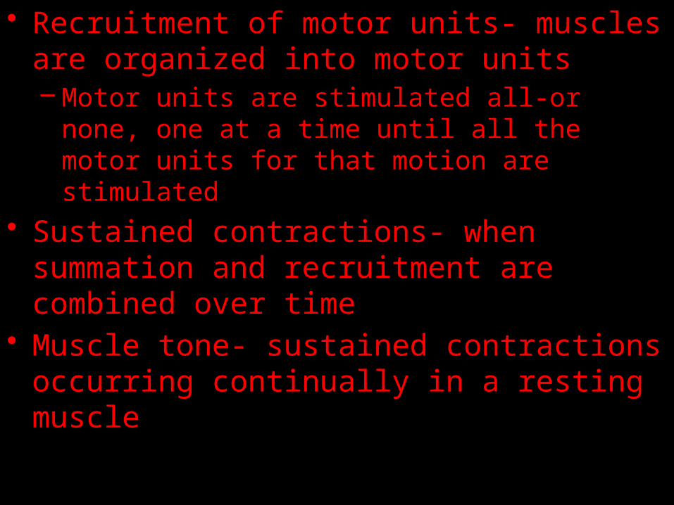
• Recruitment of motor units- muscles are organized into motor units– Motor units are stimulated all-or none, one at a time
until all the motor units for that motion are stimulated
• Sustained contractions- when summation and recruitment are combined over time
• Muscle tone- sustained contractions occurring continually in a resting muscle
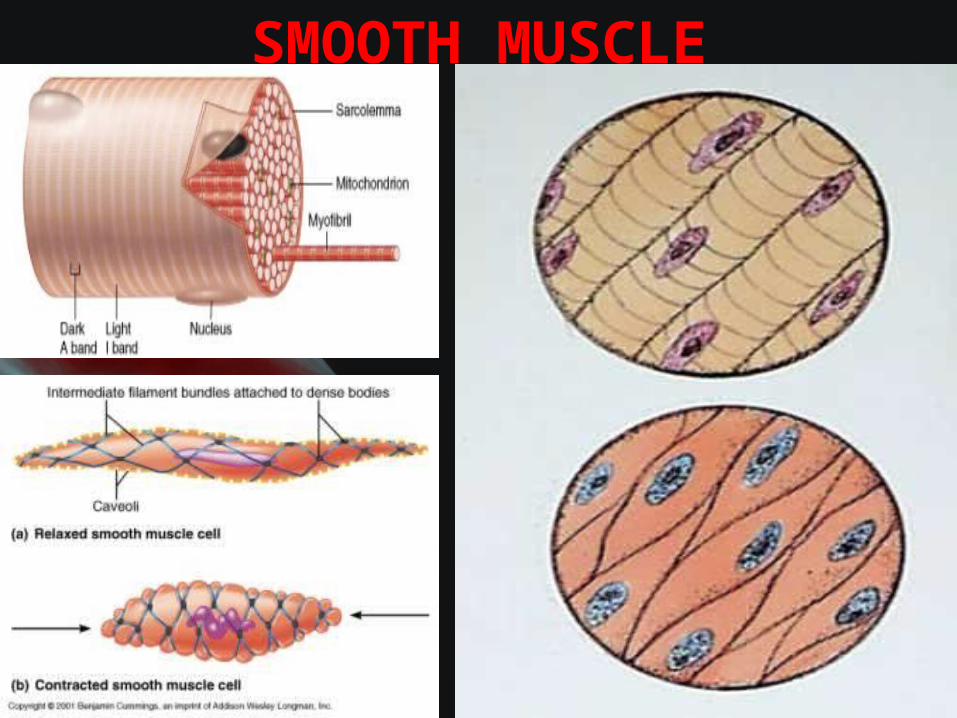
SMOOTH MUSCLE
SMOOTH MUSCLE• Contractile mechanisms for smooth and cardiac
muscles are the same as those of skeletal muscles• Smooth muscle fibers- no striations
– Multiunit smooth muscle- occur as separate fibers rather than in sheets • Irises of the eyes and blood vessels
– Visceral smooth muscle- sheets of spindle shaped cells- more common• Walls of hollow organs- stomach, intestine, bladder• Peristalsis- rhythmic contraction of smooth muscles
• Smooth muscle- slower to contract than skeletal muscle, but can maintain contraction longer with the same amount of ATP

CARDIAC MUSCLE• Occurs only in the heart• Striated cells joined end to
end• Long contractions• Intercolated Discs- cross
bands that connect opposing ends of cardiac muscle cells
• Cardiac muscle cells form a network and act as a single unit when the all-or-none stimuli response occurs
• Cardiac muscle is self exciting and rhythmic
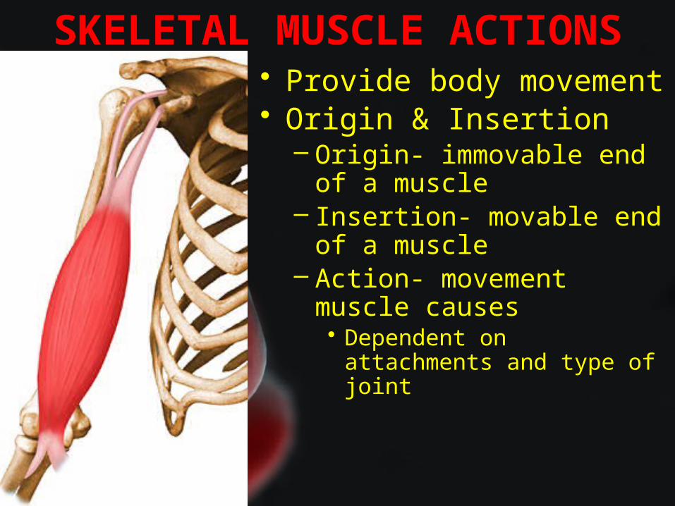
SKELETAL MUSCLE ACTIONS• Provide body movement• Origin & Insertion
– Origin- immovable end of a muscle
– Insertion- movable end of a muscle
– Action- movement muscle causes• Dependent on attachments and
type of joint
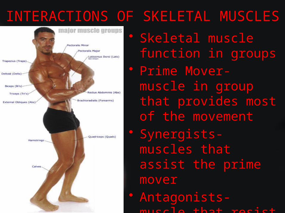
INTERACTIONS OF SKELETAL MUSCLES• Skeletal muscle function
in groups• Prime Mover- muscle in
group that provides most of the movement
• Synergists- muscles that assist the prime mover
• Antagonists- muscle that resist the prime mover and move in the opposite direction