anatomy and physiology i bones of the pelvic girdle and lower limb instructor: mary holman
TRANSCRIPT
Anatomy and Physiology I
Bones of the Pelvic Girdle
And
Lower Limb
Instructor: Mary Holman
Pelvic girdle
2 coxal (hip) bones united anteriorly at the pubic symphysis
Bony Pelvis
The complete ring consisting of the pelvic girdle, the pubic symphysis and the sacrum
Copyright © The McGraw-Hill Companies, Inc. Permission required for reproduction or display.
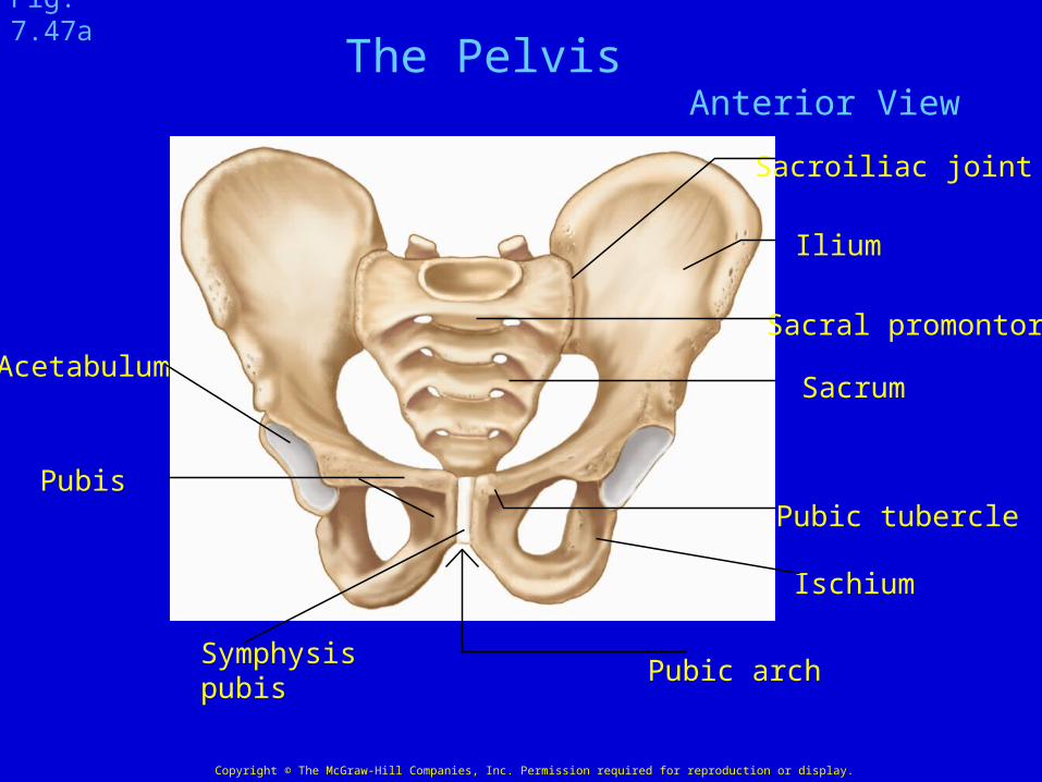
Sacrum
Sacral promontory
Sacroiliac joint
Acetabulum
Pubis
Pubic arch
Ischium
Pubic tubercle
Ilium
Symphysispubis
Fig. 7.47a
Anterior ViewThe Pelvis

Copyright © The McGraw-Hill Companies, Inc. Permission required for reproduction or display.
Obturator foramen
Ischium
CoccyxSacral hiatus
Sacrum
IliumSacral canal
Pubis
Fig. 7.47b
Posterior View
The Pelvis
Distance betweenIschial spines
Copyright © The McGraw-Hill Companies, Inc. Permission required for reproduction or display.
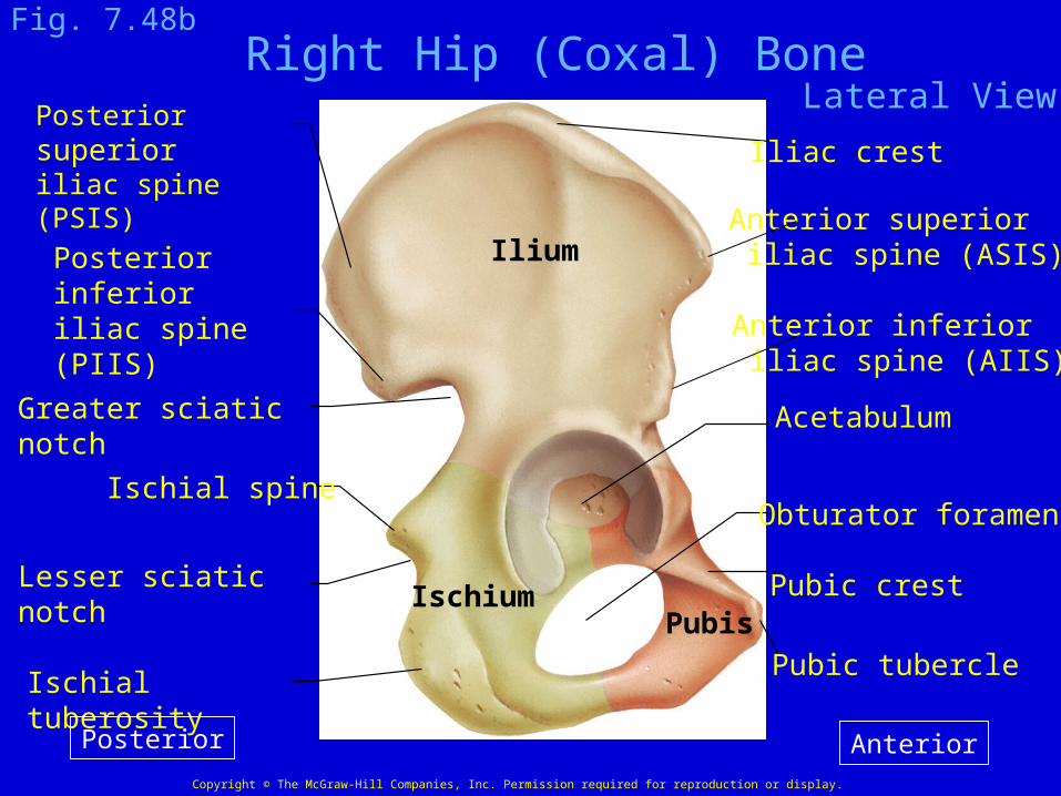
Iliac crest
Ilium
IschiumPubis
Ischial tuberosity
Ischial spineObturator foramen
Acetabulum
Pubic crest
Pubic tubercle
Posterior superioriliac spine (PSIS)
Posterior inferioriliac spine (PIIS)
Greater sciatic notch
Lesser sciatic notch
Fig. 7.48bRight Hip (Coxal) Bone
Lateral View
AnteriorPosterior
Anterior superior iliac spine (ASIS)
Anterior inferior iliac spine (AIIS)

Copyright © The McGraw-Hill Companies, Inc. Permission required for reproduction or display.
Anterior superioriliac spine (ASIS)
Anterior inferioriliac spine (AIIS)
Iliac crest
Posterior superioriliac spine (PSIS)
Posterior inferioriliac spine (PIIS)
Greater sciatic notch
Lesser sciatic notch
Iliac fossa
Ilium
IschiumPubis Ischial spineObturator foramen
Fig. 7.48a Right Hip Bone (Coxal) Bone
Medial Surface
AnteriorPosteriorIschial tuberosity
Auricular articulating surface
Copyright © The McGraw-Hill Companies, Inc. Permission required for reproduction or display.
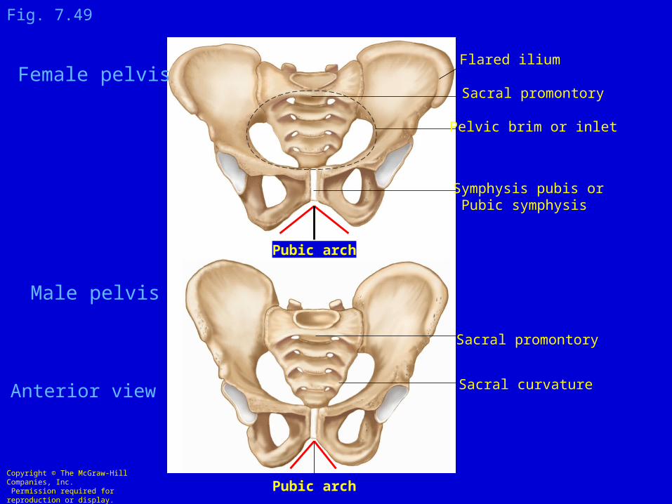
Sacral promontory
Flared ilium
Pelvic brim or inlet
Symphysis pubis or Pubic symphysis
Pubic arch
Pubic arch
Female pelvis
Male pelvis
Sacral promontory
Sacral curvature
Fig. 7.49
Anterior view
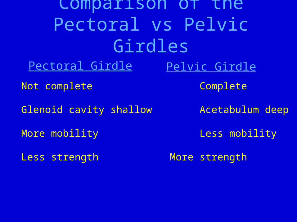
Comparison of the Pectoral vs Pelvic Girdles
Pectoral Girdle Pelvic Girdle
Not complete Complete
Glenoid cavity shallow Acetabulum deep
More mobility Less mobility
Less strength More strength
Copyright © The McGraw-Hill Companies, Inc. Permission required for reproduction or display.
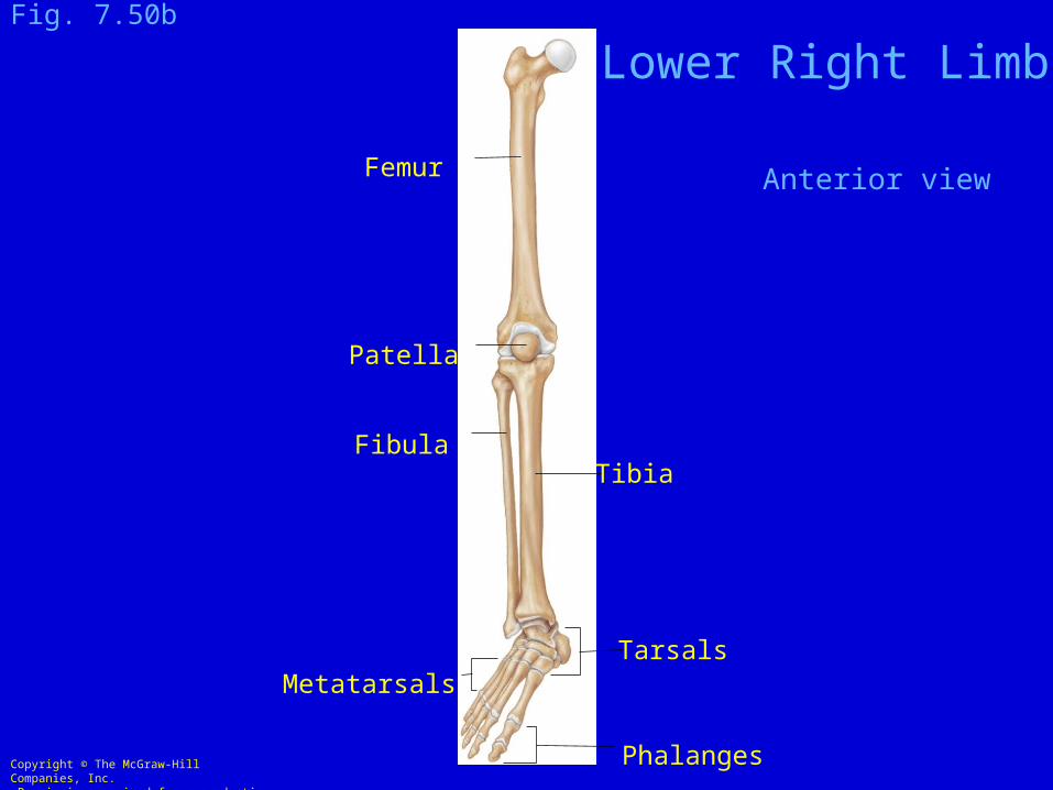
Metatarsals
FibulaTibia
Tarsals
Phalanges
Femur
Patella
Fig. 7.50b
Lower Right Limb
Anterior view

Copyright © The McGraw-Hill Companies, Inc. Permission required for reproduction or display.
© Martin Rotker
Fig. 7.47c
Copyright © The McGraw-Hill Companies, Inc.
Permission required for reproduction or display.
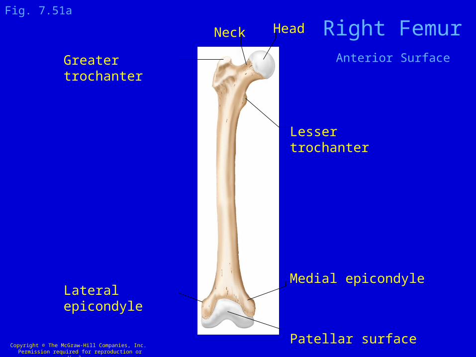
Medial epicondyle
Patellar surface
Lateral epicondyle
Greater trochanter
Lesser trochanter
Neck Head
Fig. 7.51a
Right FemurAnterior Surface

Copyright © The McGraw-Hill Companies, Inc. Permission required for reproduction or display.
Courtesy of John W. Hole, Jr.
Fig. 8.17b
Fovea capitis on the head of a Femur
From:Principles of Anatomy & Physiology by Tortora and Grabowski
Copyright © The McGraw-Hill Companies, Inc. Permission required for reproduction or display.
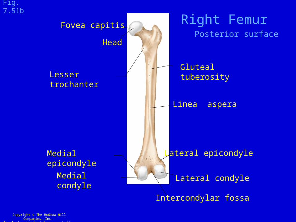
Lateral condyleMedial condyle
Intercondylar fossa
Medial epicondyle
Gluteal tuberosityLesser trochanter
Head
Fovea capitis
Linea aspera
Fig. 7.51b
Lateral epicondyle
Right FemurPosterior surface

Articulations of Lower
Lower Right Limb
Anterior View

Anterior View
Quadriceps Angleor
Q angle
Copyright © The McGraw-Hill Companies, Inc. Permission required for reproduction or display.
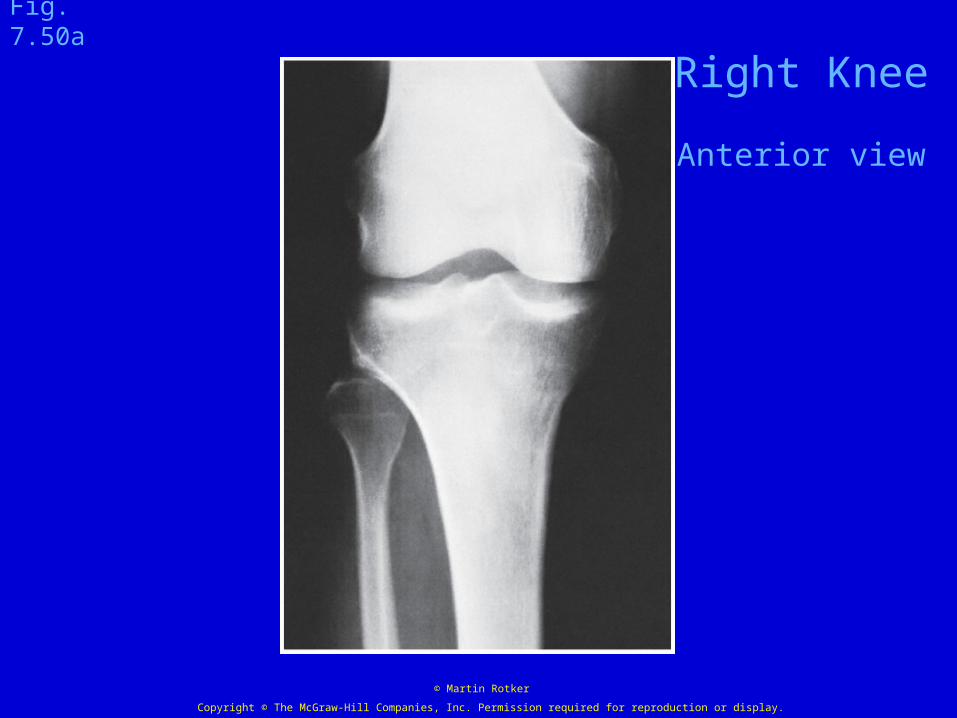
© Martin Rotker
Fig. 7.50a
Right Knee
Anterior view

Copyright © The McGraw-Hill Companies, Inc. Permission required for reproduction or display.
Tibia
Patella
Femur
Fibula
Lateral view
Fig. 7.50c
Right Knee Joint
Copyright © The McGraw-Hill Companies, Inc. Permission required for reproduction or display.
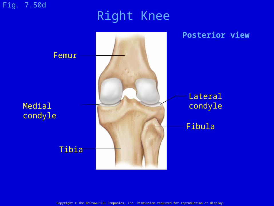
Fibula
Tibia
Lateral condyle
Posterior view
Medial condyle
Femur
Fig. 7.50d
Right Knee
Copyright © The McGraw-Hill Companies, Inc. Permission required for reproduction or display.
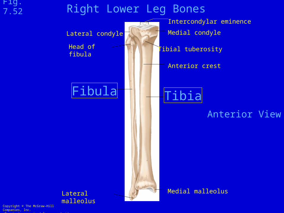
Medial malleolus
Tibia
Tibial tuberosity
Anterior crest
Medial condyle
Intercondylar eminence
Lateral malleolus
Lateral condyle
Head of fibula
Fibula
Fig. 7.52
Anterior View
Right Lower Leg Bones

Copyright © The McGraw-Hill Companies, Inc. Permission required for reproduction or display.
Fibula
Interosseus membrane of leg
Tibia
Medial malleolus
Anterior tibiofibular ligament(interosseus ligament)
Lateral malleolus
Fig. 8.1
Syndesmosis (fibrous joint) between the Tibia and Fibula

Copyright © The McGraw-Hill Companies, Inc. Permission required for reproduction or display.
© Martin Rotker
Fig. 7.53a
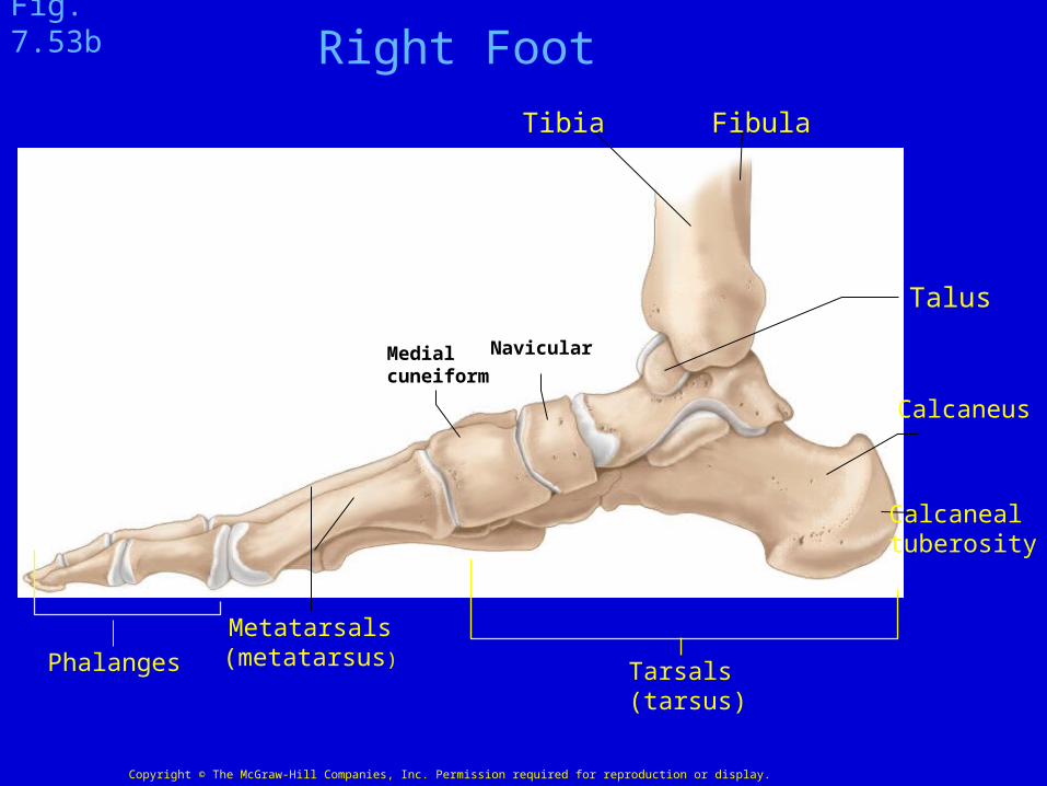
Tibia Fibula
NavicularMedialcuneiform
Metatarsals(metatarsus)Phalanges Tarsals
(tarsus)
Calcaneus
Calcanealtuberosity
Talus
Fig. 7.53b
Copyright © The McGraw-Hill Companies, Inc. Permission required for reproduction or display.
Right Foot
Copyright © The McGraw-Hill Companies, Inc. Permission required for reproduction or display.
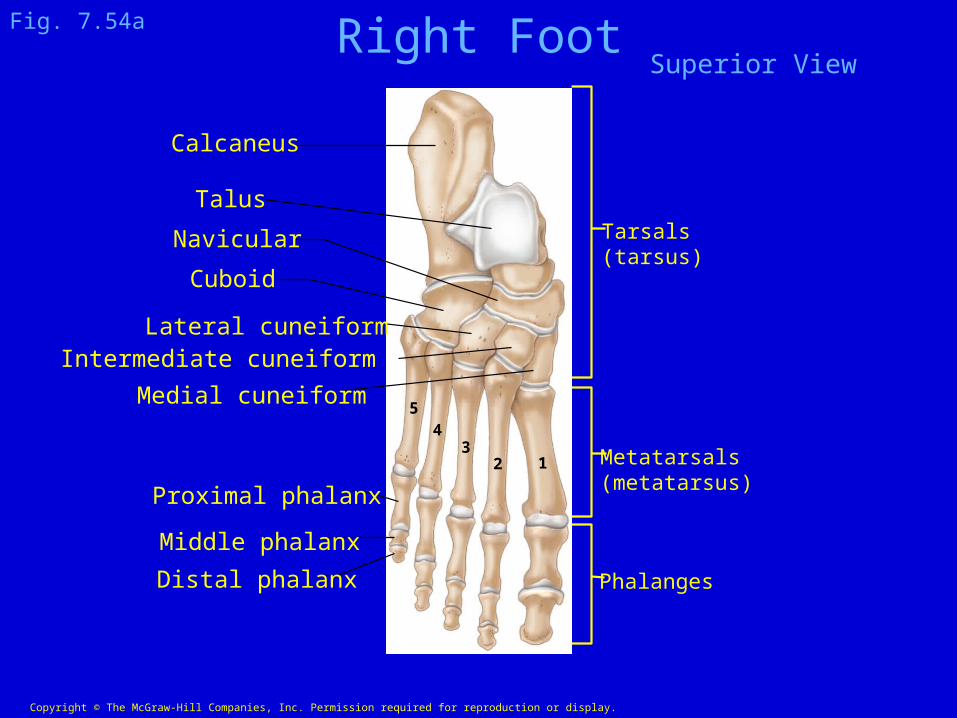
Calcaneus
Talus
Navicular
Cuboid
Lateral cuneiformIntermediate cuneiform
Proximal phalanx
Middle phalanx
Distal phalanx Phalanges
Metatarsals(metatarsus)
Tarsals(tarsus)
54
32 1
Medial cuneiform
Fig. 7.54a Right FootSuperior View

Arches of the Right Foot
From:Principles of Anatomy & Physiology by Tortora and Grabowski