anatomical landmarks for transnasal endoscopie …bib_dcd600fbc39d...keyword transnasal endoscopic...
TRANSCRIPT
Unicentre
CH-1015 Lausanne
http://serval.unil.ch
Year : 2011
Anatomical landmarks for transnasal endoscopie skull base surgery
Sandu Kishore
Sandu Kishore, 2011, Anatomical landmarks for transnasal endoscopie skull base surgery Originally published at : Thesis, University of Lausanne Posted at the University of Lausanne Open Archive. http://serval.unil.ch
Droits d’auteur L'Université de Lausanne attire expressément l'attention des utilisateurs sur le fait que tous les documents publiés dans l'Archive SERVAL sont protégés par le droit d'auteur, conformément à la loi fédérale sur le droit d'auteur et les droits voisins (LDA). A ce titre, il est indispensable d'obtenir le consentement préalable de l'auteur et/ou de l’éditeur avant toute utilisation d'une oeuvre ou d'une partie d'une oeuvre ne relevant pas d'une utilisation à des fins personnelles au sens de la LDA (art. 19, al. 1 lettre a). A défaut, tout contrevenant s'expose aux sanctions prévues par cette loi. Nous déclinons toute responsabilité en la matière.
Copyright The University of Lausanne expressly draws the attention of users to the fact that all documents published in the SERVAL Archive are protected by copyright in accordance with federal law on copyright and similar rights (LDA). Accordingly it is indispensable to obtain prior consent from the author and/or publisher before any use of a work or part of a work for purposes other than personal use within the meaning of LDA (art. 19, para. 1 letter a). Failure to do so will expose offenders to the sanctions laid down by this law. We accept no liability in this respect.

UNIVERSITE DE LAUSANNE -FACULTE DE BIOLOGIE ET DE MEDECINE
Département des services de chirurgie et d'anesthésiologie
Service d'ORL et chirurgie cervicofaciale
Anatomical landmarks for transnasal endoscopie skull base surgery
THE SE
préparée sous la direction du Professeur Philippe Pasche, médecin chef (avec la co-direction du Professeur Philippe Monnier, chef de service)
et présentée à la Faculté de biologie et de médecine de l'Université de Lausanne pour l'obtention du grade de
DOCTEUR EN MEDECINE
par
Kishore SANDU
Médecin diplômé de la Confédération Suisse obtenue en Sept 2011, FMH - ORL et Chirurgie Cervico-Faciale, Suisse obtenue en Nov 2011,
Fellow of European Board of Otorhinolaryngology and Head & Neck Surgery, 2010 Originaire de Bombay (lndia)
Lausanne
2011

Resumé :
La résection par voie endoscopique transnasale de tumeurs envahissant la base du crâne
antérieure a été récemment décrite. Cette chirurgie requiert une connaissance précise des
repères anatomiques endoscopiques afin réduire le risque de complications vasculaires et
neurologiques.
Nous avons réalisé une étude anatomique endoscopique sur 6 têtes dont 3 injectées avec du
silicone coloré. Les repères anatomiques pour les abords de 3 régions d’importance clinique
ont été étudiés. Les repères pour l’abord de l’apex orbitaire sont le recessus carotidien latéral,
l’empreinte du nerf optique, « l’optic strut » et le V2. Leurs rapports avec le canal optique,
l’artère carotide interne et les fentes orbitaires supérieures et inférieures sont décrits. Les
repères pour l’abord de l’apex pétreux sont le V2 et le nerf vidien qui permettent repérer la
portion intrapétreuse de l’artère carotide interne. Les repères pour l’abord de la fosse ptérygo-
maxillaire sont le V2 et le foramen rotundum, l’artère et le trou sphénopalatins et l’artère
maxillaire interne.
Cette nouvelle approche permettant d’aborder des lésions médianes et paramédianes ouvre de
nouvelles perspectives pour des équipes de neurochirurgiens et d’ORL. Ces voies d’abords
s’appliquent aussi bien à des résections décompressives à but palliatif qu’à l’exérèse de
tumeurs benignes et malignes, bien que les résultats à long terme doivent encore être validés
pour cette dernière indication.
Eur Arch OtorhinolaryngolDOI 10.1007/s00405-011-1698-4
123
RHINOLOGY
Anatomical landmarks for transnasal endoscopic skull base surgery
Kishore Sandu · Philippe Monnier · Philippe Pasche
Received: 12 April 2011 / Accepted: 27 June 2011© Springer-Verlag 2011
Abstract Resection of midline skull base lesions involveapproaches needing extensive neurovascular manipulation.Transnasal endoscopic approach (TEA) is minimally inva-sive and ideal for certain selected lesions of the anteriorskull base. A thorough knowledge of endonasal endoscopicanatomy is essential to be well versed with its surgicalapplications and this is possible only by dedicated cadav-eric dissections. The goal in this study was to understandendoscopic anatomy of the orbital apex, petrous apex andthe pterygopalatine fossa. Six cadaveric heads (3 injectedand 3 non injected) and 12 sides, were dissected using aTEA outlining systematically, the steps of surgical dissec-tion and the landmarks encountered. Dissection done by the“2 nostril, 4 hands” technique, allows better transnasalinstrumentation with two surgeons working in unison witheach other. The main surgical landmarks for the orbitalapex are the carotid artery protuberance in the lateral sphe-noid wall, optic nerve canal, lateral optico-carotid recess,optic strut and the V2 nerve. Orbital apex includes struc-tures passing through the superior and inferior orbitalWssure and the optic nerve canal. Vidian nerve canal and theV2 are important landmarks for the petrous apex. IdentiW-cation of the sphenopalatine artery, V2 and foramen rotun-dum are important during dissection of the pterygopalatinefossa. In conclusion, the major potential advantage of TEA
to the skull base is that it provides a direct anatomical routeto the lesion without traversing any major neurovascularstructures, as against the open transcranial approacheswhich involve more neurovascular manipulation and brainretraction. Obviously, these approaches require close coop-eration and collaboration between otorhinolaryngologistsand neurosurgeons.
Keyword Transnasal endoscopic skull base anatomy · Anatomical landmarks · Endonasal · Skull base surgery
Introduction
Detailed knowledge of nasal and paranasal anatomy,including the anterior and middle skull base, is a fundamen-tal requirement for surgical treatment of pathologiesinvolving these anatomical areas. Cadaver dissectionassisted by endoscopy is a valid approach for acquiring atrue three-dimensional mental image rather than schematicanatomic knowledge. Also, otorhinolaryngologists are ableto acquire appropriate practical skills that serve as basis formastering these complex interventions. Disorientation ofthe surgical Weld is the major risk faced by a surgeon duringendoscopy. An in-depth knowledge of endonasal anatomyimproves the sense of depth to identify anatomic landmarksand sites at risk, and thus can avoid iatrogenic damage.Another crucial prerequisite for mastering the most com-plex surgical situations is knowledge of the neighbouringanatomical structures delimiting the sinonasal structureslike the orbit, anterior and middle skull base.
The endoscopic transnasal transsphenoidal approach hasnow been adopted for treatment of several midline anteriorskull base lesions [1, 2]. It is versatile and minimally inva-sive as compared to open neurosurgical procedures. It oVers
Presented as an oral contribution at the Annual meeting of the Swiss Otorhinolaryngological Society, 2009 Geneva.
K. Sandu (&) · P. Monnier · P. PascheDepartment of Otorhinolaryngology, University Hospital CHUV, Lausanne, Switzerlande-mail: [email protected]

Eur Arch Otorhinolaryngol
123
a lower morbidity and mortality rate when compared toopen transcranial procedures leading to shorter hospitalstay. A variety of innovative skull base approaches (includ-ing anterior, anterolateral and posterolateral routes, whichoften require extensive neurovascular manipulation to gainaccess to the lesion) have been developed to allow resectionof extra-axial lesions of the anterior skull base that are out-side the sella, immediate parasellar area, or orbit but maybe in close contact with the cavernous sinus or the carotidartery. In contrast to the traditional cranial base surgicalapproaches, the endonasal technique oVers a directapproach without brain retraction oVering the surgeon anability to operate safely and eVectively in a precarious sur-gical Weld [3, 4]. Furthermore, the constant improvementsin diagnostic imaging techniques and the increasing use ofimage guidance systems during endoscopic endonasal pro-cedures have provided increasing accuracy and safety forthis approach, allowing improved, constant surgical orien-tation in an anatomically complex area.
We present an anatomical dissection study to deWneapplication of extended endoscopic endonasal approach tothe midline skull base concentrating mainly on threeareas—orbital apex, petrous apex and pterygopalatinefossa. The study was performed to understand better thecomplex anatomical relationships of the structures involvedin the approach, and become more familiar with the endo-scopic views and associated skills which are quite diVerentfrom traditional microscopic approach. Surgery wouldmean collaboration between otorhinolaryngologists andneurosurgeons who need to be equally acquainted with theendonasal anatomy and endoscopic skills so as to excise thelesions correctly and manage possible complications.
Materials and methods
Transnasal endoscopic dissection was performed in sixcadaveric heads (12 sides), of which three were non-injected and three heads had an intravascular injection ofcoloured liquid silicone using a previously described tech-nique [1]. The heads were stored in 75% alcohol solution.Cadaveric dissections were performed and the specimenstored in the Anatomy laboratory of the University Hospi-tal, Lausanne. We used StorzR 0°, 30°, 70° 4 mm, 18 and30 cm rod lens rigid endoscopes Wtted with an endoscoperinsing system (Karl Storz and Co., Tuttlingen, Germany).The endoscope was connected to a Xenon cold light sourcevia Wbreoptic cable and to a StrykerR video viewing andrecording column to carefully document every surgical stepand photograph important landmarks. The photographswere edited with AdobeR Photoshop CS3 program. Duringedition, additional colouring of important bony landmarks andneurovascular structures was done for better identiWcation of
the surface anatomy. Routine Storz sinuscopic and pituitaryinstruments were used for dissection. Long neurosurgicalhigh-speed cutting and diamond burrs were used for bonedrilling. Cadaveric specimens were placed supine with thehead in neutral position.
Surgical dissection
Nasal preparation and diagnostic nasal endoscopy
The dissection begins by cleaning of the nasal cavities withQ tips and cotton pledgets. The 1st diagnostic pass is alongthe Xoor of the nasal cavity between the inferior turbinateand the nasal septum up to the posterior choana. The mid-dle turbinate is identiWed rostrally to the inferior turbinate.Eustachian tube opening with its tubal elevation is seen inthe cavum. The 2nd pass is made to identify the middle tur-binate with its meatus. The 3rd pass is performed to visual-ise the natural sphenoid ostium passing the endoscopebetween the septum and middle turbinate. The ostium ispositioned in the sphenoethmoidal recess, medial to themiddle turbinate and behind the superior turbinate typically1 cm above the roof of the posterior choana. All surgicalsteps are performed using a 0° endoscope. The 30° and 70°scopes are used to see hidden areas needing angled visual-isation.
Preparation of surgical corridor
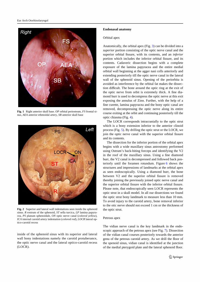
The right middle turbinate is medialised and resectedleaving a small stub attached to the skull base which canbe used for future reference. Uncinate process is excisedto visualise the hiatus semilunaris leading to the ethmoi-dal bulla. A complete anterior and posterior ethmoidec-tomy with excision of the basal lamella of the middleturbinate is performed. Lamina papyracea and the orbitalperiosteum, roof of the ethmoid and skull base with ante-rior ethmoid artery and the frontonasal recess are visual-ised (Fig. 1). Maxillary sinus ostium is identiWed with acurved suction tip following the superior edge of theinferior turbinate. The suction tip dips into the ostium atthe junction of middle and posterior third of the inferiorturbinate on the lateral nasal wall. Posterior ethmoidartery is identiWed on the skull base just anterior to theanterior sphenoid wall. A large posterior septectomy isdone to allow a 2 nostril, 4 hands technique which in turnallows increased instrumentation and introduction of twoor three instruments in the nasal cavity along with theendoscope at the same time. Anterior wall of the sphe-noid sinus is excised bilaterally leaving rostrum of thesphenoid intact. Intersphenoidal septum and the entiresphenoid sinus mucosa is removed. Figure 2 shows the
Eur Arch Otorhinolaryngol
123
inside of the sphenoid sinus with its superior and lateralwall bony indentations namely the carotid protuberance,the optic nerve canal and the lateral optico-carotid recess(LOCR).
Endonasal anatomy
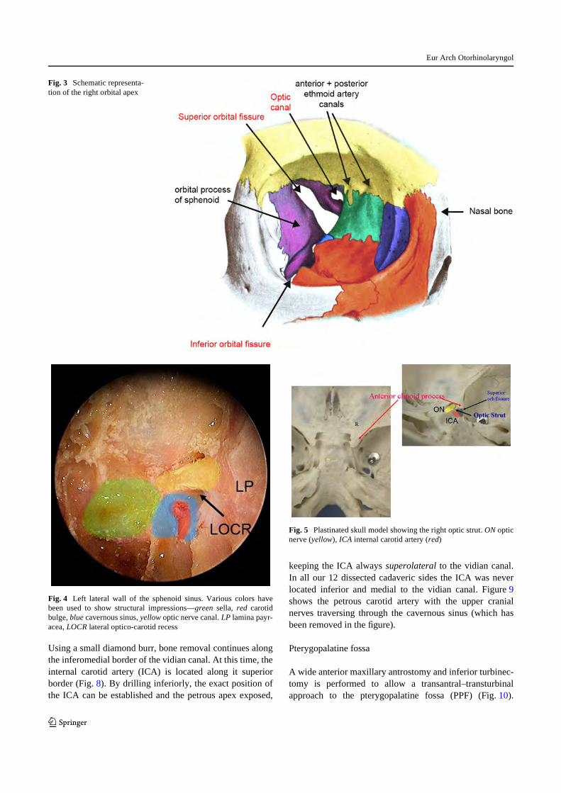
Orbital apex
Anatomically, the orbital apex (Fig. 3) can be divided into asuperior portion consisting of the optic nerve canal and thesuperior orbital Wssure, with its contents, and an inferiorportion which includes the inferior orbital Wssure, and itscontents. Cadaveric dissection begins with a completeexposure of the lamina papyracea and the entire medialorbital wall beginning at the agger nasi cells anteriorly andextending posteriorly till the optic nerve canal in the lateralwall of the sphenoid sinus. Opening of the periorbita isavoided as interference by the orbital fat makes the dissec-tion diYcult. The bone around the optic ring at the exit ofthe optic nerve from orbit is extremely thick. A Wne dia-mond burr is used to decompress the optic nerve at this exitexposing the annulus of Zinn. Further, with the help of aWne curette, lamina papyracea and the bony optic canal areremoved, decompressing the optic nerve along its entirecourse exiting at the orbit and continuing posteriorly till theoptic chiasma (Fig. 4).
The LOCR corresponds intracranially to the optic strutwhich is a bony extension inferior to the anterior clinoidprocess (Fig. 5). By drilling the optic strut or the LOCR, wejoin the optic nerve canal with the superior orbital Wssureand its contents.
The dissection for the inferior portion of the orbital apexbegins with a wide maxillary sinus antrostomy performedusing Ostrum’s back-biting forceps and identifying the V2in the roof of the maxillary sinus. Using a Wne diamondburr, the V2 canal is decompressed and followed back pos-teriorly until the foramen rotundum. Figure 6 shows thestructures and impressions of landmarks at the orbital apexas seen endoscopically. Using a diamond burr, the bonebetween V2 and the superior orbital Wssure is removedthereby joining the previously joined optic nerve canal andthe superior orbital Wssure with the inferior orbital Wssure.Please note, that endoscopically seen LOCR represents theoptic strut in a skull model. In all our dissections we foundthe optic strut bony landmark to measure less than 10 mm.To avoid injury to the carotid artery, bone removal inferiorto the otic nerve should not exceed 1 cm or the thickness ofthe optic strut.
Petrous apex
The vidian nerve canal is the key landmark in the endo-scopic approach of the petrous apex (see Fig. 7). Dissectionof the vidian canal courses posteriorly towards the anteriorgenu of the petrous carotid artery. As we drill the Xoor ofthe spenoid sinus, vidian canal is identiWed at the junctionof the medial pterygoid plate and the lateral sphenoid Xoor.
Fig. 1 Right anterior skull base. OP orbital periosteum, FS frontal si-nus, AEA anterior ethmoidal artery, SB anterior skull base
Fig. 2 Superior and lateral wall indentations seen inside the sphenoidsinus. R rostrum of the sphenoid, ST sella turcica, LP lamina papyra-cea, PS planum sphenoidale, ON optic nerve canal (colored yellow),ICA internal carotid artery indentation (colored red), LOCR lateral op-tico-carotid recess
Eur Arch Otorhinolaryngol
123
Using a small diamond burr, bone removal continues alongthe inferomedial border of the vidian canal. At this time, theinternal carotid artery (ICA) is located along it superiorborder (Fig. 8). By drilling inferiorly, the exact position ofthe ICA can be established and the petrous apex exposed,
keeping the ICA always superolateral to the vidian canal.In all our 12 dissected cadaveric sides the ICA was neverlocated inferior and medial to the vidian canal. Figure 9shows the petrous carotid artery with the upper cranialnerves traversing through the cavernous sinus (which hasbeen removed in the Wgure).
Pterygopalatine fossa
A wide anterior maxillary antrostomy and inferior turbinec-tomy is performed to allow a transantral–transturbinalapproach to the pterygopalatine fossa (PPF) (Fig. 10).
Fig. 3 Schematic representa-tion of the right orbital apex
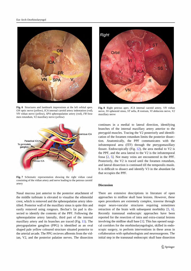
Fig. 4 Left lateral wall of the sphenoid sinus. Various colors havebeen used to show structural impressions—green sella, red carotidbulge, blue cavernous sinus, yellow optic nerve canal. LP lamina payr-acea, LOCR lateral optico-carotid recess
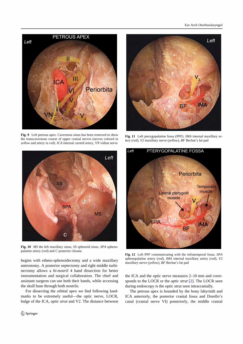
Fig. 5 Plastinated skull model showing the right optic strut. ON opticnerve (yellow), ICA internal carotid artery (red)
Eur Arch Otorhinolaryngol
123
Nasal mucosa just anterior to the posterior attachment ofthe middle turbinate is elevated to visualise the ethmoidalcrest, which is removed and the sphenopalatine artery iden-tiWed. Posterior wall of the maxillary sinus is quite thin andeasily removed using rongeurs. Bechat’s fat pad is dis-sected to identify the contents of the PPF. Following thesphenopalatine artery laterally, third part of the internalmaxillary artery and its branches are traced (Fig. 11). Thepterygopalatine ganglion (PPG) is identiWed as an ovalshaped pale yellow coloured structure situated posterior tothe arterial arcade. The PPG recieves aVerents from the vid-ian, V2, and the posterior palatine nerves. The dissection
continues in a medial to lateral direction, identifyingbranches of the internal maxillary artery anterior to thepterygoid muscles. Tracing the V2 posteriorly and identiW-cation of the foramen rotundum limits the posterior dissec-tion. Anatomically, the PPF communicates with theinfratemporal area (ITF) through the pterygomaxillaryWssure. Endoscopically (Fig. 12), the area medial to V2 isthe PPF, and the area lateral to the V2 is the infratemporalfossa [2, 5]. Not many veins are encountered in the PPF.Posteriorly, the V2 is traced until the foramen rotundum,and lateral dissection is continued till the temporalis musle.It is diYcult to dissect and identify V3 in the abundant fatthat occupies the PPF.
Discussion
There are extensive descriptions in literature of openapproaches to midline skull base lesions. However, theseopen procedures are extremely complex, traverse throughmajor neuro-vascular structures requiring sometimesretraction of the brain with subsequent morbidity [5, 6].Recently transnasal endoscopic approaches have beenreported for the resection of intra and extra-cranial lesionsinvolving the midline skull base [1]. This has opened surgi-cal corridors for the otorhinolaryngologist, skilled in endo-scopic surgery, to perform interventions in these areas incollaboration with opthalmologists and neurosurgeons. Theinitial step in the transnasal endoscopic skull base dissection
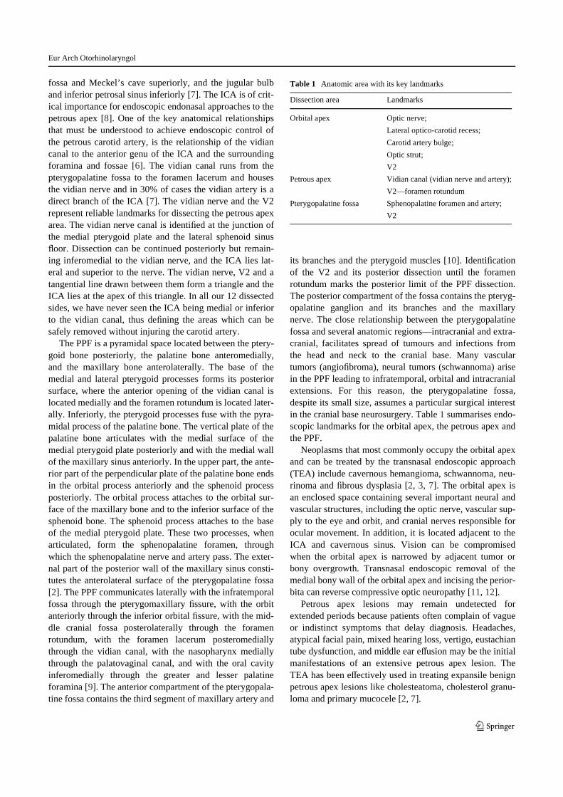
Fig. 6 Structures and landmark impressions at the left orbital apex.ON optic nerve (yellow), ICA internal carotid artery indentation (red),VN vidian nerve (yellow), SPA sphenopalatine artery (red), FR fora-men rotundum, V2 maxillary nerve (yellow)
Fig. 7 Schematic representation showing the right vidian canalconsisting of the vidian artery and nerve leading to the petrous carotidartery
Fig. 8 Right petrous apex. ICA internal carotid artery, VN vidiannerve, SS sphenoid sinus, ST sella, R rostrum, VI abducens nerve, V2maxillary nerve
Eur Arch Otorhinolaryngol
123
begins with ethmo-sphenoidectomy and a wide maxillaryantrostomy. A posterior septectomy and right middle turbi-nectomy allows a bi-nostril 4 hand dissection for betterinstrumentation and surgical collaboration. The chief andassistant surgeon can use both their hands, while accessingthe skull base through both nostrils.
For dissecting the orbital apex we Wnd following land-marks to be extremely useful—the optic nerve, LOCR,bulge of the ICA, optic strut and V2. The distance between
the ICA and the optic nerve measures 2–10 mm and corre-sponds to the LOCR or the optic strut [2]. The LOCR seenduring endoscopy is the optic strut seen intracranially.
The petrous apex is bounded by the bony labyrinth andICA anteriorly, the posterior cranial fossa and Dorello’scanal (cranial nerve VI) posteriorly, the middle cranial
Fig. 9 Left petrous apex. Cavernous sinus has been removed to showthe transcavernous course of upper cranial nerves (nerves colored inyellow and artery in red). ICA internal carotid artery, VN vidian nerve
Fig. 10 MS the left maxillary sinus, SS sphenoid sinus, SPA spheno-palatine artery (red) and C posterior choana
Fig. 11 Left pterygopalatine fossa (PPF). IMA internal maxillary ar-tery (red), V2 maxillary nerve (yellow), BF Bechat’s fat pad
Fig. 12 Left PPF communicating with the infratemporal fossa. SPAsphenopalatine artery (red), IMA internal maxillary artery (red), V2maxillary nerve (yellow), BF Bechat’s fat pad
Eur Arch Otorhinolaryngol
123
fossa and Meckel’s cave superiorly, and the jugular bulband inferior petrosal sinus inferiorly [7]. The ICA is of crit-ical importance for endoscopic endonasal approaches to thepetrous apex [8]. One of the key anatomical relationshipsthat must be understood to achieve endoscopic control ofthe petrous carotid artery, is the relationship of the vidiancanal to the anterior genu of the ICA and the surroundingforamina and fossae [6]. The vidian canal runs from thepterygopalatine fossa to the foramen lacerum and housesthe vidian nerve and in 30% of cases the vidian artery is adirect branch of the ICA [7]. The vidian nerve and the V2represent reliable landmarks for dissecting the petrous apexarea. The vidian nerve canal is identiWed at the junction ofthe medial pterygoid plate and the lateral sphenoid sinusXoor. Dissection can be continued posteriorly but remain-ing inferomedial to the vidian nerve, and the ICA lies lat-eral and superior to the nerve. The vidian nerve, V2 and atangential line drawn between them form a triangle and theICA lies at the apex of this triangle. In all our 12 dissectedsides, we have never seen the ICA being medial or inferiorto the vidian canal, thus deWning the areas which can besafely removed without injuring the carotid artery.
The PPF is a pyramidal space located between the ptery-goid bone posteriorly, the palatine bone anteromedially,and the maxillary bone anterolaterally. The base of themedial and lateral pterygoid processes forms its posteriorsurface, where the anterior opening of the vidian canal islocated medially and the foramen rotundum is located later-ally. Inferiorly, the pterygoid processes fuse with the pyra-midal process of the palatine bone. The vertical plate of thepalatine bone articulates with the medial surface of themedial pterygoid plate posteriorly and with the medial wallof the maxillary sinus anteriorly. In the upper part, the ante-rior part of the perpendicular plate of the palatine bone endsin the orbital process anteriorly and the sphenoid processposteriorly. The orbital process attaches to the orbital sur-face of the maxillary bone and to the inferior surface of thesphenoid bone. The sphenoid process attaches to the baseof the medial pterygoid plate. These two processes, whenarticulated, form the sphenopalatine foramen, throughwhich the sphenopalatine nerve and artery pass. The exter-nal part of the posterior wall of the maxillary sinus consti-tutes the anterolateral surface of the pterygopalatine fossa[2]. The PPF communicates laterally with the infratemporalfossa through the pterygomaxillary Wssure, with the orbitanteriorly through the inferior orbital Wssure, with the mid-dle cranial fossa posterolaterally through the foramenrotundum, with the foramen lacerum posteromediallythrough the vidian canal, with the nasopharynx mediallythrough the palatovaginal canal, and with the oral cavityinferomedially through the greater and lesser palatineforamina [9]. The anterior compartment of the pterygopala-tine fossa contains the third segment of maxillary artery and
its branches and the pterygoid muscles [10]. IdentiWcationof the V2 and its posterior dissection until the foramenrotundum marks the posterior limit of the PPF dissection.The posterior compartment of the fossa contains the pteryg-opalatine ganglion and its branches and the maxillarynerve. The close relationship between the pterygopalatinefossa and several anatomic regions—intracranial and extra-cranial, facilitates spread of tumours and infections fromthe head and neck to the cranial base. Many vasculartumors (angioWbroma), neural tumors (schwannoma) arisein the PPF leading to infratemporal, orbital and intracranialextensions. For this reason, the pterygopalatine fossa,despite its small size, assumes a particular surgical interestin the cranial base neurosurgery. Table 1 summarises endo-scopic landmarks for the orbital apex, the petrous apex andthe PPF.
Neoplasms that most commonly occupy the orbital apexand can be treated by the transnasal endoscopic approach(TEA) include cavernous hemangioma, schwannoma, neu-rinoma and Wbrous dysplasia [2, 3, 7]. The orbital apex isan enclosed space containing several important neural andvascular structures, including the optic nerve, vascular sup-ply to the eye and orbit, and cranial nerves responsible forocular movement. In addition, it is located adjacent to theICA and cavernous sinus. Vision can be compromisedwhen the orbital apex is narrowed by adjacent tumor orbony overgrowth. Transnasal endoscopic removal of themedial bony wall of the orbital apex and incising the perior-bita can reverse compressive optic neuropathy [11, 12].
Petrous apex lesions may remain undetected forextended periods because patients often complain of vagueor indistinct symptoms that delay diagnosis. Headaches,atypical facial pain, mixed hearing loss, vertigo, eustachiantube dysfunction, and middle ear eVusion may be the initialmanifestations of an extensive petrous apex lesion. TheTEA has been eVectively used in treating expansile benignpetrous apex lesions like cholesteatoma, cholesterol granu-loma and primary mucocele [2, 7].
Table 1 Anatomic area with its key landmarks
Dissection area Landmarks
Orbital apex Optic nerve;
Lateral optico-carotid recess;
Carotid artery bulge;
Optic strut;
V2
Petrous apex Vidian canal (vidian nerve and artery);
V2—foramen rotundum
Pterygopalatine fossa Sphenopalatine foramen and artery;
V2

Eur Arch Otorhinolaryngol
123
Certain PPF lesions like angioWbroma, schwannoma,neuroma are ideal to be treated by the TEA [1, 2]. Thisapproach is minimally invasive and better suited for surgi-cal debridement of fungal infections (aspergillosis, mucor-mycosis), than the open transfacial approaches. Eventhough this region is relatively small and its location isdeep, in the presence of tumors, especially benign ones, allthe neurovascular structures are displaced by the lesionitself, which creates or enlarges the surgical corridors.Under these conditions, endoscopically it is relatively easyto manipulate the blood vessels and nerves and then excisethese lesions.
Malignant tumors (primary/recurrent/metastatic epider-moid, adenoid cystic) at the orbital apex, petrous apex andPPF can be treated by transnasal endoscopic debulking fol-lowed by radiotherapy, and this has been accepted as aneVective means of surgical palliation [2], though results andsurgical aims are debatable. Complications of the expandedendonasal approach are the same as open approaches: neu-ral and vascular injury, meningitis and CSF leak. The mainproblem faced during surgery is bleeding, and only anexperienced team can eVectively use the bipolar electrocau-tery or manage cavernous sinus bleeding with precise focalpacking without increasing the neurovascular injury.Another important factor in consideration is the reconstruc-tion of ventral skull base defects after endoscopic excisions.Multilayered closures, vascularised nasoseptal mucosalXaps, balloon stenting and surgical bioglues have been sen-tinel events in reconstruction that have been eVective inclosing these defects reducing chances of CSF leaks [10].
At present, these surgical resection techniques are beingpractised at very few centers and long term results areawaited. The most critical issue to be addressed is avoidingtrauma to the carotid artery and the optic nerve. One musthave a thorough knowledge of various radiological investi-gations like the CT, MRI and 3D reconstructional imagesof the skull base. Periodic discussions with radiologicalcollegues with special interests in the skull base betters ourunderstanding of this complex anatomy. Intraoperatively,use of navigational techniques and a systematic use of thedoppler have been very useful in our limited experience.This approach adds choice, variety and versatility to thearmamentarium of a skull base surgeon and may serve as agood alternative to transfacial approaches which are com-plex and more morbid.
Conclusions
Surgical interventions in complex areas of orbital apex,petrous apex and the pterygopalatine fossa is a long learn-
ing curve and begins in the dissection rooms. One must rea-lise that anatomy is a science of recognition and not ofdiscovery, hence surgeons need to have adequate knowl-edge of endoscopic nasal anatomy before embarking ontreating lesions of these complex regions. Each of theseareas can be approached systematically, by deWning andidentifying reliable landmarks. These approaches could beeVectively used for certain selected lesions of the anteriorskull base, which otherwise would require more complexand morbid procedures. We intend to plastinate our dis-sected specimens for teaching purposes. Obviously, theseapproaches require collaboration between otorhinolaryng-ologists and neurosurgeons.
ConXict of interest None.
References
1. Cavallo LM, Messina A, Cappabianca P, Esposito F, De DevitiisE, Gardner P, Tschabitcher M (2007) Extended endoscopic endo-nasal transsphenoidal approach to the suprasellar area: anatomicconsiderations—part 1. Neurosurgery 61:24–34
2. Anand VK, Schwartz TH (2007) Practical endoscopic skull basesurgery. Plural Publishing, San Diego
3. Jho HD, Ha HG (2004) Endoscopic endonasal skull base surgery:part 1-the midline anterior fossa skull base. Minim Invasive Neu-rosurg 47:1–8
4. Cavallo LM, Mesina A, Cappabianca P, Gardner P, TschabitscherM (2005) Endoscopic endonasal surgery of the midline skull base:anatomical study and clinical considerations. Neurosurg Focus19(1):E2
5. Malgro F, Solari D, Cavallo LM, Samii A, Cappabianca P, PaternoV (2006) Endoscopic endonasal approach to the lateral recess ofthe sphenoid sinus via the pterygopalatine fossa: comparison ofendoscopic and radiological landmarks. Neurosurgery 59(ONSSuppl 4):237–243
6. Kassam AB, Gardner P, Snyderman C, Carrau R (2008) Expandedendonasal approach: vidian canal as a landmark to the petrousinternal carotid artery. J Neurosurg 108:177–183
7. Kassam AB, Gardner P, Snyderman C et al. (2005) Expandedendonasal approach: fully endoscopic, completely transnasalapproach to the middle third of the clivus, petrous bone, middlecranial fossa, and infratemporal fossa. Neurosurg Focus 19:E6,1–10
8. Brackmann DE, Toh EH (2003) Surgical management of petrousapex cholesterol granulomas. Otol Neurotol 23:529–533
9. Osborn AG (1980) The vidian artery: normal and pathologic anat-omy. Radiology 136:373–378
10. Snyderman CH, Kasam AB, Carrau R, Mintz A (2007) Endo-scopic reconstruction of cranial base defects following endonasalskull base surgery. Skull base: an interdisciplinary approach17(1):75–76
11. Yeh S, Foroozan R (2004) Orbital apex syndrome. Curr Opin Oph-thalmol 15(6):490–498
12. Schick U, Dott U, Hassler W (2003) Surgical treatment of orbitalcavernomas. Surg Neurol 60(3):234–244