anatomical and histochemical characterization of dipteryx ...and histochemical characterization of...
TRANSCRIPT
O
Aa
PLa
b
c
d
e
a
ARAA
KFILMS
I
NdtBedewratc
0c
Revista Brasileira de Farmacognosia 29 (2019) 425–433
ww w.elsev ier .com/ locate /b jp
riginal Article
natomical and histochemical characterization of Dipteryx odoratand Taralea oppositifolia, two native Amazonian species
aulo Marcos Ferreira da Silva a, Eduardo Oliveira Silva b,c,∗, Marleide de Sousa Chaves Rêgo a,aísa Maria de Resende Castro d, Advanio Inácio Siqueira-Silva e
Programa de Pós-graduac ão em Ciências Biológicas, Universidade Federal Rural da Amazônia/Museu Paraense Emílio Goeldi, Belém, PA, BrazilCoordenac ão de Licenciatura em Ciências Naturais, Universidade Federal do Maranhão, Codó, MA, BrazilPrograma de Pós-graduac ão em Botânica Aplicada, Universidade Federal de Lavras, Lavras, MG, BrazilPrograma de Pós-graduac ão em Botânica, Universidade de Brasília, DF, BrazilCoordenac ão de Agronomia, Universidade Federal do Oeste do Pará, Juriti, PA, Brazil
r t i c l e i n f o
rticle history:eceived 4 February 2019ccepted 15 May 2019vailable online 3 July 2019
eywords:abaceaedioblastseaflet blade
a b s t r a c t
Dipteryx odorata (Aubl.) Willd. and Taralea oppositifolia Aubl., Fabaceae: Dipterygeae, are two Amazo-nian species of great economic and pharmacological potential. The anatomy of these species, however,remains poorly studied. The aim of this work was to inventory leaf anatomical characteristics of D. odor-ata and T. oppositifolia and to locate and identify secretory structures and determine the main classes ofmetabolites they store. Vegetative branches were collected in Parque Ecológico de Gunma, Belém, stateof Pará, Brazil. Some of the branches were destined for herborization while the remainder was submit-ted to standard protocols for anatomical analysis and histochemical tests. Both species were found topossess an unstratified epidermis, with D. odorata being amphistomatic and T. oppositifolia being hypos-
edicinal plantsecretory structures
tomatic, and dorsiventral mesophyll with spongy parenchyma and wide cellular space. The two specieswere also found to possess idioblasts and secretory cavities that produce a heterogeneous exudate con-sisting of polysaccharides, lipids, alkaloids and phenolic compounds. The species presented differencesin leaf anatomy and chemical composition of the secretory structures, which may be useful in theirdifferentiation.
© 2019 Sociedade Brasileira de Farmacognosia. Published by Elsevier Editora Ltda. This is an openhe CC
access article under tntroduction
Dipterygeae (Leguminosae, Papilionoideae) is an exclusivelyeotropical clade of approximately 25 species of woody legumesistributed among four genera whose representatives occur in phy-ogeographical domains of the Amazon: Monopteryx Spruce exenth, Pterodon Vogel, Dipteryx Schreb. and Taralea Aubl. (Cardosot al., 2013). Among these genera, Dipteryx and Taralea stand outue to the economic potential of their species. Although these gen-ra share a number of vegetative characters, such as thick leaves,inged rachises, terminal appendices probable arising from the
eduction of the terminal leaflet, 4–18 alternate leaflets, leafletpexes ranging from cuspidate to obtuse and the presence of
ranslucent punctuations, their fruits have distinctive morphologi-al characteristics, including drupe-type fruit with late dehiscence∗ Corresponding author.E-mail: [email protected] (E.O. Silva).
https://doi.org/10.1016/j.bjp.2019.05.004102-695X/© 2019 Sociedade Brasileira de Farmacognosia. Published by Elsevier Editreativecommons.org/licenses/by-nc-nd/4.0/).
BY-NC-ND license (http://creativecommons.org/licenses/by-nc-nd/4.0/).
in Dipteryx and legume fruit with elastic dehiscence in Taralea(Barroso et al., 1999; Francisco, 2010).
Dipteryx odorata (Aubl.) Willd and Taralea oppositifolia Aubl.are popularly known in Brazil as “cumaru” or “cumaru-ferro”(Sousa et al., 2007; Carvalho, 2009) and “cumarurana” or “cumaru-amarelo” (Faria and Lima, 2002; Sousa et al., 2007), respectively.Both species have multiple uses, and have been used in medicine,pharmacology, and the perfumery and cosmetics industries(Uchida and Campos, 2000; Bessa et al., 2001; Takemoto et al., 2001;Pesce, 2009; Breitbach et al., 2013), as well as in the timber sec-tor (Sousa et al., 2007; Herrero-Jáuregui et al., 2012; Soriano et al.,2012). Almonds of Dipteryx odorata have also been used as a rawmaterial in the production of biodiesel (Ramalingam et al., 2018).
Studies related to the anatomy of vegetative and reproduc-tive organs involving genera of Leguminosae have provided newcharacters to support both taxonomic (Lackey, 1978; Leelavathi
et al., 1980; Coutinho et al., 2016), and phylogenetic investigationssuch as, for example, morphological data combined with molecularmarkers for the reconstruction of phylogenies and the indicationof possible synapomorphies for genera (Francisco, 2010). In gen-ora Ltda. This is an open access article under the CC BY-NC-ND license (http://
4 de Fa
ep12c2impD
tDdsl
M
S
dmnafep2va
P
aalt
osoc
lsn
S
et(aa2fJnwagr
26 P.M. Silva et al. / Revista Brasileira
ral, anatomical studies and studies of the chemical compoundsroduced by these species have mainly emphasize wood (Gasson,999) and seeds (Bessa et al., 2001; Takemoto et al., 2001; Pesce,009), leaving little known about the anatomical and histochemi-al characterization of the leaves (Palermo et al., 2017; Silva et al.,018). Nonetheless, anatomical and histochemical characterization
s necessary for identifying potentially useful species and for deter-ining sites of accumulation and/or secretion of biologically active
roducts (Thadeo et al., 2009; Palermo et al., 2017), since species ofipteryx and Taralea possess considerable pharmacological value.
In view of the above considerations, the aim of this study waso characterize the anatomy and histochemistry of the leaves of. odorata and T. oppositifolia, looking for diagnostic characters toelimit the taxa since the occurrence and distribution of secretorypaces varies in the Dipterygeae clade and the species are morpho-ogically similar.
aterials and methods
tudy area
Material of the two species was collected in Parque Ecológicoe Gunma – PEG (1◦13′86′′S; 48◦17′41.18′′W), located in theunicipality of Santa Bárbara, about 48 km from Belém, in the
ortheastern region of the state of Pará, Brazil. The park possessespproximately 580 ha with vegetation composed of “terra-firme”orest along with environments of igapós and várzeas (Almeidat al., 2009). The climate of the region is Afi – tropical humid (Köep-en classification), with a mean annual temperature of around6 ◦C, a minimum of 22 ◦C and a maximum of 31 ◦C. Annual rainfallaries from 2500 to 3000 mm, with relative air humidity reachingbout 85% (Sudam, 1984).
lant material
Dipteryx odorata (Aubl.) Willd., Fabaceae, leaves composite,lternate, rachis winged and protruding without leaflets in thepical zone; leaflets sub-opposite, 3–4 pairs per pinna, ovate-anceolate, margin entire, apex acuminate and base round, andranslucent punctuations present.
Taralea oppositifolia Aubl., Fabaceae, leaves composite, alternate,pposite, rachis winged, appendix terminal; leaflets alternating,ub-opposed, rarely opposite, 4–8 pairs per pinna, elliptic, lance-late, ovate to oval-elliptical, margin entire, apex acuminate touspidate, and translucent punctuations present.
Flowering branches of D. odorata and T. oppositifolia were col-ected in Parque Ecológico do Gunma with vouchers for the twopecies being deposited in Herbarium MG, with the registrationumbers MG 20.0490 and MG 20.1435, respectively.
tructural and histochemical characterization
For anatomical and histochemical characterization, fullyxpanded leaflets and petiolules were collected form the thirdo the fourth node of the medial portions of vegetative branchesincluding the main rib, margin and region between the marginnd main rib) and petiolules and fixed in FAA (formaldehyde, glacialcetic acid and 70% ethyl alcohol; 1:1:18 v/v; Johansen, 1940) for4 h. Portions of these samples (3–4) were fixed in neutral-bufferedormalin [NBF; Lillie (1965)] or ferrous sulphate in formalin [FSF;ohansen (1940)] for 48 h for the detection of lipophilic and phe-olic substances, respectively. After fixation, the samples were
ashed in distilled water, dehydrated in a tertiary butyl seriesnd embedded in paraffin (Johansen, 1940). Transverse and lon-itudinal sections (12–14 �m) were made using a semiautomaticotary microtome (Leica RM 2245), stained with 1.5% alcoholic
rmacognosia 29 (2019) 425–433
safranin and 1% aqueous astra blue (Bukatsch, 1972), and mountedin Permount® synthetic resin.
The epidermis of leaflets was dissociated using the solution ofFranklin (1945), followed by washing in water, staining with astrablue and 1% safranin (Bukatsch, 1972), and mounting in 1:1 v/vwater/glycerin temporary medium (Purvis et al., 1964).
Histochemical tests of fresh samples were performed using PAS(Periodic Acid Schiff reagent) for total polysaccharides (McManus,1946); Ruthenium red for the detection of acidic mucilages(Johansen, 1940); tannic acid/ferric chloride for mucilages(Pizzolato and Lillie, 1973); Lugol for starch (Johansen, 1940);Sudan black B for total lipids (Pearse, 1980); Nile blue for acidicand neutral lipids (Cain, 1947); NADI reagent for essential oilsand oil resin (David and Carde, 1964); Dragendorff (Sverdsen andVerpoorte, 1983) and Wagner reagents (Furr and Mahlberg, 1981)for alkaloids, and ferric chloride for total phenolic compounds(Johansen, 1940).
For the control for tests for lipophilic substances, samples werestored in extraction solution (methanol/chloroform/water/HCl;High, 1985) for 48 h, fixed in neutral buffered formalin (NBF) andsubmitted to the aforementioned reagents and dyes. The controlfor the tests for hydrophilic substances was performed accordingto the respective techniques. Samples were also analyzed withoutany treatment (blank), for visualization of color in natura. The testswere applied to sections cut by free hand and then mounted inglycerin jelly (Kaiser, 1880).
Photographic documentation was performed using an AxiolabZeiss microscope coupled to a Canon Powershot A640 digital cam-era.
Scanning electron microscopy (SEM)
For SEM analyses, leaflets samples were fixed in FAA, dehydratedin an ethanolic series and critical point dried with CO2 (Bozzolaand Russel, 1991). The leaf fragments were then glued to a metallicsupport, metalized with gold and examined under a LEO 1450 VPscanning electron microscope.
Results
Anatomical characterization of leaves
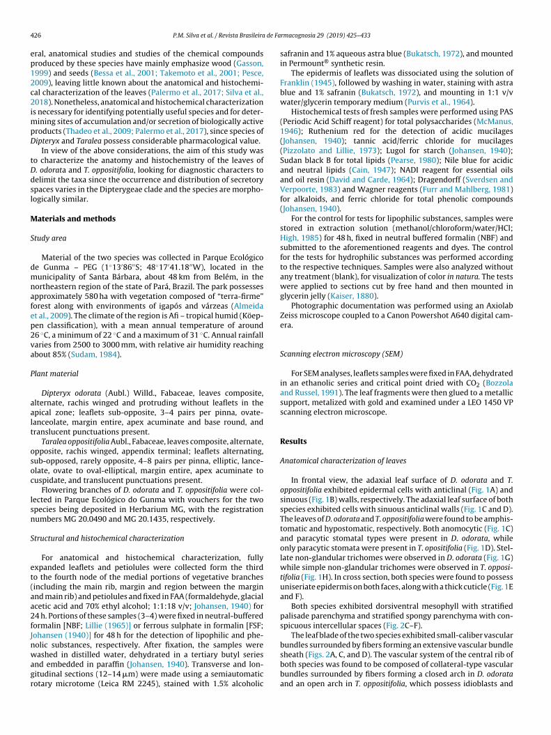
In frontal view, the adaxial leaf surface of D. odorata and T.oppositifolia exhibited epidermal cells with anticlinal (Fig. 1A) andsinuous (Fig. 1B) walls, respectively. The adaxial leaf surface of bothspecies exhibited cells with sinuous anticlinal walls (Fig. 1C and D).The leaves of D. odorata and T. oppositifolia were found to be amphis-tomatic and hypostomatic, respectively. Both anomocytic (Fig. 1C)and paracytic stomatal types were present in D. odorata, whileonly paracytic stomata were present in T. opositifolia (Fig. 1D). Stel-late non-glandular trichomes were observed in D. odorata (Fig. 1G)while simple non-glandular trichomes were observed in T. opposi-tifolia (Fig. 1H). In cross section, both species were found to possessuniseriate epidermis on both faces, along with a thick cuticle (Fig. 1Eand F).
Both species exhibited dorsiventral mesophyll with stratifiedpalisade parenchyma and stratified spongy parenchyma with con-spicuous intercellular spaces (Fig. 2C–F).
The leaf blade of the two species exhibited small-caliber vascularbundles surrounded by fibers forming an extensive vascular bundle
sheath (Figs. 2A, C, and D). The vascular system of the central rib ofboth species was found to be composed of collateral-type vascularbundles surrounded by fibers forming a closed arch in D. odorataand an open arch in T. oppositifolia, which possess idioblasts and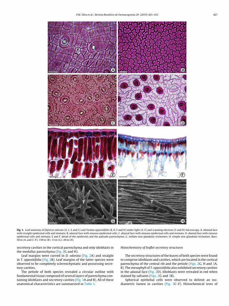
P.M. Silva et al. / Revista Brasileira de Farmacognosia 29 (2019) 425–433 427
Fig. 1. Leaf anatomy of Dipteryx odorata (A, C, E and G) and Taralea oppositifolia (B, D, F and H) under light (A–F) and scanning electron (G and H) microscopy. A. Adaxial facew lls; C.e nchym5
st
iot
fta
ith straight epidermal cells and stomata; B. adaxial face with sinuous epidermal cepidermal cells and stomata; E and F. detail of the epidermis and the palisade pare0 m (A, and C–F); 150 m (B); 13 m (G); 20 m (H).
ecretory cavities in the cortical parenchyma and only idioblasts inhe medullar parenchyma (Fig. 2G and H).
Leaf margins were curved in D. odorata (Fig. 2A) and straightn T. oppositifolia (Fig. 2B). Leaf margins of the latter species werebserved to be completely sclerenchymatic and possessing secre-ory cavities.
The petiole of both species revealed a circular outline withundamental tissue composed of several layers of parenchyma con-aining idioblasts and secretory cavities (Fig. 3A and B). All of thesenatomical characteristics are summarized in Table 1.
abaxial face with sinuous epidermal cells and stomata; D. abaxial face with sinuousa; G. stellate non-glandular trichomes; H. simple non-glandular trichomes. Bars:
Histochemistry of leaflet secretory structures
The secretory structures of the leaves of both species were foundto comprise idioblasts and cavities, which are located in the corticalparenchyma of the central rib and the petiole (Figs. 2G, H and 3A,B). The mesophyll of T. oppositifolia also exhibited secretory cavities
in the adaxial face (Fig. 2D). Idioblasts were revealed in red whenstained by safranin (Figs. 2G and 3B).Spherical epithelial cells were observed to delimit an iso-diametric lumen in cavities (Fig. 3C–F). Histochemical tests of
428 P.M. Silva et al. / Revista Brasileira de Farmacognosia 29 (2019) 425–433
Fig. 2. Leaf anatomy of Dipteryx odorata (A, C, E and G) and Taralea oppositifolia (B, D, F and H) under light (A–D, G and H) and scanning electron (E and F) microscopy. A.Curved downward leaf margin; B. straight leaf margin with secretory cavity (SC); C. general view of mesophyll; D. general view of mesophyll with secretory cavity; E and F.d ) pali(
iotSl(sp
etail of spongy parenchyma (SP); G and H. general view of central rib. Legend: (PPE and F); 300 m (G and H).
dioblasts and secretory cavities revealed that they contain a seriesf chemical compounds, namely: total lipids (acidic and neu-ral), polysaccharides, alkaloids and phenolic compounds (Table 2).ecretions were seen preserved in idioblasts and cavities, with the
atter being present both in epithelial cells and inside the lumenFigs. 4A–F and 5A–F). The exudates present in the idioblasts pos-essed a dark brown coloration in both species, evidencing theresence of phenolic compounds (Fig. 5F).sade parenchyma; (VB) vascular bundle. Bars: 150 m (A, C and D); 50 �m (B); 60 m
Discussion
Among taxa of the Dipterygeae clade, the outline of the epider-mal cells of both leaf surfaces varies from rectilinear to sinuous
(Silva et al., 2018), as observed in the present study. Cells withstraight and sinuous walls have been widely reported amongothers species of Fabaceae, such as Aldina heterophylla Spruceex Benth. (Araújo and Mendonc a, 1998), Bauhinia microstachya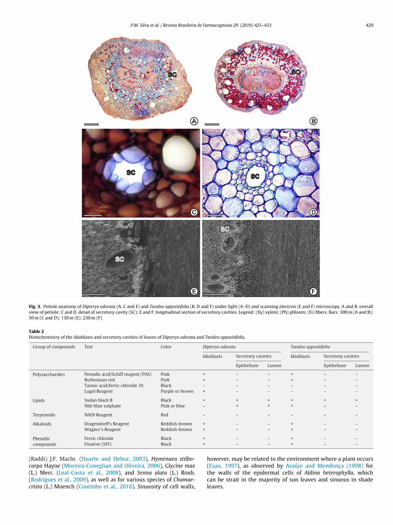
P.M. Silva et al. / Revista Brasileira de Farmacognosia 29 (2019) 425–433 429
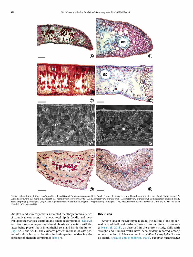
Fig. 3. Petiole anatomy of Dipteryx odorata (A, C and E) and Taralea oppositifolia (B, D and F) under light (A–D) and scanning electron (E and F) microscopy. A and B. overallview of petiole; C and D. detail of secretory cavity (SC); E and F. longitudinal section of secretory cavities. Legend: (Xy) xylem; (Ph) phloem; (Fi) fibers. Bars: 300 m (A and B);50 m (C and D); 130 m (E); 230 m (F).
Table 2Histochemistry of the idioblasts and secretory cavities of leaves of Dipteryx odorata and Taralea oppositifolia.
Group of compounds Test Color Dipteryx odorata Taralea oppositifolia
Idioblasts Secretory cavities Idioblasts Secretory cavities
Epithelium Lumen Epithelium Lumen
Polysaccharides Periodic acid/Schiff reagent (PAS) Pink + – – + – –Ruthenium red Pink + – – + – –Tannic acid/ferric chloride 3% Black – – – – – –Lugol Reagent Purple or brown + – – – – –
Lipids Sudan black B Black + + + + + +Nile blue sulphate Pink or blue – + + + – –
Terpenoids NADI Reagent Red – – – – – –
Alkaloids Dragendorff’s Reagent Reddish-brown + – – + – –Wagner’s Reagent Reddish-brown + – – + – –
+
+
(c((c
Phenoliccompounds
Ferric chloride Black
Fixative (SFF) Black
Raddi) J.F. Macbr. (Duarte and Debur, 2003), Hymenaea stilbo-
arpa Hayne (Moreira-Coneglian and Oliveira, 2006), Glycine maxL.) Merr. (Leal-Costa et al., 2008), and Senna alata (L.) Roxb.Rodrigues et al., 2009), as well as for various species of Chamae-rista (L.) Moench (Coutinho et al., 2016). Sinuosity of cell walls,– – + – –– – + – –
however, may be related to the environment where a plant occurs
(Esau, 1997), as observed by Araújo and Mendonc a (1998) forthe walls of the epidermal cells of Aldina heterophylla, whichcan be strait in the majority of sun leaves and sinuous in shadeleaves.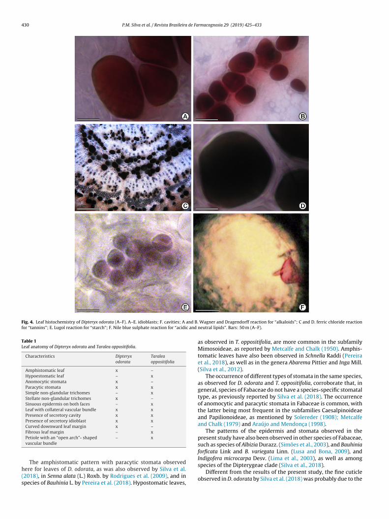
430 P.M. Silva et al. / Revista Brasileira de Farmacognosia 29 (2019) 425–433
Fig. 4. Leaf histochemistry of Dipteryx odorata (A–F). A–E. idioblasts; F. cavities; A and B.for “tannins”; E. Lugol reaction for “starch”; F. Nile blue sulphate reaction for “acidic and
Table 1Leaf anatomy of Dipteryx odorata and Taralea oppositifolia.
Characteristics Dipteryxodorata
Taraleaoppositifolia
Amphistomatic leaf x –Hypoestomatic leaf – xAnomocytic stomata x –Paracytic stomata x xSimple non-glandular trichomes – xStellate non-glandular trichomes x –Sinuous epidermis on both faces – xLeaf with collateral vascular bundle x xPresence of secretory cavity x xPresence of secretory idioblast x xCurved downward leaf margin x –Fibrous leaf margin – x
h(s
Indigofera microcarpa Desv. (Lima et al., 2003), as well as among
Petiole with an “open arch”- shapedvascular bundle
– x
The amphistomatic pattern with paracytic stomata observedere for leaves of D. odorata, as was also observed by Silva et al.2018), in Senna alata (L.) Roxb. by Rodrigues et al. (2009), and inpecies of Bauhinia L. by Pereira et al. (2018). Hypostomatic leaves,
Wagner and Dragendorff reaction for “alkaloids”; C and D. ferric chloride reactionneutral lipids”. Bars: 50 m (A–F).
as observed in T. oppositifolia, are more common in the subfamilyMimosoideae, as reported by Metcalfe and Chalk (1950). Amphis-tomatic leaves have also been observed in Schnella Raddi (Pereiraet al., 2018), as well as in the genera Abarema Pittier and Inga Mill.(Silva et al., 2012).
The occurrence of different types of stomata in the same species,as observed for D. odorata and T. oppositifolia, corroborate that, ingeneral, species of Fabaceae do not have a species-specific stomataltype, as previously reported by Silva et al. (2018). The occurrenceof anomocytic and paracytic stomata in Fabaceae is common, withthe latter being most frequent in the subfamilies Caesalpinoideaeand Papilionoideae, as mentioned by Solereder (1908); Metcalfeand Chalk (1979) and Araújo and Mendonc a (1998).
The patterns of the epidermis and stomata observed in thepresent study have also been observed in other species of Fabaceae,such as species of Albizia Durazz. (Simões et al., 2003), and Bauhiniaforficata Link and B. variegata Linn. (Lusa and Bona, 2009), and
species of the Dipterygeae clade (Silva et al., 2018).Different from the results of the present study, the fine cuticle
observed in D. odorata by Silva et al. (2018) was probably due to the
P.M. Silva et al. / Revista Brasileira de Farmacognosia 29 (2019) 425–433 431
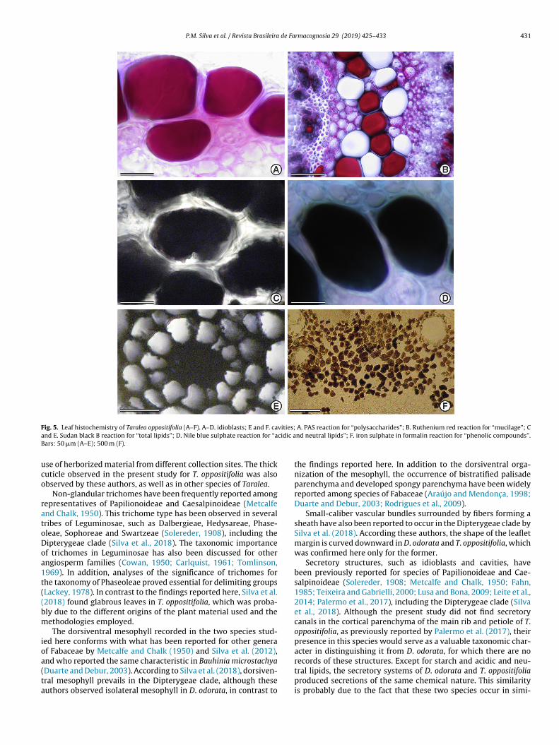
F vities;a cidic aB
uco
ratoDoa1t((bm
ioa(ta
ig. 5. Leaf histochemistry of Taralea oppositifolia (A–F). A–D. idioblasts; E and F. cand E. Sudan black B reaction for “total lipids”; D. Nile blue sulphate reaction for “aars: 50 �m (A–E); 500 m (F).
se of herborized material from different collection sites. The thickuticle observed in the present study for T. oppositifolia was alsobserved by these authors, as well as in other species of Taralea.
Non-glandular trichomes have been frequently reported amongepresentatives of Papilionoideae and Caesalpinoideae (Metcalfend Chalk, 1950). This trichome type has been observed in severalribes of Leguminosae, such as Dalbergieae, Hedysareae, Phase-leae, Sophoreae and Swartzeae (Solereder, 1908), including theipterygeae clade (Silva et al., 2018). The taxonomic importancef trichomes in Leguminosae has also been discussed for otherngiosperm families (Cowan, 1950; Carlquist, 1961; Tomlinson,969). In addition, analyses of the significance of trichomes forhe taxonomy of Phaseoleae proved essential for delimiting groupsLackey, 1978). In contrast to the findings reported here, Silva et al.2018) found glabrous leaves in T. oppositifolia, which was proba-ly due to the different origins of the plant material used and theethodologies employed.The dorsiventral mesophyll recorded in the two species stud-
ed here conforms with what has been reported for other generaf Fabaceae by Metcalfe and Chalk (1950) and Silva et al. (2012),
nd who reported the same characteristic in Bauhinia microstachyaDuarte and Debur, 2003). According to Silva et al. (2018), dorsiven-ral mesophyll prevails in the Dipterygeae clade, although theseuthors observed isolateral mesophyll in D. odorata, in contrast toA. PAS reaction for “polysaccharides”; B. Ruthenium red reaction for “mucilage”; Cnd neutral lipids”; F. iron sulphate in formalin reaction for “phenolic compounds”.
the findings reported here. In addition to the dorsiventral orga-nization of the mesophyll, the occurrence of bistratified palisadeparenchyma and developed spongy parenchyma have been widelyreported among species of Fabaceae (Araújo and Mendonc a, 1998;Duarte and Debur, 2003; Rodrigues et al., 2009).
Small-caliber vascular bundles surrounded by fibers forming asheath have also been reported to occur in the Dipterygeae clade bySilva et al. (2018). According these authors, the shape of the leafletmargin is curved downward in D. odorata and T. oppositifolia, whichwas confirmed here only for the former.
Secretory structures, such as idioblasts and cavities, havebeen previously reported for species of Papilionoideae and Cae-salpinoideae (Solereder, 1908; Metcalfe and Chalk, 1950; Fahn,1985; Teixeira and Gabrielli, 2000; Lusa and Bona, 2009; Leite et al.,2014; Palermo et al., 2017), including the Dipterygeae clade (Silvaet al., 2018). Although the present study did not find secretorycanals in the cortical parenchyma of the main rib and petiole of T.oppositifolia, as previously reported by Palermo et al. (2017), theirpresence in this species would serve as a valuable taxonomic char-acter in distinguishing it from D. odorata, for which there are no
records of these structures. Except for starch and acidic and neu-tral lipids, the secretory systems of D. odorata and T. oppositifoliaproduced secretions of the same chemical nature. This similarityis probably due to the fact that these two species occur in simi-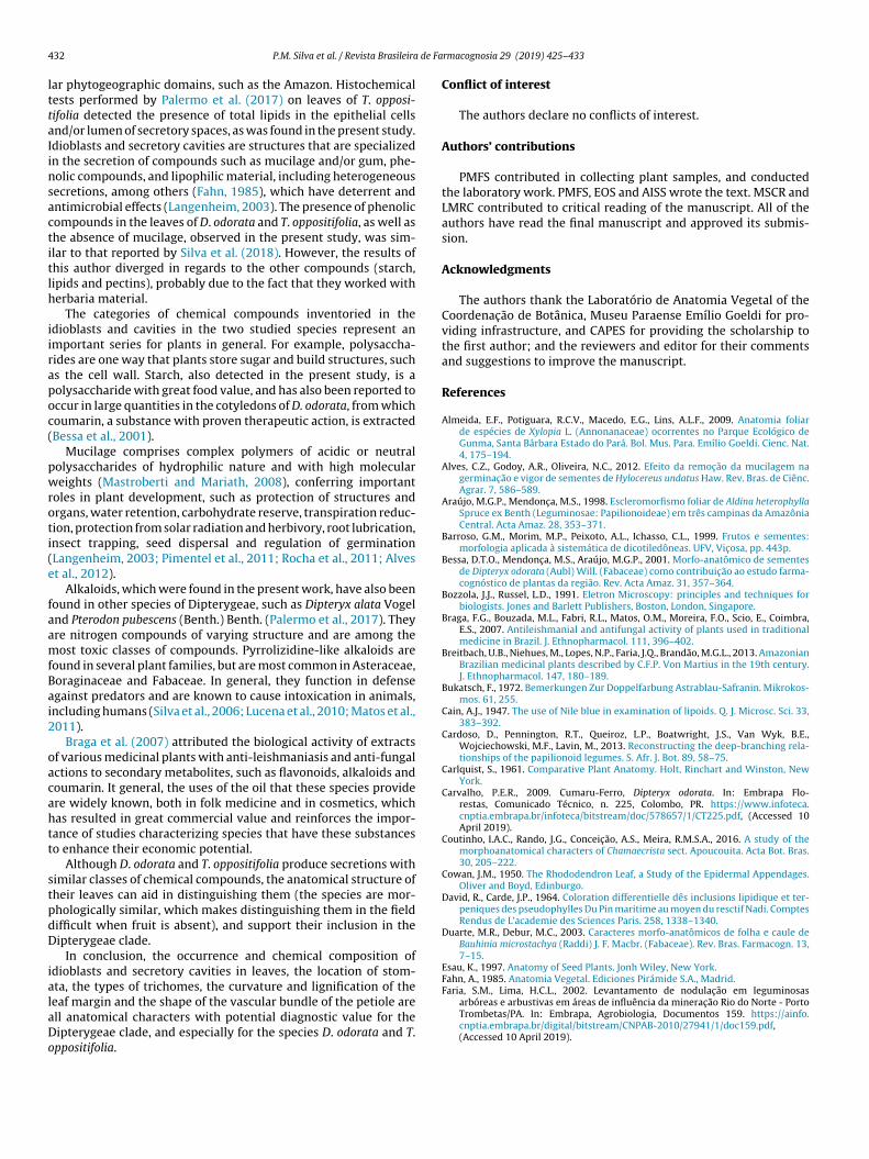
4 de Fa
lttaIinsactitlh
iirapoc(
pwroti(e
faamfBai2
oacahtt
stpdD
ialaDo
Faria, S.M., Lima, H.C.L., 2002. Levantamento de nodulac ão em leguminosasarbóreas e arbustivas em áreas de influência da minerac ão Rio do Norte - PortoTrombetas/PA. In: Embrapa, Agrobiologia, Documentos 159. https://ainfo.
32 P.M. Silva et al. / Revista Brasileira
ar phytogeographic domains, such as the Amazon. Histochemicalests performed by Palermo et al. (2017) on leaves of T. opposi-ifolia detected the presence of total lipids in the epithelial cellsnd/or lumen of secretory spaces, as was found in the present study.dioblasts and secretory cavities are structures that are specializedn the secretion of compounds such as mucilage and/or gum, phe-olic compounds, and lipophilic material, including heterogeneousecretions, among others (Fahn, 1985), which have deterrent andntimicrobial effects (Langenheim, 2003). The presence of phenolicompounds in the leaves of D. odorata and T. oppositifolia, as well ashe absence of mucilage, observed in the present study, was sim-lar to that reported by Silva et al. (2018). However, the results ofhis author diverged in regards to the other compounds (starch,ipids and pectins), probably due to the fact that they worked witherbaria material.
The categories of chemical compounds inventoried in thedioblasts and cavities in the two studied species represent anmportant series for plants in general. For example, polysaccha-ides are one way that plants store sugar and build structures, suchs the cell wall. Starch, also detected in the present study, is aolysaccharide with great food value, and has also been reported toccur in large quantities in the cotyledons of D. odorata, from whichoumarin, a substance with proven therapeutic action, is extractedBessa et al., 2001).
Mucilage comprises complex polymers of acidic or neutralolysaccharides of hydrophilic nature and with high moleculareights (Mastroberti and Mariath, 2008), conferring important
oles in plant development, such as protection of structures andrgans, water retention, carbohydrate reserve, transpiration reduc-ion, protection from solar radiation and herbivory, root lubrication,nsect trapping, seed dispersal and regulation of germinationLangenheim, 2003; Pimentel et al., 2011; Rocha et al., 2011; Alvest al., 2012).
Alkaloids, which were found in the present work, have also beenound in other species of Dipterygeae, such as Dipteryx alata Vogelnd Pterodon pubescens (Benth.) Benth. (Palermo et al., 2017). Theyre nitrogen compounds of varying structure and are among theost toxic classes of compounds. Pyrrolizidine-like alkaloids are
ound in several plant families, but are most common in Asteraceae,oraginaceae and Fabaceae. In general, they function in defensegainst predators and are known to cause intoxication in animals,ncluding humans (Silva et al., 2006; Lucena et al., 2010; Matos et al.,011).
Braga et al. (2007) attributed the biological activity of extractsf various medicinal plants with anti-leishmaniasis and anti-fungalctions to secondary metabolites, such as flavonoids, alkaloids andoumarin. It general, the uses of the oil that these species providere widely known, both in folk medicine and in cosmetics, whichas resulted in great commercial value and reinforces the impor-ance of studies characterizing species that have these substanceso enhance their economic potential.
Although D. odorata and T. oppositifolia produce secretions withimilar classes of chemical compounds, the anatomical structure ofheir leaves can aid in distinguishing them (the species are mor-hologically similar, which makes distinguishing them in the fieldifficult when fruit is absent), and support their inclusion in theipterygeae clade.
In conclusion, the occurrence and chemical composition ofdioblasts and secretory cavities in leaves, the location of stom-ta, the types of trichomes, the curvature and lignification of theeaf margin and the shape of the vascular bundle of the petiole arell anatomical characters with potential diagnostic value for the
ipterygeae clade, and especially for the species D. odorata and T.ppositifolia.rmacognosia 29 (2019) 425–433
Conflict of interest
The authors declare no conflicts of interest.
Authors’ contributions
PMFS contributed in collecting plant samples, and conductedthe laboratory work. PMFS, EOS and AISS wrote the text. MSCR andLMRC contributed to critical reading of the manuscript. All of theauthors have read the final manuscript and approved its submis-sion.
Acknowledgments
The authors thank the Laboratório de Anatomia Vegetal of theCoordenac ão de Botânica, Museu Paraense Emílio Goeldi for pro-viding infrastructure, and CAPES for providing the scholarship tothe first author; and the reviewers and editor for their commentsand suggestions to improve the manuscript.
References
Almeida, E.F., Potiguara, R.C.V., Macedo, E.G., Lins, A.L.F., 2009. Anatomia foliarde espécies de Xylopia L. (Annonanaceae) ocorrentes no Parque Ecológico deGunma, Santa Bárbara Estado do Pará. Bol. Mus. Para. Emílio Goeldi. Cienc. Nat.4, 175–194.
Alves, C.Z., Godoy, A.R., Oliveira, N.C., 2012. Efeito da remoc ão da mucilagem nagerminac ão e vigor de sementes de Hylocereus undatus Haw. Rev. Bras. de Ciênc.Agrar. 7, 586–589.
Araújo, M.G.P., Mendonc a, M.S., 1998. Escleromorfismo foliar de Aldina heterophyllaSpruce ex Benth (Leguminosae: Papilionoideae) em três campinas da AmazôniaCentral. Acta Amaz. 28, 353–371.
Barroso, G.M., Morim, M.P., Peixoto, A.L., Ichasso, C.L., 1999. Frutos e sementes:morfologia aplicada à sistemática de dicotiledôneas. UFV, Vic osa, pp. 443p.
Bessa, D.T.O., Mendonc a, M.S., Araújo, M.G.P., 2001. Morfo-anatômico de sementesde Dipteryx odorata (Aubl) Will. (Fabaceae) como contribuic ão ao estudo farma-cognóstico de plantas da região. Rev. Acta Amaz. 31, 357–364.
Bozzola, J.J., Russel, L.D., 1991. Eletron Microscopy: principles and techniques forbiologists. Jones and Barlett Publishers, Boston, London, Singapore.
Braga, F.G., Bouzada, M.L., Fabri, R.L., Matos, O.M., Moreira, F.O., Scio, E., Coimbra,E.S., 2007. Antileishmanial and antifungal activity of plants used in traditionalmedicine in Brazil. J. Ethnopharmacol. 111, 396–402.
Breitbach, U.B., Niehues, M., Lopes, N.P., Faria, J.Q., Brandão, M.G.L., 2013. AmazonianBrazilian medicinal plants described by C.F.P. Von Martius in the 19th century.J. Ethnopharmacol. 147, 180–189.
Bukatsch, F., 1972. Bemerkungen Zur Doppelfarbung Astrablau-Safranin. Mikrokos-mos. 61, 255.
Cain, A.J., 1947. The use of Nile blue in examination of lipoids. Q. J. Microsc. Sci. 33,383–392.
Cardoso, D., Pennington, R.T., Queiroz, L.P., Boatwright, J.S., Van Wyk, B.E.,Wojciechowski, M.F., Lavin, M., 2013. Reconstructing the deep-branching rela-tionships of the papilionoid legumes. S. Afr. J. Bot. 89, 58–75.
Carlquist, S., 1961. Comparative Plant Anatomy. Holt, Rinchart and Winston, NewYork.
Carvalho, P.E.R., 2009. Cumaru-Ferro, Dipteryx odorata. In: Embrapa Flo-restas, Comunicado Técnico, n. 225, Colombo, PR. https://www.infoteca.cnptia.embrapa.br/infoteca/bitstream/doc/578657/1/CT225.pdf, (Accessed 10April 2019).
Coutinho, I.A.C., Rando, J.G., Conceic ão, A.S., Meira, R.M.S.A., 2016. A study of themorphoanatomical characters of Chamaecrista sect. Apoucouita. Acta Bot. Bras.30, 205–222.
Cowan, J.M., 1950. The Rhododendron Leaf, a Study of the Epidermal Appendages.Oliver and Boyd, Edinburgo.
David, R., Carde, J.P., 1964. Coloration differentielle dês inclusions lipidique et ter-peniques des pseudophylles Du Pin maritime au moyen du resctif Nadi. ComptesRendus de L’academie des Sciences Paris. 258, 1338–1340.
Duarte, M.R., Debur, M.C., 2003. Caracteres morfo-anatômicos de folha e caule deBauhinia microstachya (Raddi) J. F. Macbr. (Fabaceae). Rev. Bras. Farmacogn. 13,7–15.
Esau, K., 1997. Anatomy of Seed Plants. Jonh Wiley, New York.Fahn, A., 1985. Anatomia Vegetal. Ediciones Pirámide S.A., Madrid.
cnptia.embrapa.br/digital/bitstream/CNPAB-2010/27941/1/doc159.pdf,(Accessed 10 April 2019).
de Fa
F
F
F
G
H
HJK
L
L
L
L
L
L
L
L
L
M
M
M
M
M
M
P
P.M. Silva et al. / Revista Brasileira
rancisco, V.M.C.R., 2010. Filogenia Molecular e Morfológica da Tribo Dipterygeae(Papilionoideae, Leguminosae). In: Rio de janeiro, Dissertac ão de Mestrado,Instituto de Pesquisas Jardim Botânico do Rio de Janeiro., pp. 91.
ranklin, G.L., 1945. Preparation of thin sections of synthetic resins and wood-resincomposites, and a new macerating method for wood. Nature 51, 24–39.
urr, M., Mahlberg, P.G., 1981. Histochemical analyses of laticifers and glandulartrichomes in Cannabis sativa. J. Nat. Prod. 44, 153–159.
asson, P., 1999. Wood anatomy of the tribe Dipterygeae with comments on relatedPapilionoid and Caesalpinioid Leguminosae. IAWA J. 20, 441–455.
errero-Jáuregui, C., Sist, P., Casado, M.A., 2012. Population structure of two low-density neotropical tree species under different management systems. ForestEcol. Manag. 280, 31–39.
igh, O.B., 1985. Lipid Histochemistry. Oxford University Press, New York.ohansen, D.A., 1940. Plant Microtechnique. Mcgraw-Hill, New York.aiser, E., 1880. Verfahren zur herstellung einer tadellosen glycerin-gelatine.
Botanisch Zentralb, Stuttgart 180, 25–26.ackey, J.A., 1978. Leaflet anatomy of Phaseoleae (Leguminosae: papilionoideae) and
its relation to taxonomy. Bot. Gaz. 139, 436–446.angenheim, J.H., 2003. Plant Resins: Chemistry, Evolution, Ecology and Ethnob-
otany. Timber Press, Portland, Cambridge.eal-Costa, M.V., Aragão, F.J.L., Reinert, F., Tavares, E.S., 2008. Anatomia foliar de
plantas transgênicas e não transgênicas de Glycine max (L.) Merrill (Fabaceae).Rev. Biociênc. 14, 23–31.
eelavathi, P., Ramayya, N., Prabhakar, M., 1980. Foliar stomatal distribution pat-terns in Leguminosae and their taxonomic significance. Phytomorphology 30,195–204.
eite, V.G., Mansano, V.F., Teixeira, S.P., 2014. Floral ontogeny in Dipterygeae(Fabaceae) reveals new insights into one of the earliest branching tribes inpapilionoid legumes. Bot. J. Linn. Soc. 174, 529–550.
illie, R.D., 1965. Histopathologic Technic and Pratical Histochemistry, 3rd ed.McGraw Hill, New York.
ima, A.K., Elba, L.C.A., Aquino, T.M., Lima, C.S.A., Pimentel, R.M.M., Higino, J.S.,Albuquerque, U.P., 2003. Estudo farmacognóstico de Indigofera microcarpa Desv.(Fabaceae). Rev. Bras. Cienc. Farm. 39, 373–379.
ucena, R.B., Rissi, D.R., Maia, L.A., Flores, M.F., Dantas, A.F.M., Nobre, V.M.T.,Riet-Correa, F., Claudio, S.L., Barros, C.S.L., 2010. Intoxicac ão por alcaloides pir-rolizidínicos em ruminantes e equinos no Brasil. Pesqui. Vet. Bras. 3, 447–452.
usa, M.G., Bona, C., 2009. Análise morfoanatômica comparativa da folha de Bauhiniaforficata Link e Bauhinia variegata Linn. (Leguminosae, Caesalpinioideae). ActaBot. Bras. 23, 196–211.
astroberti, A.A., Mariath, J.E.A., 2008. Immunocytochemistry of the mucilage cellsof Araucaria angustifolia (Bertol.) Kuntze (Araucariaceae). Rev. Bras. Bot. 31, 1–13.
atos, F.J.A., Lorenzi, H., Santos, L.F.L., Matos, M.E.O., Silva, M.G.V., Sousa, M.P., 2011.Plantas Tóxicas: Estudo de Fitotoxicologia Química de Plantas Brasileiras. Plan-tarum, Flora, Nova Odessa.
cManus, J.F.A., 1946. Histological demonstration of mucin after periodic acid.Nature 158, 202–211, http://dx.doi.org/10.1038/158202a0.
etcalfe, C.R., Chalk, L., 1950. Anatomy of the Dicotyledons Leaves, Stem and Woodin Relation to Taxonomy With Notes on Economy Uses. Clarendon Press, Oxford.
etcalfe, C.R., Chalk, L., 1979. Anatomy of the Dicotyledons, 2nd ed. Claredon Press,Oxford.
oreira-Coneglian, I.R., Oliveira, D.T., 2006. Anatomia comparada dos limbos cotile-
donares e eofilares de dez espécies de Caesalpinoideae (Fabaceae). Rev. Bras.Bot. 29, 193–207.alermo, F.H., Teixeira, S.P., Mansano, V.F., Leite, V.G., Rodrigues, T.M., 2017. Secre-tory spaces in species of the clade Dipterygeae (Leguminosae, papilionoideae).Acta Bot. Bras. 31, 374–381.
rmacognosia 29 (2019) 425–433 433
Pearse, A.G.E., 1980. Histochemistry Theretical and Applied, 4 ed. Longman groupLimited., Baltimore.
Pereira, L.B.S., Costa-Silva, R., Felix, L.P., Agra, M.F., 2018. Leaf morphoanatomy of“mororó” (Bauhinia and Schnella, Fabaceae). Rev. Bras. Farmacogn. 28, 383–392.
Pesce, C., 2009. Oleaginosas da Amazônia, 2nd ed. Museu Paraense Emílio Goeldi,Belém-PA http://repiica.iica.int/docs/B2252p/B2252p.pdf.
Pimentel, R.R., Machado, S.R., Rocha, J.F., 2011. Estruturas secretoras de Pavonia alni-folia (Malvaceae), uma espécie ameac ada de extinc ão. Rodrigusia 62, 253–262.
Pizzolato, T.D., Lillie, R.D., 1973. Mayer’s tannic acid-ferric chloride stain for mucins.J. Histochem. Cytochem. 21, 56–64.
Purvis, M.J., Collier, D.C., Walls, D., 1964. Laboratory Techniques in Botany. Butter-worths, London.
Ramalingam, S., Rajendran, S., Ganesan, P., Govindasamy, M., 2018. Effect of oper-ating parameters and antioxidant additives with biodiesels to improve theperformance and reducing the emissions in a compression ignition engine—areview. Renew. Sust. Energ. Rev. 81, 775–788.
Rocha, J.F., Pimentel, R.R., Machado, S.R., 2011. Estruturas secretoras de mucilagemem Hibiscus pernambucensis Arruda (Malvaceae): distribuic ão, caracterizac ãomorfoanatômica e histoquímica. Acta Bot. Bras. 25, 51–763.
Rodrigues, I.M.C., Souza Filho, A.P.S., Ferreira, F.A., Ilkiu-Borges, F., Gurgel, E.S.C.,2009. Anatomia e histoquímica das folhas de Senna alata. Planta Daninha 27,515–526.
Silva, C.M., Bolzan, A.A., Heinzmann, B.M., 2006. Alcalóides pirrolizidínicos em espé-cies do gênero Senecio. Quím. Nova. 29, 1047–1053.
Silva, M.S., Reis, C., Pontes-Pires, A.F.P., 2012. Anatomical characteristics of leaf offive species of family Fabaceae found in Sinop, MT. Sci. Electron. Arch. 1, 16–19.
Silva, N.F., Arruda, R.C.O., Alves, F.M., Sartori, A.L.B., 2018. Leaflet anatomy of theDipterygeae clade (Faboideae: fabaceae): evolutionary implications and system-atics. Bot. J. Linn. Soc. 187, 99–117.
Simões, M.O.M., Lopes, P.S.N., Oliveira, M.N.S., Junior, E.M.F., Ribeiro, L.M., 2003.Estudo anatômico do mesofilo foliar de Albizia spp (Leguminosae/Mimosoideae).Rev. Unimontes Científica 5, 1–9.
Solereder, H., 1908. Systematic anatomy of the dicotyledons. Introduction, polypeta-lae, Gamopetalae. Oxford at the Carendon press.
Soriano, M., Kainer, K.A., Staudhammer, C.L., Soriano, E., 2012. Implementing multi-ple forest management in Brazil nut-rich community forests: effects of loggingon natural regeneration and forest disturbance. Forest Ecol. Manag. 268, 92–102.
Sousa, M.A.R., Moutinho, V.H.P., Silva, S.S., 2007. Levantamento das espécies comer-cializadas vernacularmente como cumaru no Estado do Pará. Rev. Bras. Biocienc.5, 81–83.
Sudam, 1984. Atlas Climatológico da Amazônia Brasileira. SUDAM/PHCA, Belém.Sverdsen, A.B., Verpoorte, R., 1983. Chromatography of Alkaloids. Elsevier Scientific
Publish Company, New York.Takemoto, E., Okada, I.A., Garbelotti, M.M., Tavares, M., Aued-Pimenel, S., 2001.
Composic ão química da semente e do óleo de baru (Dipteryx alada Vog.) nativodo município de Pirenópolis, Estado de Goiás. Rev. Inst. Adolfo Lutz 60, 113–117.
Teixeira, S.P., Gabrielli, A.C., 2000. Anatomia de eixo vegetativo de Dahlstedtia pinna(Benth.) Malme e D. pentaphylla (Taub.) Burk (Leguminosae, Papilionoideae).Rev. Bras. Bot. 23, 1–11.
Thadeo, M., Meira, R.M.S.A., Azevedo, A.A., Araújo, J.M., 2009. Anatomia e his-toquímica das estruturas secretoras da folha de Casearia decandra Jacq.(Salicaceae). Rev. Bras. Bot. 32, 329–338.
Tomlinson, P.B., 1969. Anatomy of the Monocotyledons III. Commelinales - Zingib-erales. Clarendon press, Oxford.
Uchida, T., Campos, M.A., 2000. Influência do sombreamento no crescimento demudas de cumaru (Dipteryx odorata (Aubl.) Willd. Fabaceae), cultivadas emviveiro. Acta Amaz. 30, 107–114.