analysis of nasal breathing function dynamics...
TRANSCRIPT
Indexed by Scopus (Elsevier)
Co-Publisher: OMICS Group, www.omicsonline.org
Volume 6, Issue 1, Article ID: BM-007-14, 2014
eISSN: 09748369
Analysis of nasal breathing function dynamics during rhinoplasty
Biology and Medicine
Research Article
Article ID: BM-007-14 Page 1 of 6
Research Article Biology and Medicine, 6(1), 2014
Introduction
The nose has a central position on the face play-ing a huge role in the realization of the human per-sonality and appeal. Therefore, we pay so much attention to proper shape of the nose, harmoni-ous with the type of face as well as analysis of the aesthetic shape of the nose [1-3]. However, a beautiful but poorly functioning nose will be also uncomfortable for the person, as well as asym-metric one, but for a different reason – because in addition to the aesthetic malfunction, the other nasal functions will be also affected: absorptive, excretory, respiratory, safety, air heater, etc. [4]. Deformation of the “external” nose is usually accompanied by intranasal deformities, which, in turn, in 42-77% of cases lead to a violation of nasal breathing [5,6].
At the present time, the plastic surgeons practice mainly aesthetic approach to the rhi-noplasty: a number of specialists focus on the correction of external nasal structures as well as pre and postoperative analysis of the aesthetic shape of the nose [7,8]. A few people pay atten-tion to the functional aspects, developing and applying techniques that improve breathing, but do not pay enough attention to the objective pre and postoperative assessment of nasal breathing function [9,10]. On the other hand, correction of
www.biolmedonline.com
Analysis of nasal breathing function dynamics during rhinoplasty
Alexey Drobyshev1, Leonid Pavlyuk-Pavlyuchenko2, Katerina Krasavtseva1*1Moscow State University of Medicine and Dentistry, Delegatskaya St. 20,
p.1, Moscow 127473, Russia.2Peoples’ Friendship University of Russia, Miklukho-Maklay St. 21, case 3,
Moscow 117198, Russia.
*Corresponding author: Moscow State University of Medicine and Dentistry, Delegatskaya St. 20, p.1, Moscow 127473, Russia.
Received: 19th Jul 2014; Accepted: 2nd Sep 2014; Published: 12th Sep 2014
AbstractThe aim of the study was to assess the dynamics of nasal breathing in patients with congenital and acquired deformi-ties of nose as a result of combined functional and aesthetic rhinoplasty. Using the method of front active rhinoman-ometry (FARM) in 52 patients with deformities of the nose, we have found special features of nasal breathing, which allowed to diagnose changes of intranasal structures in preoperative period and to correct algorithm of the surgery. The study of nasal breathing in the postoperative period has confirmed the adequacy of the surgery.
Keywords: Nose deformation; rhinoplasty; FARM.
the internal structures of the nose and, accord-ingly, correction of nasal breathing is completely impossible without a preliminary study on nasal functions [11].
Objectives of the Study
To assess the dynamics of nasal breathing in patients suffering from congenital and acquired deformities of the nose as a result of combina-tion of functional and aesthetic rhinoplasty.
Materials and Methods
We have observed 52 patients with nasal deformities of congenital and acquired etiolo-gies from 2011 to 2013 on the clinical bases of the Department of Plastic Surgery of the Medical Personnel Training Faculty at the Peoples’ Friendship University of Russia and Dentistry and Maxillofacial Surgery Center of Moscow State University of Medicine and Dentistry named after A.I. Evdokimov. The study enrolled 43 women and 9 men aged from 17 to 46 yrs.
Depending on the type of the “pri-mary” deformation of external nasal struc-tures patients were divided into three groups:
Research Article Biology and Medicine, 6(1), 2014
Article ID: BM-007-14 Page 2 of 6
orthoscoliosis – 7 (13.46%) patients, kyphoscoli- osis – 40 (76.92%) patients, and lordoscoliosis – 5 (9.62%) subjects. The main complaints of patients on admission were the following: complaints on the deformation of the nose accompanied by nasal breathing difficulties or without them. Nasal defor-mations in patients were divided into two etiologi-cal groups: acquired – 34 subjects (65.4%) and congenital – 18 subjects (34.6%).
All patients were examined preopera-tively using the following algorithm: anamnesis collection, medical examination, rhinoscopy, taking pictures in seven projections (front, left and right profile, semiprofile, basal and semihe-licopter projection), front active rhinomanometry (FARM) by the below described scheme. In addi-tion, we have performed multi-slice computed tomography (MSCT).
Rhinoplasty was performed as three-stage intervention procedure.
1. Septoplasty
- Partial resection of osteochondral frag-ments of limited area.
- Resection of the quadrangular cartilage, its straightening and replantation.
- Medialization of the septum.
2. Conchoplasty
- Lateroposition of inferior turbinates. - Submucosal conchotomy of the lower
and/or middle turbinates. - Coagulation of submucosal vessels of the
lower and/or middle turbinates.
3. External nose plastic reconstruction
- Plastic reconstruction of the terminal nasal department using the method of “fenestrated resection” of crus laterale of the major alar cartilage.
- Correction of dorsum of nose using “hump on leg” method.
- Correction of dorsum of nose using rasp. - Osteotomy of the nasal bones. - Using special fillers such as bone, carti-
lage and soft-tissue, auto- or allografts.
In 1 yr after rhinoplasty, all patients underwent the control study of respiratory func-tion completeness by FARM method and the study of nasal structures condition by MSCT method.
FARM: In our research, we have applied a rhinomanometry device PTS-14P-01 “Rinolan” (CJSC “Lana Medica”, St. Petersburg). The device determines the variable airway pressure on inspiration and expiration, as well as the amount of air used by the patient for breathing. Based on these data, the computer software allows to study the following basic parameters:
- Total expiratory flow at 150 Pa (normally total volume flow (TVF) is 700 ml/s and more);
- Percent increase in the volume flow in each half of the nose, when the pressure increases from 75 to 150 Pa (normally more than 35%);
- Symmetry indicator of maximum amount of airflow on inspiration on each side (physiologic ratio is considered less than 1.5).
The last two indicators show an asym-metric structure of intranasal components and the presence of a nonphysiological obsta-cle in the nasal cavity, usually confirmed by MSCT (septal crest, distortions of the septum, hypertrophic conches, their bullous or hamate deformation, etc.).
At the end of the study, the data are printed on paper or stored in electronic archives.
For examination purpose, we have used disposable foam inserts of three sizes. Their right choice or forming an elliptic indi-vidual shape contributed to avoid deformation of the nasal vestibule and to obtain the reliable results.
Preoperative examination of the breath-ing consisted of three phases: (1) patient (sitting) closed one nostril by a disposable insert and fixed a face mask tightly. Then he performed 3-5 calm breaths through the nose with the mouth closed following by a pause of 1-2 s between inhalation and exhalation. At the same time, the pressure was registered in the closed half of the nose, while the airflow was registered in the free half. A similar procedure was repeated on the other side. (2) A patient laid for 30 min in horizon-tal position in order to determine vascular hydro-static mucosal response, then the examination was repeated. If the parameters obtained in first and second examinations were normal, the study ended and the nasal was regarded as normal. (3) Two drops of adrenomimetic drug were instilled into each nostril, if there were results deterioration at the second stage of the study followed by the third assessment in 10 min.
Research Article Biology and Medicine, 6(1), 2014
Article ID: BM-007-14 Page 3 of 6
Results and Discussion
Depending on the degree of respiratory disorder based on the total volume flow (TVF), all patients were divided into the following groups: (1) with-out impairment of respiratory function (100% of volume and above) – 38.5% of patients; (2) with minor respiratory dysfunction (99-90% of volume) – 23.1% of patients; (3) with moderately reduced respiratory function (89-56%) – 25.0%; and (4) with a considerable reduction in respira-tory function (55% or less) – in 13.4% of patients. In 67.3% of cases we have observed an allow-able breathing asymmetry (flow ratio 1.0-1.49), in 32.7% cases there was a considerable asym-metry; increase in the airflow in 28.85% of patients was within normal limits (an increase of over 35%), while the remaining patients had one- or two-sided decrease in airflow. Only two patients had marked symmetrical increments of airflow.
We have found that half of the patients (14 out of 28 persons) admitted with a desire for aesthetic correction of the nose, respira-tory function parameters were reduced in vari-ous ranges: in six patients (21.43%) there was a slight decrease in the TVF, in seven patients (25.0%) there was a moderate decrease in TVF, and in one patient (3.57%) – a considerable reduction of respiration was diagnosed. One of five patients (20.0%) consulted with a symptoms of impaired nasal breath showed absolutely full nasal breathing.
At comprehensive analysis of all the data of an active anterior rhinomanometry in patients without impairment of respiratory function, aver-age indicators at the first stage of the study were as follows: TVF – 800.8 6 10.9 ml/s, increase in the airflow – 36.5 6 3.1% on the right side and 34.0 6 2.0% on the left side, while an average asymmetry of airflows was 1.19 6 0.04, which reasonably showed almost complete health of the nasal cavity with a few exceptions: the growth rate of an airflow in some cases was lower than normal. This indicated the presence of small structural changes in the nasal cavity, resulting in an increased airflow resistance. This fact in our study was the reason for performing preoperative MSCT for visual diagnostic imaging of possible changes of internal structures.
TVF of the nasal cavity in patients with a considerable reduction in respiratory function prior to treatment was 313.5 6 26.2 ml/s in the first study, which is lower by 487.3 ml/s (60.85%)
than the respective value in patients without impairment of respiratory function. When com-paring these figures with a group of subjects with minor impairments of respiratory function and also with moderately reduced respiratory func-tion, we have found that TVF values in patients in all groups were significantly lower than in the normal group by 129.5 ml/s (16.17%) and 301.8 ml/s (37.69%), respectively. It was also noted that the decrease in TVF values in groups of patients was associated with proportionally increased asymmetry in the airflows from 1.19 in patients without respiratory dysfunction to 1.86 in patients with a considerable decrease in res-piratory function. Breathing symmetry indicators in groups of patients with moderate and severe decrease in the volume of respiration were within the essential asymmetry of breath (aspect ratio greater than 1.5).
At the second stage of the study, the patients with an initially modest decline in breathing and severe decline in respiratory func-tion were not enrolled. Skipping this stage, they were immediately transferred to the third stage of testing a vasoconstrictor response.
A total reduction in TVF in groups of patients with initially normal or slightly reduced breathing was detected as a result of conduct-ing the second stage of the study: the reduction amounted to 56.2 ml/s (7.02%) and 83.5 ml/s (12.44%), respectively, which allowed to reveal a positive hydrostatic reaction of nasal mucosa in these two groups of patients. If the first and second stages of respiration were within the normal range, the test was completed, while the state of nasal cavity was considered as normal. If the state was worsened after repeated meas-urements, the patient was given vasoconstric-tor (Adrianol), and 10 min later a third study was conducted.
TVF in the third phase of the study (in 10 min after instillation of adrenomimetic drug) was improved in all groups of patients:
- In the first and second group of patients as compared with the results of Austrian Azacitidine Registry (AAR) test at the second phase of the study (after being in the horizon-tal position for 30 min) in the group without impairment of respiratory function – by 97.8 ml/s (13.14%), in the group with minimally impaired respiratory function by 191 ml/s (32.49%);
- In the third and fourth groups com-pared with the results of the first phase of FARM
Research Article Biology and Medicine, 6(1), 2014
Article ID: BM-007-14 Page 4 of 6
test: in the group with moderately reduced respi-ratory function – by 194.4 ml/s (28.96%), and in the group with a considerable decrease in respi-ratory function by 241.4 ml/s (77.0%).
Although the first and second groups’ parameters of the TVF returned to normal values (and even exceeded it) indicating just a labile vascular reaction of nasal mucosa, the respec-tive indicators were improved in persons of the third and fourth groups (and even exceeded the values obtained in the first phase of the study). However, the values did not reach normal lev-els. Such reaction allowed us to diagnose the presence of hypertrophic component of internal structures of the nose in addition to the vascular component. Subsequently, the vascular compo-nent was confirmed by the data of MSCT.
The data of the three- or two-stage study coupled with MSCT data contributed to the full diagnosis of nasal state and making the right algorithm for surgical correction of the deformity of the external nasal structures with the mandatory use of septoconchoplasty tech-niques. We have performed intraoperative vasot-omy of the lower and/or middle turbinates in all cases with confirmed mucosal vascular reac-tion. And in all cases of the proved hypertrophic reaction, we have performed lateropexia and/or submucosal conchotomy of the lower and/or middle turbinates. The choice for surgical correc-tion was made after receiving the MSCT results. Conchoplasty was mandatory associated by a
septoplasty due to a consensual deformation of intranasal structures. The surgical correction of osteochondral structures of an external nose was also a mandatory procedure, depending on the type of the main deformation (lordo-, kifo-, ortho-scoliosis), and included only functional methods, which, however, are not the subject of this article.
The TVF study after surgical treatment in 48 of 52 patients showed an improvement of breathing to the normal volume (700 ml/s or more), TVF didn’t reach the normal volume only in 7.7% of cases (four patients), rising from con-siderable to moderate decline in a respiratory function and moderate to slight (two patients (3.85%), respectively). These changes were subjectively interpreted by the patients as a considerable improvement in breathing. Among the 48 patients who showed excellent data of nasal breathing, four had not changed and some showed worse results after treatment and that also fit with the normal range (two patients (3.85%) from each category), and the patients did not feel subjectively. Among 32 patients (61.5%) who initially had reduced values of respiration, the complete TVF restoration was achieved in 28 subjects (87.5% efficiency).
Clinical examplePatient L., 21 yrs old, was admitted with a com-plaint for aesthetically unappealing appearance of the nose and a slight decline in nasal breathing. On the breathing examination, a total flow rate before the surgery at the first stage of the study
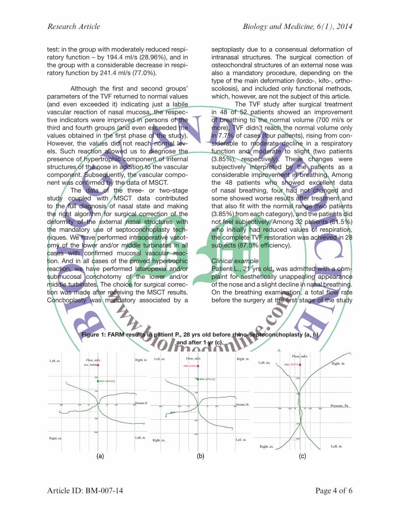
Figure 1: FARM results in patient P., 28 yrs old before rhino-septoconchoplasty (a, b) and after 1 yr (c).
Research Article Biology and Medicine, 6(1), 2014
Article ID: BM-007-14 Page 5 of 6
was 410 ml/s (59% of the normal flow – a moder-ate decrease in passability). Also a serious asym-metry of nasal breathing was diagnosed – the volume ratio on the right side to such parameter on the left side was 1.99 with predominance on the left side. There was also two-sided decrease in flows increment. Due to initial unsatisfactory result, the patient was immediately given two drops of Adrianol (Tramazoline1Phenylephrine) in each nostril. After 10 min, the TVF was equal to 468 ml/s (67% of norm (Figure 1b)), it increased slightly, allowing to diagnose mainly hypertrophic changes of the nasal mucosa. Aggravation of the flow asymmetry after applying of adrenomimetic indicated persistent asymmetric deformation of the nasal structures (asymmetric hypertrophy of the turbinates and septum deformation process with a predominance on the right side), subse-quently confirmed by MSCT. During the surgical correction, in addition to the traditional methods of correction of external nasal structures we per-formed correction of deformity of the nasal sep-tum (resection of the septum crest), lateropexy, submucosal conchotomy, and coagulation of vessels of both inferior turbinates (more on the right side) and the right middle turbinate. As a result of the TVF recovery treatment, the nor-mal values achieved after correction in a year (1059 ml/s-151%). Asymmetry of the volume flow after the treatment was within the normal range (1.06), while increase in the flow incre-ments was normal (Figure 1c).
Thus, we can conclude that the use of FARM in the preoperative period allowed a more thorough examination and contributed to the development of an optimal surgical treat-ment to achieve stable positive results in most cases. FARM results do not always coincide with the feelings of the patients themselves, which requires a careful approach to the diagno-sis and the right treatment of the study results. The control of nasal breathing by an FARM method in 1 yr after the surgery helped to con-firm the correct choice of the surgical technique and the quality of its implementation, as well as to ascertain and document postoperative results.
Conclusions
1. During the stepwise comprehensive analysis of the results, the FARM method allows to deter- mine not only the completeness of the volume
of nasal breathing but also the hyperreactivity of nasal mucosa, to diagnose subjectivity of assessment of patient’s breath and anatomical defects of the internal nasal structures with a predominance on the affected side.
2. The study of nasal breathing quality (in cases of breathing malfunction) in the preoperative period should be accompanied by MSCT test for a complete diagnosis of changes of the nasal structures and to optimize an algorithm of the rhinoplasty.
3. In all cases of nasal deformation with a proven decrease in the volume of breathing aesthetic techniques, rhinoplasty should be supplemented by the functional methods of correction of the nasal septum and turbinates. Their comprehensive application will give positive predictable postoperative results.
References
1. Pshenisnov KP, et al. (2010) Course of plastic surgery: A guide for physicians (in 2 volumes), Rybinsk Printing House, Yaroslavl, Rybinsk, pp. 577-684.
2. Ezrokhin VM, et al. (2007) Surgical treatment of nasal deformities. Medkniga, Moscow, p. 144.
3. Pshenisnov KP, Gagarin VV (2000) Rhinoplasty I: Surgical anatomy of the nose and facial proportions analysis. Selected Topics of Plastic Surgery 1(4): 48.
4. Piskunov SZ, Kharchenko V, Piskunov VS (2000) Clinical significance of some anomalies of the nasal cavity and paranasal sinuses. Russian Rhinology 4: 8-10.
5. Clement PA, Gordts F (2005) Consensus report on acoustic rhinometry and rhinomanometry. Rhinology 43(3): 169-179.
6. Passàli D, Mezzedimi C, Passàli GC (2000) The role of rhinomanometry, acoustic rhinometry, and mucociliary transport time in the assessment of nasal patency. Journal of Ear, Nose, & Throat 79(5): 397-400.
7. Ewart CJ, Leonard CJ, Harper JG, Yu J (2006) A simple and inexpensive method of preoperative computer imaging for rhinoplasty. Annals of Plastic Surgery 56: 46-49.
Research Article Biology and Medicine, 6(1), 2014
Article ID: BM-007-14 Page 6 of 6
8. Palma P, Khodaei I, Tasman A (2011) A guide to the assessment and analysis of the rhinoplasty patient. Facial Plastic Surgery 27(2): 146-159.
9. Belousov AE (2010) Functional rhinoplasty. Publishing House of the Polytechnic University, St. Petersburg, p. 512.
10. Huizing EH, Groot JM (2003) Functional reconstructive nasal surgery. Thieme, p. 386.
11. Yunusov AS, Popova OI (2011) Additional research methods that improve the effectiveness of reconstructive endonasal surgeries. Doctor.ru 65(6): 23-26.
Citation: Drobyshev A, Pavlyuk-Pavlyuchenko L, Krasavtseva K (2014) Analysis of nasal breathing function dynamics during rhinoplasty. Biol Med 6(1): BM-007-14.