anaerobic clostridia chair of medical biology, microbiology, virology, and immunology lecturer prof....
TRANSCRIPT
Anaerobic Clostridia
Chair of Medical Biology, Microbiology, Virology, and Immunology
Lecturer Prof. S.I. Klymnyuk
Clostridia Responsible for Anaerobic Infections
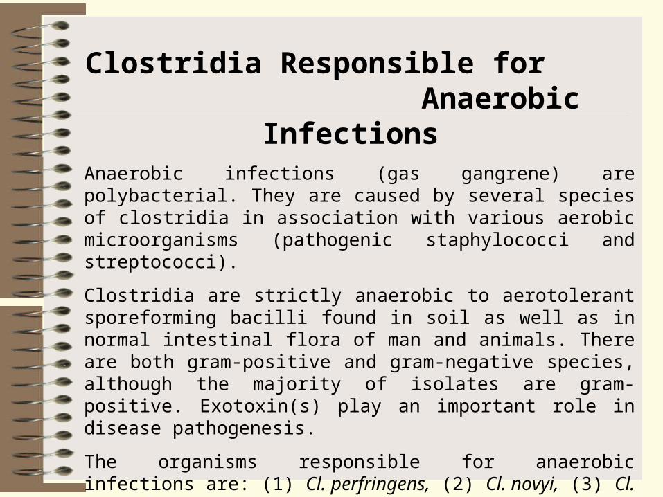
Anaerobic infections (gas gangrene) are polybacterial. They are caused by several species of clostridia in association with various aerobic microorganisms (pathogenic staphylococci and streptococci).
Clostridia are strictly anaerobic to aerotolerant sporeforming bacilli found in soil as well as in normal intestinal flora of man and animals. There are both gram-positive and gram-negative species, although the majority of isolates are gram-positive. Exotoxin(s) play an important role in disease pathogenesis.
The organisms responsible for anaerobic infections are: (1) Cl. perfringens, (2) Cl. novyi, (3) Cl. septicum, (4) Cl. histolyticum, and (5) Cl. sordellii. Cl. chauvoei, Cl. fallax, and Cl. sporogenes are pathogenic for animals. Cl. aerofoetidum and Cl. tertium are non-pathogenic organisms which have significance in the pathogenesis of anaerobic infections only in association with pathogenic bacteria.
Anaerobic infections may be caused by any one of the first four species mentioned above but usually several members of a parasitocoenosis acting in a particular combination are responsible for them. The less pathogenic and non-pathogenic species cannot be responsible for anaerobic infections by themselves, but they cause tissue destruction, lower the oxidation-reduction potential, and thus create favourable conditions for the growth of pathogenic species.
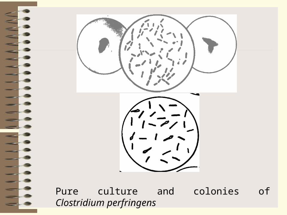
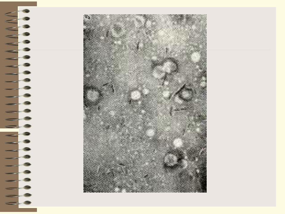
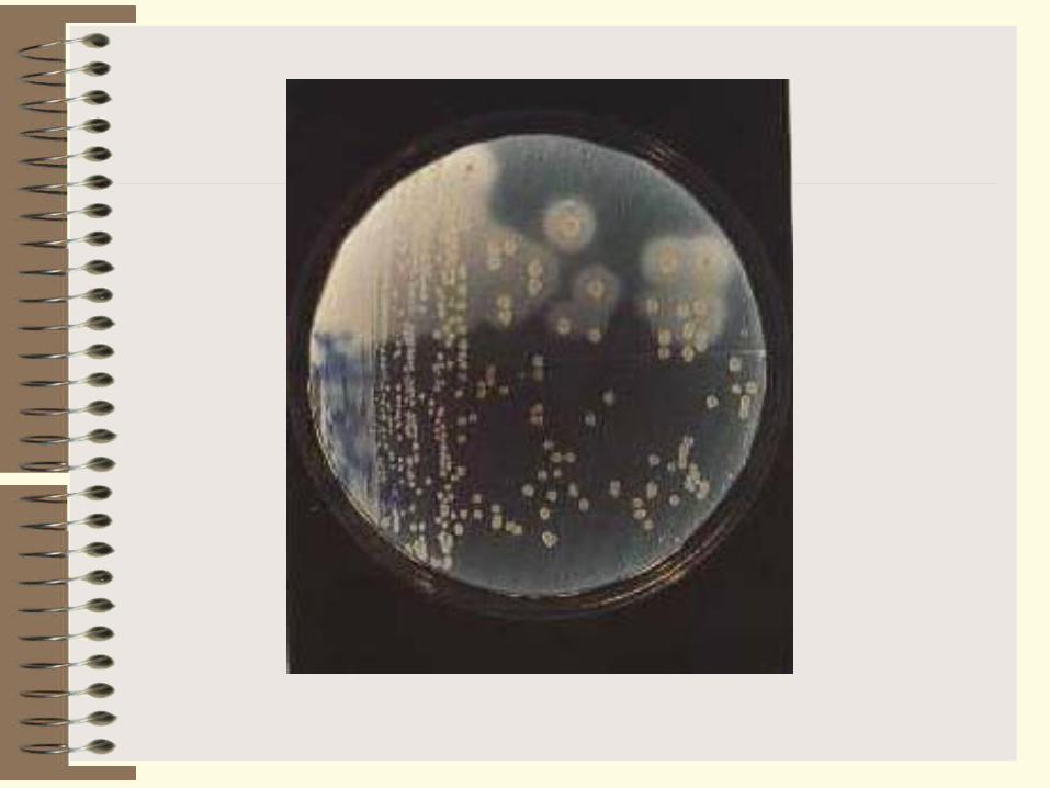
Pure culture and colonies of Clostridium perfringens
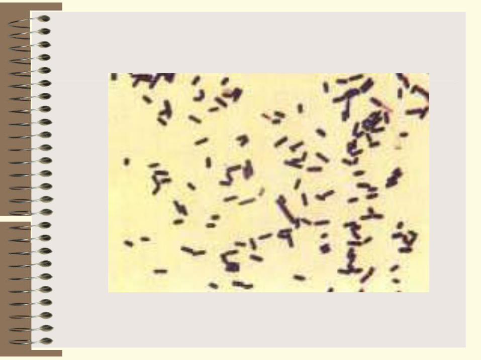

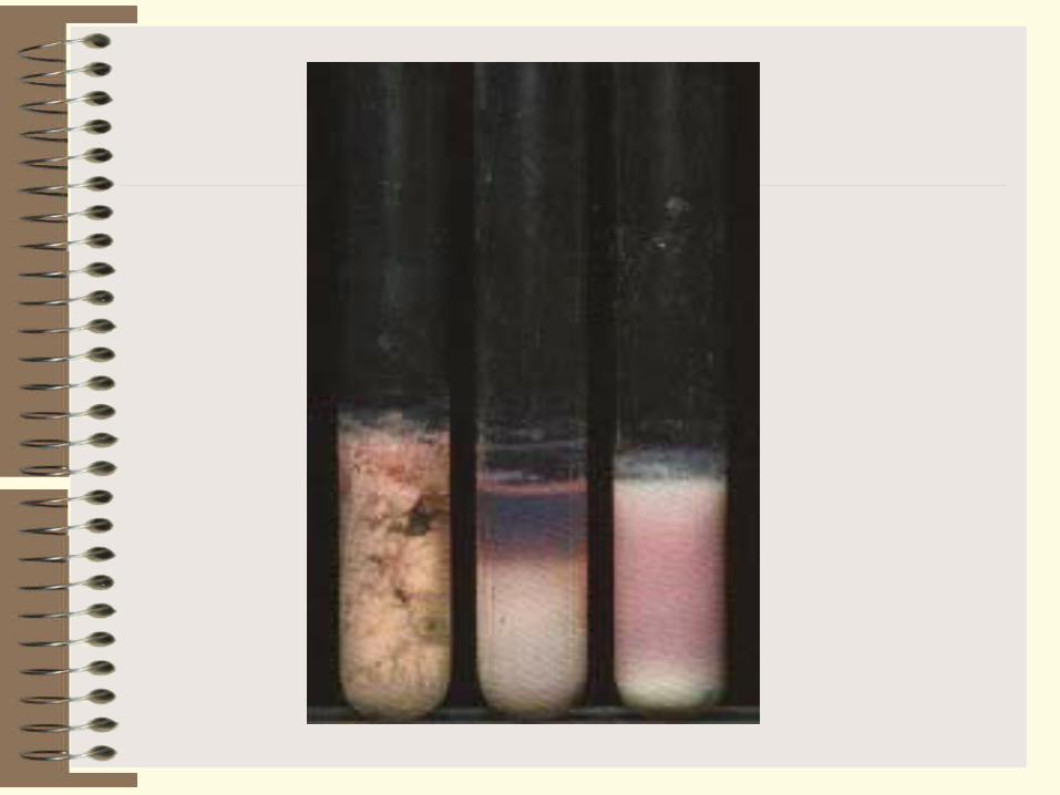
Cultivation. Cl. perfringens is less anaerobic than the other causative agents of anaerobic infections. It grows on all nutrient media which are used for cultivation of anaerobes. The optimum temperature for growth is 35-37 0 (it does not grow below 16 and above 50°C), and optimal pH of medium is 6.0-8.0. A uniform turbidity and large volumes of gas are produced in cultures grown on Kitt-Tarozzi medium.
Brain medium is not blackened. The colonies resemble discs or lentils deep in agar stabcultures (see Fig. 1). On blood agar containing glucose smooth disc-like grey colonies are formed, with smooth edges and a raised centre.

Many strains of Cl. perfringens lose their anaerobic properties on exposure to antibiotics, bacteriophage, and X-rays and may be cultivated under aerobic conditions. Catalase and peroxidase, enzymes typically present in aerobic organisms, were revealed in the variants thus obtained. The aerobic variants are non-toxic and non-pathogenic for laboratory animals.

Fermentative properties. Cl. perfringens slowly liquefies gelatin, coagulated blood serum and egg albumen The organism reduces nitrates to nitrites and normally no indole or only traces are produced. Volatile amines, aldehydes, ketones, and acetyl methyl carbinol, are produced. Milk is vigorously coagulated and a sponge-like clot is formed. In meat medium the organism yields butyric and acetic acids and large quantities of gases (CO2 H2, H2S, NH3). It ferments
glucose, levulose, galactose, maltose, saccharose, lactose, starch, and glycogen with acid and gas formation. Mannitol is not fermented.
Toxin production. The organism produces a toxin which has a complex chemical structure (lethal toxin, haemotoxin, neurotoxin, and necrotic toxin). The toxins and enzymes produced by the various species of the gas gangrene group are similar from one species to another. Actually, many of them have not been purified or characterized, and are grouped together under the general name lethal toxins. The products produced by C perfringens have received the most study: at least 12 different toxins and enzymes have been described and labeled with Greek letters, but not all serologic strains of C perfringens produce all 12 products or even similar quantities of certain toxins and enzymes.
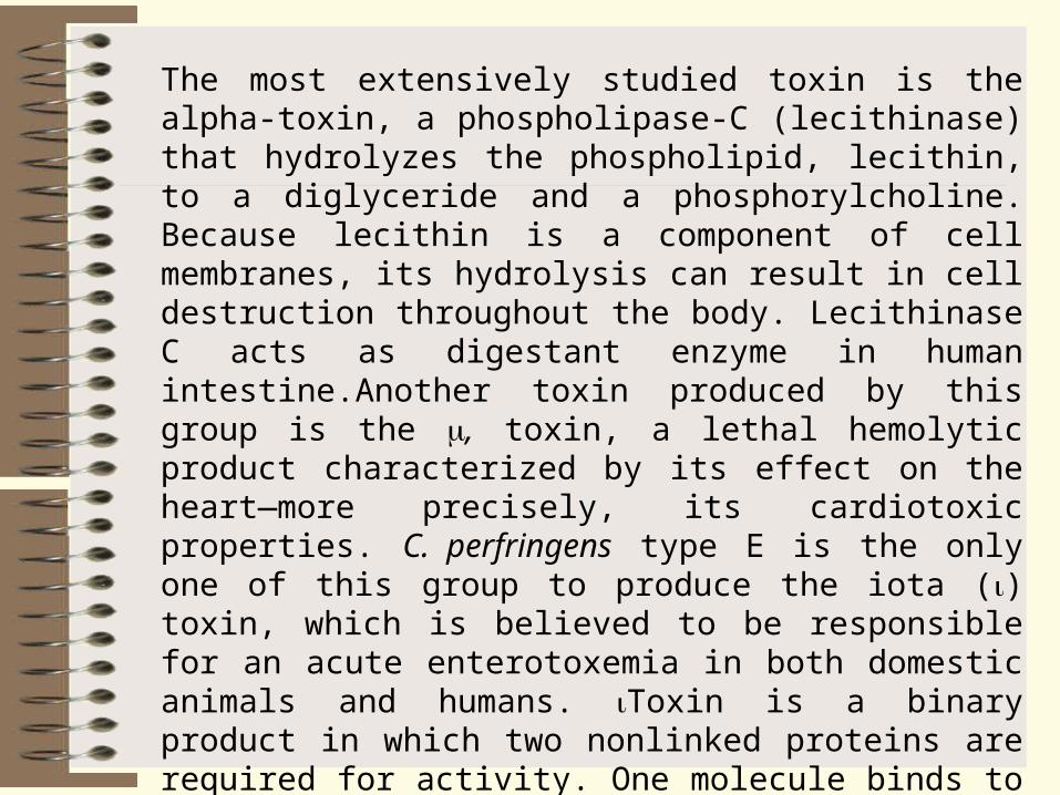
The most extensively studied toxin is the alpha-toxin, a phospholipase-C (lecithinase) that hydrolyzes the phospholipid, lecithin, to a diglyceride and a phosphorylcholine. Because lecithin is a component of cell membranes, its hydrolysis can result in cell destruction throughout the body. Lecithinase C acts as digestant enzyme in human intestine.Another toxin produced by this group is the , toxin, a lethal hemolytic product characterized by its effect on the heart—more precisely, its cardiotoxic properties. C. perfringens type E is the only one of this group to produce the iota () toxin, which is believed to be responsible for an acute enterotoxemia in both domestic animals and humans. Toxin is a binary product in which two nonlinked proteins are required for activity. One molecule binds to a cell (iota–b), functioning as a receptor to transport the active toxin molecule (iota–a)across the membrane. Like botulism C2 toxin, toxin will ADP–ribosylate poly L–arginine and skeletal muscle and nonmuscle actin, but its true substrate within the cell is unknown
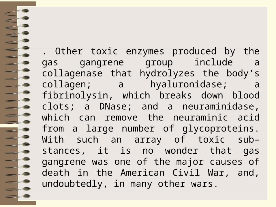
. Other toxic enzymes produced by the gas gangrene group include a collagenase that hydrolyzes the body's collagen; a hyaluronidase; a fibrinolysin, which breaks down blood clots; a DNase; and a neuraminidase, which can remove the neuraminic acid from a large number of glycoproteins. With such an array of toxic sub–stances, it is no wonder that gas gangrene was one of the major causes of death in the American Civil War, and, undoubtedly, in many other wars.
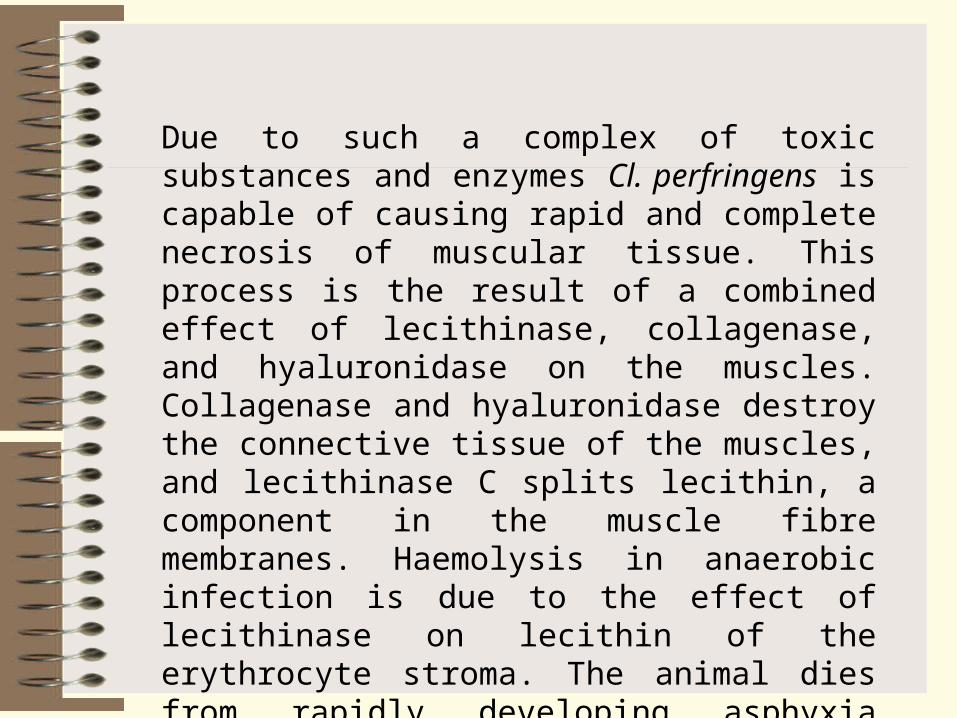
Due to such a complex of toxic substances and enzymes Cl. perfringens is capable of causing rapid and complete necrosis of muscular tissue. This process is the result of a combined effect of lecithinase, collagenase, and hyaluronidase on the muscles. Collagenase and hyaluronidase destroy the connective tissue of the muscles, and lecithinase C splits lecithin, a component in the muscle fibre membranes. Haemolysis in anaerobic infection is due to the effect of lecithinase on lecithin of the erythrocyte stroma. The animal dies from rapidly developing asphyxia which is the result of intensive erythrocyte destruction and disturbance of the nerve centres.
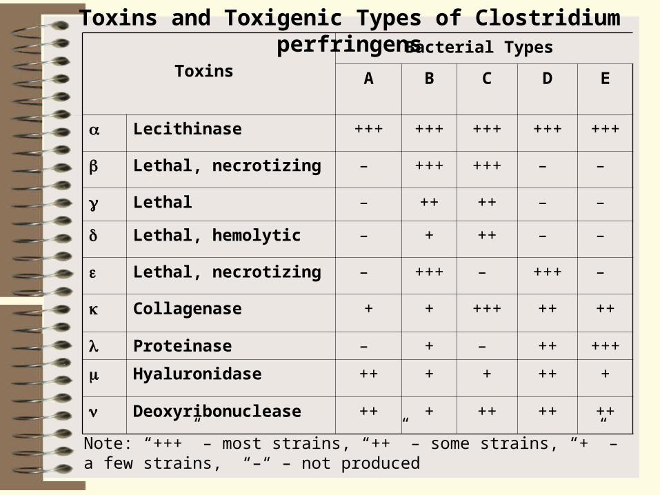
Toxins and Toxigenic Types of Clostridium perfringens
Toxins
Bacterial Types
A B C D E
Lecithinase +++ +++ +++ +++ +++
Lethal, necrotizing – +++ +++ – –
Lethal – ++ ++ – –
Lethal, hemolytic – + ++ – –
Lethal, necrotizing – +++ – +++ –
Collagenase + + +++ ++ ++
Proteinase – + – ++ +++
Hyaluronidase ++ + + ++ +
Deoxyribonuclease ++ + ++ ++ ++
Note: “+++” – most strains, “++” – some strains, “+” – a few strains, “–“ – not produced
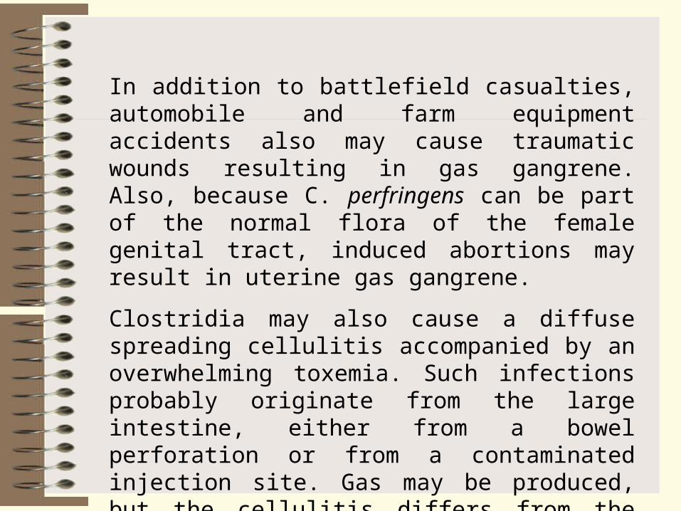
In addition to battlefield casualties, automobile and farm equipment accidents also may cause traumatic wounds resulting in gas gangrene. Also, because C. perfringens can be part of the normal flora of the female genital tract, induced abortions may result in uterine gas gangrene.
Clostridia may also cause a diffuse spreading cellulitis accompanied by an overwhelming toxemia. Such infections probably originate from the large intestine, either from a bowel perforation or from a contaminated injection site. Gas may be produced, but the cellulitis differs from the classic gas gangrene in that muscle necrosis is not involved.
Antigenic structure and classification. Six variants of Cl. perfringens are distinguished: A, B, C, D, E, and F. These variants are differentiated by their serological properties and specific toxins.
Variant A is commonly found as a commensal in the human intestine, but it produces anaerobic infections when it penetrates into the body by the parenteral route. Variant B is responsible for dysentery in lambs and other animals. Variant C causes hemorrhagic enterotoxaemia in sheep, goats, sucking pigs, and calves. Variant D is the cause of infectious enterotoxaemia in man and animals, and variant E causes enterotoxaemia in lambs and calves. Variant F is responsible for human necrotic enteritis.
Resistance. The spores withstand boiling for period of 8 to 90minutes. The vegetative forms are most susceptible to hydrogen peroxide, silver ammonia, and phenol in concentrations commonly employed for disinfection.
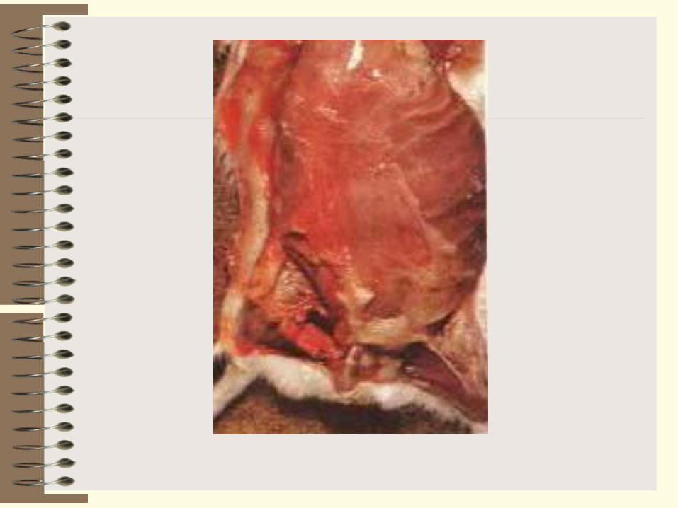
Pathogenicity for animals. Among laboratory animals, guinea pigs, rabbits, pigeons, and mice are most susceptible to infection. Postmortem examination of infected animals reveals oedema and tissue necrosis with gas accumulation at the site of penetration of the organism. Most frequently clostridia are found in the blood.
Clostridium novyi. The organism was discovered by F. Novy in 1894. Its role in the aetiology of anaerobic infections was shown in 1915 by M. Weinbergand P. Seguin. It ranks second among the causative agents of anaerobic infections. Soil examination reveals the presence of the organism in 64per cent of the cases.
Morphology. Cl. novyi is a large pleomorphous rod with rounded ends, 4.7-22.5 mcm in length and 1.4-2.5 mcm in width, and occurs often in short chains (Fig. 2). The organism is motile, peritrichous, and may possess as many as 20 flagella. It forms oval, normally subterminal spores in the external environment. In the body of man and animals it is non-capsulated. The organism is Gram-positive. The G+C content in DNA amounts to 23 per cent.
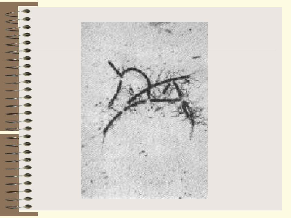
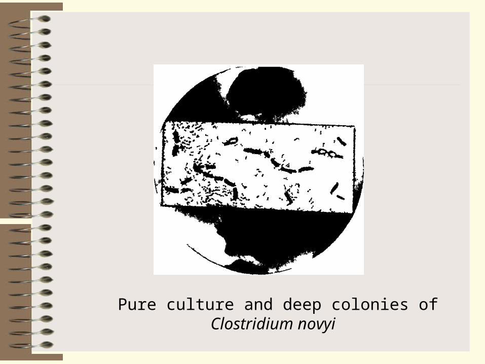
Pure culture and deep colonies of Clostridium novyi
Fermentative properties. The organisms slowly liquefy and blacken gelatin. They coagulate milk, producing small flakes. Glucose, maltose, and glycerin are fermented with acid and gas formation. Acetic, butyric, and lactic acids as well as aldehydes and alcohols are evolved as a result of the breakdown of carbohydrates.
Toxin production. Cl. novyi A produces alpha, gamma, delta, and epsilon toxins; Cl. novyi B produces alpha, beta, zeta, and eta toxins. Cl.novyi C is marked by low toxigenicity. In cultures Cl. novyi liberates active haemolysin which possesses the properties of lecithinase.
Clostridium septicum. The organism was isolated from the blood of a cow in 1877 by L. Pasteur and J. Joubert. In 1881 R. Koch proved the organism to be responsible for malignant oedema. It is found in 8 per cent of examined soil specimens.
Morphology. The clostridia are pleomorphous and may be from3.1-14.1 mcm long and from 1.1-1.6 mcm thick; filamentous forms, measuring up to 50 mcm in length, also occur. The organisms are motile, peritrichous, and produce no capsules in the animal body. The spores are central or subterminal. The clostridia are Gram-positive but Gram-negative organisms occur in old cultures.
Fermentative properties. Cl. septicum liquefies gelatin slowly, produces no indole, reduces nitrates to nitrites, and decomposes proteins, with hydrogen sulphide and ammonia formation. Force-meat is reddened but not digested; the culture evolving a rancid odour. Levulose, glucose, galactose, maltose, lactose, and salicin are fermented with acid and gas formation. Milk is coagulated- slowly.
Toxin production. Cl. septicum produces a lethal exotoxin, necrotic toxin, haemotoxin, hyaluronidase, deoxyribonuclease, and collagenase. The organism haemolyses human, horse, sheep, rabbit, and guinea pig erythrocytes.
Antigenic structure and classification. On the basis of the agglutination reaction, serovars of Cl. septicum can be distinguished, which produce identical toxins, the differential properties being associated with the structure of the H-antigen Cl. septicum possesses antigens common to Cl. chauvoei which is responsible for anaerobic infections in animals.
Resistance is similar to that of Cl novyi.
Pathogenicity for animals. Among domestic animals horses, sheep, pigs, and cattle may contract the disease. Infected guinea pigs die in18-48 hours. Postmortem examination reveals crepitant haemorrhagic oedema and congested internal organs. The affected muscles have a moist appearance and are light brown in colour. Long curved filaments which consist of clostridia are found in impression smears of microscopical sections of the liver.
Clostridium histolyticum. The organism was isolated in 1916 by M. Wemberg and P. Segum. It produces fibrinolysin, a proteolytic enzyme, which causes lysis of the tissues in the infected body. An intravenous injection of the exotoxin into an animal is followed shortly by death. The fact that the organisms are pathogenic for man has not met with general acceptance in the recent years The organism's responsibility for anaerobic infections during World War II was insignificant.
Pathogenesis and diseases in man. Anaerobic infections are characterized by a varied clinical picture, depending on a number of factors. These include the number of pathogenic anaerobic species and their concomitant microflora, i. e. non-pathogenic or conditionally pathogenic anaerobes and aerobes which occur in particular association reflecting the complex process of parasitocoenosis. The type of wound and the immunobiological condition of the body are also among the factors.
The causative agents of anaerobic infections require certain conditions for their development after they have gained entrance into the body, i. e. favourable medium (the presence of dead or injured tissues)and a low oxidation-reduction potential (state of anaerobiosis) which arises due to the presence of necrotized cells of the affected tissues and aerobic microflora. Later the pathogenic anaerobes cause the necrosis of the healthy tissues themselves.
This process develops particularly intensively in the muscles owing to the fact that they contain large amounts of glycogen which serves as a favourable medium for pathogenic anaerobes responsible for anaerobic infections. Oedema is produced during the first phase of the infection, and gangrene of the muscles and connective tissue, during the second phase.
The exotoxins which are produced by clostridia anaerobic infections exert not only a local effect, causing destruction of muscular and connective tissues, but affect the entire body. This results in severe toxaemia. The body is attacked also by toxic substances produced by the decaying tissues. Investigations have shown that exotoxins produced by the causative agents of anaerobic infections possess potentiation activity. Simultaneous injections of one-fourth of a lethal dose of both Cl. perfringens and Cl. novyi toxins produce a reaction which is more marked than that produced by separate injections of the toxins into different parts of the body.
As a result of the vasoconstrictive effect of the toxins, development of oedema, and gas formation, the skin becomes pale and glistening at first and bronze-coloured later. The temperature of the affected tissues is always lower than that of the healthy areas. Deep changes occur in the subcutaneous cellular, muscle, and connective tissues, and degenerative changes take place in the internal organs.
The organisms themselves play an essential part in the pathogenesis of anaerobic infections owing to their high invasive activity. An extremely important role in the development of the disease is attributed to the reactivity state of the macro-organism (trauma, concomitant diseases, etc.).
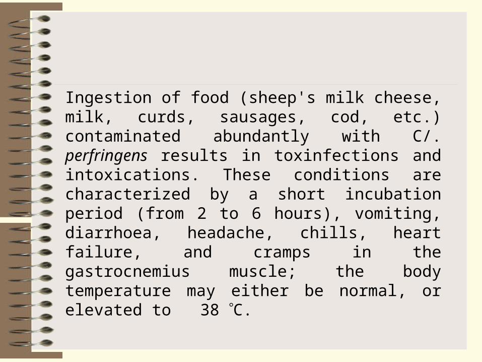
Ingestion of food (sheep's milk cheese, milk, curds, sausages, cod, etc.) contaminated abundantly with C/. perfringens results in toxinfections and intoxications. These conditions are characterized by a short incubation period (from 2 to 6 hours), vomiting, diarrhoea, headache, chills, heart failure, and cramps in the gastrocnemius muscle; the body temperature may either be normal, or elevated to 38 C.
Immunity. The immunity produced by anaerobic infections is associated mainly with the presence of antitoxins which act against the most commonly occurring causative agents of the wound infection. For example, Cl. perfringens loses its lecithinase activity completely in the presence of a sufficient amount of antitoxin against its alpha-toxin.
The toxin-antitoxin reaction depends to a great extent on the presence of lecithin which acts as substratum for toxin activity. The antitoxin cannot neutralize lecithinase if the former is added at certain periods of time after the toxin had been in the presence of lecithin, the reaction being simply somewhat delayed in such cases. A definite role is played by the antibacterial factor, since the existence of bacteraemia in the pathogenesis of anaerobic infections has been shown.
Laboratory diagnosis. Material selected for examination include spieces of affected and necrotic tissues, oedematous fluid, dressings, surgical silk, catgut, clothes, soil, etc. The specimens are examined in stages:
(1) microscopic examination of the wound discharge for the presence of C/. perfringens;
(2) isolation of the pure culture and its identification according to the morphological characteristics of clostridia, capsule production, motility, milk coagulation, growth on iron-sulphite agar, gelatin liquefaction, and fermentation of carbohydrates;
(3) inoculation of white mice with broth culture filtrates or patient's blood for toxin detection;
(4) performance of the antitoxin-toxin neutralization reaction on white mice (a rapid diagnostic method).
C. perfringens is found in 70% to 80% of all cases of gas gangrene, and of the five serologic types of this organism, type A is the most prevalent. Any exudate is cultivated on thioglycolate broth and on blood–agar plates that are incubated both aerobically and anaerobically. The presence of large gram–positive rods that grow only anaerobically is strong evidence for clostridia C. perfringens is characterized by a stormy fermentation in milk, in which the coagulated milk is blown apart by gas formed during the fermentation of the lactose in milk. Organisms producing an toxin hydrolyze the lecithin in an egg yolk medium, breaking down the lipid emulsion and, in turn, causing an opaque area to appear around the colony. Individual clostridial species are identified by a series of biochemical tests.
Transfusion of blood, oxygen therapy, administration of inhibitors of proteolytic enzymes and biologically active preparations which normalize metabolism are auxiliary therapeutic measures. Hyperbaric oxygen chambers, in which an infected area is placed in a chamber containing pure oxygen under pressure, have been used with some success to stop the growth of these obligate anaerobes.

Causative agents of Tetanus and Botulism
Tetanus Clostridia
A. Nicolaier discovered the causative agent of tetanus in 1884, and S.Kitasato isolated the pure culture in 1889.
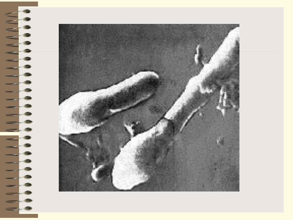
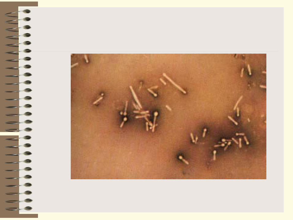
Morphology. The causative agent of tetanus (Clostridium tetani) is a thin motile rod, 2.4-5 mcm in length and 0.5-1.1 mcm in breadth. It has peritrichous flagellation and contains granular inclusions which occur centrally and at the ends of the cell. The organism produces round terminal spores which give it the appearance of a drumstick. Cl. tetani is Gram-positive.
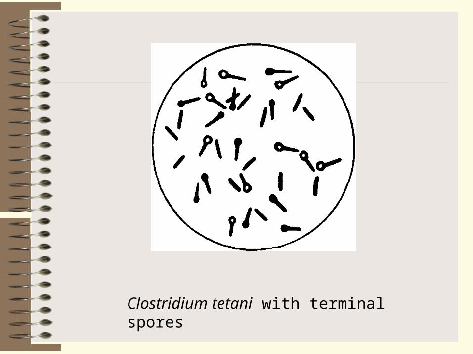
Clostridium tetani with terminal spores


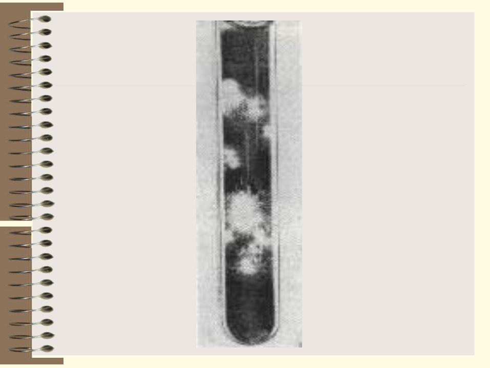
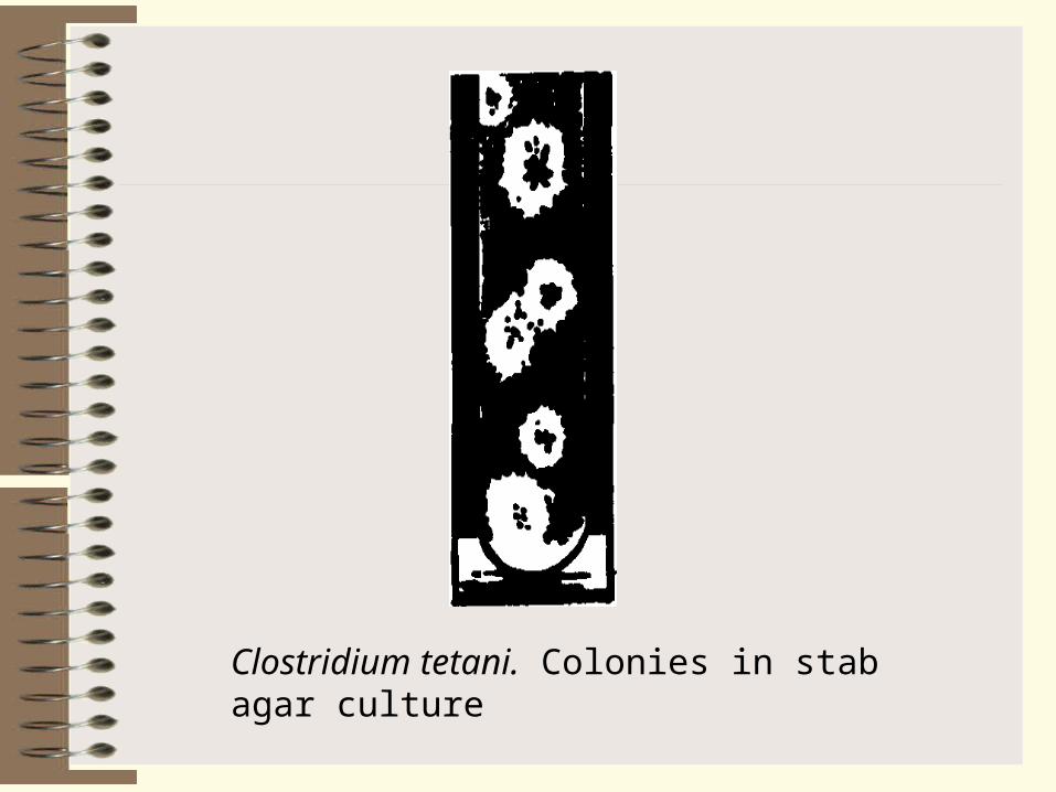
Clostridium tetani. Colonies in stab agar culture
Fermentative properties. Cl. tetani causes slow gelatin liquefaction and produces no indole. Nitrates are rapidly reduced to nitrites. The organisms coagulate milk slowly, forming small flakes. No carbohydrates are usually fermented
Toxin production. Cl tetani produces an extremely potent exotoxin which consists of two fractions, tetanospasmin, which causes muscle contraction, and tetanolysin, which haemolyses erythrocytes.
A 0.0000005 ml dose of toxin obtained from a broth culture filtrate kills a white mouse which weighs 20 g; and 0.000000005 g of dry toxin obtained by ammonium sulphate precipitation is fatal to the mouse. Several million lethal mouse doses are contained in 1 mg of crystalline toxin.
The mode of action of the tetanus toxin is similar to that of enzymes which catalyse chemical reactions in the bodies of affected animals.
Tetanus toxin (also termed tetanospasmin) is synthesized in the bacterium as a single polypeptide chain, but after its release by lysis of the organism, a bacterial protease cleaves one peptide bond to yield two chains that remain linked together through a disulfide bond. The larger chain (H chain) has a molecular weight of 100,000 daltons, and it possesses the specific receptors that bind the toxin to the neuronal gangliosides. The smaller peptide (L chain) has a molecular weight of 50,000 daltons and is thought to exert the biologic effect of the toxin.
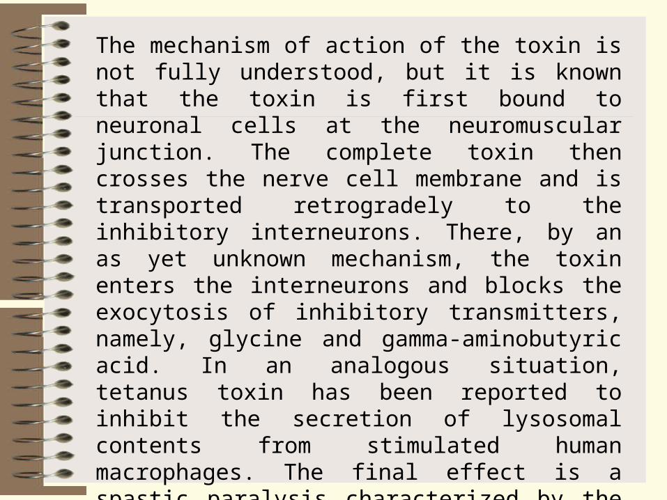
The mechanism of action of the toxin is not fully understood, but it is known that the toxin is first bound to neuronal cells at the neuromuscular junction. The complete toxin then crosses the nerve cell membrane and is transported retrogradely to the inhibitory interneurons. There, by an as yet unknown mechanism, the toxin enters the interneurons and blocks the exocytosis of inhibitory transmitters, namely, glycine and gamma-aminobutyric acid. In an analogous situation, tetanus toxin has been reported to inhibit the secretion of lysosomal contents from stimulated human macrophages. The final effect is a spastic paralysis characterized by the convulsive contractions of voluntary muscles. Because the spasms frequently involve the neck and jaws, the disease had been referred to as lockjaw. Death ordinarily results from muscular spasms affecting the mechanics of respiration.
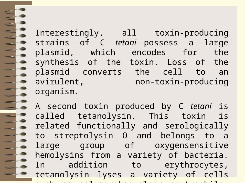
Interestingly, all toxin-producing strains of C tetani possess a large plasmid, which encodes for the synthesis of the toxin. Loss of the plasmid converts the cell to an avirulent, non-toxin-producing organism.
A second toxin produced by C tetani is called tetanolysin. This toxin is related functionally and serologically to streptolysin O and belongs to a large group of oxygensensitive hemolysins from a variety of bacteria. In addition to erythrocytes, tetanolysin lyses a variety of cells such as polymorphonuclear neutrophils, macrophages, fibroblasts, ascites tumor cells, and platelets. It is unknown, however, whether it plays any significant role in infections by C tetani.
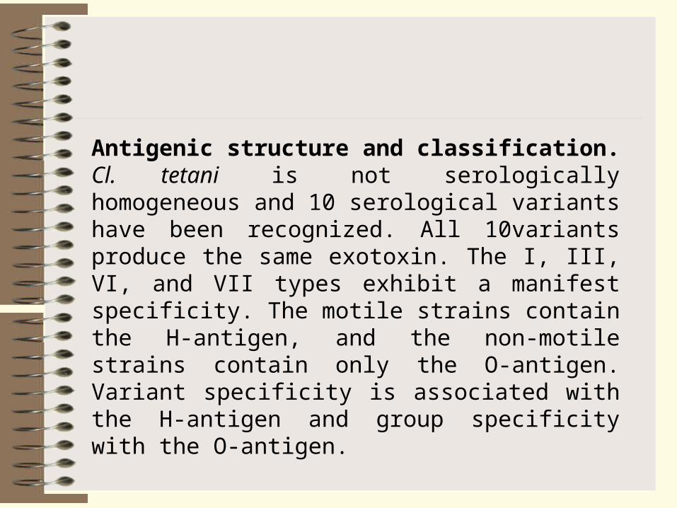
Antigenic structure and classification. Cl. tetani is not serologically homogeneous and 10 serological variants have been recognized. All 10variants produce the same exotoxin. The I, III, VI, and VII types exhibit a manifest specificity. The motile strains contain the H-antigen, and the non-motile strains contain only the O-antigen. Variant specificity is associated with the H-antigen and group specificity with the O-antigen.
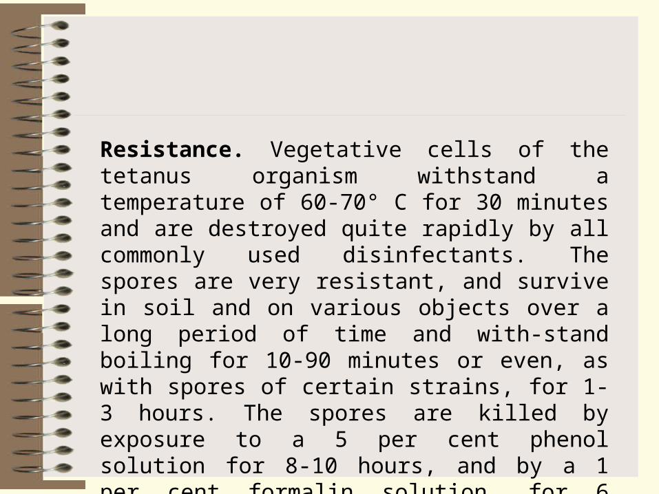
Resistance. Vegetative cells of the tetanus organism withstand a temperature of 60-70° C for 30 minutes and are destroyed quite rapidly by all commonly used disinfectants. The spores are very resistant, and survive in soil and on various objects over a long period of time and with-stand boiling for 10-90 minutes or even, as with spores of certain strains, for 1-3 hours. The spores are killed by exposure to a 5 per cent phenol solution for 8-10 hours, and by a 1 per cent formalin solution, for 6 hours. Direct sunlight destroys them in 3-5 days.
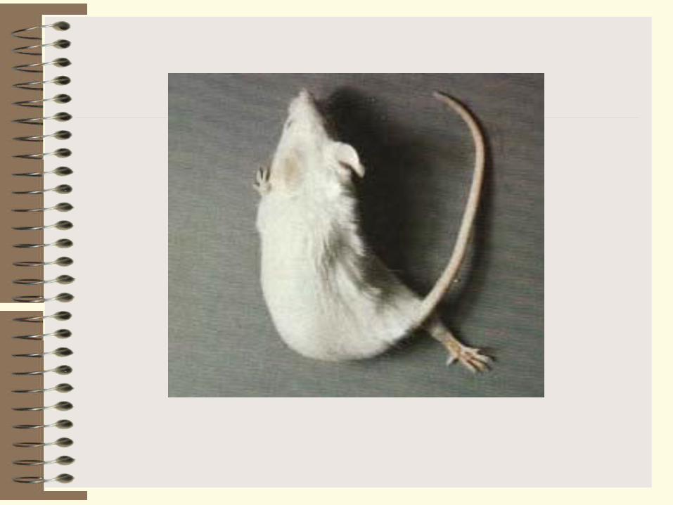
Pathogenicity for animals. Horses and small cattle acquire the disease naturally, and many animals may act as carriers of Cl. tetani.
Among experimental animals, white mice, guinea pigs, rats, rabbits, and hamsters are susceptible to tetanus.
The disease in animals is manifested by tonic contractions of the striated muscles and lesions in the pyramid cells of the anterior cornua of the spinal cord. The extremities are the first to be involved in the process, the trunk being affected later (ascending tetanus).
Pathogenesis and disease in man. Healthy people and animals, who discharge the organisms in their faeces into the soil, are sources of the infection. Spores of Cl. tetani can be demonstrated in 50-80 per cent of examined soil specimens, and some soils contain the spores in all test samples (manured soil is particularly rich in spores). The spores may be spread in dust, carried into houses, and fall on clothes, underwear, foot-wear, and other objects.
The majority of tetanus cases in adults occur among farm workers, and more than 33 per cent of the total incidence of the disease is associated with children from 1 to 15 years old. In more than 50 per cent of cases tetanus is acquired as the result of wounds of the lower extremities inflicted by spades, nails, and stubbles during work in the orchard or in the field.
Cl. tetani may gain entrance into the body of a newborn infant through the umbilical cord and into a woman during childbirth, through the injured uterine mucosa.
The organisms produce exotoxins (tetanospasmin and tetanolysin) at the site of entry. In some cases tetanus is accompanied by bacteraemia.
Microbes and spores, washed-off from the toxin, normally produce no disease and are rapidly destroyed by phagocytes.
The tetanus toxin reaches the motor centres of the spinal cord via the peripheral nerves (it moves along the axial nerve cylinders or along the ecto- and endoneural lymphatics).
According to the school of thought of A. Speransky, the specificity of the tetanus toxin is manifest only at the onset of the disease. In its further stages the infection is governed by other phenomena, primarily by the neurodystrophic factors. Sites of high and increasing excitation develop under the influence of irritation stimuli.
The toxin enters the blood and is thus distributed throughout the whole body, causing subsequent excitation of the peripheral nerve branches and the cells of the anterior cornua of the spinal cord.
Receptors situated in the neuromuscular apparatus play a significant role in the development of tetanus. Impulses sent out from these receptors give rise to a dominant excitation focus in the central nervous sys-tem The effect of the toxin produces an increased reflex excitation of the motor centres, and this, in its turn, leads to the development of attacks of reflex tonic muscular spasms which may occur often in response to any stimuli coming from the external environment (light, sound, etc.).
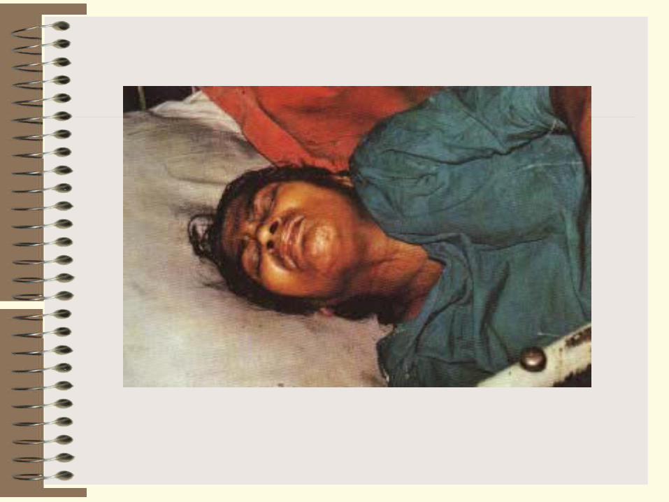
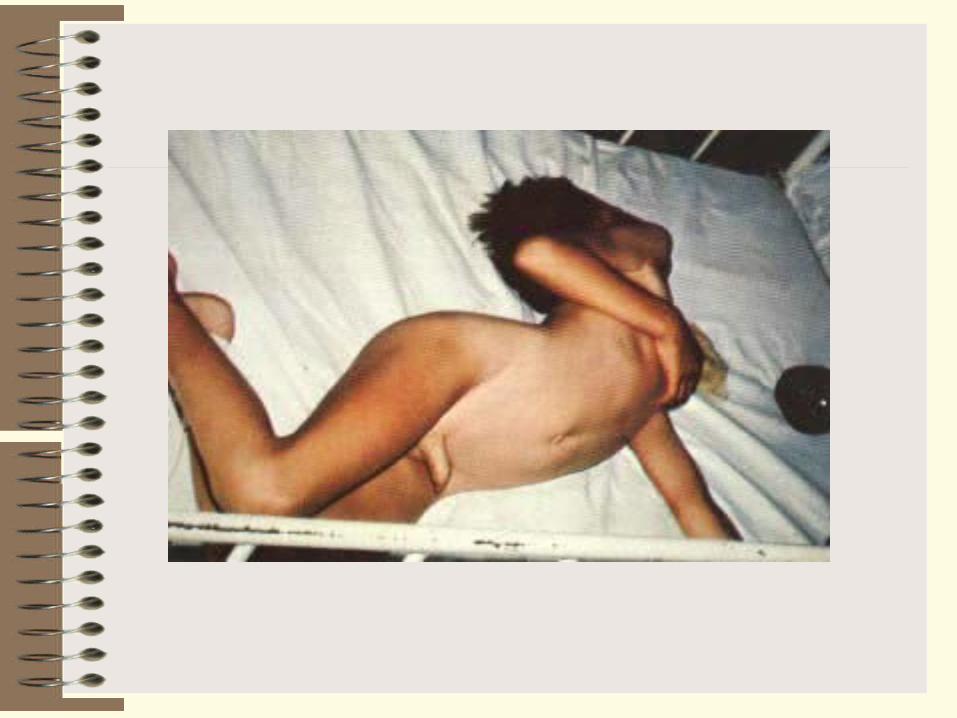
The onset of the disease is characterized by persistent tonic muscular spasms at the site of penetration of the causative agent. This is followed by tonic spasms of the jaw muscles (trismus), face muscles (risus sardonicus), and occipital muscles. After this the muscles of the back (opisthotonus) and extremities are affected. Such is the development of the clinical picture of descending tetanus. The patient lies in bed, resting on his head and hips with his body bent forward like an arc. The death rate varies from 35 to 70 per cent, being 40 per cent on the average and 65 per cent in the USA. More than 50000 people die every year from tetanus in the world. According to incomplete WHO data, more than one million people contracted the disease within a period of 10 years (1951-1960) and about 500000 of them died.


Immunity following tetanus is mainly antitoxic in character, and of low grade. Reinfections may occur.
Treatment. Intramuscular injections of large doses of antitoxic antitetanus serum are employed. The best result is produced by gamma-globulin obtained from the blood of humans immunized against tetanus. Anticonvulsant therapy includes intramuscular injections of 25 per cent solutions of magnesium sulphate, administration of diplacine, condelphine, aminazine, pipolphen or andaxine and chloral hydrate introduced in enemas. To reproduce active immunity, 2 ml of toxoid is administered two hours before injecting the serum; the same dose of toxoid is repeated within 5-6 days.
Uninoculated persons are subjected to active and passive immunization. This is achieved by injecting 0.5 ml of toxoid and 3.000 units of antitoxic serum and then 5 days later, another 0.5 ml of toxoid. The tetanus antitoxin is also introduced into previously inoculated individuals suffering from a severe wound. Injection of the total dose of antitoxin is preceded by an intracutaneous test for body sensitivity to horse protein. This is carried out by introducing 0.1 ml of antitoxin, previously diluted 1 :100, into the front part of the forearm. If the intracutaneous test proves negative, 0.1 ml of whole antitoxin is injected subcutaneously and if no reaction is produced in 30 minutes, the total immunization dose is introduced.

The complex of prophylactic measures includes adequate surgical treatment of wounds. The organisms are sensitive to penicillin, but the antibiotic has no effect on the neutralization of the toxin. However, after surgical cleansing of the wound, antibiotic therapy can be helpful in preventing any additional growth of the organisms.
Prophylaxis is ensured by prevention of occupational injuries and traumas in everyday life. Active immunization is achieved with tetanus toxoid. It is injected together with a tetravalent or polyvalent vaccine or maybe a component of an associated adsorbed vaccine. The pertussis-diphtheria-tetanus vaccine and associated diphtheria-tetanus toxoid are employed for specific tetanus prophylaxis in children. Immunization is carried out among all children from 5-6 months to 12 years of age, individuals living in certain rural regions (in the presence of epidemiological indications), construction workers, persons working at timber, water-supply, cleansing and sanitation, and peat enterprises, and railway transport workers.
Immunization with tetanus toxoid stimulates the production of sufficient amounts of antitoxin. Immunity lasts for a period of 2 or 3 years.
The effectiveness of the immunization has made tetanus a relatively rare disease in the developed countries(36 cases in the United States during 1994). In Third World countries, however, most persons are not immunized, and infections of the umbilical cord give rise to large numbers of neonatal tetanus. For example, special surveillance programs indicate death rates from neonatal tetanus to be 2% of all live births in Bangladesh, 6% in Papua, New Guinea, and 14.5% in Haiti. One study relating the proportion of pregnant females with tetanus antitoxin titres adequate to provide protection for the newborn found 96% protected in New Haven, Connecticut, and only 19% in Santiago, Chile.
Oral immunization may become possible using a live attenuated strain of Salmonella typhimurium that was transfected with a plasmid engineered to express a 50–kdfragment of the tetanus toxin. Given orally, this strain provided protective immunity in mice.
After an injury, human tetanus immune globulins should be administered to those who have never been immunized with tetanus toxoid or to those who did not receive the full three doses of toxoid. Booster injections of toxoid also are given if the immune status of the patient is unknown, or if it has been over 5 years since the last dose of toxoid.
Clostridia Responsible for Botulism
The causative agent of botulism (L. botulus sausage, botulism poisoning by sausage toxin), Closlridium botulinum, was discovered in Holland in 1896 by E. van Ermengem. The organism was isolated from ham which had been the source of infection of 34 people and from the intestine and spleen on post-mortem examination. In Western Europe botulism was due to ingestion of sausages, while in America it was caused by canned vegetables, and in Russia, by ingestion of red fish. In the recent 50 years 5635 persons contracted botulism, 1714 of them died.
Morphology. Cl. botulinum is a large pleomorphous rod with rounded ends, 4.4-8.6 mcm in length and 0.3-1.3 mcm in breadth. The organism sometimes occurs in short forms or long threads. Cl. botulinum is slightly motile and produces from 4 to 30 flagella per cell. In the external environment Cl. botulinum produces oval terminal or subterminal spores which give them the appearance of tennis rackets. The organisms are Gram-positive.
On ultrathin sections the cell wall in A, B, and E types consists of five layers, the cytoplasmatic membrane of three layers. By the time of maxi-mum exotoxin liberation (on the 5th-7th day) cell lysis with the discharge of crystalline structure occurs. The cytoplasm is granular and contains inclusions of various size. The nucleoid is compact and occupies a small part of the cytoplasm. Spore formation takes place on the3rd or 4th day of cultivation 1 he G +C content in DNA ranges between 26 and 28 per cent.
Cultivation. Cl. botulinum are strict anaerobes. The optimal growth temperature for serovars A, B, C, and D is 30-40 C, for serovar E 25-37 C, for serovar G 30-37 C They grow on all ordinary media at pH 7.3-7.6 Cultivation is best on minced meat or brain which the organisms turn darker. The cultures have an odour of rancid butter.
On Zeissler's sugar-blood agar irregular colonies are produced which possess filaments or thin thread-like outgrowths. The colonies are surrounded by a zone of haemolysis
In agar stab cultures the colonies resemble balls of cotton wool or compact clusters with thread-like filaments.
On gelatin the organisms form round translucent colonies surrounded by small areas of liquefaction. Later the colonies turn turbid, brownish, and produce thorn-like filaments.
In liver broth (Kitt-Tarozzi medium) turbidity is produced at first, but a compact precipitate forms later, and the fluid clears.

Pure culture and deep colonies of Closlridium botulinum

Fermentative properties. Cl. botulinum (serovars A and B) are proteolytic organisms, and decompose pieces of tissues and egg albumin in fluid medium. The organisms liquefy gelatin, produce hydrogen sulphide, ammonia, volatile amines, ketones, alcohols, and acetic, butyric, and lactic acids. Milk is peptonized with gas formation. Glucose, levulose, maltose, and glycerin are fermented, with acid and gas formation.
Toxin production. Cl. botulinum produces an extremely potent exotoxin. The toxin is produced in cultures and foodstuffs (meat, fish, and vegetables) under favourable conditions in the body of man and animals. Multiplication of the organism and toxin accumulation are inhibited in the presence of a 6-8 per cent concentration of common salt or in media with an acid reaction. Heating at 90 C for 40 minutes or boiling for 10 minutes destroys the toxin.
The toxin produced by Cl. botulinum, as distinct from the tetanus and diphtheria toxins, withstands exposure to gastric juice and is absorbed intact. The toxin produced by serovar A Cl. botulinum can kill 60000 million mice having a total weight of 1 200 000 tons. The toxin has been obtained in crystalline form and is the most potent of all toxins known to date. Curiously, the toxins seem to be secreted as progenitor toxins which, even though some have been crystallized, are composed of two polypeptide subunits linked by disulfide bonds. Also, the toxicity of those toxins that have been extensively studied can be increased from four–fold to 250–fold by treatment with trypsin. This phenomenon is not understood at the molecular level.
The botulinum toxin is a globulin and does not change on recrystallization. Its activity is similar to that of enzymes which catalyse chemical processes in the body of man and animals with formation of large amounts of toxic substances. These substances produce the clinical manifestations of poisoning.
The toxin acts primarily as a neurotoxin, inducing paralysis in three basic steps (1) binding of the toxin to a receptor on the nerve synapse, (2) entrance of the toxin (or possibly one polypeptide subunit) into the nerve cell, and (3) blocking of the release of acetylcholine from the cell, resulting in a flaccid muscle paralysis.
C botulinum type C produces two distinct toxins that have been designated Cl and C2 The Cl toxin functions like other botulism toxins to block the release of acetylcholine at the myoneural junction. C2 toxin, however, is a binary complex consisting of two unlinked components designated as I and II Component II recognizes the cell receptor and thus facilitates the entrance of component I into the cytoplasm The C2 toxin causes a necrotic enteritis, which seems to result in an increase in vascular leakage of the intestinal mucosa. Its mechanism of action is unclear, but it has been shown to ADP-ribosylate G-actin as well as the synthetic substrate, homo-poly L arginine
C botulinum organisms, types C and D, also produce an additional toxin which has been termed exoenzyme C3. The DNA encoding C3 is located on both phage C and phage D, the phages that also encode for botulism toxins C and D, respectively Its function is to ADP-ribosylates Rho protein, a eucaryotic member of the ras superfamily of proteins Because the ras superfamily of proteins are GTP-binding proteins involved in enzyme regulation, this exoenzyme could function as a virulence factor, but the exact consequence of the C3 ADP-ribosylation is unknown.
Antigenic structure and classification. Six serovars of Cl. botulinum are known: A, B, C, D, E, and F, serovars A, B, and F being the most toxic. Each of the serovars is characterized by specific immunogenicity associated with the H-antigen and is neutralized by the corresponding antitoxin. Variants C and D are responsible for neuroparalytic lesions in animals. As has been proved recently, serovar C may produce diseases also in man. The O-antigen is common to all variants.
Resistance. The vegetative forms of the organisms are killed in 30 minutes at 80 C, while the spores withstand boiling for periods from 90 minutes to 6 hours-and survive 115 ° C for 5-40 minutes and 120° C, for3-22 minutes. Spores remain viable in large pieces of meat and in large cans even after autoclaving for 15 minutes at 120° C. In 5 per cent phenol solutions they survive for up to 24 hours and in cultures they may live for a year.
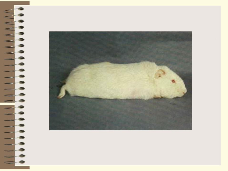
Pathogenicity for animals. Horses, cattle, minks, birds, and among the laboratory animals, guinea pigs, white mice, cats, rabbits, and dogs are susceptible to the botulinum toxin.
Paralysis of the deglutitive, mastication, and motor muscles is usually produced in horses 3 days after infection. The mortality rate reaches 100 per cent. Botulism in bovine cattle is accompanied with bulbar paralysis, and in birds it causes limbemeck and paresis of the legs.
Infection of guinea pigs results in muscular weakness which appears in 24 hours, followed by death in 3-4 days. Autopsy displays hyperaemia of the intestine, gastric flatulence, and a urinary bladder filled beyond capacity. White mice die on the second day after infection manifesting relaxed abdomen muscles and paresis of the hind limbs. Paralysis of the eye muscles, disturbances of accommodation, aphonia, pendulous and protruding tongue, and diarrhoea are caused in cats.
Pathogenesis and disease in man. Botulism is contracted by ingesting meat products, canned vegetables, sausages, ham, salted and smoked fish (red fish more frequently), canned fish, chicken and duck flesh, and other products contaminated with Cl. botulinum. The organisms enter the soil in the faeces of animals (horses, cattle, minks, and domes-tic and wild birds) and fish and survive there as spores.
Natural nidality of botulism among ducks and other wild birds has been ascertained. Extremely widespread epizootics occur in the western regions of Canada, in Uruguay, and in the USA. Natural foci develop in territories where there are stagnant reservoirs rich in vegetable debris which provides favourable conditions for anaerobiosis in warm weather. Intensive processes of decay are accompanied with oxygen uptake which contributes to the growth of serovar C botulism clostridia and production of high concentrations of the exotoxin in the water and alkaline mud in swamps. Besides birds, muskrats and frogs may also contract botulism. Migrating birds spread the causative agent of botulism from the natural foci during their flights.
Cl. botulinum spores occur both in cultivated and virgin soil. They were isolated from 70 per cent of examined soil samples in California, and from 40 percent of samples in the Northern Caucasus. Spores have been isolated from littoral soil at the Sea of Azov, sea water, and silt. They have also been found on the surface of vegetables and fruit, in the intestine of healthy animals, in 5.4 per cent of cases in the guts of fresh red fish (sturgeon, beluga, etc.), and in 15-20 per cent of cases in the guts and in 20 per cent of cases in the tissues of dead fish.
The infectious condition is caused by the exotoxin which is absorbed in the intestine, from where it invades the blood, and affects the medulla oblongata nuclei, cardiovascular system, and muscles. It has been ascertained that Cl. bolulinum may enter the body through wounds. Usually, the wounds themselves were not serious, but wound botulism should be suspected in any persons with even minor wounds who present the typical symptoms of botulism: blurred vision, weakness, and difficulty in swallowing. In the past botulism was considered to be only of a toxic nature. Recent investigations have proved the Cl. botulinum to be present in various organs of individuals who have died from botulism. Therefore, this disease is a toxinfection. The incubation period in botulism varies from 2 hours to10 days, its usual duration is 18 to 24 hours.
Botulism symptoms include dizziness, headache, and, sometimes, vomiting. Paralysis of the eye muscles, accommodation disturbances, dilatation of the pupils, and double vision occur. Difficulty in swallowing, aphonia, and deafness also arise. The death rate is very high (40-60 per cent).
Immunity. The disease does not leave a stable anti-infectious immunity (antitoxic and antibacterial).
Laboratory diagnosis. Remains of food which caused poisoning, blood, urine, vomit, faeces, and lavage waters are examined. Post-mortem examination of stomach contents, portions of the small and large intestine, lymph nodes, and the brain and spinal cord is carried out.
The test specimens are inoculated into Kitt-Tarozzi medium which has previously been held at 100 C for 10-20 minutes. To free the cultures from foreign non-sporeforming microflora, 50 per cent of the test tubes containing the inoculated medium is heated at 80 C for 20minutes and then incubated in anaerobic conditions. The isolated pure culture is identified by its cultural, biochemical, and toxigenic properties.
For toxin detection a broth culture filtrate, patient's blood or urine, or extracts of food remains, are injected subcutaneously or intraperitoneally into guinea pigs, white mice, or cats. One of the control animals is infected with unheated material, while the other animal is injected with the heated specimen. In addition, 3 laboratory animals are given injections .of the filtrate together with serovar A antitoxin, with serovar B antitoxin, and with serovar E antitoxin.
The indirect haemagglutination reaction and determination of the phagocytic index are also performed. This index is significantly lowered in the presence of the toxin.
A rapid method of detection of serovar A, B, C, D, and E toxins in water has been developed in which the toxin is absorbed by talc and a suspension of the talc and toxin is injected into the animals.
Treatment. The stomach is lavaged with potassium permanganate or soda solutions Polyvalent botulinum antitoxin is injected intramuscularly (intravenously or into the spinal canal) m doses of 10000 IU (serovars A, C, and E) and 5000 IU (serovar B). If there is no improvement, the injection is repeated at the same dosage within 5-10 hours. All individuals who had used food which caused even a single case of food poisoning are given 1000-2000 IU of antitoxin as a preventive measure. Simultaneously with the antitoxin, 0 5 ml of each serovar of botulinum toxoid is injected three times at intervals of 3-5 days, for production of active immunity. Penicillin and tetracycline are recommended
General measures include subcutaneous injections of saline and glucose solutions Camphor, caffeine, vitamin C, and thiamine are prescribed if necessary. Strychnine is given 2-3 times a day as a stimulant.
Prophylaxis. Proper organization of food processing technology at food factories, meat, fish, and vegetable canning in particular, and preparation of smoked and salted fish and sausages is essential for the prevention of botulism. Home-preserved fish products (smoked and salted)as well as canned mushrooms and canned vegetables of a low acid con-tent (cucumbers, peppers, eggplant), stewed apricots, etc. are very dangerous since they are usually prepared without observance of sanitary rules.
Fish should be gutted after being caught, and placed in the refrigerator. The established temperature regimen must be observed during transportation, and the fish must be protected from pollution with soil and bowel contents. Vegetables must be washed thoroughly. The cooking of meat and fish in small pieces is recommended. Foodstuffs (ham, fish) should not be stored in large hunks and in many layers. The weight of a canned product should not exceed 0.5 kg. Cl botulinum which have with stood sterilization cause swelling of the can lids. The contents of such cans have an odour of rancid butter Such canned goods must not be put on the market and must be withdrawn and thoroughly examined. Fish must be salted in strong salt solutions (brine) with a minimal concentration of 10 per cent. Canned goods must be stored in a cool place.
Active immunization of man, horses, and cows with the toxoid is recommended by many authors in view of Cl botulinum being wide-spread in nature
Botulism occurrence in the USSR is extremely rare as a result of continuous improvement of the standard of living of the population, and of technological methods in food processing and canning industries, observance of the rules of hygiene and strict state and sanitary control of the production, storage, and sale of foodstuffs.
Infant Botulism
A new variety of botulism was recognized during 1976with the report of five cases of infant botulism. These cases occurred in babies as young as 5 weeks, some of whom were breast fed, although all had had some exposure to other foods. Since then, hundreds of additional cases of infant botulism have been diagnosed, and it has become a significant paediatric clinical entity.
Epidemiology and pathogenesis of infant botulism. Infant botulism has been diagnosed in infants ranging from 3to 35 weeks of age. It is well-established that the disease is acquired by the ingestion of C botulinum spores that subsequently germinate in the intestine and produce botulism toxin. Such spores are ubiquitous and, in fact, soil and dust samples from many homes have been shown to contain such spores. Thus, even breast-fed infants are susceptible through contaminated dust. Honey also has been shown to contain spores of C botulinum, and a number of cases of infant botulism have followed the ingestion of honey.
The major initial symptom of infant botulism is 2to 3 days of constipation followed by flaccid paralysis, resulting in difficulty in nursing and a generalized weakness that has been described as "overtly floppy."
The mortality rate of infants admitted to the hospital has been about 3%, and some patients have required mechanical respirators because of respiratory distress. Death, however, may occur more frequently in undiagnosed cases, and considerable data link infant botulism to at least some cases of the sudden infant death syndrome.
Because, by definition, infant botulism is the result of toxin production by organisms that have colonized the gut, it is not surprising that there have been a few cases of adult-infant botulism that occurred after antibiotic therapy or gastric surgery. Even in cases of food-borne botulism, it is usual for the gut to be colonized with C. botulinum, providing a continuing source of toxin.

Diagnosis and treatment of infant botulism. A tentative clinical diagnosis of botulism can be made for an infant with several days of constipation, an unexplained weakness, difficulty in swallowing, or respiratory distress. A laboratory diagnosis, however, requires the demonstration of botulism toxin in the feces, which is determined by the injection of fecal extracts intraperitoneally into a mouse. Death of the mouse within 96 hours (which did not occur in controls in which the fecal extracts were first neutralized with botulism antitoxin) is taken as positive evidence for the presence of the toxin.
Infants are not usually treated with antitoxin, primarily because it is a horse product and may induce lifelong hypersensitivity. Attempts to eradicate the bacteria are not recommended because of the fear that the organisms might lyse in the intestine, releasing large amounts of toxin. Treatment thus far has been mostly symptomatic, requiring an average of 1 month of hospitalization.