anaerobic bacteria - jesseniova lekárska fakulta uk · oxygen by the enzyme catalase, which is...
TRANSCRIPT
ANAEROBIC BACTERIA
The oxygen requirement of bacteria reflects the mechanism used by those particular bacteria
to satisfy their energy needs. Obligate anaerobes do not carry out oxidative phosphorylation.
Furthermore, they are killed by oxygen, they lack enzymes such as catalase [which breaks
down hydrogen peroxide (H2O2 ) to water and oxygen], peroxidase [by which 1NADH + H2O2
are converted to 2NAD and O2] and superoxide dismutase [by which superoxide, O2
., is
converted to H2O2]. These enzymes detoxify peroxide and oxygen free radicals produced
during metabolism in the presence of oxygen. Anaerobic respiration includes glycolysis and
fermentation. During the latter stages of this process NADH (generated during glycolysis) is
converted back to NAD by losing a hydrogen. The hydrogen is added to pyruvate and,
depending on the bacterial species, a variety of metabolic end-products are produced.
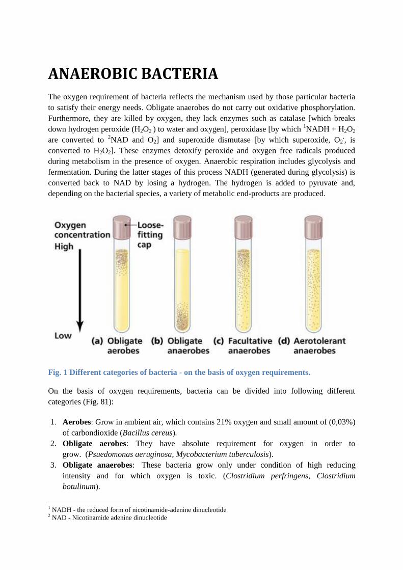
Fig. 1 Different categories of bacteria - on the basis of oxygen requirements.
On the basis of oxygen requirements, bacteria can be divided into following different
categories (Fig. 81):
1. Aerobes: Grow in ambient air, which contains 21% oxygen and small amount of (0,03%)
of carbondioxide (Bacillus cereus).
2. Obligate aerobes: They have absolute requirement for oxygen in order to
grow. (Psuedomonas aeruginosa, Mycobacterium tuberculosis).
3. Obligate anaerobes: These bacteria grow only under condition of high reducing
intensity and for which oxygen is toxic. (Clostridium perfringens, Clostridium
botulinum).
1 NADH - the reduced form of nicotinamide-adenine dinucleotide
2 NAD - Nicotinamide adenine dinucleotide
4. Facultative anaerobes: They are capable of growh under both aerobic and anaerobic
conditions. (Enterobacteriaceae group, Staphylococcus aureus).
5. Aerotolerant anaerobes: Are anaerobic bacteria that are not killed by exposure to
oxygen.
6. Capnophiles: Capnophilic bacteria require increased concentration of carbondioxide
(5% to 10%) and approximately 15% oxygen. This condition can be achieved by a candle
jar (3% carbondioxide) or carbondioxide incubator, jar or bags. (Haemophilus influenzae,
Neisseria gonorrhoeae).
7. Microaerophiles: Microaerophiles are those groups of bacteria that can grow under
reduced oxygen (5% to 10%) and increased carbondioxide (8% to 10%). Higher oxygen
tensions may be inhibitory to them. This environment can be obtained in specially
designed jars or bags. (Campylobacter jejuni, Helicobacter pylori).
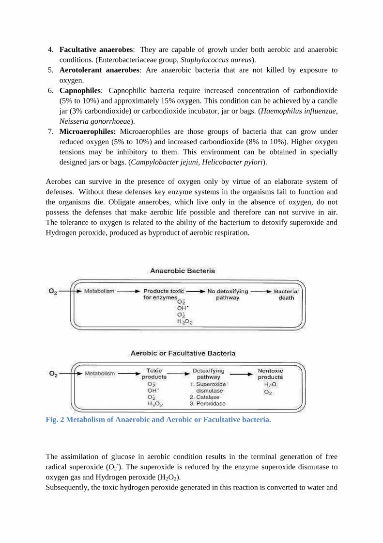
Aerobes can survive in the presence of oxygen only by virtue of an elaborate system of
defenses. Without these defenses key enzyme systems in the organisms fail to function and
the organisms die. Obligate anaerobes, which live only in the absence of oxygen, do not
possess the defenses that make aerobic life possible and therefore can not survive in air.
The tolerance to oxygen is related to the ability of the bacterium to detoxify superoxide and
Hydrogen peroxide, produced as byproduct of aerobic respiration.
Fig. 2 Metabolism of Anaerobic and Aerobic or Facultative bacteria.
The assimilation of glucose in aerobic condition results in the terminal generation of free
radical superoxide (O2-). The superoxide is reduced by the enzyme superoxide dismutase to
oxygen gas and Hydrogen peroxide (H2O2).
Subsequently, the toxic hydrogen peroxide generated in this reaction is converted to water and
oxygen by the enzyme catalase, which is found in aerobic and facultative anaerobic bacteria,
or by various peroxidases which are found in several aerotolerant anaerobes.
.ANAEROBIC NON - SPORE - FORMERS
Gram-negative rods Bacteroides
Fusobacterium
Gram-positive rods Actinomyces
Eubacterium
Bifidobacterium
Lactobacillus
Propionibacterium
Gram-positive cocci Peptostreptococcus
Peptococcus
Gram-negative cocci Veillonella
Acidominococcus
ANAEROBIC SPORE - FORMERS
Gram-positive rods
Clostridium tetani
Clostridium perfringens
Clostridium botulinum
Table 1 Anaerobic bacteria - non-spore-formers and spore-formers.
CLOSTRIDIA
Clostridium tetani (TETANUS)
Clostridium tetani, a Gram-positive rod that forms a terminal spore (Fig. 85), is commonly
found in the soil, dust and animal feces. Contamination of wounds, which provide anaerobic
conditions, can lead to spore germination and tetanus, a relatively rare, but frequently fatal
disease. Tetanus is also know as lockjaw because of the patient's inability to open the mouth
as a result of muscle paralysis.
Infection usually occurs when spores (in dirt, feces or saliva) enter wounds and scratches
where they germinate and produce tetanus toxin. The organism is non-invasive and thus
remains in the local wound.
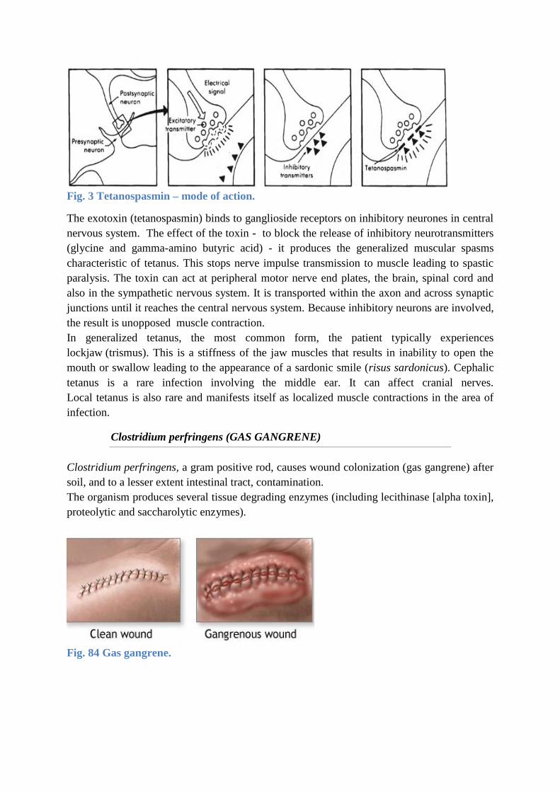
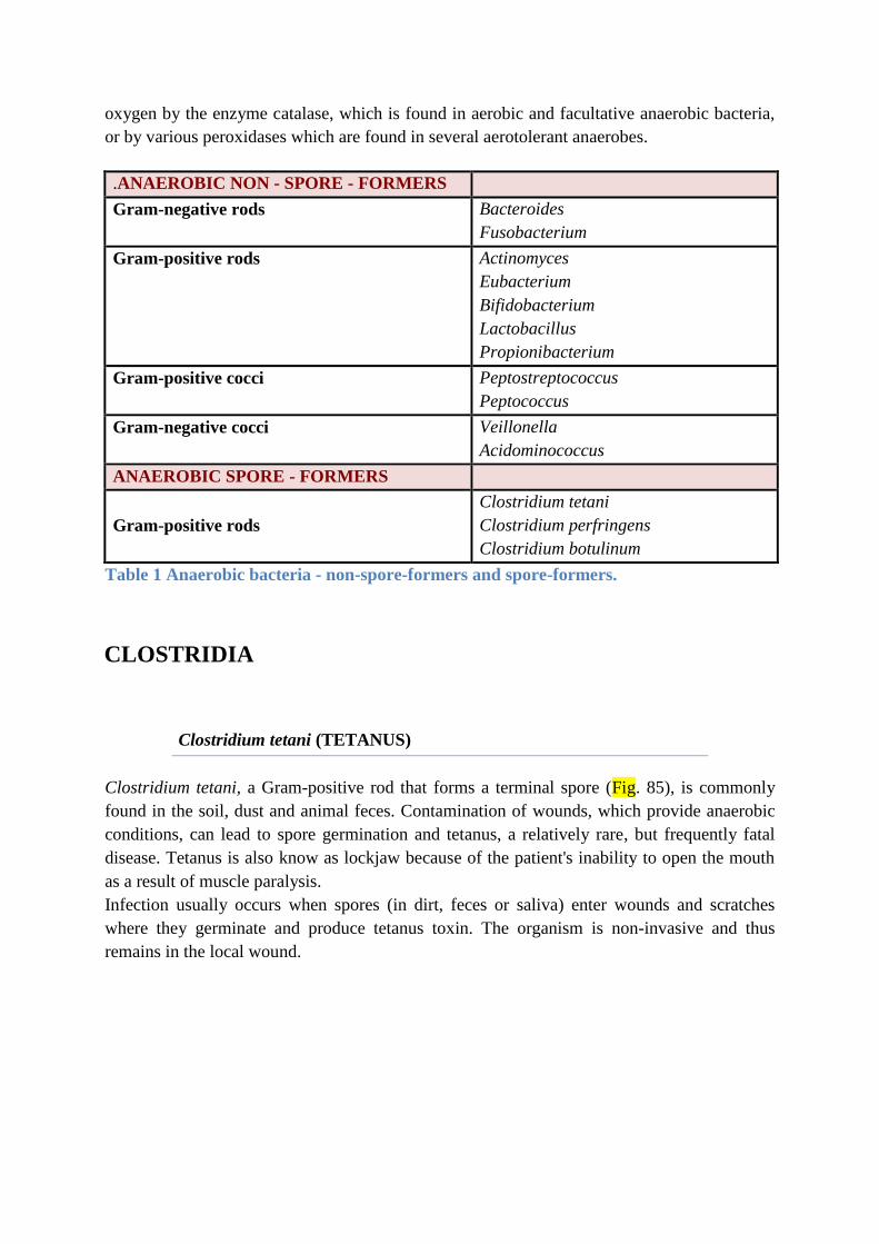
Fig. 3 Tetanospasmin – mode of action.
The exotoxin (tetanospasmin) binds to ganglioside receptors on inhibitory neurones in central
nervous system. The effect of the toxin - to block the release of inhibitory neurotransmitters
(glycine and gamma-amino butyric acid) - it produces the generalized muscular spasms
characteristic of tetanus. This stops nerve impulse transmission to muscle leading to spastic
paralysis. The toxin can act at peripheral motor nerve end plates, the brain, spinal cord and
also in the sympathetic nervous system. It is transported within the axon and across synaptic
junctions until it reaches the central nervous system. Because inhibitory neurons are involved,
the result is unopposed muscle contraction.
In generalized tetanus, the most common form, the patient typically experiences
lockjaw (trismus). This is a stiffness of the jaw muscles that results in inability to open the
mouth or swallow leading to the appearance of a sardonic smile (risus sardonicus). Cephalic
tetanus is a rare infection involving the middle ear. It can affect cranial nerves.
Local tetanus is also rare and manifests itself as localized muscle contractions in the area of
infection.
Clostridium perfringens (GAS GANGRENE)
Clostridium perfringens, a gram positive rod, causes wound colonization (gas gangrene) after
soil, and to a lesser extent intestinal tract, contamination.
The organism produces several tissue degrading enzymes (including lecithinase [alpha toxin],
proteolytic and saccharolytic enzymes).
Fig. 84 Gas gangrene.
Necrosis and destruction of blood vessels and the surrounding tissue, especially muscle, result
(myonecrosis is a condition of necrotic damage, specific to muscle tissue) (Fig. 84). This
creates an anaerobic environment in adjacent tissue and the organism spreads systemically.
Clostridium botulinum (BOTULISM)
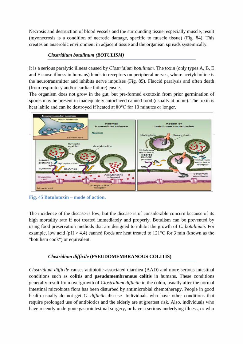
It is a serious paralytic illness caused by Clostridium botulinum. The toxin (only types A, B, E
and F cause illness in humans) binds to receptors on peripheral nerves, where acetylcholine is
the neurotransmitter and inhibits nerve impulses (Fig. 85). Flaccid paralysis and often death
(from respiratory and/or cardiac failure) ensue.
The organism does not grow in the gut, but pre-formed exotoxin from prior germination of
spores may be present in inadequately autoclaved canned food (usually at home). The toxin is
heat labile and can be destroyed if heated at 80°C for 10 minutes or longer.
Fig. 45 Botulotoxin – mode of action.
The incidence of the disease is low, but the disease is of considerable concern because of its
high mortality rate if not treated immediately and properly. Botulism can be prevented by
using food preservation methods that are designed to inhibit the growth of C. botulinum. For
example, low acid (pH > 4.4) canned foods are heat treated to 121°C for 3 min (known as the
"botulism cook") or equivalent.
Clostridium difficile (PSEUDOMEMBRANOUS COLITIS)
Clostridium difficile causes antibiotic-associated diarrhea (AAD) and more serious intestinal
conditions such as colitis and pseudomembranous colitis in humans. These conditions
generally result from overgrowth of Clostridium difficile in the colon, usually after the normal
intestinal microbiota flora has been disturbed by antimicrobial chemotherapy. People in good
health usually do not get C. difficile disease. Individuals who have other conditions that
require prolonged use of antibiotics and the elderly are at greatest risk. Also, individuals who
have recently undergone gastrointestinal surgery, or have a serious underlying illness, or who
are immunocompromised, are at risk. C. difficile produces two toxins. Toxin A is referred to
as an enterotoxin because it causes fluid accumulation in the bowel. Toxin B is an extremely
lethal (cytopathic) toxin.
LABORATORY DIAGNOSIS OF ANAEROBIC BACTERIA
Anaerobes are normally found within certain areas of the body but result in serious infection
when they have access to a normally sterile body fluid or deep tissue that is poorly
oxygenated. Some anaerobes normally live in the crevices of the skin, in the nose, mouth,
throat, intestine, and vagina. Injury to these tissues (cuts, puncture wounds, or trauma)
especially at or adjacent to the mucous membranes allows anaerobes entry into otherwise
sterile areas of the body and is the primary cause of anaerobic infection. A second source of
anaerobic infection occurs from the introduction of spores into a normally sterile site. Spore-
producing anaerobes live in the soil and water, and spores may be introduced via wounds,
especially punctures. Anaerobic infections are most likely to be found in persons who are
immunosuppressed, those treated recently with broad-spectrum antibiotics , and persons who
have a decaying tissue injury on or near a mucous membrane, especially if the site is foul-
smelling. The identification of anaerobes is highly complex, and laboratories may use
different identification systems. Organisms are identified by their: colonial and microscopic
morphology,
- growth on selective media,
- oxygen tolerance,
- biochemical characteristics (these include sugar fermentation, bile solubility, esculin,
starch, and gelatin hydrolysis, casein and gelatin digestion, catalase, lipase,
lecithinase, and indole production, nitrate reduction, volatile fatty acids as determined
by gas chromatography)
- susceptibility to antibiotics (by the microtube broth dilution method).
ANAEROBIC INFECTIONS – SPECIMEN COLLECTION
The keys to effective anaerobic bacteria cultures include collecting a contamination-free
specimen and protecting it from oxygen exposure. Anaerobic bacteria cultures should be
obtained from an appropriate site without the health care professional contaminating the
sample with bacteria from the adjacent skin, mucus membrane, or tissue. Swabs should be
avoided when collecting specimens for anaerobic culture because cotton fibers may be
detrimental to anaerobes. Abscesses or fluids can be aspirated using a sterile syringe that is
then tightly capped to prevent entry of air. Tissue samples should be placed into a degassed
bag and sealed, or into a gassed out screw top vial that may contain oxygen-free prereduced
culture medium and tightly capped. The specimens should be plated as rapidly as possible.
ANAEROBIC BACTERIA - GRAM STAIN
Gram-positive anaerobes
Gram-positive anaerobes include the following:
Actinomyces (head, neck, pelvic infections; aspiration pneumonia)
Bifidobacterium (ear infections, abdominal infections)
Clostridium (gas, gangrene, food poisoning, tetanus, pseudomembranous colitis)
Peptostreptococcus (oral, respiratory, and intra-abdominal infections)
Propionibacterium (shunt infections)
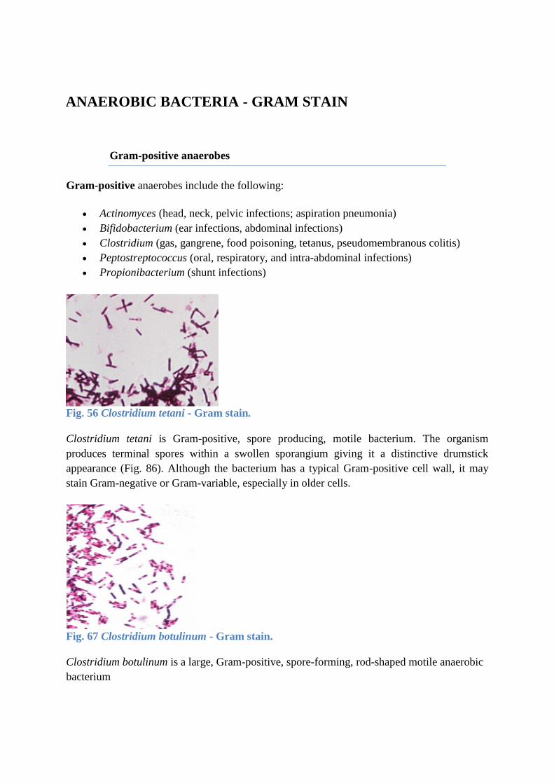
Fig. 56 Clostridium tetani - Gram stain.
Clostridium tetani is Gram-positive, spore producing, motile bacterium. The organism
produces terminal spores within a swollen sporangium giving it a distinctive drumstick
appearance (Fig. 86). Although the bacterium has a typical Gram-positive cell wall, it may
stain Gram-negative or Gram-variable, especially in older cells.
Fig. 67 Clostridium botulinum - Gram stain.
Clostridium botulinum is a large, Gram-positive, spore-forming, rod-shaped motile anaerobic
bacterium
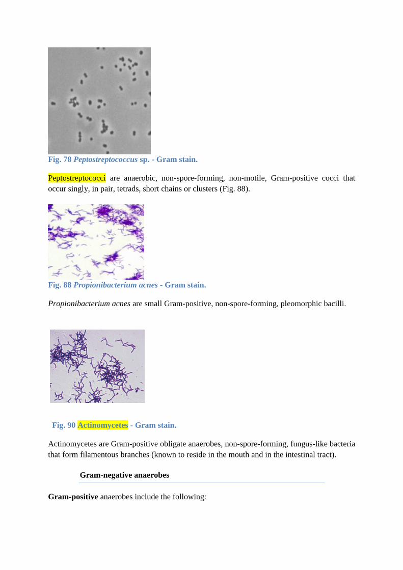
Fig. 78 Peptostreptococcus sp. - Gram stain.
Peptostreptococci are anaerobic, non-spore-forming, non-motile, Gram-positive cocci that
occur singly, in pair, tetrads, short chains or clusters (Fig. 88).
Fig. 88 Propionibacterium acnes - Gram stain.
Propionibacterium acnes are small Gram-positive, non-spore-forming, pleomorphic bacilli.
Fig. 90 Actinomycetes - Gram stain.
Actinomycetes are Gram-positive obligate anaerobes, non-spore-forming, fungus-like bacteria
that form filamentous branches (known to reside in the mouth and in the intestinal tract).
Gram-negative anaerobes
Gram-positive anaerobes include the following:
Bacteroides (the most commonly found anaerobes in cultures; intra-abdominal
infections, rectal abscesses, soft tissue infections, liver infection)
Fusobacterium (abscesses, wound infections, pulmonary and intracranial infections)
Porphyromonas (aspiration pneumonia, periodontitis)
Prevotella (intra-abdominal infections, soft tissue infections)
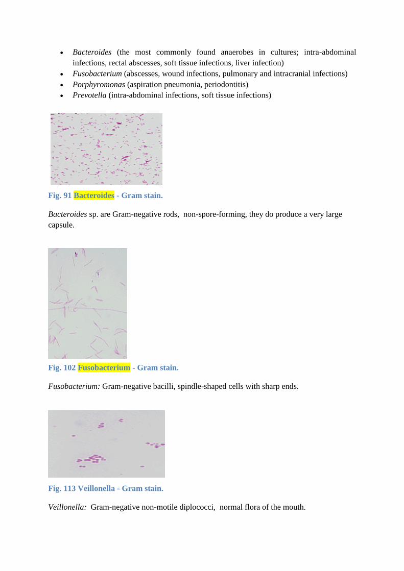
Fig. 91 Bacteroides - Gram stain.
Bacteroides sp. are Gram-negative rods, non-spore-forming, they do produce a very large
capsule.
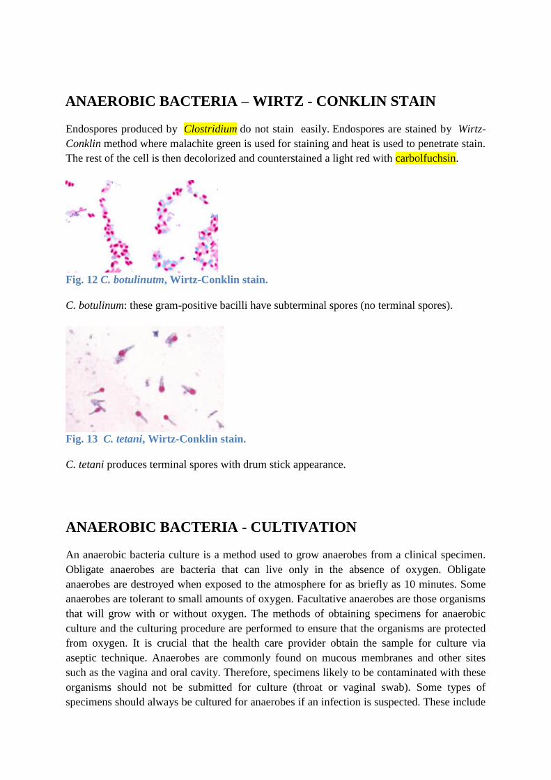
Fig. 102 Fusobacterium - Gram stain.
Fusobacterium: Gram-negative bacilli, spindle-shaped cells with sharp ends.
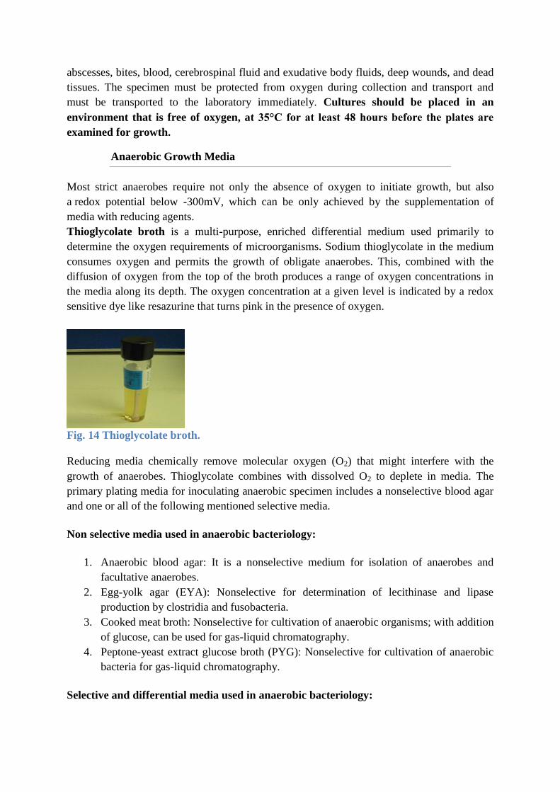
Fig. 113 Veillonella - Gram stain.
Veillonella: Gram-negative non-motile diplococci, normal flora of the mouth.
ANAEROBIC BACTERIA – WIRTZ - CONKLIN STAIN
Endospores produced by Clostridium do not stain easily. Endospores are stained by Wirtz-
Conklin method where malachite green is used for staining and heat is used to penetrate stain.
The rest of the cell is then decolorized and counterstained a light red with carbolfuchsin.
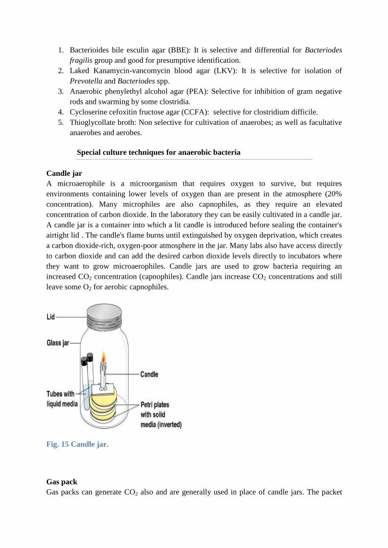
Fig. 12 C. botulinutm, Wirtz-Conklin stain.
C. botulinum: these gram-positive bacilli have subterminal spores (no terminal spores).
Fig. 13 C. tetani, Wirtz-Conklin stain.
C. tetani produces terminal spores with drum stick appearance.
ANAEROBIC BACTERIA - CULTIVATION
An anaerobic bacteria culture is a method used to grow anaerobes from a clinical specimen.
Obligate anaerobes are bacteria that can live only in the absence of oxygen. Obligate
anaerobes are destroyed when exposed to the atmosphere for as briefly as 10 minutes. Some
anaerobes are tolerant to small amounts of oxygen. Facultative anaerobes are those organisms
that will grow with or without oxygen. The methods of obtaining specimens for anaerobic
culture and the culturing procedure are performed to ensure that the organisms are protected
from oxygen. It is crucial that the health care provider obtain the sample for culture via
aseptic technique. Anaerobes are commonly found on mucous membranes and other sites
such as the vagina and oral cavity. Therefore, specimens likely to be contaminated with these
organisms should not be submitted for culture (throat or vaginal swab). Some types of
specimens should always be cultured for anaerobes if an infection is suspected. These include
abscesses, bites, blood, cerebrospinal fluid and exudative body fluids, deep wounds, and dead
tissues. The specimen must be protected from oxygen during collection and transport and
must be transported to the laboratory immediately. Cultures should be placed in an
environment that is free of oxygen, at 35°C for at least 48 hours before the plates are
examined for growth.
Anaerobic Growth Media
Most strict anaerobes require not only the absence of oxygen to initiate growth, but also
a redox potential below -300mV, which can be only achieved by the supplementation of
media with reducing agents.
Thioglycolate broth is a multi-purpose, enriched differential medium used primarily to
determine the oxygen requirements of microorganisms. Sodium thioglycolate in the medium
consumes oxygen and permits the growth of obligate anaerobes. This, combined with the
diffusion of oxygen from the top of the broth produces a range of oxygen concentrations in
the media along its depth. The oxygen concentration at a given level is indicated by a redox
sensitive dye like resazurine that turns pink in the presence of oxygen.
Fig. 14 Thioglycolate broth.
Reducing media chemically remove molecular oxygen (O2) that might interfere with the
growth of anaerobes. Thioglycolate combines with dissolved O2 to deplete in media. The
primary plating media for inoculating anaerobic specimen includes a nonselective blood agar
and one or all of the following mentioned selective media.
Non selective media used in anaerobic bacteriology:
1. Anaerobic blood agar: It is a nonselective medium for isolation of anaerobes and
facultative anaerobes.
2. Egg-yolk agar (EYA): Nonselective for determination of lecithinase and lipase
production by clostridia and fusobacteria.
3. Cooked meat broth: Nonselective for cultivation of anaerobic organisms; with addition
of glucose, can be used for gas-liquid chromatography.
4. Peptone-yeast extract glucose broth (PYG): Nonselective for cultivation of anaerobic
bacteria for gas-liquid chromatography.
Selective and differential media used in anaerobic bacteriology:
1. Bacterioides bile esculin agar (BBE): It is selective and differential for Bacteriodes
fragilis group and good for presumptive identification.
2. Laked Kanamycin-vancomycin blood agar (LKV): It is selective for isolation of
Prevotella and Bacteriodes spp.
3. Anaerobic phenylethyl alcohol agar (PEA): Selective for inhibition of gram negative
rods and swarming by some clostridia.
4. Cycloserine cefoxitin fructose agar (CCFA): selective for clostridium difficile.
5. Thioglycollate broth: Non selective for cultivation of anaerobes; as well as facultative
anaerobes and aerobes.
Special culture techniques for anaerobic bacteria
Candle jar
A microaerophile is a microorganism that requires oxygen to survive, but requires
environments containing lower levels of oxygen than are present in the atmosphere (20%
concentration). Many microphiles are also capnophiles, as they require an elevated
concentration of carbon dioxide. In the laboratory they can be easily cultivated in a candle jar.
A candle jar is a container into which a lit candle is introduced before sealing the container's
airtight lid . The candle's flame burns until extinguished by oxygen deprivation, which creates
a carbon dioxide-rich, oxygen-poor atmosphere in the jar. Many labs also have access directly
to carbon dioxide and can add the desired carbon dioxide levels directly to incubators where
they want to grow microaerophiles. Candle jars are used to grow bacteria requiring an
increased CO2 concentration (capnophiles). Candle jars increase CO2 concentrations and still
leave some O2 for aerobic capnophiles.
Fig. 15 Candle jar.
Gas pack
Gas packs can generate CO2 also and are generally used in place of candle jars. The packet
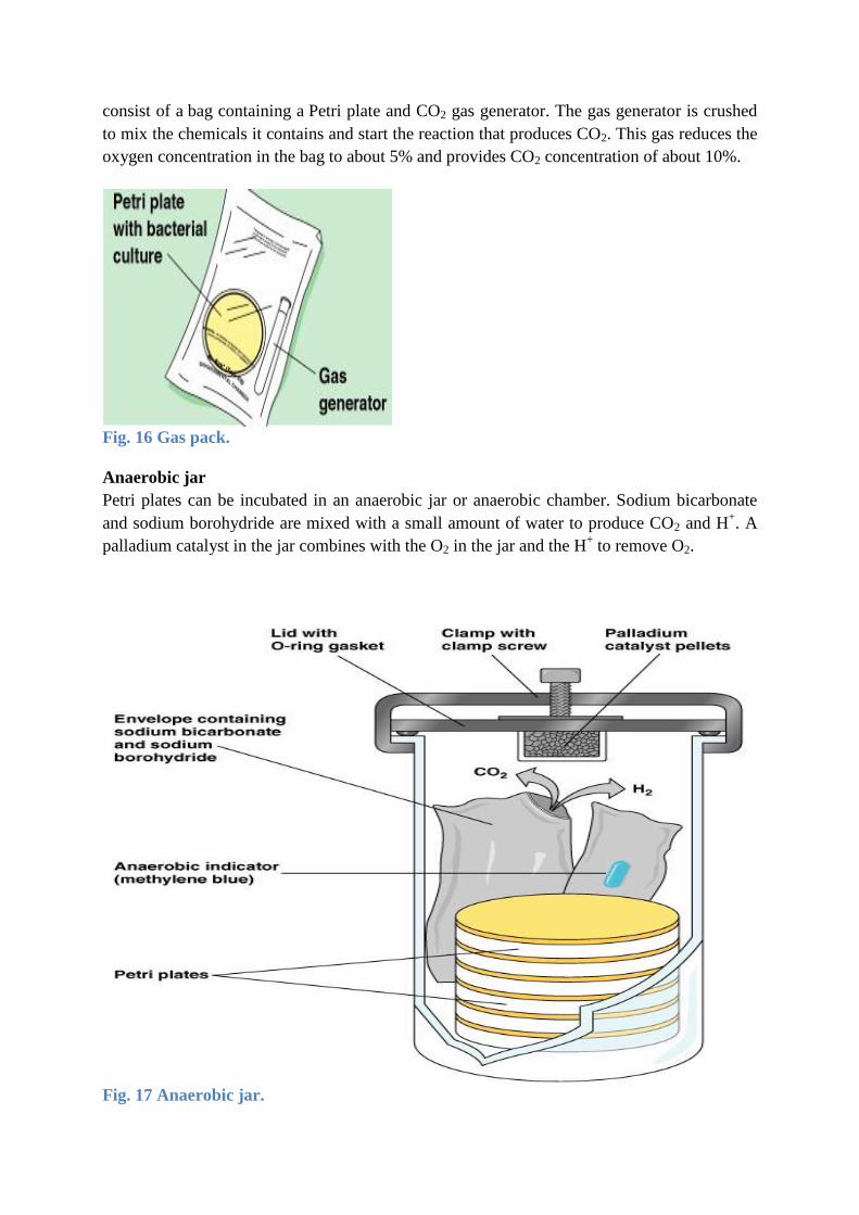
consist of a bag containing a Petri plate and CO2 gas generator. The gas generator is crushed
to mix the chemicals it contains and start the reaction that produces CO2. This gas reduces the
oxygen concentration in the bag to about 5% and provides CO2 concentration of about 10%.
Fig. 16 Gas pack.
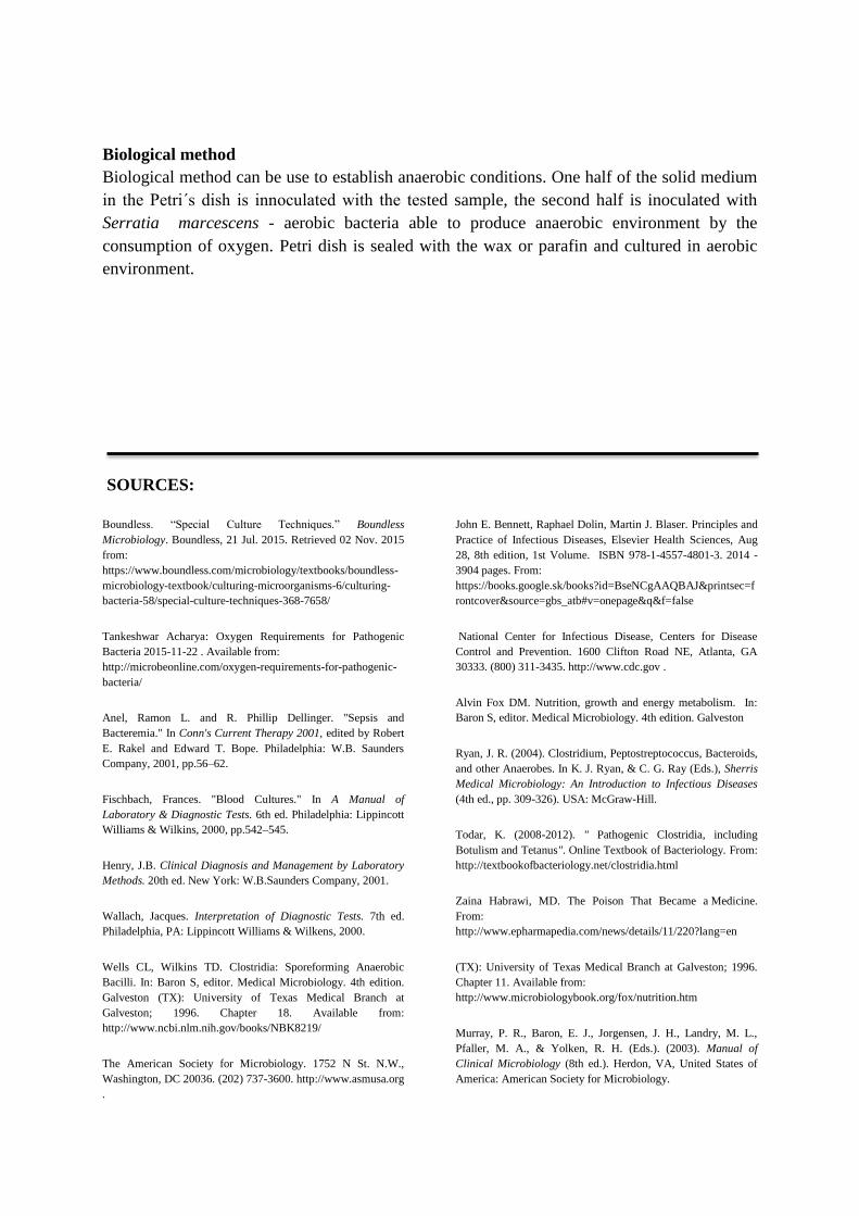
Anaerobic jar
Petri plates can be incubated in an anaerobic jar or anaerobic chamber. Sodium bicarbonate
and sodium borohydride are mixed with a small amount of water to produce CO2 and H+. A
palladium catalyst in the jar combines with the O2 in the jar and the H+ to remove O2.
Fig. 17 Anaerobic jar.
Biological method
Biological method can be use to establish anaerobic conditions. One half of the solid medium
in the Petri´s dish is innoculated with the tested sample, the second half is inoculated with
Serratia marcescens - aerobic bacteria able to produce anaerobic environment by the
consumption of oxygen. Petri dish is sealed with the wax or parafin and cultured in aerobic
environment.
SOURCES:
Boundless. “Special Culture Techniques.” Boundless
Microbiology. Boundless, 21 Jul. 2015. Retrieved 02 Nov. 2015
from:
https://www.boundless.com/microbiology/textbooks/boundless-
microbiology-textbook/culturing-microorganisms-6/culturing-
bacteria-58/special-culture-techniques-368-7658/
Tankeshwar Acharya: Oxygen Requirements for Pathogenic
Bacteria 2015-11-22 . Available from:
http://microbeonline.com/oxygen-requirements-for-pathogenic-
bacteria/
Anel, Ramon L. and R. Phillip Dellinger. "Sepsis and
Bacteremia." In Conn's Current Therapy 2001, edited by Robert
E. Rakel and Edward T. Bope. Philadelphia: W.B. Saunders
Company, 2001, pp.56–62.
Fischbach, Frances. "Blood Cultures." In A Manual of
Laboratory & Diagnostic Tests. 6th ed. Philadelphia: Lippincott
Williams & Wilkins, 2000, pp.542–545.
Henry, J.B. Clinical Diagnosis and Management by Laboratory
Methods. 20th ed. New York: W.B.Saunders Company, 2001.
Wallach, Jacques. Interpretation of Diagnostic Tests. 7th ed.
Philadelphia, PA: Lippincott Williams & Wilkens, 2000.
Wells CL, Wilkins TD. Clostridia: Sporeforming Anaerobic
Bacilli. In: Baron S, editor. Medical Microbiology. 4th edition.
Galveston (TX): University of Texas Medical Branch at
Galveston; 1996. Chapter 18. Available from:
http://www.ncbi.nlm.nih.gov/books/NBK8219/
The American Society for Microbiology. 1752 N St. N.W.,
Washington, DC 20036. (202) 737-3600. http://www.asmusa.org
.
John E. Bennett, Raphael Dolin, Martin J. Blaser. Principles and
Practice of Infectious Diseases, Elsevier Health Sciences, Aug
28, 8th edition, 1st Volume. ISBN 978-1-4557-4801-3. 2014 -
3904 pages. From:
https://books.google.sk/books?id=BseNCgAAQBAJ&printsec=f
rontcover&source=gbs_atb#v=onepage&q&f=false
National Center for Infectious Disease, Centers for Disease
Control and Prevention. 1600 Clifton Road NE, Atlanta, GA
30333. (800) 311-3435. http://www.cdc.gov .
Alvin Fox DM. Nutrition, growth and energy metabolism. In:
Baron S, editor. Medical Microbiology. 4th edition. Galveston
Ryan, J. R. (2004). Clostridium, Peptostreptococcus, Bacteroids,
and other Anaerobes. In K. J. Ryan, & C. G. Ray (Eds.), Sherris
Medical Microbiology: An Introduction to Infectious Diseases
(4th ed., pp. 309-326). USA: McGraw-Hill.
Todar, K. (2008-2012). " Pathogenic Clostridia, including
Botulism and Tetanus". Online Textbook of Bacteriology. From:
http://textbookofbacteriology.net/clostridia.html
Zaina Habrawi, MD. The Poison That Became a Medicine.
From:
http://www.epharmapedia.com/news/details/11/220?lang=en
(TX): University of Texas Medical Branch at Galveston; 1996.
Chapter 11. Available from:
http://www.microbiologybook.org/fox/nutrition.htm
Murray, P. R., Baron, E. J., Jorgensen, J. H., Landry, M. L.,
Pfaller, M. A., & Yolken, R. H. (Eds.). (2003). Manual of
Clinical Microbiology (8th ed.). Herdon, VA, United States of
America: American Society for Microbiology.

University of Texas - Houston Medical School, DPALM
MEDIC. Gram-positive bacili (Non-spore-forming). 1995.
Available from:
http://www.uaz.edu.mx/histo/pathology/ed/ch_9b/path/00001503
.htm
Ids Education: Rod-Shaped Bacteria, Spore-Forming (Bacilli).
http://www.inds.co.uk/slides/slides.php?category=280&class=96
0
http://www.surgeryencyclopedia.com/A-Ce/Anaerobic-Bacteria-
Culture.html
http://parasites.czu.cz/food/_data/176.jpg
http://visualsunlimited.photoshelter.com/image/I0000.mJ4vwiH
wYU
http://anaerobe.shellab.com/section4-slide5.php
http://classes.midlandstech.edu/carterp/courses/bio225/chap06/
Microbial%20Growth%20ss2.htm
http://iws2.collin.edu/dcain/CCCCD%20Micro/thioglycollatebro
th.htm
http://www.atsu.edu/faculty/chamberlain/website/lects/toxins.ht
m
http://iws2.collin.edu/dcain/CCCCD%20Micro/tutorial.htm
https://www.nlm.nih.gov/medlineplus/ency/imagepages/17190.h
tm
http://atlas.sund.ku.dk/microatlas/veterinary/bacteria/Peptostrept
ococcus_indolicus/phasecon.html
http://www.microbiologyinpictures.com/bacteria-
micrographs/gram-stain/gram-positive/propionibacterium.html
