an innate bactericidal oleic acid effective against skin ...nano.ucsd.edu/~l7zhang/publications/45-j...
TRANSCRIPT
J. Microbiol. Biotechnol. (2011), 21(4), 391–399doi: 10.4014/jmb.1011.11014First published online 16 January 2011
An Innate Bactericidal Oleic Acid Effective Against Skin Infection ofMethicillin-Resistant Staphylococcus aureus: A Therapy Concordant withEvolutionary Medicine
Chen, Chao-Hsuan1,2,3†
, Yanhan Wang1†
, Teruaki Nakatsuji1, Yu-Tsueng Liu
4, Christos C. Zouboulis
5,
Richard L. Gallo1,2
, Liangfang Zhang4,6*, Ming-Fa Hsieh
3*, and Chun-Ming Huang
1,2,4*
1Division of Dermatology, Department of Medicine, University of California, San Diego, La Jolla, CA 92093-0671, USA2VA San Diego Healthcare Center, San Diego, CA 92161-0002, USA 3Department of Biomedical Engineering and R&D Center for Biomedical Microdevice Technology, Chung Yuan Christian University,Chung Li, 32023 Taiwan4Moores Cancer Center, University of California, San Diego, La Jolla, CA 92093, USA5Department of Dermatology, Venereology, Allergology and Immunology, Dessau Medical Center, 06847 Dessau, Germany6Department of Nanoengineering, University of California, San Diego, La Jolla, CA 92093-0448, USA
Received: November 15, 2010 / Revised: January 20, 2011 / Accepted: January 21, 2011
Free fatty acids (FFAs) are known to have bacteriocidal
activity and are important components of the innate
immune system. Many FFAs are naturally present in
human and animal skin, breast milk, and in the
bloodstream. Here, the therapeutic potential of FFAs
against methicillin-resistant Staphylococcus aureus (MRSA)
is demonstrated in cultures and in mice. Among a series of
FFAs, only oleic acid (OA) (C18:1, cis-9) can effectively
eliminate Staphylococcus aureus (S. aureus) through cell
wall disruption. Lauric acid (LA, C12:0) and palmitic
acid (PA, C16:0) do not have this ability. OA can inhibit
growth of a number of Gram-positive bacteria, including
hospital and community-associated MRSA at a dose
that did not show any toxicity to human sebocytes. The
bacteriocidal activities of FFAs were also demonstrated in
vivo through injection of OA into mouse skin lesions
previously infected with a strain of MRSA. In conclusion,
our results suggest a promising therapeutic approach
against MRSA through boosting the bacteriocidal activities
of native FFAs, which may have been co-evolved during
the interactions between microbes and their hosts.
Keywords: Staphylococcus aureus, oleic acid
Staphylococcus aureus (S. aureus) is a Gram-positive bacterium
and one of the major causes of fatal nosocomial infections
[23]. It is estimated that annually in the United States (US)
alone, S. aureus accounts for 12 million outpatient visits
and 292,000 hospitalizations of which 126,000 are due to
methicillin-resistant Staphylococcus aureus (MRSA) [19].
Recent studies estimated that more people die from the
MRSA bacterium than from human immunodeficiency
virus (HIV) in the US [31, 42], and the Center for Disease
Control and Prevention estimated that more than 90,000
people die from hospital-acquired bacterial infections in
the US each year [42]. Historically, MRSA infections have
occurred in nosocomial settings. However, recently MRSA
infections have increasingly been found among individuals
in the community without healthcare exposures [55].
Although S. aureus including MRSA and community-
associated (CA)-MRSA can cause life-threatening and
systemic infections (bacteremia), skin and soft tissues are
the most common sites of S. aureus infection and comprise
more than 75% of MRSA disease [9]. Unfortunately, it
remains as an unmet challenge to develop effective therapeutic
approaches for MRSA treatment because of its formidable
resistance against multiple traditional antibiotics such as
methicillin, cloxacillin, and flucloxacillin [49, 50]. Decades
of selective pressure with β-lactam antibiotics and close
proximity of susceptible hosts have resulted in a high
prevalence of MRSA in hospitals worldwide. Although
*Corresponding authorL. ZhangPhone: +858-246-0999; Fax: +858-534-9553;E-mail: [email protected]. HsiehPhone: +886-3265-4550; Fax: +886-3265-4599;E-mail: [email protected]. HuangPhone: +858-822-4627; Fax: +858-642-1435;E-mail: [email protected]†These Authors contributed equally to this
392 Chen et al.
these factors logically explain the high incidence of hospital-
associated MRSA infections, the molecular basis for the
increased incidence remains incompletely defined. Recent
studies indicate that strains (e.g., USA300 and 400) that are
the leading causes of CA-MRSA disease in the US [1,
13, 53] have enhanced virulence compared with strains that
are the leading causes of hospital infections (e.g., USA200).
The need for new therapy, which can effectively cripple
bacterial infection and lower the risk of creating S. aureus
resistance, is indisputable. Although vaccination is one of
the modalities against S. aureus infection, it generally takes
a long time to develop an effective vaccine. Furthermore,
people may hesitate to be immunized with a preventive
vaccine because they are unsure whether S. aureus bacteria
will infect them. Even if a therapeutic vaccine is administrated,
protective antibodies may not be produced quickly enough
to suppress bacterial spread through the body. Although
systemic antibiotic therapy is currently used for treatment of
S. aureus infections, it nonspecifically kills the majority of
microbes and disrupts the homeostasis of body-resident
microflora [51]. In addition, antibiotic use has a potential
risk of selecting for antibiotic-resistant bacteria.
Free fatty acids (FFAs) are known to possess anti-S. aureus
activity [8, 45, 46] and are important components of the
innate immune system [10]. Many FFAs including oleic
acid (OA) (a C18:1 FFA, cis-9) naturally exist in humans
(e.g., skin, breast milk, and bloodstream) and mice [8, 21,
48]. These FFAs thus can function as “innate bactericides.”
It has also been known that the FFAs can anchor in the
bacterial membranes to damage the cell wall structure [30].
Drugs that anchor in the bacterial membrane (e.g., ceragenins
and lipopeptides) or that target the bacterial membrane and
proteic (lipoglycopeptides) or lipidic (glycodepsipeptides)
cell wall precursors seem to have the most potential. They
show a fast and extensive bactericidal effect and are
probably less prone to select for resistance owing to the
difficulty that bacteria would have in modifying these
targets in a way that is compatible with survival [52]. It has
been known that endogenous bactericides are largely
nonspecific and hold great promise to avert the development
of bacterial resistance [17, 44, 46]. It has been proposed
that innate bactericides and innate bactericides-resistance
mechanisms have co-evolved, leading to a transient host-
pathogen balance that has shaped the existing repertoire of
innate bactericides [44]. More intriguingly, it has been
found that two FFAs (linoleic acid and dehydrocrepenynic
acid) inhibit the bacterial drug resistance by decreasing the
transfer frequency of the conjugal DNA [14, 46].
In this study, we found that OA exerts excellent
antimicrobial activity against various S. aureus strains,
including a hospital-acquired MRSA strain (MRSA252)
and a CA-MRSA strain (USA300). The use of OA as an
innate bactericide is in compliance with evolutionary medicine
because it is endogenously present in human skin.
MATERIALS AND METHODS
Bacterial Strains and Culture Conditions
S. aureus [MRSA252, USA200, USA300, and a S. aureus mprF and
S. epidermidis (ATCC1228; Manasass, VA, USA)] were cultured on
3% tryptic soy broth (TSB) (Sigma, St. Louis, MO, USA) agar
overnight at 37oC. B. anthracis Sterne 34F2 spores (Colorado Serum
company, Denver, CO, USA) were heated at 65oC for 30 min and then
cultured in a modified nutrient broth medium [0.8% Bacto nutrient
broth, 0.1% KCl, 0.012% MgSO4, 1 mM Ca(NO3)2, 10 µM MnCl2,
1 µM FeSO4, pH 7.6], followed by incubation at 37oC with shaking.
L. monocytogenes (EGD strain BUG) was grown overnight in
Brain-heart infusion medium (Difco) at 37oC without shaking. E. coli
BL21 (DE3) (Invitrogen, Carlsbad, CA, USA) was cultured on Luria
broth agar (Difco) at 26oC for 48 to 72 h. All bacteria from a single
colony were cultured in their media overnight at 37oC. The overnight
culture was diluted 1:100 and cultured until reaching around
OD600=1.0. Bacteria were harvested by centrifugation at 5,000 ×g
for 10 min, washed with PBS, and suspended to an appropriate
amount of PBS for the further experiments.
Minimal Bactericidal Concentration (MBC) Assay
To determine the MBC of OA, bacteria (1×10⁶
CFU/ml) were
incubated with OA at various concentrations (0.078125-200 µg/ml)
in 5% DMSO in PBS on a 96-well microplate (100 µl per well) for
10 min, 30 min, 1 h, 3 h, or 5 h. The control received only 5% (v/v)
of DMSO. After incubation, the bacteria were diluted 1:10-1:106
with PBS. MBC was finally examined at a 99.9% killing level and
determined by spotting the dilution (5 µl) on an agar plate supplemented
with bacterial media for the counting of CFUs.
Radial Diffusion Assay (RDA)
RDA with minor modifications has been described previously [36].
Briefly, bacteria in mid-log phase were centrifuged at 2,000 ×g for
10 min and washed with PBS. Bacteria (1×10⁵ CFU/ml) were
dispersed in agar consisting of 1% (w/v) agarose (Sigma) and 1%
(w/v) TSB in 10 mM PBS at 42oC. Subsequently, the agar was
poured into Petri dishes and solidified. The wells (3 mm in diameter)
with a 10-µl capacity were created by poking a pipette tip into the
semi-solidified agar. Six serially diluted samples of each OA ranging
in concentration from 0.625 to 200 µg/ml were prepared, and 5-µl
OA aliquots solubilized in 5% (v/v) of DMSO were added to the
wells. After 3 h of incubation, a 10-ml overlay gel composed of 6%
TSB, 1% agarose, and 10 mM PBS (pH 7.4) was poured onto the
plates, and the plates were incubated overnight to allow the surviving
organisms to form microcolonies. The growth inhibition zones
reflecting the antimicrobial activity of OA were observed.
Transmission Electron Microscopy
After incubation at 37oC, bacteria were harvested, washed twice
with PBS, and fixed in Karnovsky’s fixative (4% paraformaldehyde,
2.5% glutaraldehyde, 5 mM CaCl2 in 0.1 M Na cacodylate buffer,
pH 7.4) overnight at 4oC, followed by 1% OsO4 in 0.1 M Na
cacodylate buffer, pH 7.4, en bloc staining with 4% uranyl acetate
in 50% ethanol, and subsequent dehydration using a graded series of
ethanol solutions followed by propylene oxide and infiltration with
epoxy resin (Scipoxy 812, Energy Beam Sciences, Agawam, MA,
USA). After polymerization at 65oC overnight, thin sections were
cut and stained with uranyl acetate (4% uranyl acetate in 50%
OLEIC ACID AGAINST SKIN INFECTION OF MRSA 393
ethanol) followed by bismuth subnitrate. Sections were examined at
an accelerating voltage of 60 kV using a Zeiss EM10C electron
microscope (Carl Zeiss, Thornwood, NY, USA).
Skin USA300 Infection Treated with OA
USA300 bacteria (1×10⁷ CFU in PBS) mixed with the microcarrier
(Cytodex) beads [1:1 (v/v); Sigma] were subcutaneously injected
into the dorsal skins of 8- to 12-week-old female ICR mice (Harlan,
Indianapolis, IN, USA). Then 100 µl of OA (100 µg/ml in 5% DMSO
in PBS) or 5% DMSO in PBS was injected into the same location
right after bacterial injection. For histological observation, the dorsal
skins were cross-sectioned, stained with H&E (Sigma), and viewed
on a microscope (Carl Zeiss). The lesion sizes were measured using a
microcaliper (Mitutoyo, Kanagawa, Japan) 24 and 48 h after injection.
The lesion sizes were examined and quantified by using Image
software [National Institutes of Health (NIH), Bethesda, MD, USA].
To determine the bacterial number in lesional skin, the dorsal skin
was punched with an 8-mm biopsy 24 h after bacterial injection.
The punch biopsy was homogenized in 1 ml of sterile PBS with a
hand tissue grinder. Bacterial CFUs in the skin were enumerated by
plating serial dilutions (1:101-1:106) of the homogenate on a TSB
agar plate. The plate was incubated for 24 h at 37oC to count
colonies. All experiments using mice were conducted in a Biosafety
Level 2 (BSL-2) facility and with accordance to institutional guidelines
for animal experiments.
Statistical Analysis
To determine significances between groups, comparisons were made
using the two-tailed t-test. For all statistical tests, a P-value of <0.05
was accepted for statistical significance.
RESULTS
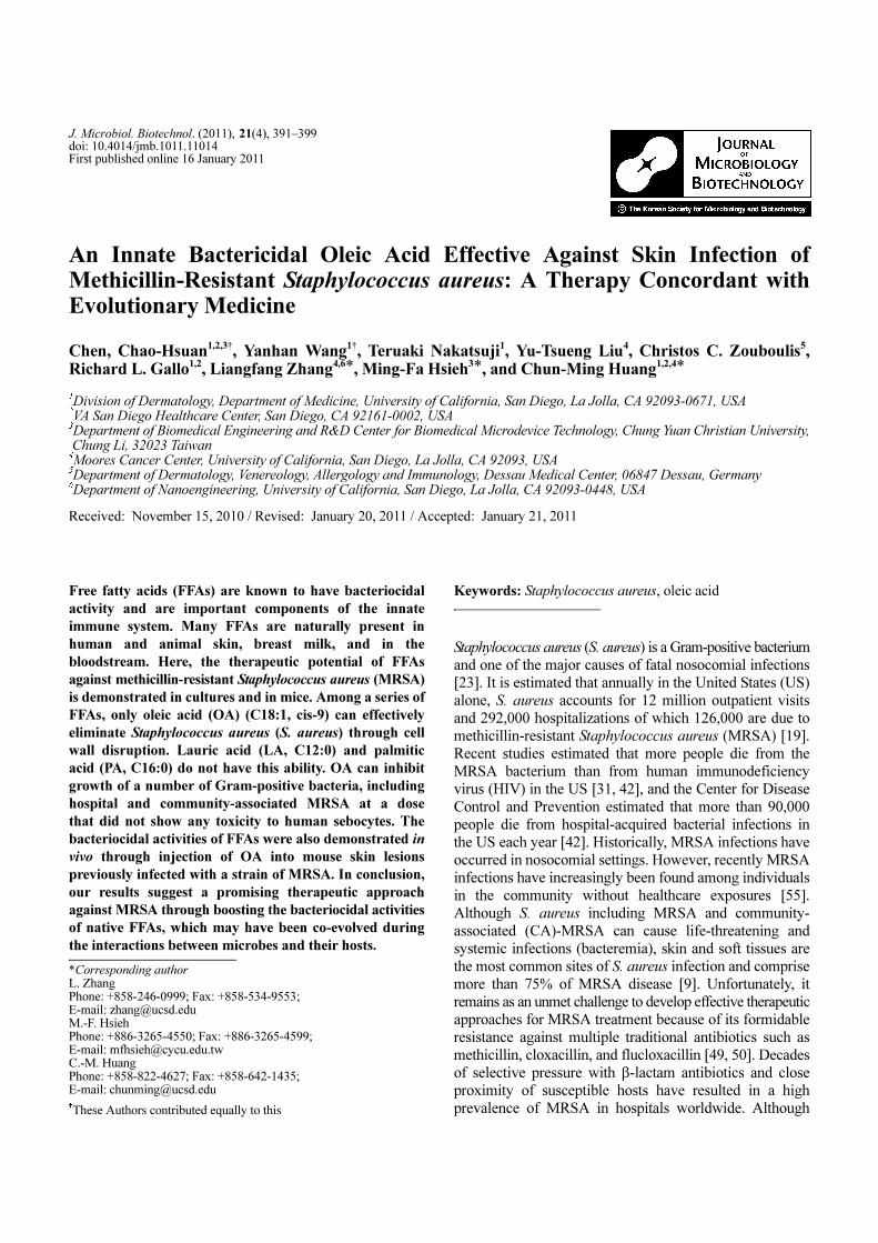
OA is an Effective FFA to Inhibit S. aureus Growth
We have investigated a series of FFAs including OA
(C18:1, cis-9), lauric acid (LA, C12:0), and palmitic acid
(PA, C16:0) for anti-S. aureus activity and discovered that
only OA can effectively kill S. aureus at reasonable doses.
To determine the minimal bactericidal concentration (MBC)
of FFAs, S. aureus multiple peptide resistance factor
mutant (∆mprF) that lacks the ability to modify anionic
membrane lipids with L-lysine [40] was incubated in
phosphate buffer saline (PBS) with several concentrations
of LA, PA, or OA for 5 h at 37oC. After incubation, the
bacteria were diluted with PBS and spotted on an agar
plate to count colony-forming units (CFU). Since all FFAs
were dissolved in 5% (v/v) dimethylsulfoxide (DMSO),
bacteria incubated with DMSO served as a control group.
As shown in Fig. 1, no killing was detected when S. aureus
was incubated with 5% (v/v) DMSO (Vehicle). The
incubation of PA (1.25 to 100 µg/ml) did not influence the
growth of S. aureus mprF (data not shown). In contrast, we
found that when the concentrations of OA and LA are
higher than 3.125 and 70 µg/ml, respectively, they started
killing S. aureus mprF (Fig. 1A, 1B). Furthermore, MBC
assays indicated that LA (0.1-200 µg/ml) did not kill a
MRSA252 strain (data not shown). To determine the
cytotoxicity of FFAs, a human skin sebocyte SZ95 cell line
[56] was incubated overnight with 100 µg/ml of LA, OA,
or PA. Although LA has a slight toxic effect on sebocytes,
this was only seen at a concentration far exceeding that
needed to kill S. aureus. PA and OA were not cytotoxic for
SZ95 cells (Supplementary Fig. S1).
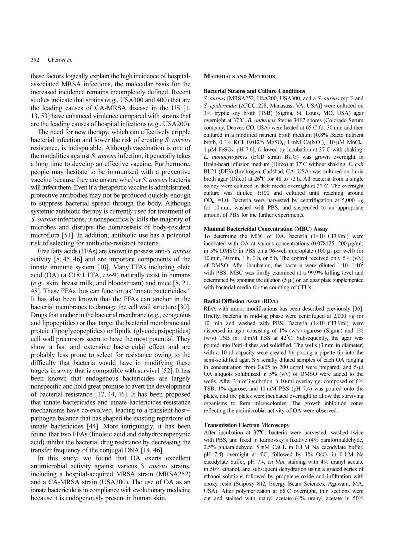
OA Kills S. aureus Bacteria by Breaking Down the Cell
Walls
We next examine if OA can effectively inhibit the MRSA
bacteria. A hospital-acquired MRSA bacterial strain
(MRSA252) was incubated with OA at various concentrations
in PBS for 5 h at 37oC in MBC assays. Our recent publication
demonstrated that killing of MRSA252 occurred at an OA
concentration greater than 5 µg/ml, and complete killing of
MRSA252 occurred at concentrations greater than 10 µg/ml
Fig. 1. OA exerts bactericidal activity against S. aureus. S. aureus mprF was cultured on Tryptic soy broth (TSB) (Sigma). For
MBC assays, the bacteria (1×10⁷
CFU/ml) was incubated with OA (0.375-
100 µg/ml) (A), LA (6.25-100 µg/ml) (B), and PA (6.25-100 µg/ ml)
(data not shown) in 5% DMSO in PBS on a 96-well microplate (100 µl/
well) for 5 h. Bacteria incubated with 5% (v/v) DMSO served as a control
(vehicle). After incubation, the bacterial suspension was diluted 1:10-1:106
with PBS, and 5 µl of the dilutions were spotted on a TSB agar plate for
CFU counts. **P<0.01. P-values were evaluated using two-tailed t-tests.
Data are the mean ± SD of three individual experiments. UD, undetectable.
394 Chen et al.
[24]. A sensitive radial diffusion assay (RDA) [36] as
described in Materials and Methods was used to further
verify the antimicrobial activity of OA against MRSA252
(Fig. 2). In consistence with MBC assays [24], the growth
inhibition zones were clearly observed when bacteria were
incubated with OA at a minimum effective concentration
of 10 µg/ml.
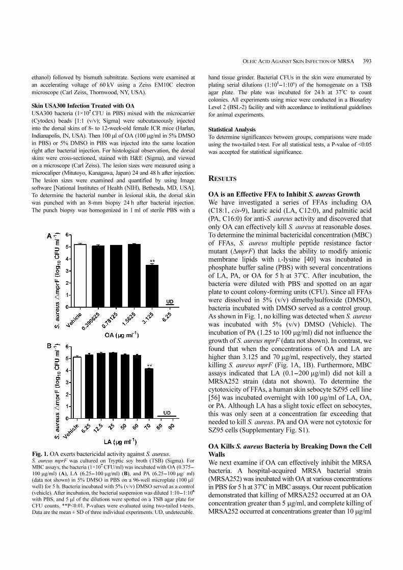
Because it has been reported that OA kills bacteria by
disrupting the cell wall [30], we investigated OA-induced
cell wall damage to S. aureus by transmission electron
microscopy (Fig. 3). Vehicle control [5% (v/v) DMSO] treated
bacteria showed uniform density in cytoplasmic compartments
and cell separation by prominent high contrast septa (Fig. 3a).
After OA treatment, disruption of the cell wall architecture
was observed (Fig. 3b). The cell walls in many bacteria
were completely separated from cytoplasmic compartments.
This finding suggests that OA breaks down the cell wall of
S. aureus bacteria.
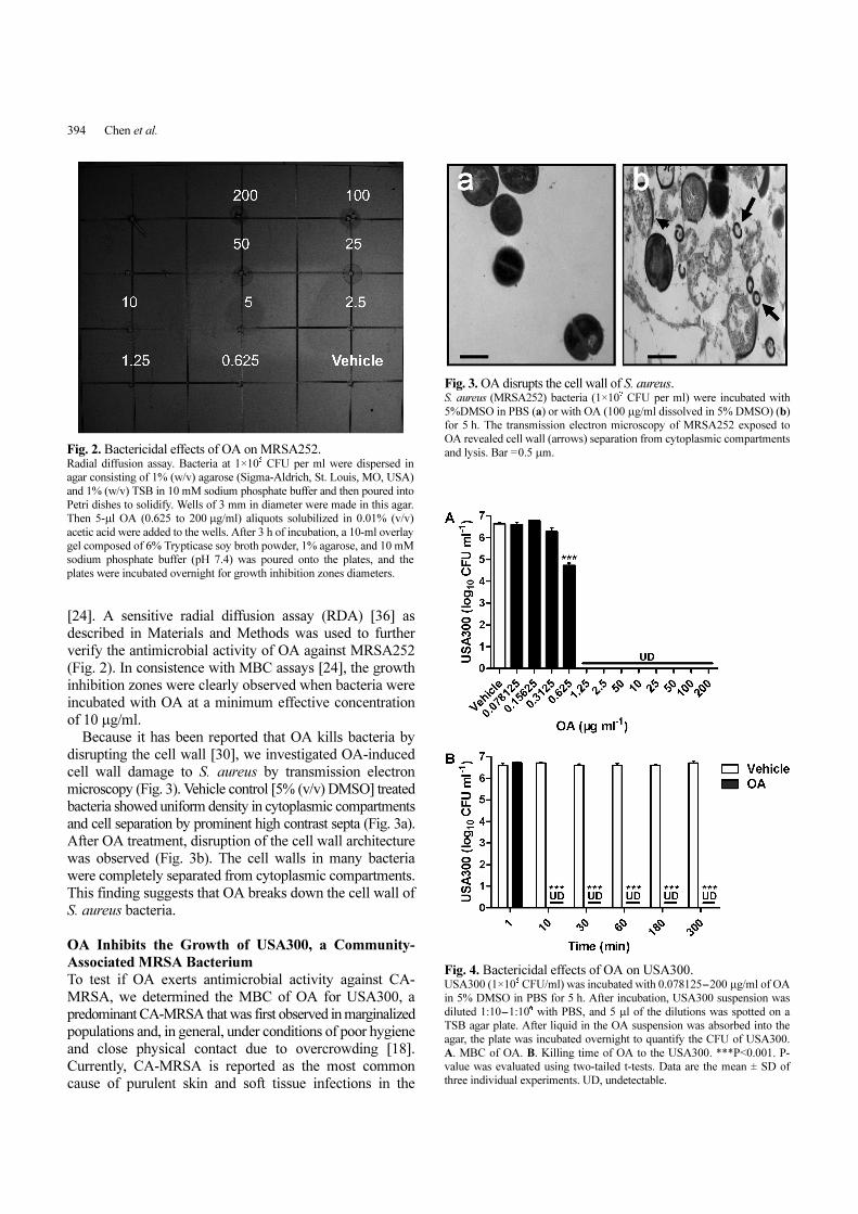
OA Inhibits the Growth of USA300, a Community-
Associated MRSA Bacterium
To test if OA exerts antimicrobial activity against CA-
MRSA, we determined the MBC of OA for USA300, a
predominant CA-MRSA that was first observed in marginalized
populations and, in general, under conditions of poor hygiene
and close physical contact due to overcrowding [18].
Currently, CA-MRSA is reported as the most common
cause of purulent skin and soft tissue infections in the
Fig. 3. OA disrupts the cell wall of S. aureus. S. aureus (MRSA252) bacteria (1×10
7 CFU per ml) were incubated with
5%DMSO in PBS (a) or with OA (100 µg/ml dissolved in 5% DMSO) (b)
for 5 h. The transmission electron microscopy of MRSA252 exposed to
OA revealed cell wall (arrows) separation from cytoplasmic compartments
and lysis. Bar =0.5 µm.Fig. 2. Bactericidal effects of OA on MRSA252.Radial diffusion assay. Bacteria at 1×10
5 CFU per ml were dispersed in
agar consisting of 1% (w/v) agarose (Sigma-Aldrich, St. Louis, MO, USA)
and 1% (w/v) TSB in 10 mM sodium phosphate buffer and then poured into
Petri dishes to solidify. Wells of 3 mm in diameter were made in this agar.
Then 5-µl OA (0.625 to 200 µg/ml) aliquots solubilized in 0.01% (v/v)
acetic acid were added to the wells. After 3 h of incubation, a 10-ml overlay
gel composed of 6% Trypticase soy broth powder, 1% agarose, and 10 mM
sodium phosphate buffer (pH 7.4) was poured onto the plates, and the
plates were incubated overnight for growth inhibition zones diameters.
Fig. 4. Bactericidal effects of OA on USA300. USA300 (1×10
6CFU/ml) was incubated with 0.078125-200 µg/ml of OA
in 5% DMSO in PBS for 5 h. After incubation, USA300 suspension was
diluted 1:10-1:106 with PBS, and 5 µl of the dilutions was spotted on a
TSB agar plate. After liquid in the OA suspension was absorbed into the
agar, the plate was incubated overnight to quantify the CFU of USA300.
A. MBC of OA. B. Killing time of OA to the USA300. ***P<0.001. P-
value was evaluated using two-tailed t-tests. Data are the mean ± SD of
three individual experiments. UD, undetectable.
OLEIC ACID AGAINST SKIN INFECTION OF MRSA 395
United States [28]. To determine the MBC of OA for
USA300, bacteria were incubated with OA at various
concentrations in PBS for 5 h at 37oC. After incubation,
the bacteria were diluted with PBS and spotted on an agar
plate to count CFUs. We found that OA effectively suppressed
the growth of USA300 at an OA concentration greater
than 0.625 µg/ml, and completely killed the USA300 at
concentrations greater than 1.25 µg/ml (Fig. 4A). Taken
together with data in Fig. 1A, 2, and 4A, the findings
suggest that OA may have broad-spectrum antimicrobial
activity against S. aureus bacteria. To reveal how fast OA
can efficiently kill USA300, bacteria were incubated for
1 min, 10 min, 30 min, 1 h, 3 h, and 5 h with OA, respectively.
The results illustrated that the USA300 bacteria were
completely eliminated after incubation with OA (100 µg/ml)
for 10 min (Fig. 4B).
To determine if OA exerts this antimicrobial activity to
other bacteria, we measured the MBC values of OA for
Gram-positive bacteria including Staphylococcus epidermidis
(S. epidermidis; American Type Culture Collection (ATCC)
12228], Listeria monocytogenes (L. monocytogenes; EGD
strain BUG), Bacillus anthracis (B. anthracis; Sterne 34F2
spores), and a Gram-negative bacterium [Escherichia coli
(E. coli); BL21 (DE3)] (Table 1). The growth of all Gram-
positive bacteria, but not E. coli, could be significantly
suppressed by OA at a concentration lower than 200 µg/ml,
suggesting that Gram-positive bacteria are more susceptible
to OA than are Gram-negative bacteria.
OA Alleviates USA300-Induced Skin Lesions
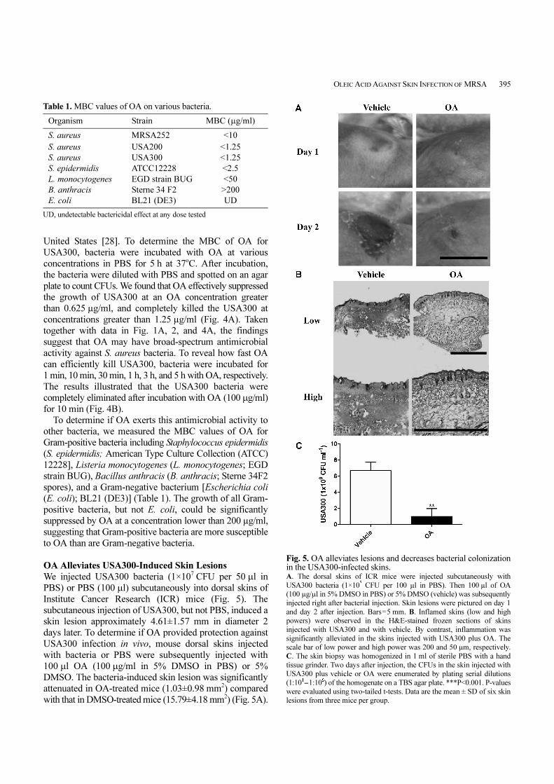
We injected USA300 bacteria (1×10⁷ CFU per 50 µl in
PBS) or PBS (100 µl) subcutaneously into dorsal skins of
Institute Cancer Research (ICR) mice (Fig. 5). The
subcutaneous injection of USA300, but not PBS, induced a
skin lesion approximately 4.61±1.57 mm in diameter 2
days later. To determine if OA provided protection against
USA300 infection in vivo, mouse dorsal skins injected
with bacteria or PBS were subsequently injected with
100 µl OA (100 µg/ml in 5% DMSO in PBS) or 5%
DMSO. The bacteria-induced skin lesion was significantly
attenuated in OA-treated mice (1.03±0.98 mm2) compared
with that in DMSO-treated mice (15.79±4.18 mm2) (Fig. 5A).
Table 1. MBC values of OA on various bacteria.
Organism Strain MBC (µg/ml)
S. aureus MRSA252 <10
S. aureus USA200 <1.25
S. aureus USA300 <1.25
S. epidermidis ATCC12228 <2.5
L. monocytogenes EGD strain BUG <50
B. anthracis Sterne 34 F2 >200
E. coli BL21 (DE3) UD
UD, undetectable bactericidal effect at any dose tested
Fig. 5. OA alleviates lesions and decreases bacterial colonizationin the USA300-infected skins. A. The dorsal skins of ICR mice were injected subcutaneously with
USA300 bacteria (1×10⁷ CFU per 100 µl in PBS). Then 100 µl of OA
(100 µg/µl in 5% DMSO in PBS) or 5% DMSO (vehicle) was subsequently
injected right after bacterial injection. Skin lesions were pictured on day 1
and day 2 after injection. Bars=5 mm. B. Inflamed skins (low and high
powers) were observed in the H&E-stained frozen sections of skins
injected with USA300 and with vehicle. By contrast, inflammation was
significantly alleviated in the skins injected with USA300 plus OA. The
scale bar of low power and high power was 200 and 50 µm, respectively.
C. The skin biopsy was homogenized in 1 ml of sterile PBS with a hand
tissue grinder. Two days after injection, the CFUs in the skin injected with
USA300 plus vehicle or OA were enumerated by plating serial dilutions
(1:101-1:10
6) of the homogenate on a TBS agar plate. ***P<0.001. P-values
were evaluated using two-tailed t-tests. Data are the mean ± SD of six skin
lesions from three mice per group.
396 Chen et al.
Histological observation in hematoxylin and eosin (H&E)
stained tissue sections revealed that injection of USA300
inflamed the skin and broke down the epidermal layer
(Fig. 5B). Treatment with OA, but not DMSO, alleviated the
skin damage response to USA300 infection. To determine
the intensity of bacterial colonization, skins treated with
OA or DMSO were homogenized to estimate the CFU. The
bacterial numbers in skins injected with USA300/DMSO
and USA300/OA were 6.7×10⁹ ± 1×10⁹ and 1.0×10⁹ ±
9.8×10⁸ CFU, respectively, suggesting that OA considerably
decreased the growth of USA300 in the skin lesions (Fig. 5C).
DISCUSSION
Evolutionary (Darwinian) medicine has proven valuable in
explaining the reasons for the development of antibiotic
resistance. The use of non-endogenous antibiotics for bacterial
treatments may not be in compliance with evolutionary
medicine since bacteria can fight back by developing the
ability to neutralize these antibiotics. For example, overuse
of broad-spectrum antibiotics, such as second- and third-
generation cephalosporins, greatly hastens the development
of methicillin resistance [33]. It has been reported that
FFAs endogenously exist in human organs [27] including
skin [4] and can function as innate bactericides against
various pathogens by altering the hydrophobicity of the
bacterial cell wall [30]. Here, we demonstrated that OA,
but not LA and PA, can effectively kill S. aureus bacteria
including both MRSA252 and USA300 in vitro (Fig. 2 and 4)
and significantly suppress USA300-induced skin lesions
and bacterial colonization in mice (Fig. 5). USA300 is one
of the aggressive CA-MRSA that became notable for extreme
antibiotic resistance and being responsible for rapidly
progressive, fatal diseases including necrotizing pneumonia
and severe sepsis [5]. As shown in Fig. 4, complete
eradication of USA300 bacteria can be found as early as 10
min after incubation with OA, suggesting that OA may be
able to efficiently clear bacteria at the early stage of CA-
MRSA infection. OA is an unsaturated FFA, whereas LA
and PA are saturated FFAs. In general, unsaturated FFAs
tend to have greater potency against bacteria than saturated
FFAs [11]. However, our previous studies demonstrated
that, compared with OA and PA, LA showed the strongest
bacteriocidal activity against P. acnes, a Gram-positive
bacterium [56]. It has been reported that the hydrophobicity
of the bacterial cell wall can affect the interactions between
bacteria and FFAs [30]. Thus, we speculate that, besides
the structure of FFAs, the hydrophobicity of the bacterial
cell wall may influence the bacteriocidal activities of FFAs.
Our data have demonstrated that OA, PA, and LA are not
detrimental to a human skin cell (sebocyte) (Fig. S1). The
bacteriocidal actions of FFAs are typically broad spectrum
and of potencies comparable to antimicrobial peptides
(AMPs). Although both FFAs and AMPs are endogenous
antimicrobial agents, FFAs with diverse structures are
relatively unstable and they also have a tendency to bind
nonspecifically to proteins [12]. Treatment of sebocytes
with OA or PA up-regulated the expression of human beta-
defensin (hBD)-2 and -3 and induced a release of hBD-2
[38]. An in vivo study from our laboratory [38] demonstrated
the up-regulation of mouse β-defensin 4, a mouse ortholog
for hBD-2, in mouse ear skin after epicutaneous application
of OA. In addition, hBD-2 synergistically killed P. acnes in
combination with LA [38], suggesting cooperative effects
between FFAs and AMPs. Recently, it has been reported
that FFA induced faster wound closure in mice [7] and
increased the wound healing tissue mass in rats [43]. FFAs
induced vascular endothelial growth factor-alpha (VEGF-
alpha) and interleukin-1beta (IL-1beta) in the inflammatory
phase of wound healing in rats [43]. In addition, FFAs
were able to stimulate the production of cytokine-induced
neutrophil chemoattractant in inflammation 2 alpha/beta
[43], suggesting that the pro-inflammatory effect of OA
may speed up the wound healing process [7]. The above
results suggest that endogenous FFAs not only function as
bactericides but also as native enhancers of innate immunity.
Moreover, the OA-induced cytokine profiles in infected
and non-infected mice may be worth establishing in future
experiments.
Previous studies have demonstrated that FFAs of various
chain lengths and with different levels of unsaturation
were primarily effective against Gram-positive bacteria,
but not against a number of Gram-negative bacteria [3, 15,
22, 25, 29, 32, 40, 47]. Consistent with these studies, our
results indicate that OA has antibacterial activity against a
range of Gram-positive bacteria, but not a Gram-negative
E. coli bacterium (Table 1). Cell walls in Gram-positive
bacteria consist of many layers of peptidoglycan without a
lipid outer membrane. Cell walls in Gram-negative bacteria,
on the other hand, have only one or a few layers of
peptidoglycan but possess an outer membrane consisting of
various lipid complexes. It has been shown that the outer
membrane layer of Gram-negative organisms may screen
the cells against fatty acids to prevent their accumulation
on the inner cell membrane. Therefore, Gram-negative
bacteria, protected by their outer lipid membrane, are
resistant to the destructive powers of OA [47]. It has been
reported that use of a chelating agent to remove the outer
lipid membrane will overcome the limitation of FFAs to
Gram-negative organisms [26], raising the possibility of
using OA for treatment of Gram-negative organism-
infected skin. On the surface of the skin, triglycerides
produce FFAs by catalytic reactions in the presence of
bacterial hydrolases [34]. Although the FFA content in
human and mouse skins is different, it has been reported
that human skin surface is rich in FFAs including LA,
myristic acids (8.7 nmol/cm2), PA (15 nmol/cm2), stearic acids
OLEIC ACID AGAINST SKIN INFECTION OF MRSA 397
(4.8 nmol/cm2), linoleic, and linolenic acids, palmitoleate,
and OA (9.5 nmol/cm2) [39, 54]. As shown in Fig. 5,
subcutaneous injection of 100 µg (approximately 354 nmol)
OA significantly decreased the growth of USA300 in the
skin lesions. The incomplete elimination of bacteria by OA
may be due to the possibility that OA cannot access
endocytosed USA300 within immune cells. Recently, flk/
flk mutant mice (an N-ethyl-N-nitrosourea-induced recessive
germ line mutation of C57BL/6 mice) showed reduced
sebum production and were unable to synthesize the
palmitoleate and OA [16]. The mutant mice had a defect in
the clearance of skin infections by Streptococcus pyogenes
(S. pyogenes) and S. aureus. Positional cloning and
sequencing revealed that flk is a novel allele of the stearoyl
coenzyme A desaturase 1 gene (Scd1), which is an enzyme
responsible for the biosynthesis of OA and POA [41] and
can be strongly up-regulated by Toll-like receptor 2
(TLR2) [16]. Thus, the essential role of endogenous OA in
combating S. aureus/MRSA infection can be addressed in
the future by comparing the severity of S. aureus/MRSA-
induced skin lesions in flk/flk mutant and wild-type mice.
It is also unknown how many endogenous FFAs in skin
can actively eliminate infected S. aureus. The amount of
endogenous FFAs in the human body was insufficient to
completely eradicate the overgrowth of S. aureus and/or
bacteria with mutations or drug resistance. The use of
exogenous FFAs including OA at different doses can
efficiently alleviate mild to severe stages of S. aureus/
MRSA infection. Unfortunately, one challenge of using
FFAs for medical practice stems from their poor water-
solubility. DMSO was used to dissolve OA in this study.
Although DMSO is an anti-inflammatory agent that has
been shown clinically to relieve pain [6], it has been
reported that topical application of DMSO can cause skin
irritation [57]. To circumvent this problem, we have
recently incorporated OA onto the surface of a liposome
(LipoOA) [56]. Our published result demonstrated that
LipoOA exhibited higher potency than free OA in terms of
the suppression of the growth of MRSA252 [56]. Without
toxicity to normal skin, LipoOA was highly effective in
curing skin infections caused by MRSA252 [24]. Previous
studies demonstrated that liposomes enriched in OA are
less susceptible to oxidation and show less pro-inflammatory
activity when exposed to oxidizing conditions [35]. In
addition, it has been known that liposomal antibacterial
agents are able to kill intracellular bacteria [2]. Thus,
LipoOA may be able to optimize the potency of OA against
S. aureus infection in humans.
Resistance of S. aureus to β-lactam antibiotics is usually
caused by the production of β-lactamases. Production of β-
lactamase is controlled by the bla regulatory apparatus,
which is homologous to the mec system responsible for the
regulation of mecA, the gene encoding methicillin resistance,
in many strains with inducible resistance [20]. It has been
reported that FFAs in sebum have the capability of
inhibiting the induction of β-lactamase in S. aureus [8].
Here, we emphasize that OA is an endogenous molecule
that may be part of innate immunity against S. aureus/
MRSA infection. Novel bactericides using OA and its
derivatives may gain a new set of modalities for fighting
antibiotic resistance.
Acknowledgments
This work was supported by several NIH grants
(1R21AI088147-01, R01-AI067395-01, R21-R022754-
01, R21-I58002-01, and 1R41AR056169-01). We thank
Dr. Stuart Ibsen for critical reading of the manuscript.
REFERENCES
1. Adem, P. V., C. P. Montgomery, A. N. Husain, T. K. Koogler,
V. Arangelovich, M. Humilier, S. Boyle-Vavra, and R. S. Daum.
2005. Staphylococcus aureus sepsis and the Waterhouse-
Friderichsen syndrome in children. N. Engl. J. Med. 353: 1245-
1251.
2. Bakker-Woudenbera, I. A. J. M., G. Storm, and M. C. Woodle.
1994. Liposomes in the treatment of infections. J. Drug Target.
2: 363-371.
3. Bergsson, G., J. Arnfinnsson, O. Steingrimsson, and H.
Thormar. 2001. Killing of Gram-positive cocci by fatty acids
and monoglycerides. APMIS 109: 670-678.
4. Bodoprost, J. and H. Rosemeyer. 2007. Analysis of
phenacylester derivatives of fatty acids from human skin surface
sebum by reversed-phase HPLC: Chromatographic mobility as
a function of physico-chemical properties. Int. J. Mol. Sci. 8:
1111-1124.
5. Boyle-Vavra, S. and R. S. Daum. 2007. Community-acquired
methicillin-resistant Staphylococcus aureus: The role of Panton-
Valentine leukocidin. Lab. Invest. 87: 3-9.
6. Brien, S., P. Prescott, N. Bashir, H. Lewith, and G. Lewith.
2008. Systematic review of the nutritional supplements dimethyl
sulfoxide (DMSO) and methylsulfonylmethane (MSM) in the
treatment of osteoarthritis. Osteoarthr. Cartilage 16: 1277-1288.
7. Cardoso, C. R. B., M. A. Souza, E. A. V. Ferro, S. Favoreto,
and J. D. O. Pena. 2004. Influence of topical administration of
n-3 and n-6 essential and n-9 nonessential fatty acids on the
healing of cutaneous wounds. Wound Repair Regen. 12: 235-243.
8. Clarke, S. R., R. Mohamed, L. Bian, A. F. Routh, J. F. Kokai-
Kun, J. J. Mond, A. Tarkowski, and S. J. Foster. 2007. The
Staphylococcus aureus surface protein IsdA mediates resistance
to innate defenses of human skin. Cell Host Microbe 1: 199-212.
9. Cohen, A. L., C. Shuler, S. McAllister, G. E. Fosheim, M. G.
Brown, D. Abercrombie, et al. 2007. Methamphetamine use and
methicillin-resistant Staphylococcus aureus skin infections. Emerg.
Infect. Dis. 13: 1707-1713.
10. de Pablo, M. A. and G. Alvarez de Cienfuegos. 2000. Modulatory
effects of dietary lipids on immune system functions. Immunol.
Cell Biol. 78: 31-39.
398 Chen et al.
11. Desbois, A. P., T. Lebl, L. Yan, and V. J. Smith. 2008. Isolation
and structural characterization of two antibacterial free fatty
acids from the marine diatom, Phaeodactylum tricornutum.
Appl. Microbiol. Biotechnol. 81: 755-764.
12. Desbois, A. P. and V. J. Smith. 2010. Antibacterial free fatty
acids: Activities, mechanisms of action and biotechnological
potential. Appl. Microbiol. Biotechnol. 85: 1629-1642.
13. Diep, B. A., H. A. Carleton, R. F. Chang, G. F. Sensabaugh, and
F. Perdreau-Remington. 2006. Roles of 34 virulence genes in
the evolution of hospital- and community-associated strains of
methicillin-resistant Staphylococcus aureus. J. Infect. Dis. 193:
1495-1503.
14. Fernandez-Lopez, R., C. Machon, C. M. Longshaw, S. Martin,
S. Molin, E. L. Zechner, M. Espinosa, E. Lanka, and F. de la
Cruz. 2005. Unsaturated fatty acids are inhibitors of bacterial
conjugation. Microbiology 151: 3517-3526.
15. Galbraith, H. and T. B. Miller. 1973. Effect of long chain fatty
acids on bacterial respiration and amino acid uptake. J. Appl.
Bacteriol. 36: 659-675.
16. Georgel, P., K. Crozat, X. Lauth, E. Makrantonaki, H. Seltmann,
S. Sovath, et al. 2005. A Toll-like receptor 2-responsive lipid
effector pathway protects mammals against skin infections with
Gram-positive bacteria. Infect. Immun. 73: 4512-4521.
17. Gibbons, M. A., D. M. Bowdish, D. J. Davidson, J. M. Sallenave,
and A. J. Simpson. 2006. Endogenous pulmonary antibiotics.
Scot. Med. J. 51: 37-42.
18. Gilbert, M., J. MacDonald, D. Gregson, J. Siushansian, K.
Zhang, S. Elsayed, et al. 2006. Outbreak in Alberta of
community-acquired (USA300) methicillin-resistant Staphylococcus
aureus in people with a history of drug use, homelessness or
incarceration. CMAJ 175: 149-154.
19. Goetghebeur, M., P. A. Landry, D. Han, and C. Vicente. 2007.
Methicillin-resistant Staphylococcus aureus: A public health
issue with economic consequences. Can. J. Infect. Dis. Med.
18: 27-34.
20. Hackbarth, C. J. and H. F. Chambers. 1993. Blai and Blar1
regulate beta-lactamase and Pbp 2a production in methicillin-
Resistant Staphylococcus aureus. Antimicrob. Agents Chemother.
37: 1144-1149.
21. Hamosh, M. 1998. Protective function of proteins and lipids in
human milk. Biol. Neonate 74: 163-176.
22. Heczko, P. B., R. Lutticken, W. Hryniewicz, M. Neugebauer,
and G. Pulverer. 1979. Susceptibility of Staphylococcus aureus
and group A, B, C, and G streptococci to Free fatty acids. J.
Clin. Microbiol. 9: 333-335.
23. Homa, D. G. and M. A. Palfreyman. 2000. Infectious diseases
in the operating room. CRNA 11: 8-14.
24. Huang, C. M., C. H. Chen, D. Pornpattananangkul, L. Zhang,
M. Chan, M. F. Hsieh, and L. F. Zhang. 2011. Eradication of
drug resistant Staphylococcus aureus by liposomal oleic acids.
Biomaterials 32: 214-221.
25. Kabara, J. J., D. Swieczkowski, A. J. Conley, and J. P. Truant.
1972. Fatty acids and derivatives as antimicrobial agents.
Antimicrob. Agents Chemother. 2: 23-28.
26. Kabara, J. J. 1984. Antimicrobial agents derived from fatty
acids. J. Am. Oil. Chem. Soc. 61: 397-403.
27. Kangani, C. O., D. E. Kelley, and J. P. DeLany. 2008. New
method for GC/FID and GC-C-IRMS analysis of plasma free
fatty acid concentration and isotopic enrichment. J. Chromatogr.
B 873: 95-101.
28. Kaplan, S. L., K. G. Hulten, B. E. Gonzalez, W. A. Hammerman,
L. Lamberth, J. Versalovic, and E. O. Mason Jr. 2005. Three-
year surveillance of community-acquired Staphylococcus aureus
infections in children. Clin. Infect. Dis. 40: 1785-1791.
29. Kelsey, J. A., K. W. Bayles, B. Shafii, and M. A. McGuire.
2006. Fatty acids and monoacylglycerols inhibit growth of
Staphylococcus aureus. Lipids 41: 951-961.
30. Kenny, J. G., D. Ward, E. Josefsson, I. M. Jonsson, J. Hinds, H.
H. Rees, J. A. Lindsay, A. Tarkowski, and M. J. Horsburgh.
2009. The Staphylococcus aureus response to unsaturated long
chain free fatty acids: Survival mechanisms and virulence
implications. PLoS ONE 4: e4344.
31. Klevens, R. M., M. A. Morrison, J. Nadle, S. Petit, K. Gershman,
S. Ray, et al. 2007. Invasive methicillin-resistant Staphylococcus
aureus infections in the United States. JAMA 298: 1763-1771.
32. Knapp, H. R. and M. A. Melly. 1986. Bactericidal effects of
polyunsaturated fatty acids. J. Infect. Dis. 154: 84-94.
33. Kollef, M. H. 2009. New antimicrobial agents for methicillin-
resistant Staphylococcus aureus. Crit. Care Resusc. 11: 282-286.
34. Kotani, A., Y. Hayashi, R. Matsuda, and F. Kusu. 2003. Prediction
of measurement precision of apparatus using a chemometric
tool in electrochemical detection of high-performance liquid
chromatography. J. Chromatogr. A 986: 239-246.
35. Lee, C., J. Barnett, and P. D. Reaven. 1998. Liposomes enriched
in oleic acid are less susceptible to oxidation and have less
proinflammatory activity when exposed to oxidizing conditions.
J. Lipid Res. 39: 1239-1247.
36. Lehrer, R. I., M. Rosenman, S. S. Harwig, R. Jackson, and
P. Eisenhauer. 1991. Ultrasensitive assays for endogenous anti-
microbial polypeptides. J. Immunol. Methods 137: 167-173.
37. Martin, A. and M. Clynes. 1991. Acid phosphatase: Endpoint
for in vitro toxicity tests. In Vitro Cell Dev. Biol. 27A: 183-
184.
38. Nakatsuji, T., M. C. Kao, L. Zhang, C. C. Zouboulis, R. L.
Gallo, and C. M. Huang. 2010. Sebum free fatty acids enhance
the innate immune defense of human sebocytes by upregulating
beta-defensin-2 expression. J. Invest. Dermatol. 130: 985-994.
39. Nicollier, M., T. Massengo, J. P. Remy-Martin, R. Laurent, and
G. L. Adessi. 1986. Free fatty acids and fatty acids of
triacylglycerols in normal and hyperkeratotic human stratum
corneum. J. Invest. Dermatol. 87: 68-71.
40. Nieman, C. 1954. Influence of trace amounts of fatty acids on
the growth of microorganisms. Bacteriol. Rev. 18: 147-163.
41. Ntambi, J. M. 1995. The regulation of stearoyl-CoA desaturase
(SCD). Prog. Lipid Res. 34: 139-150.
42. Payne, D. J. 2008. Microbiology. Desperately seeking new
antibiotics. Science 321: 1644-1645.
43. Pereira, L. M., E. Hatanaka, E. F. Martins, F. Oliveira, E. A.
Liberti, S. H. Farsky, R. Curi, and T. C. Pithon-Curi. 2008.
Effect of oleic and linoleic acids on the inflammatory phase of
wound healing in rats. Cell Biochem. Funct. 26: 197-204.
44. Peschel, A., R. W. Jack, M. Otto, L. V. Collins, P. Staubitz, G.
Nicholson, et al. 2001. Staphylococcus aureus resistance to
human defensins and evasion of neutrophil killing via the novel
virulence factor MprF is based on modification of membrane
lipids with L-lysine. J. Exp. Med. 193: 1067-1076.
OLEIC ACID AGAINST SKIN INFECTION OF MRSA 399
45. Sado Kamdem, S., M. E. Guerzoni, J. Baranyi, and C. Pin.
2008. Effect of capric, lauric and alpha-linolenic acids on the
division time distributions of single cells of Staphylococcus
aureus. Int. J. Food Microbiol. 128: 122-128.
46. Smith, P. A. and F. E. Romesberg. 2007. Combating bacteria
and drug resistance by inhibiting mechanisms of persistence and
adaptation. Nat. Chem. Biol. 3: 549-556.
47. Speert, D. P., L. W. Wannamaker, E. D. Gray, and C. C.
Clawson. 1979. Bactericidal effect of oleic acid on group A
streptococci: Mechanism of action. Infect. Immun. 26: 1202-1210.
48. Stewart, M. E. 1992. Sebaceous gland lipids. Semin. Dermatol.
11: 100-105.
49. Takano, T., K. Saito, L. J. Teng, and T. Yamamoto. 2007. Spread
of community-acquired methicillin-resistant Staphylococcus
aureus (MRSA) in hospitals in Taipei, Taiwan in 2005, and
comparison of its drug resistance with previous hospital-
acquired MRSA. Microbiol. Immunol. 51: 627-632.
50. Takizawa, Y., I. Taneike, S. Nakagawa, T. Oishi, Y. Nitahara,
N. Iwakura, et al. 2005. A Panton-Valentine leucocidin (PVL)-
positive community-acquired methicillin-resistant Staphylococcus
aureus (MRSA) strain, another such strain carrying a multiple-
drug resistance plasmid, and other more-typical PVL-negative
MRSA strains found in Japan. J. Clin. Microbiol. 43: 3356-3363.
51. Tancrede, C. 1992. Role of human microflora in health and
disease. Eur. J. Clin. Microbiol. 11: 1012-1015.
52. Van Bambeke, F., M. P. Mingeot-Leclercq, M. J. Struelens, and
P. M. Tulkens. 2008. The bacterial envelope as a target for
novel anti-MRSA antibiotics. Trends Pharmacol. Sci. 29: 124-134.
53. Weigel, L. M., D. B. Clewell, S. R. Gill, N. C. Clark, L. K.
McDougal, S. E. Flannagan, et al. 2003. Genetic analysis of a
high-level vancomycin-resistant isolate of Staphylococcus aureus.
Science 302: 1569-1571.
54. Wilkinson, D. I. and J. T. Walsh. 1974. Effect of various
methods of epidermal-dermal separation on distribution of acetate-
C-14-labeled polyunsaturated fatty acids in skin compartments.
J. Invest. Dermatol. 62: 517-521.
55. Wilson, P. C. and B. Rinker. 2009. The incidence of methicillin-
resistant Staphylococcus aureus in community-acquired hand
infections. Ann. Plas. Surg. 62: 513-516.
56. Yang, D. R., D. Pornpattananangkul, T. Nakatsuji, M. Chan, D.
Carson, C. M. Huang, and L. F. Zhang. 2009. The antimicrobial
activity of liposomal lauric acids against Propionibacterium
acnes. Biomaterials 30: 6035-6040.
57. Zesch, A. 1983. Skin irritation by topical drugs. Derm. Beruf.
Umwelt. 31: 74-78.
58. Zouboulis, C. C., H. Seltmann, H. Neitzel, and C. E. Orfanos.
1999. Establishment and characterization of an immortalized
human sebaceous gland cell line (SZ95). J. Invest. Dermatol.
113: 1011-1020.