an emerging tick-borne disease of humans is...
TRANSCRIPT
Pathogens 2013, 2, 544-555; doi:10.3390/pathogens2030544
pathogens
ISSN 2076-0817
www.mdpi.com/journal/pathogens
Article
An Emerging Tick-Borne Disease of Humans Is Caused by a
Subset of Strains with Conserved Genome Structure
Anthony F. Barbet 1,2,†,
*, Basima Al-Khedery 1,†
, Snorre Stuen 3, Erik G. Granquist
4,
Roderick F. Felsheim 5 and Ulrike G. Munderloh
5
1 Department of Infectious Diseases and Pathology, University of Florida, Gainesville, FL 32611,
USA; E-Mail: [email protected] 2 Emerging Pathogens Institute, University of Florida, Gainesville, FL 32611, USA
3 Department of Production Animal Clinical Sciences, Norwegian School of Veterinary Science,
Sandnes N-4325, Norway; E-Mail: [email protected] 4 Department of Production Animal Clinical Sciences, Norwegian School of Veterinary Science,
Oslo N-0033, Norway; E-Mail: [email protected] 5 Department of Entomology, University of Minnesota, St. Paul, MN 55108, USA;
E-Mails: [email protected] (R.F.F.); [email protected] (U.G.M.)
† These authors contributed equally to this work.
* Author to whom correspondence should be addressed; E-Mail: [email protected];
Tel.: +1-352-294-4119; Fax: +1-352-392-9704.
Received: 17 July 2013; in revised form: 29 August 2013 / Accepted: 2 September 2013 /
Published: 10 September 2013
Abstract: The prevalence of tick-borne diseases is increasing worldwide. One such emerging
disease is human anaplasmosis. The causative organism, Anaplasma phagocytophilum, is
known to infect multiple animal species and cause human fatalities in the U.S., Europe and
Asia. Although long known to infect ruminants, it is unclear why there are increasing
numbers of human infections. We analyzed the genome sequences of strains infecting
humans, animals and ticks from diverse geographic locations. Despite extensive variability
amongst these strains, those infecting humans had conserved genome structure including
the pfam01617 superfamily that encodes the major, neutralization-sensitive, surface
antigen. These data provide potential targets to identify human-infective strains and have
significance for understanding the selective pressures that lead to emergence of disease in
new species.
OPEN ACCESS
Pathogens 2013, 2 545
Keywords: anaplasmosis; tick-borne diseases; high-throughput sequencing; pfam01617;
msp2/p44; comparative genomics
1. Introduction
Human exposure to vectors carrying disease agents has been increased by climate and land-use
changes causing more contact between humans and domestic animals with wildlife reservoirs [1]. One
such recently emerging disease is tick-borne anaplasmosis that causes infections in multiple animal
species [2]. These include cattle, sheep, goats, horses, dogs, foxes, cats, rodents and most recently,
humans. Different strains of the causative organism, Anaplasma phagocytophilum, have different host
predilections and not all strains can infect all hosts. Small mammals are thought to be a reservoir,
infecting immature stages of ticks which act as bridge vectors transferring infection to humans and
domestic animals. Cases reported to the U.S. Centers for Disease Control and Prevention comprise a
flu-like febrile illness with some severe sequelae such as multiple organ failure, or severe acute
respiratory distress syndrome. In the U.S., case reports increased from 348 cases in 2000 to 1,761 cases
in 2010 [3]. The reported hospitalization rate is 36% [4]. Granulocytic anaplasmosis (GA) can be
treated with antibiotics, but the symptoms, such as headache, fever, and muscle aches are non-specific
and can be confused with other common diseases such as Lyme, transmitted by the same species of
tick. If left untreated GA can be severe [5], resulting in a case fatality rate in the U.S. of up to 3%
(CDC data for 2003). Increasingly, there are reports of infections transmitted by blood transfusions in
the U.S. and Europe [6,7]. Here we show that, in contrast to the extensive worldwide genomic
diversity of A. phagocytophilum strains, human-infective strains are a conserved subset. This has
implications for understanding the selective pressures that lead to emergence of disease in new species
and for control of this infection.
2. Results and Discussion
2.1. Comparative Genomics of Nine Strains of A. phagocytophilum
Underlying the ecological complexity and variable host tropism of A. phagocytophilum is a large
degree of genetic variability. European strains of A. phagocytophilum are highly pathogenic in sheep
and cattle, whereas North American strains do not cause disease in domestic ruminants. Some U.S.
strains, defined as Ap-variant 1 because of a 2 base pair difference in 16S rRNA, are infectious to deer
and other ruminants, but do not infect mice and are thought not to infect humans [8,9]. Highly unusual
for an obligate intracellular pathogen with a small, 1.5 Mb genome, A. phagocytophilum contains
approximately 100 “functional pseudogenes” of the major surface protein 2 gene (msp2/p44) that
codes for the major antigen on its surface [10]. These hypervariable pseudogene cassettes are
recombined into a single expression site which provides N- and C-terminal conserved sequences,
allowing the bacterium to serially express variable antigens and evade host immunity [11,12]. We
applied high-throughput genome sequencing to nine strains, derived from humans and different host
animal species from the U.S. and Europe. These strains are: two human origin strains from New York
Pathogens 2013, 2 546
and Minnesota, a rodent- and dog-origin strain from Minnesota, two Ap-variant 1 strains from
Minnesota, a horse-origin strain from California and two sheep-origin strains from Norway, for which
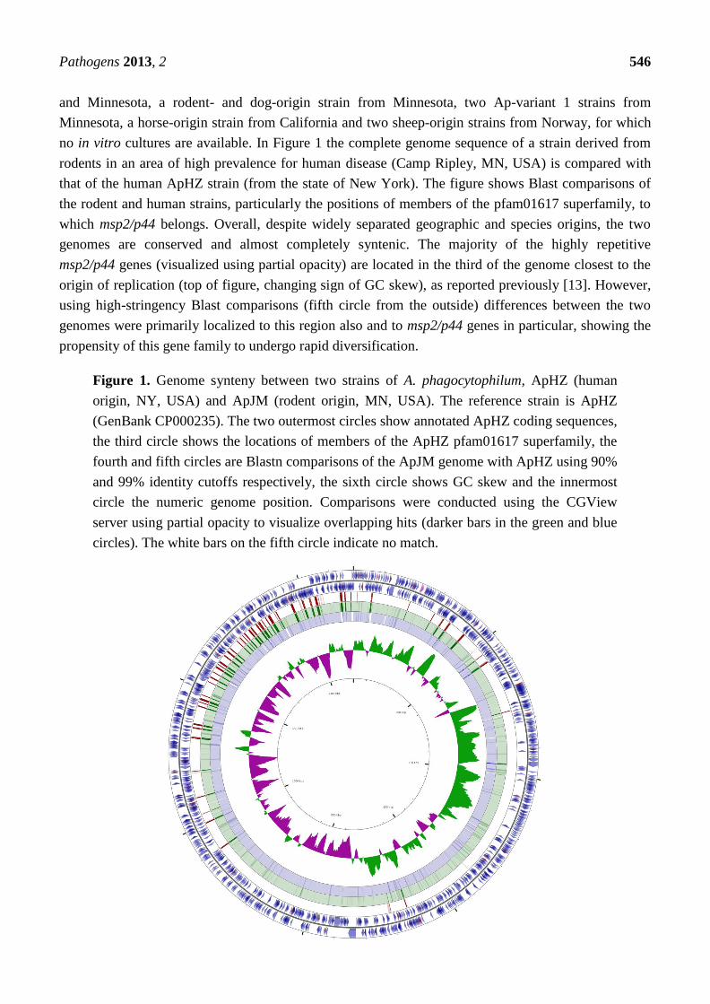
no in vitro cultures are available. In Figure 1 the complete genome sequence of a strain derived from
rodents in an area of high prevalence for human disease (Camp Ripley, MN, USA) is compared with
that of the human ApHZ strain (from the state of New York). The figure shows Blast comparisons of
the rodent and human strains, particularly the positions of members of the pfam01617 superfamily, to
which msp2/p44 belongs. Overall, despite widely separated geographic and species origins, the two
genomes are conserved and almost completely syntenic. The majority of the highly repetitive
msp2/p44 genes (visualized using partial opacity) are located in the third of the genome closest to the
origin of replication (top of figure, changing sign of GC skew), as reported previously [13]. However,
using high-stringency Blast comparisons (fifth circle from the outside) differences between the two
genomes were primarily localized to this region also and to msp2/p44 genes in particular, showing the
propensity of this gene family to undergo rapid diversification.
Figure 1. Genome synteny between two strains of A. phagocytophilum, ApHZ (human
origin, NY, USA) and ApJM (rodent origin, MN, USA). The reference strain is ApHZ
(GenBank CP000235). The two outermost circles show annotated ApHZ coding sequences,
the third circle shows the locations of members of the ApHZ pfam01617 superfamily, the
fourth and fifth circles are Blastn comparisons of the ApJM genome with ApHZ using 90%
and 99% identity cutoffs respectively, the sixth circle shows GC skew and the innermost
circle the numeric genome position. Comparisons were conducted using the CGView
server using partial opacity to visualize overlapping hits (darker bars in the green and blue
circles). The white bars on the fifth circle indicate no match.

Pathogens 2013, 2 547
This comparative genomics analysis was extended to all nine strains (Figure 2). Except for
Ap-variant 1 strains, the U.S. strains had high identities across their entire genomes, with the ApDog
strain most similar to human ApHZ by Blast analysis. The Norwegian sheep and U.S. Ap-variant 1
strains had the lowest identities with ApHZ in Blast comparisons, especially in members of the
pfam01617 superfamily.
Figure 2. Similarities and differences between nine A. phagocytophilum genomes.
Roche/454 reads from each genomic DNA were compared with the human ApHZ genome
as reference using Blastn and the CGView Comparison Tool. The first to ninth circles
compare, respectively, sequencing reads from ApHZ, ApDog, ApJM, ApHGE1, ApMRK,
ApCRT35, ApCRT38-1, ApNorV2, ApNorV1 genomic DNAs. The 10th circle shows the
locations of members of the ApHZ pfam01617 superfamily. Blast parameters used a query
size of 500 bp segments of the reference genome and a Blast expect value of 10−100
. Circles
are colored according to the percent identities of matches (black to light red, 100%–90%
identical, dark to light blue, 88%–82% identical, colorless, 0% identical).
The differences in this superfamily were examined more closely in order to determine their
msp2/p44 genomic repertoires and if differences were primarily located in the known pseudogene
hypervariable regions (Figure 3).

Pathogens 2013, 2 548
Figure 3. Gene variation in a region rich in p44/msp2 pseudogenes close to the origin of
replication in nine genomes of A. phagocytophilum. Roche/454 pyrosequencing reads from
each of the nine genomes are shown aligned with the annotated reference ApHZ genome
(bottom) between positions 1,450 and 1,462 kbp. The sequencing reads are derived from:
ApHZ, ApDog, ApHGE1, ApJM, ApMRK, ApCRT35, ApCRT38-1, ApNorV1, ApNorV2
(bottom to top panels respectively). The positions of six msp2/p44 pseudogenes are marked
by arrows at the top and shown in their respective reading frames at the bottom. The
hypervariable regions LAKT.. LAKT, present in five of the six pseudogenes, are indicated
in red. Gaps in coverage indicate no aligning reads over the central hypervariable regions
in some strains.
Similar to the rodent strain ApJM, the msp2/p44 repertoires of a U.S. human strain, ApHGE1 and
the ApDog strain from the same area of Minnesota were closely related and slightly different from the
New York human strain ApHZ (8/95 or 9/95 different msp2/p44 pseudogenes determined as <90%
identical, Table 1). In contrast, strains infecting horses or ruminants (Ap-variant 1 strains) in the U.S.
or Norway (representing “classical” strains long known to be pathogenic to sheep [14] shared fewer
msp2/p44 pseudogenes with human strains, with the two Norwegian sheep strains having almost
totally different repertoires from human strains. Differences between individual members of the
msp2/p44 repertoire were located in the known central hypervariable region between flanking
consensus amino acids LAKT.. LAKT (Figure 3).

Pathogens 2013, 2 549
Table 1. Genome structural relationships between Anaplasma strains.
Strain ApHZ ApHGE1 ApJM ApDog ApMRK ApCRT35 ApCRT38 ApNorV1 ApNorV2
Different msp2/p44
pseudogenes from ApHZ 0/95 8/95 8/95 9/95 49/95 71/95 75/95 92/95 89/95
ANIm 100 98.81 98.84 98.79 97.76 96.28 96.21 94.92 95.87
Tetra 1.000 0.999 0.999 0.999 0.998 0.996 0.996 0.995 0.996
Strain AmFL AmStM AcIs ApHZ
ANIm 100 98.86 88.89 77.82
Tetra 1.000 0.999 0.989 0.821
The top panel compares strains with ApHZ, the lower panel compares strains and species with AmFL. Abbreviations:
ANIm, Average % genome nucleotide identity using Mummer; Tetra, correlation coefficient of tetranucleotide signature
frequencies; AmFL, Anaplasma marginale Florida strain; AmStM, A. marginale St. Maries Idaho strain; AcIs,
Anaplasma centrale Israel strain; Ap, A. phagocytophilum strains.
Two additional quantitative measures of genome structural relationships compare the average
nucleotide identities or the tetranucleotide frequencies between two genomes. The latter method is
independent of alignment algorithms. The proposed threshold for prokaryotic species separation is 94%
for average nucleotide identity and 0.990 for the correlation coefficient of tetranucleotide signature
frequencies [15]. It is evident from Table 1 that the existing taxonomy of Anaplasma species meets these
definitions. However, it is apparent that by these measures also, the U.S. human, dog and rodent strains
are closely related (average nucleotide identity 98.79%–98.84% with ApHZ). The Norwegian ruminant
strains are again most distinct from ApHZ (average nucleotide identity 94.92%–95.87%), agreeing with
the similar divergence observed in their pfam01617 superfamily genes.
2.2. Comparison of the msp2/p44 Family among Strains of A. phagocytophilum
Although these data showed that two U.S. A. phagocytophilum strains infecting humans, as well as
a dog and rodent strain were similar to one another and different from strains infecting ruminants, we
wanted to analyze this more globally using a larger dataset. To do this, we took advantage of the fact
that conserved regions of msp2/p44 have been used as sensitive PCR diagnostic targets because of the
large copy number of the pfam01617 superfamily. More than 500 partial pseudogene sequences are
present in GenBank, from humans, ticks and animal strains derived from multiple regions throughout
the U.S., Europe and Asia. From these sequences it is possible to extract the hypervariable region of
msp2/p44 flanked by sequence encoding a consensus LAKT on either end, facilitating alignment [16].
Importantly, for this analysis it is necessary to recognize that multiple different hypervariable regions
exist in a single genome because of the ~100 distinct pseudogene cassettes present. Therefore, it is not
sufficient to simply analyze phylogenetic trees and apparent evolutionary relationships between all
msp2/p44 sequences. If one wishes to compare global repertoires with the genome-sequenced human
ApHZ strain, it is necessary to align each msp2/p44 sequence with all ~100 ApHZ strain genomic
pseudogene cassettes to find the best fit (maximum sequence identity). If strains are related to ApHZ,
one expects to find one or more ApHZ pseudogenes with high identity to other strain msp2/p44s. Our
analyses, therefore, employed a matrix where every available msp2/p44 polypeptide (661 total

Pathogens 2013, 2 550
including those encoded by ApHZ pseudogene cassettes) was aligned with every other, generating
218,791 alignments having a mean amino acid sequence identity of 48.2%. These percentage sequence
identities can be rapidly analyzed using a 661 (row) × 661 (column) spreadsheet. Surprisingly, despite
the differences in msp2/p44 repertoires observed previously, all 91 available msp2/p44 human-origin
sequences from widely dispersed U.S. locations (states of New York, Massachusetts, Wisconsin and
Minnesota) encoded polypeptides with a mean maximum percentage identity with ApHZ of 97.2%
(Table 2). Further, in analyzing the available msp2/p44 sequences it was notable that, world-wide,
other human-origin strains also achieved similar levels of sequence identity with ApHZ (e.g., 98.1%
mean maximum percentage amino acid identity of a dataset from Japan comprising 27 human-origin
msp2/p44 variants, no significant difference with ApHZ). In contrast, A. phagocytophilum strains from
Europe and Asia from non-human sources had mean maximum identities of <76% (significantly
different from ApHZ, Table 2).
Table 2. Comparison of msp2/p44 variants from different geographic locations with the
human ApHZ strain.
Country Source Number of msp2/p44
Variants Analyzed
Mean Maximum % a.a. Identity
with ApHZ (+/−Std.Dev.)
Significantly different
from U.S. Human *
U.S.A. Human 91 97.2 (7.0)
U.S.A. Dog 27 94.8 (9.8) No
U.S.A. Horse 29 94.2 (7.3) No
U.S.A. Bear 4 92.4 (4.1) N/A
U.S.A. Woodrat 88 78.4 (13.8) Yes
U.S.A. Ruminant, tick (Ap-variant 1) 34 86.5 (14.7) Yes
Norway Sheep 54 66.6 (6.4) Yes
Sweden Dog 19 64.3 (7.9) Yes
U.K. Goat 20 67.2 (8.2) Yes
U.K. Sheep 19 65.1 (6.7) Yes
Czech
Republic
Human, Roe deer, Perdix,
Ixodes ricinus 6 86.6 (14.5) N/A
[Human only] [2] [97.8][(2.0)] N/A
Japan Sika deer 17 39.3 (3.0) Yes
Japan Ixodes persulcatus 87 69.0 (6.9) Yes
Japan Ixodes ovatus 22 71.8 (15.5) Yes
Japan Haemaphysalis formosensis 9 75.6 (14) N/A
Japan Human 27 98.1 (7.2) No
China Human 2 100 (0) N/A
* Kruskal-Wallis One Way Analysis of Variance on Ranks (p ≤ 0.001) followed by Dunn’s method for Multiple
Comparisons versus a Control group (p ≤ 0.05). N/A: Not applied to sample sizes <10.
3. Experimental
3.1. Origin of A. phagocytophilum Strains
The origins of the A. phagocytophilum strains used in this study are as follows: ApHZ, human,
Westchester County, New York; HGE1, human, Minnesota; ApJM, meadow jumping mouse (Zapus

Pathogens 2013, 2 551
hudsonius), Camp Ripley, Minnesota; ApDog, Minnesota; ApCRT35, Camp Ripley tick (Ixodes
scapularis), Minnesota (defined previously as Ap-variant 1); ApCRT38-1, Camp Ripley tick (I.
scapularis), Minnesota (Ap-variant 1); ApMRK, horse, California; ApNorV1, sheep, Norway;
ApNorV2, sheep, Norway. A. phagocytophilum genomic DNA was prepared from in vitro cultured
organisms or directly from infected sheep (Norwegian strains), as described previously [17].
3.2. Ethics Statement
The experimental study in sheep was approved by the Norwegian Animal Research Authority.
3.3. Genome Sequencing and Bioinformatics
Genomic DNA was sequenced on the Roche/454 Genome Sequencer using non-paired and 3 kb
paired-end libraries, also as described [17]. Mean genome coverage with respect to ApHZ varied
between 31.3X and 72.1X. The ApJM sequence was finished using manual inspection for conflicts and
mismatched paired ends and PCR to fill gaps. All sequences were compared with ApHZ for regions of
identity using Blastn analysis in CGView [18,19]. Differences in the msp2/p44 repertoires between
strains were defined using a method previously validated using the pfam01617 superfamily and two
completely Sanger-sequenced genomes of Anaplasma marginale [20]. Briefly, this method uses
Mosaik to align individual reads and generate BAM format files to detect gaps in alignment (no
coverage) with respect to the annotated reference sequence. In A. marginale, this method detected all
different pseudogenes having <90% nucleotide identity with the reference genome. In homologous
comparisons between ApHZ Roche/454 reads and the ApHZ genome all msp2/p44 pseudogenes were
detected as present (Table 1). Comparisons of average nucleotide identities and the correlation
coefficients of tetranucleotide signature frequencies between genomes were conducted using Jspecies
software, as described [15].
3.4. Analysis of msp2/p44 Repertoires
To analyze global msp2/p44 repertoires, deposited msp2/p44 sequences were downloaded from
GenBank following Blast searches; several large datasets are also available from published
studies [16,21–25]. The sequences were each trimmed to that encoding the hypervariable region,
where possible using the consensus flanking LAKT residues, and the polypeptides were aligned (all
against all) with MATGAT [26]. This generates the alignments that can be inspected for accuracy, and
allows export of all percent identity values to a spreadsheet. This spreadsheet was analyzed in Excel
for the relatedness of the different datasets. The percent identity of each variant msp2/p44 sequence
with every ApHZ msp2/p44 polypeptide encoded by a pseudogene was reported and, from that, the
mean best match with ApHZ (maximum percent identity) determined for each dataset. As the
percentage identities were not normally distributed and the population variances were unequal,
significant differences between groups were determined by nonparametric methods (Table 2).

Pathogens 2013, 2 552
4. Conclusions
These data show that there is genome diversity worldwide within the A. phagocytophilum species
that extends close to some proposed guidelines for species discrimination. However, strains infecting
humans are a subset with more conserved genome structure than the species overall, and this includes
their repertoires of msp2/p44 genes. This subset of A. phagocytophilum strains does not differ
significantly from the U.S. ApHZ strain and is closely related to strains infecting U.S. domestic dogs.
The mean seroprevalence (using a conserved msp2/p44 peptide as antigen) in the U.S. among 479,640
dogs was 4.8% with prevalence >50% in some counties in the Northeast and Midwest, corresponding
to the location of the majority of human cases [27]. These data have significance in several areas. First,
msp2/p44 encodes a protein that induces neutralizing antibodies against homologous strains of
A. phagocytophilum [28,29]. The repertoire differences between strains reflect an evolving antigenic
system with adaptive pressures imposed by an array of different persistently infected hosts. In contrast,
humans are incidental dead-end hosts that do not impose significant pressure for change. The
similarities between human-origin strains in the U.S., Europe and Asia suggest that humans may not be
susceptible to many of the circulating wildlife strains. They may become susceptible when selection
pressures in small mammal reservoir hosts cause evolution of novel strains that allow invasion and survival
in humans. Rodents are important reservoirs for a multitude of human disease agents, vector-borne or not,
and are known to be infected with strains of A. phagocytophilum having different species tropisms [30].
Rodent control has historically been emphasized as a means to control human disease outbreaks. The
underlying evolutionary drivers remain to be identified but could reflect the long-standing association
between “men and mice”. Second, these data have practical significance for control of this emerging
infection. Despite the ubiquity of A. phagocytophilum strains and positive serology world-wide, it is
necessary to focus on the prevalence and transmission of a smaller, human-infective subset.
Differentiation of such strains from the high-prevalence background may be possible using genomic
PCR targets or selected msp2/p44 polypeptides for serology. In order to accomplish this goal it is
necessary to acquire genome sequences from multiple human-origin strains on different continents.
The technology now exists to do this.
Acknowledgments
We thank Anna M. Lundgren for technical support, Savita Shanker for high-throughput DNA
sequencing, David Allred and Dan Brown for reading the manuscript and to Dan Brown for suggesting
the use of Jspecies software. This investigation received support from grants RO1 GM081714
and GM081714-03S1.
Draft and complete sequences obtained in this project are being made available through the NCBI
Bioproject portal [31] as bioprojects PRJNA158483, 163167, 163169, 171710, 183838, 216999,
217002, 217033, and 217037.
Conflicts of Interest
The authors declare no conflict of interest.

Pathogens 2013, 2 553
References
1. Patz, J.A.; Olson, S.H.; Uejio, C.K.; Gibbs, H.K. Disease emergence from global climate and land
use change. Med. Clin. North Am. 2008, 92, 1473–1491.
2. Dumler, J.S.; Choi, K.S.; Garcia-Garcia, J.C.; Barat, N.S.; Scorpio, D.G.; Garyu, J.W.; Grab, D.J.;
Bakken, J.S. Human granulocytic anaplasmosis and Anaplasma phagocytophilum. Emerg. Infect.
Dis. 2005, 11, 1828–1834.
3. CDC. Statistics and Epidemiology. Annual Cases of Anaplasmosis in the United States; Available
online: www.cdc.gov/anaplasmosis/stats/ (accessed on 5 September 2013).
4. Dahlgren, F.S.; Mandel, E.J.; Krebs, J.W.; Massung, R.F.; McQuiston, J.H. Increasing incidence
of Ehrlichia chaffeensis and Anaplasma phagocytophilum in the United States, 2000–2007. Am. J.
Trop. Med. Hyg. 2011, 85, 124–131.
5. Zhang, L.; Wang, G.; Liu, Q.; Chen, C.; Li, J.; Long, B.; Yu, H.; Zhang, Z.; He, J.; Qu, Z.; et al.
Molecular analysis of Anaplasma phagocytophilum isolated from patients with febrile diseases of
unknown etiology in China. PLoS One 2013, 8, e57155.
6. Annen, K.; Friedman, K.; Eshoa, C.; Horowitz, M.; Gottschall, J.; Straus, T. Two cases of
transfusion-transmitted Anaplasma phagocytophilum. Am. J. Clin. Pathol. 2012, 137, 562–565.
7. Jereb, M.; Pecaver, B.; Tomazic, J.; Muzlovic, I.; Avsic-Zupanc, T.; Premru-Srsen, T.;
Levicnik-Stezinar, S.; Karner, P.; Strle, F. Severe human granulocytic anaplasmosis transmitted
by blood transfusion. Emerg. Infect. Dis. 2012, 18, 1354–1357.
8. Massung, R.F.; Priestley, R.A.; Miller, N.J.; Mather, T.N.; Levin, M.L. Inability of a variant strain
of Anaplasma phagocytophilum to infect mice. J. Infect. Dis. 2003, 188, 1757–1763.
9. Massung, R.F.; Mather, T.N.; Levin, M.L. Reservoir competency of goats for the Ap-variant 1
strain of Anaplasma phagocytophilum. Infect. Immun. 2006, 74, 1373–1375.
10. Dunning Hotopp, J.C.; Lin, M.; Madupu, R.; Crabtree, J.; Angiuoli, S.V.; Eisen, J.A.; Seshadri, R.;
Ren, Q.; Wu, M.; Utterback, T.R.; et al. Comparative genomics of emerging human ehrlichiosis
agents. PLoS Genet. 2006, 2, e21.
11. Barbet, A.F.; Meeus, P.F.; Bélanger, M.; Bowie, M.V.; Yi, J.; Lundgren, A.M.; Alleman, A.R.;
Wong, S.J.; Chu, F.K.; Munderloh, U.G.; et al. Expression of multiple outer membrane protein
sequence variants from a single genomic locus of Anaplasma phagocytophilum. Infect. Immun.
2003, 71, 1706–1718.
12. Lin, Q.; Rikihisa, Y. Establishment of cloned Anaplasma phagocytophilum and analysis of p44 gene
conversion within an infected horse and infected SCID mice. Infect. Immun. 2005, 73, 5106–5114.
13. Foley, J.E.; Nieto, N.C.; Barbet, A.F.; Foley, P. Antigen diversity in the parasitic bacterium
Anaplasma phagocytophilum arises from selectively-represented, spatially clustered functional
pseudogenes. PLoS One 2009, 4, e8265.
14. Stuen, S. Anaplasma phagocytophilum—The most widespread tick-borne infection in animals in
Europe. Vet. Res. Commun. 2007, 31, 79–84.
15. Richter, M.; Rosselló-Móra, R. Shifting the genomic gold standard for the prokaryotic species
definition. Proc. Natl. Acad. Sci. USA 2009, 106, 19126–19131.

Pathogens 2013, 2 554
16. Barbet, A.F.; Lundgren, A.M.; Alleman, A.R.; Stuen, S.; Bjöersdorff, A.; Brown, R.N.;
Drazenovich, N.L.; Foley, J.E. Structure of the expression site reveals global diversity in MSP2
(P44) variants in Anaplasma phagocytophilum. Infect. Immun. 2006, 74, 6429–6437.
17. Al-Khedery, B.; Lundgren, A.M.; Stuen, S.; Granquist, E.G.; Munderloh, U.G.; Nelson, C.M.;
Alleman, A.R.; Mahan, S.M.; Barbet, A.F. Structure of the type IV secretion system in different
strains of Anaplasma phagocytophilum. BMC Genomics 2012, 13, e678.
18. Grant, J.R.; Stothard, P. The CGView Server: A comparative genomics tool for circular genomes.
Nucleic Acids Res. 2008, 36, W181–W184.
19. Grant, J.R.; Stothard, P. Comparing thousands of circular genomes using the CGView
Comparison Tool. BMC Genomics 2012, 13, e202.
20. Dark, M.J.; Al-Khedery, B.; Barbet, A.F. Multistrain genome analysis identifies candidate vaccine
antigens of Anaplasma marginale. Vaccine 2011, 29, 4923–4932.
21. Casey, A.N.J.; Birtles, R.J.; Radford, A.D.; Bown, K.J.; French, N.P.; Woldehiwet, Z.;
Ogden, N.H. Groupings of highly similar major surface protein (p44)-encoding paralogues: A
potential index of genetic diversity amongst isolates of Anaplasma phagocytophilum.
Microbiology 2004, 150, 727–734.
22. Gaowa; Wuritu; Wu, D.; Yoshikawa, Y.; Ohashi, N.; Kawamori, F.; Sugiyama, K.; Ohtake, M.;
Ohashi, M.; Yamamoto, S.; et al. Detection and characterization of p44/msp2 transcript variants
of Anaplasma phagocytophilum from naturally infected ticks and wild deer in Japan. Jpn. J.
Infect. Dis. 2012, 65, 79–83.
23. Wuritu; Gaowa; Kawamori, F.; Aochi, M.; Masuda, T.; Ohashi, N. Characterization of p44/msp2
multigene family of Anaplasma phagocytophilum from two different tick species, Ixodes
persulcatus and Ixodes ovatus, in Japan. Jpn. J. Infect. Dis. 2009, 62, 142–145.
24. Ohashi, N.; Gaowa; Wuritu; Kawamori, F.; Wu, D.; Yoshikawa, Y.; Chiya, S.; Fukunaga, K.;
Funato, T.; Shiojiri, M.; et al. Human granulocytic anaplasmosis, Japan. Emerg. Infect. Dis. 2013,
19, 289–292.
25. Caspersen, K.; Park, J.H.; Patil, S.; Dumler, J.S. Genetic variability and stability of Anaplasma
phagocytophila msp2 (p44). Infect. Immun. 2002, 70, 1230–1234.
26. Campanella, J.J.; Bitincka, L.; Smalley, J. MatGAT: An application that generates
similarity/identity matrices using protein or DNA sequences. BMC Bioinformatics 2003, 4, e29.
27. Bowman, D.; Little, S.E.; Lorentzen, L.; Shields, J.; Sullivan, M.P.; Carlin, E.P. Prevalence and
geographic distribution of Dirofilaria immitis, Borrelia burgdorferi, Ehrlichia canis, and
Anaplasma phagocytophilum in dogs in the United States: Results of a national clinic-based
serologic survey. Vet. Parasitol. 2009, 160, 138–148.
28. Kim, H.Y.; Rikihisa, Y. Characterization of monoclonal antibodies to the 44-kilodalton major
outer membrane protein of the human granulocytic ehrlichiosis agent. J. Clin. Microbiol. 1998,
36, 3278–3284.
29. Wang, X.; Kikuchi, T.; Rikihisa, Y. Two monoclonal antibodies with defined epitopes of P44
major surface proteins neutralize Anaplasma phagocytophilum by distinct mechanisms. Infect.
Immun. 2006, 74, 1873–1882.

Pathogens 2013, 2 555
30. Foley, J.E.; Nieto, N.C.; Massung, R.; Barbet, A.; Madigan, J.; Brown, R.N. Distinct ecologically
relevant strains of Anaplasma phagocytophilum. Emerg. Infect. Dis. 2009, 15, 842–843.
31. The NCBI Bioproject Portal. Available online: www.ncbi.nlm.nih.gov/bioproject (accessed on 5
September 2013).
© 2013 by the authors; licensee MDPI, Basel, Switzerland. This article is an open access article
distributed under the terms and conditions of the Creative Commons Attribution license
(http://creativecommons.org/licenses/by/3.0/).