an effective surgical approach for the management of ... · first branchial cleft anomalies ......
TRANSCRIPT
J Int Adv Otol 2017; 13(3): 419-21 • DOI: 10.5152/iao.2017.3781
Case Report
INTRODUCTIONFirst branchial cleft anomalies (FBCA) are relatively uncommon congenital malformations of the head and neck, accounting for only 1−8% of all branchial anomalies [1, 2]. Anatomically, they can be classified as cysts, sinuses, and fistulae [3]. Their rarity and diverse presentations often lead to misdiagnosis and inappropriate treatment. Even if the diagnosis is correct, complete removal is a challenge for otologists because the lesion can be involved with the facial nerve (FN) in the parotid gland. One of the most frequent complications is facial palsy postoperatively. Souza et al. [4] found an incidence of this complication ranging from 21% to 41%, including temporary and permanent facial palsy.
In this study, we describe the effective surgical approach to remove the lesions with preservation of both hearing and FN function and the parotid gland.
CASE PRESENTATIONAfter the written informed consent was obtained, the clinical data was reviewed. A 27-year-old woman presented in November 2015 with recurrent right postauricular pain, a red and swollen ear, discharge of pus, and fever. These symptoms appeared 20 years prior to her presentation in our clinic, and she had been operated on twice in the local hospital. However, she still suffered from this disorder, and it had become worse two months prior to her visit.
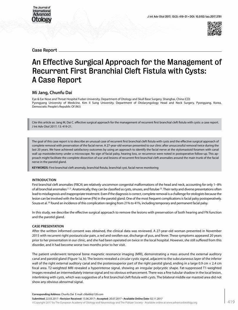
The patient underwent temporal bone magnetic resonance imaging (MRI), demonstrating a mass around the external auditory canal and parotid gland (Figure 1a, b). The lesions revealed a circular cystic signal, adjacent to the subcutaneous layer of the inferior wall of the right external auditory canal and the posterosuperior part of the right parotid gland, ending in a large 0.9 cm × 2.4 cm focal area. T2-weighted MRI revealed a hyperintense signal, showing an irregular polycystic shape. Fat-suppressed T1-weighted images revealed an intermediately intense signal and no obvious enhancement. There was a fine tubular shadow in the local lesion, interlinking with cysts, which was suggestive of a first branchial cleft fistula with cysts. The bilateral middle ear mastoid area did not show any obvious abnormal signal.
An Effective Surgical Approach for the Management of Recurrent First Branchial Cleft Fistula with Cysts: A Case Report
The goal of this case report is to describe an unusual case of recurrent first branchial cleft fistula with cysts and the effective surgical approach of complete removal with preservation of the facial nerve. A 27-year-old woman presented to our clinic after unsuccessful removal twice during the last 20 years. We have achieved satisfactory outcomes by using an approach to identify the facial nerve at the stylomastoid foramen with canal wall up mastoidectomy under a microscope. No sign of facial palsy, hearing loss, or recurrence were noted in postoperative follow-up. This ap-proach might facilitate the complete dissection of scar and lesions of recurrent first branchial cleft anomalies around the main trunk of the facial nerve in the parotid gland.
KEYWORDS: First branchial cleft anomaly, branchial fistula, branchial cyst, facial nerve monitoring
Mi Jang, Chunfu DaiEye & Ear Nose and Throat Hospital Fudan University, Department of Otology and Skull Base Surgery, Shanghai, China (CD)Pyongyang University of Medicine, Kim Il Sung University, Department of Otolaryngology Head and Neck Surgery, Pyongyang, Korea, Democratic People's Republic Of (MJ)
Corresponding Address: Chunfu Dai E-mail: [email protected]
Submitted: 22.03.2017 • Revision Received: 15.06.2017 • Accepted: 20.07.2017 • Available Online Date: 02.11.2017©Copyright 2017 by The European Academy of Otology and Neurotology and The Politzer Society - Available online at www.advancedotology.org
Cite this article as: Jang M, Dai C. effective surgical approach for the management of recurrent first branchial cleft fistula with cysts: a case report. J Int Adv Otol 2017; 13: 419-21.
419
Under general anesthesia, the patient was subjected to total mass excision. A “C” shaped postauricular incision was performed, extend-ing to below the lower jaw, and then the surface of the parotid gland was made into a skin flap on the anterosuperior margin of the ster-nocleidomastoid, which we separated from the caudate lobe of the parotid gland. This exposed the posterior belly of the digastric mus-cle. Furthermore, we exposed the stylomastoid foramen by removing the mastoid tip and a part of the tympanic bone with canal wall up mastoidectomy. The fistula with cysts was located in the anteroinfe-rior region of the external auditory canal. The FN trunk was identified at the stylomastoid foramen. The fistula with cysts was located in the superficial region of the main trunk of the FN in the parotid gland, further involving the deep lobe of the parotid gland, and the lesions
were tightly adhered to the main trunk of the FN in the parotid gland. Under the electrophysiological monitoring of the FN, the lesions were carefully separated from the nerve. The fistula and cysts were then separated from the parotid gland and completely removed with preservation of the FN and parotid gland. Histopathological exam-ination revealed Work’s type I first branchial cleft fistula with cysts.
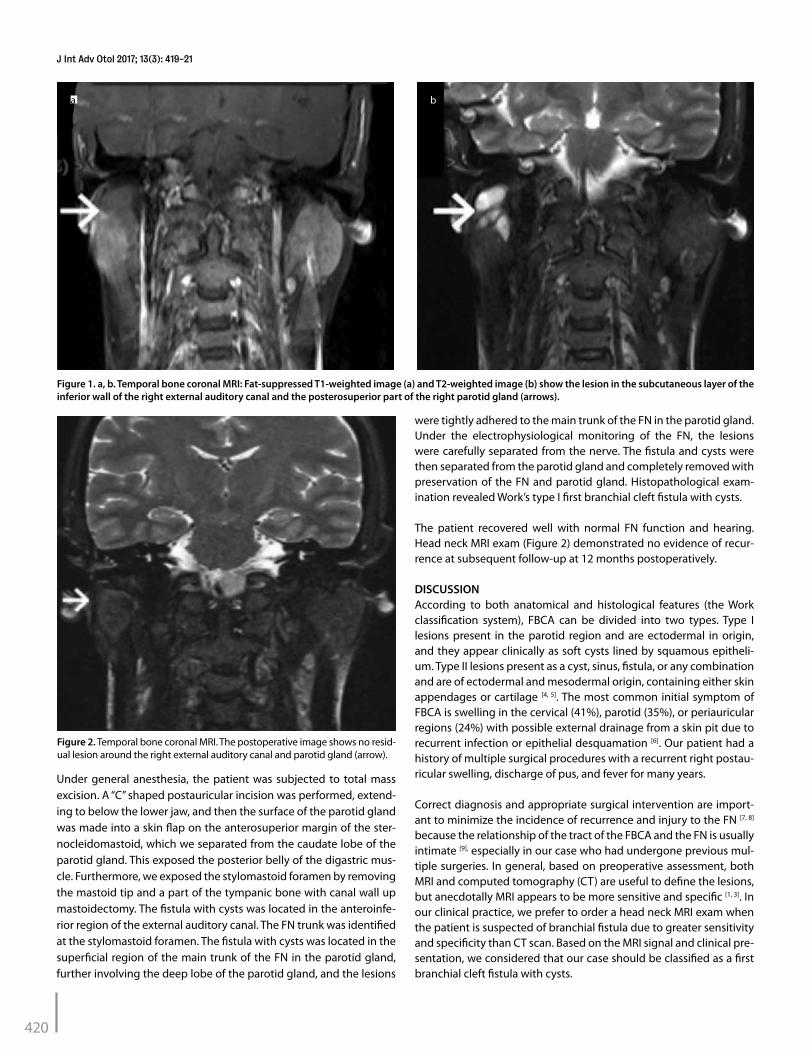
The patient recovered well with normal FN function and hearing. Head neck MRI exam (Figure 2) demonstrated no evidence of recur-rence at subsequent follow-up at 12 months postoperatively.
DISCUSSIONAccording to both anatomical and histological features (the Work classification system), FBCA can be divided into two types. Type I lesions present in the parotid region and are ectodermal in origin, and they appear clinically as soft cysts lined by squamous epitheli-um. Type II lesions present as a cyst, sinus, fistula, or any combination and are of ectodermal and mesodermal origin, containing either skin appendages or cartilage [4, 5]. The most common initial symptom of FBCA is swelling in the cervical (41%), parotid (35%), or periauricular regions (24%) with possible external drainage from a skin pit due to recurrent infection or epithelial desquamation [6]. Our patient had a history of multiple surgical procedures with a recurrent right postau-ricular swelling, discharge of pus, and fever for many years.
Correct diagnosis and appropriate surgical intervention are import-ant to minimize the incidence of recurrence and injury to the FN [7, 8] because the relationship of the tract of the FBCA and the FN is usually intimate [9], especially in our case who had undergone previous mul-tiple surgeries. In general, based on preoperative assessment, both MRI and computed tomography (CT) are useful to define the lesions, but anecdotally MRI appears to be more sensitive and specific [1, 3]. In our clinical practice, we prefer to order a head neck MRI exam when the patient is suspected of branchial fistula due to greater sensitivity and specificity than CT scan. Based on the MRI signal and clinical pre-sentation, we considered that our case should be classified as a first branchial cleft fistula with cysts.
a b
Figure 1. a, b. Temporal bone coronal MRI: Fat-suppressed T1-weighted image (a) and T2-weighted image (b) show the lesion in the subcutaneous layer of the inferior wall of the right external auditory canal and the posterosuperior part of the right parotid gland (arrows).
Figure 2. Temporal bone coronal MRI. The postoperative image shows no resid-ual lesion around the right external auditory canal and parotid gland (arrow).
420
J Int Adv Otol 2017; 13(3): 419-21
Traditionally, surgical removal of FBCA will involve either superfi-cial or total parotidectomy with injury to the FN [9]. Intraoperatively, it is necessary to completely expose the FN for a safe total excision because the lesions can be variably associated with the nerve [9, 10]. Several approaches to identify the FN have been described in the literature: (1) directly identifying the main trunk of the FN between the posterior belly of the digastric muscle and the anterior wall of external auditory canal, and (2) identifying the mandibular marginal branch of the FN at the lower jaw.
According to the literature, the tract of anomalies may pass super-ficial to the FN, being deep or between the branches of the FN. A review of 73 patients showed that the majority of anomalies passed superficial to the FN (63%) [1]. In our case, the tract of fistula and cysts was located on the surface of the FN, but they were tightly adhered to each other due to multiple surgeries and infections. We achieved a satisfactory outcome by using retroauricular C-shaped incision and identifying the FN at the stylomastoid foramen with canal wall up mastoidectomy under a microscope.
In our case, because there were many scars in the surgical field, we could not apply the conventional approaches to identify the FN (ap-proaches 1 and 2). Therefore, we were determined to identify the ver-tical segment of the FN at the stylomastoid foramen with canal wall up mastoidectomy. As well, we drilled partially through the tympanic bone to further expose the lesion and get more space to dissect the lesion from the FN, then we carefully separated the lesion and scar from the FN. Intraoperatively, the fistula with cysts was observed on the surface of the main trunk of the FN, but they were tightly ad-hered each other. Electrophysiological monitoring was carried out throughout the procedure to minimize the risk of injury to the FN.
Pathological examination of the surgical specimen showed the diag-nosis of first branchial cleft fistula with cysts (type I). This result was consistent with preoperative diagnosis. No sign of facial palsy or re-currence were noted in the postoperative follow-up.
In summary, patients with FBCA should be diagnosed and undergo surgical intervention as soon as possible because recurrent infec-tions and repeated drainage will result in scar and fibrotic adhesion. To our knowledge, this is the first report to apply the approach to identify the FN in the stylomastoid foramen with canal wall up mas-toidectomy to remove the recurrent FBCA, and this might facilitate the complete dissection of the scar and lesion around the main trunk of the FN in the parotid gland.
Informed Consent: Written informed consent was obtained from patient who participated in this case.
Peer-review: Externally peer-reviewed.
Author Contributions: Concept - C.D.; Design - C.D.; Supervision - C.D.; Re-sources - C.D.; Materials - C.D.; Data Collection and/or Processing - M.J.; Anal-ysis and/or Interpretation - M.J.; Literature Search - M.J.; Writing Manuscript - M.J.; Critical Review - C.D.
Conflict of interest: No conflict of interest was declared by the authors.
Financial Disclosure: The authors declared that this study has received no financial support.
REFERENCES1. Roche P, Saunders S, Naunheim M, Kovach A, Herrington H, Robson CD,
et al. First branchial cleft anomaly presenting with a complete duplica-tion of the external auditory canal-A photo anatomic review. Int J Pediatr Otorhinolaryngol Extra 2016; 12: 1-4. [CrossRef]
2. Maithani T, Pandey A, Dey D, Bhardwaj A, Singh VP. First Branchial Cleft Anomaly: Clinical Insight into its Relevance in Otolaryngology with Pe-diatric Considerations. Indian J Otolaryngol Head Neck Surg 2014; 66: S271-6. [CrossRef]
3. Chen Z, Wang Z, Dai C. An effective surgical technique for the excision of first branchial cleft fistula: make-inside-exposed method by tract inci-sion. Eur Arch Otorhinolaryngol 2010; 267: 267-71. [CrossRef]
4. Souza AR, Uppal HS, Zeitoun RD. Updating concepts of first branchial cleft defects: a literature review. Int J Pediatr Otorhinolaryngol 2002; 62: 103-9. [CrossRef]
5. Triglia JM, Nicollas R, Ducroz V, Koltai PJ, Garabedian EN. First branchial cleft anomalies: a study of 39 cases and a review of the literature. Arch Otolaryngol Head Neck Surg 1998; 124: 291-5. [CrossRef]
6. Ada m, Korkut N, Güvenç MG, Acioğlu E, Yilmaz S, Çevikbaş U. Unusual extension of the first branchial cleft anomaly. Eur Arch Otorhinolaryngol 2006; 263: 263-6. [CrossRef]
7. Kumar R, Sikka K, Sagar P, Kakkar A, Thakar A. First Branchial Cleft Anom-alies: Avoiding the Misdiagnosis. Indian J Otolaryngol Head Neck Surg 2013; 65: 260-3. [CrossRef]
8. Magdy EA, Ashram YA. First branchial cleft anomalies: presentation, vari-ability and safe surgical management. Eur Arch Otorhinolaryngol 2013; 270: 1917-25. [CrossRef]
9. Guo YX, Guo CB. Relation between a first branchial cleft anomaly and the facial nerve. Br J Oral Maxillofac Surg 2012; 50: 259-63. [CrossRef]
10. Miller PD, Corcoran M, Hobsley M. Surgical excision of first cleft branchial fistulae. Br J Surg 1984; 71: 696-7. [CrossRef]
421
Jang and Dai. Preservation of Facial Nerve