an atom probe study of the aging of iron- nickel- carbon martensite
TRANSCRIPT
An Atom Probe Study of the Aging of Iron-Nickel-Carbon Martensite
M.K. MILLER, P.A. BEAVEN, S.S. BRENNER, and G. D.W. SMITH
The atom probe field ion microscope has been used to study the redistribution of carbon in iron-nickel- carbon martensites aged in the temperature range 22 to 250 ~ The M~ temperature of the alloy used was - 5 0 ~ During all stages of aging a considerable amount of redistribution of carbon was detected in the form of carbon segregation to twin boundaries and other lattice defects and in the formation of small clusters and carbides. Small carbon-rich areas were detected at 40 ~ and disc-shaped carbide particles were found after 24 hours at 100 ~ Thin cementite lamellae were observed after one hour at 250 ~ It is evident that a close similarity exists between the sequence of reactions occurring during the tempering process and the sequence of precipitation reactions observed in age-hardening alloys. The main additional complications that arise in the case of martensites are due to the high densities of lattice defects that are present and to the strong interaction between carbon atoms and such defects.
I. INTRODUCTION
OVER the last few years much interest has been shown in the aging and tempering behavior of ferrous martensites. Many different techniques have been employed to gain understanding of the processes occurring. These techniques have included X-ray diffraction, dilatometry, electrical re- sistivity and hardness measurements, transmission electron microscopy, electron diffraction, and Mrssbauer spec- troscopy. These investigations have led to the proposal of a number of different views on the effects occurring during the aging process. ~-9
The atom probe provides a useful technique for direct study of the composition fluctuations that occur during the aging of martensites. This instrument combines the atomic resolution of the field ion microscope with single-atom chemical analysis sensitivity for all elements, including the light elements such as carbon. In this paper we report an extension of our earlier atom probe investigation of low- and high-carbon martensites 1~ to the processes occurring during the early stages of aging of iron-nickel-carbon martensites of subzero M~ temperature. In these alloys the experimental difficulties of observing the virgin material and the pre- liminary stages of aging are minimized.
II. EXPERIMENTAL
The materials selected for this investigation were Fe-Ni-C alloys with an M~ temperature of approximately - 5 0 ~ The two alloys used had slightly different nickel levels (23 or 29 pct Ni) and the same carbon content, 0.42 wt pct C (2.08 at. pct). The results obtained from the two alloys were essentially identical and will be discussed together.
M. K. MILLER and S. S. BRENNER, Associate Research Consultant, are both with U.S. Steel Research, 125 Jamison Lane, Monroeville, PA 15146. P. A. BEAVEN is with GKSS, 2054 Geesthacht, Federal Republic of Germany. G. D. W. SMITH is with the Department of Metallurgy and Science of Materials, University of Oxford, Oxford, United Kingdom.
This paper is based on a presentation made at the "Peter G. Winchell Symposium on Tempering of Steel" held at the Louisville Meeting of The Metallurgical Society of AIME, October 12-13, 1981, under the sponsorship of the TMS-AIME Ferrous Metallurgy and Heat Treat- ment Committees.
All specimens, in the form of 1-mm bar, were aus- tenitized at 950 ~ for 20 minutes and then were water- quenched. Those specimens that were to be aged were immediately transformed to martensite by further quenching into liquid nitrogen. This heat treatment produces a structure of predominantly twinned lenticular plates of martensite with a nonuniform twin distribution and only a small volume of retained austenite.
The specimen blanks were aged for various times at temperatures of 22, 40, 100, and 250 ~ The field ion specimens were then prepared by standard electropolishing techniques, l~ Particular care was taken to ensure that the temperature of the specimen did not exceed 20 ~ during the electropolishing procedure. All specimens were also given a short back polish to remove any deformation that might have been introduced during the final stage of electro- polishing. To examine the material in the unaged condition, some specimen blanks were electropolished immediately following the water quench, while the microstructure was still austenitic. These specimens then transformed to mar- tensite in situ after they were inserted into the atom probe, and cooled to liquid nitrogen temperature. Subsequent re- sults indicated that forming the martensite in the FIM needles rather than in bulk did not introduce any artifacts.
The atom probe facilities of both Oxford University and U.S. Steel Research, Monroeville, Pennsylvania, were used in these investigations. These instruments feature con- ventional time-of-flight mass spectrometers with fast digital time-measurement systems that are interfaced to dedicated minicomputers. A full description of this type of instrument can be found in Reference 11.
The atom probe was used in both the selected area and random area analysis modes. In the first mode the material from a selected region of the specimen is analyzed, while in the second the ions originating from a randomly selected cylinder of material parallel to the specimen axis are col- lected. In this second case, information concerning particles and clusters smaller than the effective probe aperture can be deduced by using standard statistical techniques.
Atom probe analyses were conducted under the following experimental conditions: (1)a system base pressure of <1.3 x 10 _6 Pascal, (2)a pressure during analysis of <4 • 10 -6 Pascal, (3) a specimen temperature of approxi- mately - 195 ~ (4) a pulse fraction of 15 pct, (5) a pulse
METALLURGICAL TRANSACTIONS A VOLUME 14A, JUNE 1983-- 1021
repetition rate of 50 Hz, (6) an ion collection rate of between 10 -2 and 1 ion per pulse, and (7) an effective probe aperture of between 1 and 3 nm.
The field ion micrographs were taken with neon image gas at a pressure of 133 Pascal.
100 ~ revealed the presence of extremely small carbon-rich regions or particles. The shape of these regions was deduced to be of the form of small rods 2 to 4 nm in length but only about 0.2 nm in diameter. These carbon-rich regions could have been carbon decorated dislocations.
III. RESULTS
A. Field Ion Micrographs
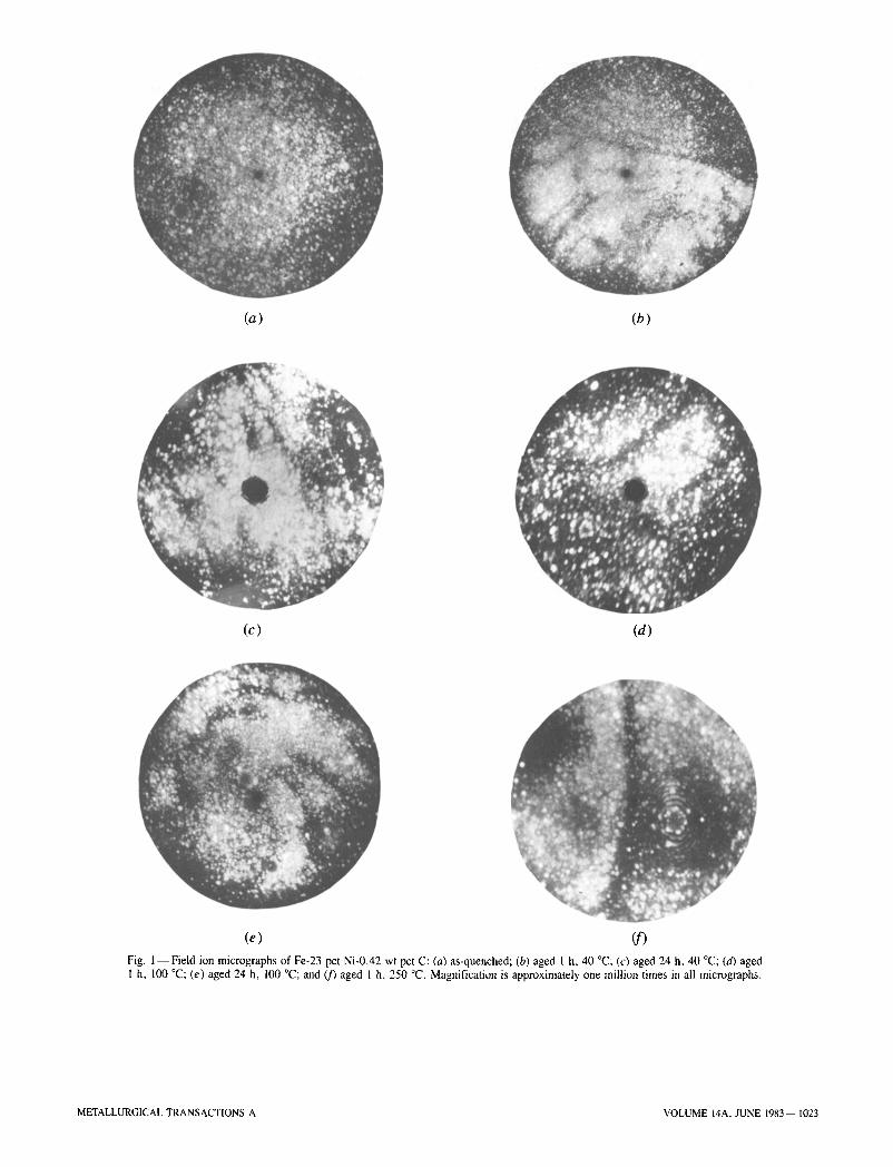
A series of field ion micrographs for the different aged specimens is shown in Figure 1. The field ion micrograph of the unaged microstructure, Figure l(a), revealed no dis- tinguishable features, whereas all the micrographs of the aged material showed darkly imaging regions. Series of darkly imaging parallel lines characteristic of carbon-rich twin interfaces 1~ are clearly demonstrated in the micrograph of the specimen aged one hour at 100 ~ Figure l(d). Some thin disk or plate shaped dark-imaging regions, 3 nm thick by - 4 0 nm diameter, were observed in the material aged 24 hours at 100 ~ Figure l(e). In the material aged one hour at 250 ~ the darkly imaging regions were in the form of lamellae (less than 5 nm thick) characteristic of cementite 1~ (Figure l(f)). The magnification of the field ion micrographs is approximately one million; however, the magnification over the specimen surface is not uniform and is influenced by the presence of the darkly imaging features. It is therefore difficult to measure the size of the small darkly imaging regions with any great precision, although the uncertainty in the measurement is likely to be less than a factor of two.
B. Atom Probe Microanalysis
The results of the selected area microanalyses are sum- marized in Table I. This table consists of two portions, the first part containing the results obtained from the brightly imaging matrix, and the second part the results obtained from the darkly imaging (or carbon-rich) regions, including twin interfaces and small carbide particles. The maximum, minimum, and the average values are given for a series of measurements taken from different areas of the matrix. In the analyses of the darkly imaging regions only the maxi- mum and minimum values are given. Each value was based on a sample size of between 1000 and 6000 ions for the matrix and between 200 and 2000 ions for the darkly imag- ing regions. Between 10 and 25 measurements were taken for each type of region. The carbon peak at mass-to-charge ratio (m/n) of 12 was taken to be exclusively C + rather than C~ +. This will lead to a small underestimate of the true carbon level.
The results of the matrix analyses revealed a progressive decrease in carbon concentration with both aging time and temperature. In the specimens aged for one hour at 250 ~ the carbon level in the matrix had fallen to that typically found in the ferrite phase. The carbon detected in the matrix was not uniformly or randomly distributed, as the incidence of small groups of carbon was found to be higher than that expected for a random distribution.
Detailed statistical analysis 12 of the ion-by-ion sequence of evaporated ions obtained from material aged one hour at
IV. DISCUSSION
These results provide direct evidence concerning some of the processes occurring during the low-temperature aging of iron-nickel-carbon martensites. The atom probe has clearly demonstrated that there is a considerable amount of redis- tribution of carbon even during the earliest stages of aging.
The first effect, which is observed even at the lowest aging temperature of 22 ~ is the depletion of the carbon in the matrix to form some small high-carbon regions. This is accompanied by the development of dark image contrast in the field ion micrographs. It appears likely that several pro- cesses contribute to this stage of aging. In addition to carbon atom clustering in the martensite matrix, some of the carbon-rich regions undoubtedly form preferentially on lat- tice defects such as twin boundaries and dislocations. The exact local concentration of these high-carbon regions is difficult to measure because of their extremely small size, the unavoidable collection of an unknown number of matrix atoms in the sample, and also the small sample size with its large statistical scatter. However, it is perhaps significant that the maximum carbon concentrations recorded from these high-carbon regions are generally of the order of 10 at. pct.
Although the atom probe has the highest spatial resolution in analysis, it is difficult to determine precisely the original lattice positions of the field evaporated ions. It can usually be assumed that adjacent atoms in the one-dimensional data chain would be at least within a few atom diameters of each other in the lattice, but they need not necessarily have been nearest neighbors. Similarly, nearest-neighbors atoms may not appear as adjacent ions in the data chain. It is therefore not practical to obtain direct information about the atomic configuration of the carbon-rich regions from the atom probe data. In the absence of any evidence to the contrary, it seems best to regard these regions as being coherent clusters (or zones) of carbon atoms, formed prior to the precipitation of the transition carbide phase. There is an obvious analogy with the sequence of processes occurring during the early stages of aging of many precipitation hardening alloys.
After 24 hours at 100 ~ the transition carbide was clearly resolved in the field ion micrographs in the form of thin plates. The very high local carbon contents observed in specimens aged for 24 hours at 40 ~ also seem to indicate the appearance of this phase. The compositions measured were also subject to the problems outlined above because of their small size. No conclusion can be drawn at this time as to which of the proposed transition carbide structures is actually present. The maximum level of carbon found in the regions was only -25 at. pct, which would seem to make Fe2C unlikely if stoichiometry is to be maintained.
The final process observed in this study was the formation of thin lamellae of cementite after aging for one hour at 250 ~ The compositions measured were within the experi- mental scatter for Fe3C. This is further supported by the very
1022--VOLUME 14A, JUNE 1983 METALLURGICAL TRANSACTIONS A
(a) (b)
(c) (d)
(e) (f)
Fig. l--Field ion micrographs of Fe-23 pct Ni-0.42 wt pct C: (a) as-quenched; (b) aged 1 h, 40 ~ (c) aged 24 h, 40 ~ (d) aged 1 h, 100 ~ (e) aged 24 h, 100 ~ and 09 aged 1 h, 250 ~ Magnification is approximately one million times in all micrographs.
METALLURGICAL TRANSACTIONS A VOLUME 14A, JUNE 1983-- 1023
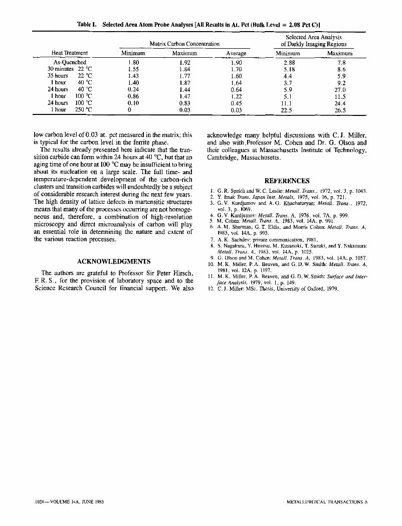
Table I. Selected Area Atom Probe Analyses [All Results in At. Pct (Bulk Level = 2.08 Pct C)]
Matrix Carbon Concentration Selected Area Analysis
of Darkly Imaging Regions
Heat Treatment Minimum Maximum Average Minimum Maximum
As-Quenched 30 minutes 22 ~ 35 hours 22 ~
I hour 40 ~ 24 hours 40 ~
I hour 100 ~ 24 hours 100 ~
1 hour 250 ~
1.80 1.92 1.90 2.88 7.8 t.55 1.84 1.70 5.18 8.6 1.43 1.77 1.60 4.4 5.9 1.40 1.87 1.64 3.7 9.2 0.24 1.44 0.64 5.9 27.0 0.86 1.47 1.22 5.1 11.5 0.10 0.83 0.45 11.1 24.4 0 0.03 0.03 22.5 26.5
low carbon level of 0.03 at. pct measured in the matrix; this is typical for the carbon level in the ferrite phase.
The results already presented here indicate that the tran- sition carbide can form within 24 hours at 40 ~ but that an aging time of one hour at 100 ~ may be insufficient to bring about its nucleation on a large scale. The full time- and temperature-dependent development of the carbon-rich clusters and transition carbides will undoubtedly be a subject of considerable research interest during the next few years. The high density of lattice defects in martensitic structures means that many of the processes occurring are not homoge- neous and, therefore, a combination of high-resolution microscopy and direct microanalysis of carbon will play an essential role in determining the nature and extent of the various reaction processes.
A C K N O W L E D G M E N T S
The authors are grateful to Professor Sir Peter Hirsch, E R. S., for the provision of laboratory space and to the Science Research Council for financial support. We also
acknowledge many helpful discussions with C.J. Miller, and also with_Professor M. Cohen and Dr. G. Olson and their colleagues at Massachusetts Institute of Technology, Cambridge, Massachusetts.
R E F E R E N C E S
1. G.R. Speich and W. C. Leslie: MetalL Trans., 1972, voi. 3, p. 1043. 2. Y. Imai: Trans. Japan Inst. Metals, 1975, vol. 16, p. 721. 3. G.V. Kurdjumov and A.G. Khachaturyan: Metall. Trans., 1972,
vol. 3, p. 1069. 4. G.V. Kurdjumov: Metall. Trans. A, 1976, vol. 7A, p. 999. 5. M. Cohen: MetaU. Trans. A, 1983, vol. 14A, p. 991. 6. A.M. Sherman, G.T. Eldis, and Morris Cohen: Metall. Trans. A,
1983, vol. 14A, p. 995. 7. A.K. Sachdev: private communication, 1981. 8. S. Nagakura, Y. Hirotsu, M. Kusunoki, T. Suzuki, and Y. Nakamura:
MetalL Trans. A, 1983, vol. 14A, p. 1025. 9. G. Olson and M. Cohen: Metall. Trans. A, 1983, vol. 14A, p. 1057.
10. M.K. Miller, P.A. Beaven, and G. D.W. Smith: Metall. Trans. A, t981, vol. 12A, p. 1197.
11. M.K. Miller, P.A. Beaven, and G. D. W. Smith: Surface and Inter- face Analysis, 1979, vol. 1, p. 149.
12. C.J. Miller: MSc. Thesis, University of Oxford, 1979.
1024--VOLUME 14A, JUNE 1983 METALLURGICAL TRANSACTIONS A