an atlas of atopic eczema - zanjan university of medical …file.zums.ac.ir/ebook/007-an atlas of...
TRANSCRIPT


An Atlas of ATOPIC ECZEMA

THE ENCYCLOPEDIA OF VISUAL MEDICINE SERIES
An Atlas of ATOPIC ECZEMA
Lionel Fry, BSC, MD, FRCPProfessor Emeritus of Dermatology
Imperial CollegeLondon, UK
Foreword by
Charles N.Ellis, MDProfessor and Associate ChairDepartment of Dermatology
University of Michigan, USA
The Parthenon Publishing GroupInternational Publishers in Medicine, Science & Technology
A CRC PRESS COMPANY
BOCA RATON LONDON NEW YORK WASHINGTON, D.C.

Published in the USA byThe Parthenon Publishing Group Inc.345 Park Avenue South, 10th Floor
New YorkNY 10010
USA
Published in the UK and Europe byThe Parthenon Publishing Group
23–25 Blades CourtDeodar Road
London SW15 2NUUK
Copyright © 2004 The Parthenon Publishing Group
Library of Congress Cataloging-in-Publication DataFry, Lionel.
An atlas of atopic eczema/Lionel Fry.p. ; cm. —(The encyclopedia of visual medicine series)
Includes bibliographical references and index.ISBN 1-84214-236-4 (hard cover : alk paper)
1. Atopic dermatitis-Atlases. I.Title: Atopic eczema. II. Title. III. Series[DNLM: 1. Dermatitis, Atopic-Atlases. WR 17 F946ab 2003]
RC593.A8F79 2003616.5'1–dc22
British Library Cataloguing in Publication DataFry, Lionel, 1933–
An atlas of atopic eczema.—[The encyclopedia of visualmedicine series)
1. Atopic dermatitisI. Title
616.5'21
ISBN 0-203-49050-9 Master e-book ISBN
ISBN 0-203-59659-5 (Adobe eReader Format)ISBN 1-84214-236-4 (Print Edition)
First published in 2004
No part of this book may be reproduced in any form without permission from the publishersexcept for the quotation of brief passages for the purposes of review
This edition published in the Taylor & Francis e-Library, 2005.
Composition by The Parthenon Publishing Group
“To purchase your own copy of this or any of Taylor & Francis or Routledge’s collection ofthousands of eBooks please go to www.eBookstore.tandf.co.uk.”

Contents
Foreword vii
Preface viii
1 Definitions 1
Dermatitis versus eczema 1
Atopic 1
Infantile eczema 2
2 Epidemiology 3
Place of domicile 3
Incidence of atopic eczema related to age 3
Increasing incidence of atopic eczema 3
3 Natural history 4
4 Genetics 6
Family studies 6
Twin studies 6
Human lymphocyte antigen system 6
Genome screens 6
Maternal effect 7
Summary 7
5 Pathogenesis 8
Histology 8
Findings in the blood 8
Immunopathogenesis 10
Inflammatory cells 11
Immunoglobulin E 12
Summary 12
6 Etiology 13
Genetics: possible candidate genes 13
Abnormal skin barrier 14
Allergens 15
Aeroallergens 15
Autoantigens 16

Bacteria 16
Fungi 18
Environmental pollution 18
Hygiene hypothesis 18
Pharmacological and vascular abnormalities 20
Emotional factors 20
Neuropeptides 21
Summary 21
7 Clinical features 22
Age of onset 22
Sex 22
Clinical features of eczema per se 22
Age-related features 22
Non-age-related features 24
Atopic eczema at specific sites 26
Other eczematous skin disorders in atopic individuals 29
Non-eczematous cutaneous features of atopy 30
Other disorders associated with atopic eczema 31
Infections 33
8 Differential diagnosis 81
Other eczematous conditions 81
Syndromes with eczematous eruptions 82
Non-eczema disorders 83
9 Management 91
Emollients 91
Bathing 91
Clothing 91
Climate 91
Diet 92
Breast-feeding 92
Delayed introduction of solid foods 92
Avoidance of food antigens in pregnancy 92
Probiotics 92
Aeroallergens 93
Animals 93
Psychotherapeutic measures 93
Investigations 93
v

Topical drugs 93
Antistaphylococcal treatment 98
Antiviral treatment 98
Antifungal treatment 98
Ultraviolet light 98
Systemic treatment 99
Biological agents 100
The future 100
References 101
Index 103
vi

Foreword
A picture shows me at a glance what it takes dozens of pages of a book to expound.
Ivan Sergeyevich Turgenev, 1862In this slim but focused book, Professor Lionel Fry has captured all we need to know about eczema. Akin to the quote above,proverbially one picture is worth more than 10 000 words. This Atlas is replete with diagrams, helpful reminders inillustrative form, and plenty of pictures of the many expressions of eczema. The teachings of the renowned Professor Fry aredelightfully presented in both word and illustration.
This book is timely because of the new information that has developed recently, particularly with regard to thepathogenesis of eczema (Chapters 5 and 6) and the treatments that are newly available or are coming shortly (Chapter 9). Justsit with this book for an hour or two, and one will feel on top of the challenge of the eczemas. I invite you to take advantageof Professor Fry’s great knowledge in this area and his clarity of writing. The eczemas, which were so mysterious before,have the veil of confusion lifted in this Atlas.
The excellent pictures and the accompanying text will be of great value to all who diagnose and treat skin diseases,including dermatologists, general practitioners, pediatricians, internists, gynecologists, and, for that matter, any physician ornurse with family or friends who seek advice about skin disorders. The eczemas, which are so common, are increasing inincidence, and therefore all of us must be cognizant of their various presentations and treatment.
Charles N.Ellis, MDProfessor and Associate Chair
Department of DermatologyUniversity of Michigan
Ann Arbor, 2004

Preface
Atopic eczema is still one of the major problems in the field of dermatology. The rising incidence has been attributed to anumber of factors including increasing urbanization, pollution and the ‘hygiene hypothesis’. The latter attributes the increasein atopy as being due to a decline of infections in early life, related to the use of antibiotics and immunizations and to smallerfamilies.
Over the last few years, advances have been made in the genetics of atopy in general; newer treatments have beenintroduced for topical use and new concepts suggested in the etiology. Thus, a new text on atopic eczema seems timely.
Atopic eczema presents to general practitioners, pediatricians, dermatologists and allergists and, therefore, I hope that thisbook will be of value to physicians in these fields, particularly those in training. The ample illustrations are intended to help inthe diagnosis and management of this common complaint.
I am grateful to Mr Bruce Mathalone for the use of illustrations on eye conditions associated with atopy and to Dr NickFrancis for the histology illustrations. I am also pleased to acknowledge the contributions of Zachary Fry and the endeavoursof my secretary, Mrs Helen Morris Crawshaw.
Lionel Fry

1Definitions
DERMATITIS VERSUS ECZEMA
The words ‘dermatitis’ and ‘eczema’ are used synonymously in the dermatological literature. Dermatitis derives from theGreek word derma, meaning skin. However, the word ‘dermis’ is now applied to the structure of the skin below the basementmembrane, whilst that above is called the epidermis. Thus, it could be argued that dermatitis, strictly speaking, refers to thedermis and not to the whole skin. Eczema is derived from the Greek word ekzein, meaning ‘to boil over’, and is a purelydescriptive term. Which term is more accurate is debatable. In this book, the term eczema is preferred, but the choice isarbitrary.
ATOPIC
The word ‘atopic’ is derived from the Greek word topos, meaning place, and was applied in the sense ‘out of place’. The termwas coined to denote an abnormal hypersensitive response to an environmental allergen and the word ‘atopen’ is nowsynonymous with allergen. Two allergists, Coco and Cooke, first used the term ‘atopic’ in 19231, and the term ‘atopicdermatitis’ originated in 1933. However, the condition that is now described by this term is considerably older and can berecognized in early Chinese and Roman writings.
At present, there is no universal agreement about the definition of atopic eczema. Some believe that immunoglobulin E(IgE)-specific antibodies to environmental antigens are part of the disease and the term atopic eczema should not be used ifIgE allergen-specific antibodies are not present2. If the clinical features suggest atopic eczema but no IgE antibodies arepresent, the term ‘atopiform’ eczema has been proposed2. Others believe that atopic eczema defines a clinical state withcharacteristic features. The clinical criteria postulated by Hanifin and Rajka3 are still the most appropriate. They give fourmajor features, of which at least three must be present. These are:
(1) Pruritus;(2) Lichenification;(3) Chronic relapsing course;(4) Personal and/or family history of asthma, allergic rhinitis/conjunctivitis and a family history of atopic eczema.
In addition, three minor features should be present. These include dryness of the skin, ichthyosis vulgaris, keratosis pilaris,immediate skin test (type 1) reactivity, elevated IgE level, tendency to cutaneous infection with Staphylococcus aureus andherpes simplex, increased incidence of hand and foot and nipple eczema, cheilitis, recurrent conjunctivitis, Dennie-Morganinfraorbital fold, anterior subcapsular cataracts, orbital darkening, facial pallor/erythema, pityriasis alba, anterior neck folds,itch when sweating, intolerance to wool, perifollicular accentuation, food intolerance, course often affected by emotional/environmental factors, and white dermographism.
More simple criteria were proposed by Williams and colleagues4. These are: a history of an itchy skin rash in the last 12months plus three or more of the following:
(1) History of flexural involvement;(2) History of asthma and/or hay fever;(3) History of generalized dry skin;(4) Onset of rash before the age of 2 years (not applicable if the patient is under 4 years);(5) Visible flexural involvement.
These criteria are not as precise as those of Hanifin and Rajka and may not be applicable to young children and infants.

It should be remembered that 100% uniformity does not exist in clinical medicine and there are always exceptions to theclinical criteria as proposed both by Hanifin and Rajka, and by Williams and colleagues.
A causative role for IgE antibodies in atopic eczema is still disputed and other pathogenetic mechanisms have beenimplicated. Until these have been fully elucidated, it would be prudent to maintain the clinical criteria for diagnosing atopiceczema.
IgE antibodies are found in 70–80% of patients with atopic eczema. The terms ‘intrinsic’ and ‘extrinsic’ have been used toimply that evidence exists for an external allergen based on IgE antibodies. The term ‘extrinsic atopic eczema’ is used whenantibodies are present and ‘intrinsic’ when the antibodies are absent. How useful this subdivision is at the clinical level isdebatable at present but, with greater understanding of the pathogenesis of atopic eczema in the future, this subdivision maythen be relevant.
Atopic eczema is strongly associated with asthma, rhinitis and conjunctivitis, due to external allergens. However, as witheczema, the demonstration that the disease is due to an external allergen is not always proven. Asthma is also divided into intrinsicand extrinsic subdivisions. Collectively, eczema, asthma and hay fever are sometimes referred to as the atopic syndrome.However, it is important to stress that the three manifestations of the atopic syndrome may occur singularly and notnecessarily all together (Figure 1).
INFANTILE ECZEMA
Atopic eczema is sometimes still referred to as infantile or flexural eczema because of its early age of onset and thecharacteristic sites of the disease. However, the term ‘atopic eczema’ is to be preferred because other types of eczema mayoccur in infancy and may affect flexures. In addition, there are prognostic and genetic implications associated with atopiceczema that are different from the other patterns of eczema seen in childhood.
Figure 1 Atopic disease may occur singularly or in combination. A, asthma; E, eczema; H, hay fever
2 AN ATLAS OF ATOPIC ECZEMA

2Epidemiology
PLACE OF DOMICILE
Epidemiological studies on atopic eczema have been carried out, particularly in European countries. However, the accuracy ofthese studies is dependent on the definitions of atopic eczema that were used, and the fact that many are based onquestionnaires rather than on an interview and examination by a dermatologist. There are also variations in the questionsposed and the age of the groups studied. Thus, some studies have only taken the incidence of eczema at a particular point intime and others have included past history.
Using point prevalence (i.e. presence of eczema at that particular point), the incidence of the disease in European countriesranges from 5.3 to 26%, with a mean of 13% in the groups aged 3–12 years5. In one of the largest studies conducted world-wide, questionnaires were completed on 458 623 individuals, aged 13–14 years. The incidence of atopic eczema ranged frommore than 15% in some northern European countries to less than 5% in China, central Asia and eastern European countries6.There was a suggestion that the line of latitude had an effect in that the closer to the equator the country was, the lower theincidence of the disease. However, it is interesting to note that, when individuals migrate from areas of low incidence to thoseof a higher incidence, the incidence of atopic eczema rises in the immigrant population and is often higher than in the localpopulation. Thus, environmental factors are of considerable importance in determining the incidence of atopic eczema. Inaddition, an increased incidence of the disease appears to be associated with a move from a rural/agricultural society to anindustrial one. It has been shown, in a study in Ibadan in Nigeria, that the incidence of atopic eczema increased with theprogressive industrialization of the area and an associated change in lifestyle. Thus, change in lifestyle may be moreimportant than the change in the latitude of the country of residence.
INCIDENCE OF ATOPIC ECZEMA RELATED TO AGE
It is generally agreed that the incidence of atopic eczema decreases as children become older and the incidence is considerablyless in adult life. As already mentioned, the incidence of atopic eczema in northern European countries is relatively high inchildren, particularly in urban areas. Several studies give an incidence of 10–15% and most of these studies are for the agegroup 3–11 years7. The studies do not distinguish between the incidences at age 3 and age 11. A few studies have examinedthe incidences in the 0–4–year age group, where they ranged from approximately 10 to 15%. Studies in teenagers have shownconsiderably lower incidences in the same countries and these range from approximately 2 to 3%. The frequency in adults iseven lower; over the age of 25, the incidence has been reported to be less than 0.2%8. Thus, to inform parents that childrenusually grow out of their eczema is correct.
INCREASING INCIDENCE OF ATOPIC ECZEMA
It has been thought that the incidence of atopic eczema has increased over the last 50 years. However, the figures supportingthis observation may not be reliable because of the criteria and techniques used to arrive at these results. The criteria fordiagnosing atopic eczema and the concept of the disease have changed over time. Many of the figures are based on parents’recall of the rash in their children and no objective examinations were carried out in most of the studies. If these limitationsare accepted, then the figures do show an increasing incidence of atopic eczema in children. In children up to 7 years, theincidence for those born before 1960 ranged from 1.4 to 3.1%, with a mean of 2.2%. For those born between 1960 and 1970,the frequency ranged from 3.8 to 8.8%, with a mean of 6.7%, and, for those born after 1970, the range was 8.9–20.4%, with amean of 12.0%9. This increased incidence of atopic eczema is supported by twin studies from Denmark. The cumulativeincidence rate for twins born between 1960 and 1964 was 3% and for those born between 1975 and 1979 it rose to 12%10.
This increase in the incidence of atopic eczema is similar to the reported increase in asthma seen over the last 40 years.

3Natural history
Although there have been several studies on the natural history of atopic eczema, there are a number of variable factors inthese studies that may influence their reliability. First, the definition of the disease was not consistent. Second, the length oftime of follow-up was variable. Third, some of the studies were hospital-based while others were community-based. It islikely that community-based studies will show milder disease than a hospital-based population and the severity of the diseasemay influence the natural history.
It is generally agreed that atopic eczema usually commences in childhood. In nearly 50% of patients, the age of onset isbefore 6 months, with 60% before 1 year and 70% before 5 years11. It is also accepted that atopic eczema will go intoremission in early life. In a general practice setting over a 40-year study period, it was found that 50% of cases had cleared bythe age of 5 years and 90% by the age of 10 (J. Fry, personal communication). In a British cohort study, 65% had cleared byage 11 and 74% by age 1612. In a hospital-based study over a 5–20-year period, the clearance rates ranged from 84 to 92%13.Thus, there is general agreement that atopic eczema is primarily a disease of childhood, with the majority clearing before orduring adolescence. However, in a smaller proportion, atopic eczema may present for the first time in adult life, and in someindividuals, whose eczema began in infancy, the disease will persist into adult life. The factors that determine these variationsare yet to be elucidated but; similar to the basic causes of the disease, they are likely to be partly constitutional and partlyenvironmental. It is also well known that, in patients whose atopic eczema has cleared, there is always a risk that it may recurbecause of inherent factors. Occupations and hobbies in later life, involving exposure to chemicals that may damage thestratum corneum, may induce eczematous reactions, either for the first time or as a recurrence in those with a past history ofatopic eczema.
Various factors have been implicated in a poor prognosis. These include severe disease at onset; extensor or inverse patternof eczema on the knees and elbows in children; family history of atopic eczema, particularly if both parents are affected;concomitant asthma; and subjects who were not breast-fed. However, these are also studies disputing these factors as poorprognostic indicators.
The risk of developing asthma and hay fever in children with atopic eczema has been reported in a number of studies14. Themain criticisms of these studies are that the period of study was too short or that the subjects were chosen from a hospital-based population, thus implying a disease more severe than in the true spectrum of atopic eczema severity. The periods of

study have mostly been 5–15 years and the incidence of asthma has ranged from 10 to 53% and that for hay fever from 28 to78%. In one large study15, in which patients were traced after 22–44 years, the incidence of asthma was 37% for patients whowere inpatients and 22% in those who were outpatients; for hay fever, the figures were 66% for inpatients and 33% foroutpatients. The two main findings of these studies are that the risk of developing hay fever is greater than that of developingasthma (Figure 2) and that the more severe the eczema, the greater the likelihood of developing asthma and hay fever.
Figure 2 Approximate percentages of atopic eczema individuals who subsequently develop asthma and/or hay fever
5
4Genetics
FAMILY STUDIES
It has been known for nearly 100 years that ‘allergy’ runs in families. Original studies in 1916 by Cooke and Van der Veerreported that, if one parent had an allergy, then 50% of the offspring would be similarly affected; if both parents had an aallergy, then there was a 75% chance of allergy in their children. Subsequent studies in the mid-twentieth century concludedthat genetic factors were more important than environmental ones in atopic disorders. All types of Mendelian inheritance wereproposed, but, by the 1960s, it was generally agreed that atopic disorders were polygenic (several genes involved) and that thediseases were multifactorial (in other words, environmental as well as genetic factors were implicated).
If one considers asthma, hay fever (allergic rhinitis) and eczema as constituting the ‘atopic diathesis’, then; in patients withatopic eczema, those with a positive family history of atopy have been reported as ranging from 43 to 83%, with a mean ofapproximately 60–65%. It is well known that, although the individual patient may present with eczema, the affected relativesmay have asthma, allergic rhinitis or eczema. However, there is usually a higher incidence of the presenting disease in the familyof the proband, i.e. if the presenting disease is asthma, then there is a higher incidence, in the family, of asthma compared toeczema and hay fever. The same is true for eczema and hay fever. This has led to the concept of ‘end-organ sensitivity’ andthe genetic determinants for variants of atopic diathesis do appear to run in families.
TWIN STUDIES
To determine the role of genetic factors in multifactorial disorders, twin studies have proved helpful in determining theimportance of genetic influence. The most recent figures for twins with atopic eczema show a concordance rate of 75% formonozygotic and 20% for dizygotic twins16. These figures imply a strong genetic component for atopic eczema and,interestingly, are very similar to those for psoriasis, another common inflammatory dermatosis.
HUMAN LYMPHOCYTE ANTIGEN SYSTEM
Immunological factors would appear to play an important role in atopic eczema and therefore, the human lymphocyte antigen(HLA) system, which is associated with immune responses, would be expected to show certain HLA antigens associated withatopic eczema. However, despite a number of studies, no specific HLA antigen or haplotype has been found to be associatedwith atopic eczema. This lack of association is supported by the genome scans that have been performed in atopic eczema inwhich no loci have been reported on chromosome 6p, which is where the HLA region is found. Interestingly, in asthma,linkage to chromosome 6p has been reported and also a significant increase in the HLA haplotype B7, SC31, DR2 has beenfound. This implies that the immune mechanisms in asthma and atopic eczema may not have common pathways.
GENOME SCREENS
To date, two genome screens have been performed in atopic eczema in childhood and one in adults17. In the first, performedin children in a German and Scandinavian population, linkage was found to chromosome 3q21. In the second, in Britishfamilies, linkage was found to 1q21, 17q25, and 20p. The latter study included some individuals with asthma as well as atopiceczema. Interestingly, in the German and Scandinavian study, linkage to the total serum IgE level was also found with the3q21 locus but the British study found linkage for the IgE level to chromosomes 5q31 and 16qtel.
In the one adult study, on Swedish individuals, linkage was reported for chromosome 3p24–22. If the IgE level wasincluded as a trait, then linkage to 18q21 was found. When the severity scores of the eczema were included, the suggestedlinkage to chromosomes 3q14, 13q14, 15q14–15, and 17q21 was found. Thus, the only common linkage for adults and

children was 17q21 and possibly on chromosome 3q. The implication of these findings is that different pathogenicmechanisms may be operative in childhood and in adult atopic eczema.
When these results on genome screens are compared to those for asthma, differences are found. The agreed loci for asthmaare 5q35, 6p, 11q13, 12q, 13q13, 14q and 16q. Interestingly, there are very few common loci for the two disorders; the onlypossible areas so far are 13q and 16q, but the regions reported are not the same. The implication of these findings is thatdifferent pathogenic mechanisms are operative in the two diseases, as suggested by the HLA findings.
Recently, it has been pointed out that similar loci have been reported (1q21, 17q15 and 20p) for psoriasis and atopiceczema, which are both inflammatory skin diseases18. Thus, these associations may be related to the control of inflammationin the skin and not to the basic causes of the diseases.
IgE has been implicated in the pathogenesis of atopic diseases because there are increased levels in the serum inapproximately 75% of patients. Atopic asthma has been linked to a locus on chromosome 11q12–13, where a gene controllingthe subunit of the high-affinity receptor for IgE (FceRI) is localized. Linkage to 11q12–13 has not been reported in genomescreens in atopic eczema, but there is a report of an association of atopic eczema and the � unit of FceR1 in thatpolymorphisms within this unit are strongly associated with atopic eczema19. This paradox has not been resolved as yet.
MATERNAL EFFECT
It has been noted in a number of studies that mothers are more likely to transmit atopic disorders, including eczema, thanfathers20. There are a number of possible reasons for this observation. First, there is a close association between the maternaland fetal immune systems, and interactions between the two might influence the development of the fetal immune system, andits response to external stimuli. The second possibility is that of genome imprinting in which the allele from one parent isdifferentially expressed. However, it has been found that the maternal linkage to atopic disease is not confined to a singlegenetic mechanism at one single gene locus21.
SUMMARY
Thus, there is strong evidence for a genetic component in the etiology of atopic eczema. However, considering that immunemechanisms are involved in the pathogenesis, it should be noted that, as yet, there is no association with a particular HLAhaplotype. In addition, despite the close association between asthma and atopic eczema, there is to date no common genotypefor the two disorders.
7

5Pathogenesis
HISTOLOGY
The histological features of atopic eczema vary, depending on whether the disease is acute, subacute or chronicIn the acute phase, spongiosis progressing to blister formation is the characteristic feature. Spongiosis is defined as an
increase in the amount of tissue fluid between the keratinocytes in the epidermis, which is visible as a widening of theintercellular space (Figure 3). Some intracellular edema also occurs. Lymphocytes migrate through the epidermis (exocytosis)and there is a perivascular infiltrate of lymphocytes and macrophages into the dermis but with no increase in number of mastcells or basophils. However, the mast cells are in various stages of degranulation, indicating activation. Occasionally,eosinophils are present.
In the subacute phase, the epidermis begins to thicken and the spongiosis lessens.In the chronic phase, the epidermis is thick (Figures 4 and 5) and similar in appearance to the epidermis in psoriasis, but the
‘folding’ of the interphase between the dermis and epidermis, present in psoriasis, is not seen in atopic eczema. Thethickening is caused by hyperplasia of the keratinocytes. The dermis is infiltrated by mast cells, lymphocytes and eosinophils(Figure 4). As in psoriasis, the blood vessels in the dermis become more prominent. Langerhans and dendritic cells areincreased in number in both the epidermis and dermis. The chronic phase (Figure 5) is often associated with persistentscratching and this has been linked to changes in the cutaneous nerves, which show demyelination, vacuolation and fibrosis.
FINDINGS IN THE BLOOD
So-called ‘reagenic’ antibodies in the blood of patients with atopic disease were first demonstrated by Prausnitz and Kustnerin 192122. These have now been shown to be IgE antibodies to foreign protein, which may be antigenic in atopics. Approximately75–80% of patients with atopic eczema have raised IgE levels and blood eosinophilia also occurs. Peripheral blood
Figure 3 Acute eczema with spongiosis in the epidermis and a dermal infiltrate of inflammatory cells, mainly lymphocytes andmacrophages

mononuclear cells have a decreased ability to produce interferon-gamma (IFN-� ), the levels of which are inversely correlatedwith those of IgE. However, there is an increase in allergen-specific lymphocytes producing the cytokines inter-leukin IL-4,IL-5, and IL-13. These are the cytokines that are capable of inducing IgE production (Figure 6). These cytokines also induceexpression of vascular adhesion molecules, for example, VCAM-1, which are involved in eosinophilic infiltrations of the skin.These findings reflect the inverse relationship between T-helper cells types 1 and 2 (Th1 and Th2) and their respectivecytokines. Th1 cells produce IFN-� , which down-regulates the production of IL-4 and IL-5 by Th2 cells. Conversely, the IL-4and Il-5 cytokines down-regulate production of IFN-� by Th1 cells (Figure 6). In atopic eczema, there is a shift in favor ofTh2 cells and their cytokines. In addition, IL-5 activates the production of eosino-phils, a characteristic feature of atopy.
Figure 4 Chronic phase of eczema with thickening of the epidermis and an intense infiltrate in the dermis of mast cells, lymphocytes,eosinophils and dendritic cells
Figure 5 Lichen simplex: chronic atopic eczema associated with continual scratching. There is more accentuation of the rete pegs of theepidermis
PATHOGENESIS 9

IMMUNOPATHOGENESIS
The eczematous response in the skin in atopic eczema is a T-lymphocyte-mediated disorder (type IV hypersensitivity) and isantigen-dependent. In theory, the antigen could be inhaled, ingested or enter through the skin. The inhaled or ingested antigenmay enter the circulation and be carried to the lymph nodes, where it is picked up by the follicular dendritic cells, antigen-presenting cells and B-cells. The follicular dendritic cells present the antigen to T-cells, which can then induce the B-cells toproduce IgE. This process will continue for the period in which allergen-specific T- and B-cells remain in the lymph nodes.
The method by which the ingested or inhaled allergen enters the skin is subject to debate. However, once the allergen is inthe skin, it can be picked up by IgE receptors on Langerhans cells, which are antigen-presenting cells.
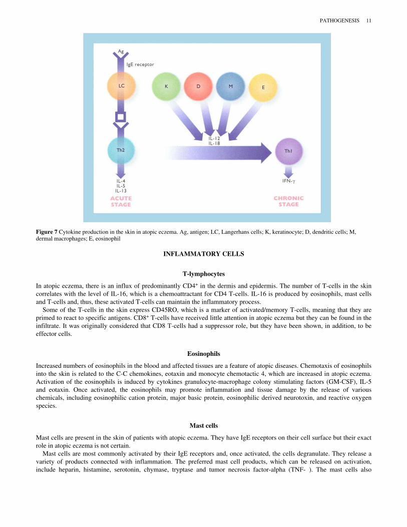
One of the characteristic features of atopic eczema is the IgE receptor on the Langerhans cells in the skin. The expressionof this receptor may possibly be under genetic control. The antigen that arrives in the skin and binds to the IgE receptor willthen be processed and presented to the T-lymphocytes in the skin. In the acute stage of atopic eczema, cytokine production isof the Th2 profile. The Th2 cytokines are IL-4, IL-5 and IL-13 (Figure 7). However, in the chronic phase of atopic eczema,there is a switch from a Th2 to a Th1 cytokine profile by the lymphocytes (Figure 7). Thus, the hypothesis that atopic eczemais a Th2 response to antigen, as opposed to a Th1 response, is not strictly correct because both responses can happen. In theso-called atopic patch test, when eczema is induced by application of house-dust allergen, a biphasic response occurs. In thefirst instance, there is a Th2 response with mainly IL-4 production, but, after 24 h, there is a Th1 response with IFN-�production. Thus, atopic individuals are able to mount both Th1 and Th2 immune responses and the concept that theresponses are mutually exclusive is only correct for a given point in time. Thoughts on the factors that govern the switch froma Th2 to a Th1 response in atopic eczema are speculative. It has been suggested that both cytokines IL-12 and IL-18 caninduce a shift from a Th2 to a Th1 response23. IL-12 is produced by constitutive cells of the skin, keratinocytes, dendritic cellsand dermal macrophages and by the infiltrating cells, i.e. eosinophils (Figure 7). However, the mechanism that controls theinhi-bition/production of these cytokines in the acute and chronic phases of the atopic eczema state requires furtherelucidation. It does appear that the initial response to antigen in atopy is Th2-mediated but, subsequently, in the more chronicphase, it is Th1-mediated. The argument that what is required to cure atopic eczema is to shift the immune response from aTh2 to a Th1 response is thus only partly correct, since, in the chronic phase, it is a Th1 response. However, the initiation islikely to be via a Th2 mechanism, so a permanent switch to a Th1 response may be helpful in clearing the eczema morepermanently.
Figure 6 In atopic eczema, there is an increase in blood of the Th2 cells producing IL-4, IL-5, IL-13 and an associated increase in IgE.Production of IFN-� by Th1 cells is decreased. E, eosinophil
10 AN ATLAS OF ATOPIC ECZEMA
INFLAMMATORY CELLS
T-lymphocytes
In atopic eczema, there is an influx of predominantly CD4+ in the dermis and epidermis. The number of T-cells in the skincorrelates with the level of IL-16, which is a chemoattractant for CD4 T-cells. IL-16 is produced by eosinophils, mast cellsand T-cells and, thus, these activated T-cells can maintain the inflammatory process.
Some of the T-cells in the skin express CD45RO, which is a marker of activated/memory T-cells, meaning that they areprimed to react to specific antigens. CD8+ T-cells have received little attention in atopic eczema but they can be found in theinfiltrate. It was originally considered that CD8 T-cells had a suppressor role, but they have been shown, in addition, to beeffector cells.
Eosinophils
Increased numbers of eosinophils in the blood and affected tissues are a feature of atopic diseases. Chemotaxis of eosinophilsinto the skin is related to the C-C chemokines, eotaxin and monocyte chemotactic 4, which are increased in atopic eczema.Activation of the eosinophils is induced by cytokines granulocyte-macrophage colony stimulating factors (GM-CSF), IL-5and eotaxin. Once activated, the eosinophils may promote inflammation and tissue damage by the release of variouschemicals, including eosinophilic cation protein, major basic protein, eosinophilic derived neurotoxin, and reactive oxygenspecies.
Mast cells
Mast cells are present in the skin of patients with atopic eczema. They have IgE receptors on their cell surface but their exactrole in atopic eczema is not certain.
Mast cells are most commonly activated by their IgE receptors and, once activated, the cells degranulate. They release avariety of products connected with inflammation. The preferred mast cell products, which can be released on activation,include heparin, histamine, serotonin, chymase, tryptase and tumor necrosis factor-alpha (TNF-� ). The mast cells also
Figure 7 Cytokine production in the skin in atopic eczema. Ag, antigen; LC, Langerhans cells; K, keratinocyte; D, dendritic cells; M,dermal macrophages; E, eosinophil
PATHOGENESIS 11

synthesize, when activated, leukotrienes, prostaglandins and the cytokines, IL-4, IL-5, IL-6, IL-13 and GM-CSF. Thesebiologically active substances can promote inflammation and lead to an influx of further inflammatory cells into the skin.
In asthma and rhinitis, mast cell degranulation occurs at an early stage and is probably involved in the acute inflammatoryresponses. In eczema, however, the mast cells are deep in the skin (dermis) in contrast to being in the epithelium of the noseand lung in asthma and rhinitis where they are thus easily accessible to allergens. It is thought that mast cell degranulation isnot part of the pathological process in atopic eczema, but that cytokine release from the mast cells, leading to an influx ofother inflammatory cells, contributes further to the inflammatory reaction.
Antigen-presenting cells
As with other immune cells, the number of antigen-presenting cells (APC) is increased in atopic eczema. These cells includeLangerhans and inflammatory dendritic cells in the epidermis and dendritic cells in the dermis. The Langerhans cellsexclusively express IgE receptors, a characteristic feature of atopic disease, and present antigens to T-cells with a Th2 cytokineprofile. The epidermal inflammatory dendritic cells present antigen to T-cells with a Th1 cytokine profile. The Langerhanscells positive for IgE receptors can migrate to lymph nodes and stimulate naïve T-cells, there by expanding the pool of Th2 cells.
IMMUNOGLOBULIN E
IgE was first described in 1967 and the radioallergosorbent test (RAST) for specific IgE antibodies was developed in 1971.IgE is elevated in the serum in 75–80% of patients with atopic eczema, and it has been claimed that there is a correlation betweenthe IgE levels and the severity of the eczema. However, it has to be remembered that IgE levels are often raised in subjectsnot suffering from eczema and there are patients with atopic eczema who have normal levels of IgE. Elevated IgE levels arealso found in other skin diseases, for example, scabies, cutaneous T-cell lymphoma and helminthic infestations. In a recentstudy24 on the levels of IgE antibodies in atopic disease, it was shown that individuals with atopic eczema have 20 timeshigher levels of antibodies to house dust mites than those with atopic asthma, even though the house dust mite is considered tobe an aeroallergen for asthma. Thus, the significance of this high level of IgE in eczema needs to be explained.
IgE is produced by B-cells and is thought to be under the control of IL-4. Atopic eczema is characterized by Th2 cellswhich produce IL-4. This, in turn, may stimulate IgE production. It is possible, therefore, that the elevated IgE level issecondary to this process and may not have an initiating role in this eczematous process. Thus, a role for IgE and itsantibodies has yet to be defined.
SUMMARY
Atopic eczema is an inflammatory skin condition that is antigen-driven and T-cell-mediated. The two typical features are thepresence of IgE receptors on Langerhans cells and a Th2 cytokine profile in the acute stages. However, in the chronic phase,there is a Th1 cytokine profile with IFN-� production that down-regulates IgE production and Th2 lymphocytes.
It should be remembered that not all patients with atopic eczema have raised serum IgE levels. Whether the pathogeneticmechanisms in these individuals are the same as those in patients with raised serum IgE levels is yet to be elucidated.
12 AN ATLAS OF ATOPIC ECZEMA

6Etiology
GENETICS: POSSIBLE CANDIDATE GENES
As discussed in Chapter 4, the role of genetic factors is important in atopic eczema and a number of chromosome loci havebeen implicated.
Chromosome 3q21
This region encodes for the co-stimulating molecules CD80 and CD86; a mutation in these genes could lead to an alteration inT-cell activation.
Chromosome 5q31
The locus 5q31 contains the gene SPINK5, which has recently been identified as the gene for Netherton’s syndrome.Netherton’s syndrome is an autosomal recessive disorder, which includes the atopic manifestations of eczema, hay fever,eosinophilia and high serum IgE levels. SPINK5 was so named as it encodes for an enzyme inhibitor, ‘serine proteinaseinhibitor, Kazal type 5’. The serine proteinase inhibitor has been termed LEKTI, as it is found in the lymphoid tissue of thethymus and epithelial tissue, including skin. (The KT is kazal type and I is inhibitor.)
Recently, six coding polymorphisms in SPINK5 have been identified; a GLu 420 � Lys variant shows significantassociation with atopy and atopic eczema25. This abnormality in the proteinase inhibitor may influence allergic disease.Proteinase activated receptors are found in keratinocytes and can serve as targets for mast cell proteinases. Proteinases areinvolved in the maturation of T- and B-cells and the expression of LEKTI in the thymus may suggest a role for theseprocesses in allergic diseases. In addition, many allergens are serine proteinases and this proteinase activity may encouragetheir allergenicity in the presence of defective inhibition.
SPINK5 also lies in the region where the cluster of cytokine genes, particularly Th2 cytokines, IL-4, IL-5, and IL-13, isfound. These Th2 cytokines are implicated in atopic eczema.
Chromosome 11q13
This region was originally linked to atopic asthma and eczema. It contains a gene that is associated with polymorphism of the� -chain of the high-affinity receptor for IgE (FceRI). This receptor acts as an allergic trigger on mast cells and basophils,leading to intracellular degranulation of these cells and the associated release of inflammatory mediators, which have a role inasthma and hay fever. In addition, IgE receptors are present on antigen-presenting cells and have been implicated in thepathogenesis of atopic eczema. However, it has recently been reported that the FceRI on antigen-presenting cells lacks theclassical � -chain, although the receptor has full function26. Thus; although the gene controlling the � -subunit of FceRI may beimportant in atopic asthma and rhinitis, it may not have a role in eczema.
Chromosome 13q14
Recently, it has been shown that serum IgE levels are related to alleles in the gene PHF11 in asthma. It is thought that thisgene regulates IgE production by B-cells in atopic disease27.

Chromosome 16q12
This locus has been linked to polymorphism in the � -subunit of the IL-4 receptor. IL-4 is considered to have a central role inatopic eczema.
Chromosome 17q11
Atopic eczema is associated with up-regulation of C-C chemokine expression. RANTES (regulated-on-activation normal T-cell, expressed and secreted) is a member of the C-C chemokine family and is considered to play a central role in allergicinflammation. A single polymorphism has been found in the promoter region for RANTES and association studies haveshown this polymorphism to contribute to the development of atopic eczema but not asthma28.
Although other loci have been linked to atopic eczema, possible candidate genes for these loci have yet to be postulated andidentified.
ABNORMAL SKIN BARRIER
Xerosis (dry skin) is a recognized feature of atopic eczema. There are two possible explanations for the dry skin. Either it is aprimary defect and part of the atopic syndrome or it is secondary to low-grade eczema, with minimal signs of inflammation.
The dry skin is associated with a defective barrier function, as manifested by increased transepidermal water loss and a lowwater content of the stratum corneum. This defect in the barrier function of the stratum corneum may lead to the passage ofchemicals (e.g. primary irritants) and microorganisms through the stratum corneum and the initiation of an eczematousresponse. It has been shown that there are alterations in the lipid content of non-eczematous skin in atopic individualscompared to those with non-atopic skin. This alteration in fat content may lead to an alteration in barrier function.
Figure 8 summarizes the possible genetic defects in atopic eczema.
Figure 8 Possible genetic abnormalities in atopic eczema
14 AN ATLAS OF ATOPIC ECZEMA

ALLERGENS
Food allergens
It is assumed that atopic eczema is an antigen-dependent disorder. However, the determination of which antigen(s) is thecause of the eczema has so far not been achieved. Parents often implicate food as a cause and it is not infrequent for childrento have been given elimination diets by the time they are seen by a dermatologist.
There are reports of some children with atopic eczema who benefit from avoidance of certain foods, and who relapse on re-introduction of these foods. These studies have been double-blind and placebo-controlled. But, in the majority of patients withatopic eczema, it has not been possible to demonstrate any benefit from elimination of particular foods29.
Elemental diets, consisting of amino acids, carbohydrates, fats, minerals and vitamins, have been tried in patients withatopic eczema. In a controlled study of an elemental diet versus a normal (liquidized diet) in adults with atopic eczema, nodifference was shown between the two diets in the clinical features, IgE levels, eosinophil counts and skin biopsies30. In astudy in children with atopic eczema, an elemental diet was shown to be of some benefit. However, the study was carried outon hospital patients and was not controlled; therefore, some or all of the improvement may have been a placebo effect31.
Breast-feeding
It has been suggested that breast-feeding is beneficial in preventing atopic eczema. This is thought to be due to the child’slack of exposure to external dietary allergens until later in infancy. However, it is well known that atopic eczema may developin purely breast-fed children and it has been demonstrated that external dietary antigens can be detected in human breast milk.Thus, breast-fed infants are exposed to dietary antigens and breast-feeding does not totally protect infants from exposure tothese antigens. In addition, specific IgE to cow’s milk and eggs has been found in infants exclusively breast-fed32.
Although dietary antigens are present in human breast milk, there may be other benefits from breast-feeding. Secretory IgAis present in breast milk and this may bind to dietary antigens and prevent their absorption. In addition, it has been proposedthat other factors present in human breast milk facilitate maturation of the gut and its responses to external antigens33.
Tests for food allergies
The two types of tests available are ‘skin-prick tests’ and tests to detect specific IgE antibodies in the serum. The validity of askin-prick test depends on the presence of antigen-specific IgE on mast cells and elicits a type I hypersensitivity response.Atopic eczema is considered to be a T-cell-mediated, type IV immune response. Thus, the test does not use the pathogenicpathway for inducing eczema and the results are therefore not likely to be relevant. This is found to be so in practice. Inroutine skin-prick tests for 20 food antigens, usually three to six foods are found to test positive in atopic eczema subjectsand, in some instances, the results yield even higher numbers. Yet, when the patient is challenged by the foods giving positiveskin-prick tests, only one or two of the foods cause exacerbation of the eczema34.
Radioallergosorbent tests (RAST) have also been shown to correlate poorly with elimination diets and subsequentchallenge for a particular food.
At present, there are no reliable tests for identifying possible foods that may be responsible for inducing or maintaining theeczema. It is possible that, in the majority of individuals with atopic eczema, it is not one or two antigens which cause theeczema but a multitude of foreign antigens, and the fault is in the way the immune system handles these substances. There is areport of finding specific T-cell clones to peanuts in the skin of an infant with atopic eczema. This has been taken to implythat this provides confirmatory evidence that food substances can induce T-cell-specific clones in the skin, thus inducingatopic eczema35. However, it has been suggested that the most likely way that infants develop peanut sensitivity is through theapplication of peanut oil in topical and bath emollients. There are likely to be many T-cell clones to various food substancesin blood and skin. Before an etiological role for T-cell clones in the skin to a particular food is accepted, it will have to beshown that removal of this food improves the eczema and that re-introduction induces the eczema.
AEROALLERGENS
Aeroallergens implicated in atopic eczema include house dust mites, molds, pollens and animal dander. The evidence comespartly from clinical data and partly from the results of tests.
ETIOLOGY 15

Clinical data
House-dust avoidance studies have been shown to benefit some individuals with atopic eczema, but the conduct of thesestudies under controlled circumstances has proved to be difficult.
There are a small group of patients who have a flare-up of their eczema on areas of skin exposed to the air in the spring andearly summer months. This flare-up is thought to be due to pollens released at this time of year. However, this pattern ofeczema in the early summer months is uncommon and most patients with atopic eczema benefit from an improvement in theirdisease condition in summer.
It has also been claimed that aeroallergens that may cause asthma and rhinitis may also cause a flare-up of eczema in atopicindividuals when the allergen is administered intranasally or by bronchial inhalation35. Yet this response does not occur in allpatients with atopic eczema and it is said only to occur in subjects with IgE antibodies to the particular allergen used forchallenge.
Patch tests
The so-called atopy patch test has been used to try to predict whether patients are sensitive to a particular aeroallergen. If thepatch test is positive, yielding an eczematous response, this implies that the aeroallergen can induce atopic eczema in theindividual, although, in this instance, the aeroallergen is acting as a contact allergen. However, the conditions under which theallergen is tested are not the same as those in which the allergen may come into contact with the skin in an atopic patient. Inthe early studies on house dust mites, positive reactions were only obtained after Sell-o-tape stripping of the stratum corneumand not with a normal intact skin barrier. The results, therefore, imply that house dust mites cannot induce eczema in patientswith an intact stratum corneum.
The most recent study on patch tests to aeroallergens in atopic individuals did not find a correlation between the clinicalfeatures and patch test results, and concluded that, at present, there was no indication for patch tests in the management ofatopic eczema36.
AUTOANTIGENS
As in most chronic inflammatory disorders, the possible role of autoantigens in maintaining the disease, even if not initiatingit, has been proposed for atopic eczema. Studies have shown that human dander can trigger hypersensitivity responses, andthis suggests an IgE-mediated response. IgE antibodies have been reported against human proteins. Although attempts havebeen made to clone and identify the autoantigens in IgE complexes, so far no specific antigen has been identified.
BACTERIA
Bacteria on the surface of the skin have been implicated in the etiology of atopic eczema for over 100 years. Gram-positivecocci were the organisms considered to be responsible for the eczema and it was said that, if Staphylococci were brought intocontact with slightly irritated skin, eczema could be induced after 24 h. Streptococci were also implicated in eczema andstreptococcal vaccines were tried as a treatment. More recently, discussion on the role of Staphylococci in atopic eczema hasbeen revived. Staphylococcus aureus is found in 100% of acute exudative lesions of atopic eczema, in approximately 90% ofchronic lesions, and in 65% of clinically normal unaffected skin of individuals with atopic eczema37. S.aureus is found on theskin in less than 5% of normal non-atopic individuals. However, the important question is whether the staphylococcalorganisms play a role in the pathogenesis of the eczema or whether the atopic skin simply provides a good environment,which encourages colonization by the organism. The presence of the organisms may be secondary to the abnormality of theatopic skin, both involved and uninvolved.
There are a number of mechanisms by which S. aureus organisms may induce the eczematous process. It has been shownthat, in a significant proportion of individuals with atopic eczema, there is a high incidence of specific IgE antibodies to S.aureus.
IgE antibodies are more common in patients with severe and chronic eczematous lesions38. It is thought that the continualscratching in chronic atopic eczema allows greater colonization by S. aureus because of the damaged barrier caused byscratching. It has been suggested that the staphylo-coccal IgE antibodies may be pathogenic and may play a role in aninflammatory reaction. Conversely, the antibodies may be of a secondary nature and may not be important to the pathogenesisof eczema.
S.aureus may also induce eczema by the same pathway as other antigens, e.g. food and aeroallergens, by binding to IgEreceptors on antigen-presenting cells in the skin and activating specific T-cells (Figure 9).
16 AN ATLAS OF ATOPIC ECZEMA

More recently, superantigens secreted by S. aureus organisms have been implicated in the pathogenesis. Superantigensinduce activation of T-cells and macrophages. In addition, they up-regulate CLA expression on lymphocytes, thus enablingthem to target the skin. They also induce a T-cell receptor V� family expansion. This results in the production of a largenumber of activated T-cells in the skin in a short space of time, and these can induce an inflammatory reaction, as seen ineczema (Figure 9). Superantigens have also been shown to increase the synthesis of allergen-specific IgE antibodies, andthese in turn increase the severity of the eczema. In support of this hypothesis, it has been shown that the application ofstaphylococcal enterotoxin B to the uninvolved skin of atopic subjects induces eczema39 (Figure 10).
The colonization of the skin by S.aureus may depend on the increased adherence of the organism in atopic skin. Adherenceof the bacteria to epithelial cells is the initial step in the pathogenesis of many infections. It has been shown that S.aureus hasa much greater degree of adherence to corneocytes affected by atopic eczema than those from normal individuals and thosewith other inflammatory skin conditions such as psoriasis37. The adhesins that enable S.aureus to bind to the skin are thoughtto be extracellular matrix components, including fibronectin. Interestingly, in binding studies of S.aureus to the skin, it wasshown that, in skin lesions with a Th2 inflammatory response compared to a Th1 response, bacterial binding was greater inthe Th2-mediated inflammation. This may be due to fibronectin induction by IL-4 (Figure 11).The innate immune system has recently been described and implicated in atopic eczema. This system in the skin is triggeredby a number of receptors for microbial components. The best-defined receptors are Toll-like receptors (TLRs). Thesereceptors on epithelial cells, when activated, can induce the release of antimicrobial peptides, which have been termed human� -defensins (HBDs) and cathelicidins. These peptides can attract T-cells, immature dendritic cells and neutrophils, and areintended to provide a protective response against invading bacteria. It has recently been shown in patients with atopic eczemathat there are low levels of two antimicrobial peptides, the human � defensin, HBD-2 and the cathelicidin, LL-3740. Boththese peptides are concerned with controlling the growth of S.aureus on the skin. It has also been shown that the cytokinesIL-4 and IL-13, which are Th2 cytokines and are increased in eczema, can inhibit the induction of antimicrobial peptidesHBD-2 and LL-37. Thus, it has been implied that the low levels of these two antimicrobial peptides may be a secondary eventin atopic eczema and not a primary one. This further implies that S.aureus colonization of skin in patients with atopic eczemais not a primary initiating cause of atopic eczema. An additional observation that argues against a primary role for S.aureus inatopic eczema is the fact that topical steroids and tacrolimus will heal the eczematous process, and the staphylococcalcolonization, present before treatment, will disappear with treatment. Thus, colonization with S. aureus would appear to bedependent on the existing eczematous process.
Figure 9 S. aureus may activate T-cell in atopic eczema, either by a superantigen or specific antigen effect
ETIOLOGY 17

FUNGI
A number of fungi that are normal inhabitants of the skin, Malassezia furfur and Candida albicans, may play some part in thepathogenesis of atopic eczema. If these organisms penetrate the skin, for example when the skin barrier is weakened or whenthe skin barrier is broken due to scratching, they can induce Th1- or Th2-dependent immune responses. Most normalindividuals will show a cell-mediated response to C.albicans. However, in atopics, there is often a type 1 hypersensitivityresponse with redness, itching, and an urticarial reaction. Although, M.furfur and C.albicans are not likely to have a primaryrole in atopic eczema, they may possibly have an aggravating effect.
ENVIRONMENTAL POLLUTION
There has been an increase in the incidence of atopic eczema over the last 50 years. Although there is a strong geneticcomponent to the disease, environmental factors also appear to play a role. Air pollutants have been suspected of playing apossible role in this increased incidence. They would act as non-specific irritants rather than allergens, but they also appear tohave an immunomodulating effect, i.e. they have been shown to enhance IgE formation in animals.
It should also be mentioned that some forms of air pollution have decreased in the last 50 years. The burning of coal, whichwas associated with smog and high sulfur content in the atmosphere, has decreased considerably. The pollution that hasincreased is caused by the oxides of nitrogen and sulfur and the photochemical oxidants such as ozone. Most of the pollutantsare from the petrochemicals used in the automobile industry. There appears to be some correlation between increasing levelsof sulfur dioxide and oxides of nitrogen and atopic eczema41. This is true for hay fever and, to a lesser extent, for asthma.Smoking during pregnancy has also been found to be a risk factor.
HYGIENE HYPOTHESIS
One of the theories suggested to account for the increase in atopic disorders over the last few decades has been the so-called‘hygiene hypothesis’. The hypothesis is that children born in the latter half of the twentieth century have less bacterial andviral infection in early life compared to children born in the first half of the century. This has resulted in the immune responsebeing Th2-skewed. Infections by bacteria and viruses induce a Th1 response and these infections in early life will convert theinfant to a Th1 response when exposed to allergens. Lack of exposure to bacteria and viruses and control of infections with
Figure 10 Effects of superantigens, which may induce atopic eczema. CLA, cutaneous lymphocyte antigen; V� , part of the T-cell receptor
18 AN ATLAS OF ATOPIC ECZEMA

antibiotics will delay the conversion of the immune system to a Th1 response and the Th2 response is maintained for a longertime. Therefore, when the subject is exposed to potential allergens, a Th2 or ‘allergic’ response results.
Support for this hygiene hypothesis comes from a study in Sweden comparing the children of families with ananthroposophic lifestyle and a control group42. Anthroposophy originates from a Greek word and means ‘wisdom about man’.Anthroposophy, as a way of life, was founded in the twentieth century by Rudolph Steiner and has been applied to art,architecture, agriculture and medicine. In anthroposophic medicine, the use of antibiotics, antipyretics, and vaccinations isrestricted. Vaccinations are usually only given against polio and tetanus and then later in life than usually recommended. Inaddition, children follow-ing the anthroposophic way of life consume local foods produced according to biodynamicprinciples. Vegetables preserved by spontaneous fermentation are a common dietary element and probably contain livelactobacilli, which are probiotics. In the study, 295 children following the anthroposophic lifestyle were compared to 380controls. There was a significant lower incidence of atopy in children following the anthroposophic lifestyle and thisincidence strongly correlated with the strictness of the lifestyle features42.
Figure 11 Antimicrobial peptides in the innate immune system are down-regulated in atopic eczema. This may be due to IL-4 produced byactivated T-cells. In addition, IL-4 induces fibronectin, which enhances adherence of S.aureus, an essential step in colonization of the skinin the pathogenesis of infections. S, S.aureus; K, keratinocyte; T, T-cell; F, fibronectin; P, innate immune system peptide
ETIOLOGY 19

PHARMACOLOGICAL AND VASCULAR ABNORMALITIES
There are a number of abnormalities that relate to the control of small blood vessels in atopic eczema. It has long been knownthat patients report pallor of the skin, and hypersensitivity to cold with vasoconstriction and low finger temperature. Whitedermographism has been attributed to an increase in catecholamines in the nerve endings in atopic skin. Yet it has beenreported that there is a decrease of adrenergic nerve fibers in atopic skin lesions. Thus, the vasoconstriction may be caused byincreased sensitivity to catecholamines or perhaps there is another pharmacological mediator causing the vasoconstriction.There is a delayed blanch to acetylcholine. The method by which these abnormalities relate to the development of atopiceczema is not yet known. It is possible they represent an abnormal neuropeptide profile in atopics.
EMOTIONAL FACTORS
There is no doubt that emotional stress can make atopic disease worse. This effect could be mediated by the immune system,hormones or neuropeptides. It is a relatively unexplored field.
Figure 12 Summary of etiological factors in atopic eczema
Figure 13 Possible triggers for atopic eczema
20 AN ATLAS OF ATOPIC ECZEMA

NEUROPEPTIDES
Peptides are released by nerve endings in tissue. This is a relatively under-researched area, but neuro-peptides are biologicallyactive substances and may well influence immune responses. Thickening of nerve fibers has been described in the skin, andrelated to continual scratching. However, whether this is the cause is not proven. Neuropeptides may also be involved in theintense pruritus associated with atopic eczema.
SUMMARY
The definite etiological factor is genetic. Allergens, or in other words, foreign proteins, either dietary or aeroallergens,probably play a role and the other possible contributory factors include micro-organisms, pollution and the ‘hygienehypothesis’ (Figure 12).
Possible triggers (Figure 13) and helpful measures (Figure 14) are discussed in more detail in the chapter on‘Management’.
Figure 14 Practical measures based on etiological factors that may be of benefit in management
ETIOLOGY 21

7Clinical features
At the present time, there is no single objective feature or investigation with which to define atopic eczema. There are certainclinical features characteristic of the disease but they are not always present. In addition, as in many disorders with amultifactorial etiology, there appears to be a spectrum of disease. At one end of the spectrum are patients with manyrecognized features associated with atopic eczema, but, at the other end, are patients with mild disease and only one or two ofthese features. Over the last two decades, there have been several meetings to attempt to define the clinical state of atopiceczema, but the best outcomes produced are lists of features, some of which have been termed major and other minor3,43. Theproblem is further complicated by the fact that the clinical features can vary in different age groups.
In this chapter, the clinical aspects will be divided in presentation according to age, localized sites of atopic skin disease,associated skin disorders with a background of atopy, non-cutaneous disorders, and stigmata associated with atopic eczema.
AGE OF ONSET
Atopic eczema may present at any age but is essentially a childhood disorder. The most common age of onset is between 2and 6 months. In the majority, the eczema will present before the age of 2 years.
SEX
Males and females appear to be equally affected.
CLINICAL FEATURES OF ECZEMA PER SE
Before considering the specific features of atopic eczema, it should be appreciated that there are other clinical patterns ofeczema. Many features are common to all these patterns of eczema, depending on the severity of the eczematous process. Inmild forms, the skin is red and scaly (Figure 15); in the severe forms (subacute eczema), the skin may be ‘edematous’, due totissue fluid, and, on the surface, there is crusting. Crusts represent the breaking down of the epidermis, with serum seepingfrom the surface but coagulating to form a yellow dried surface (Figures 16 and 17). In the most severe forms (acute eczema),the stratum corneum is lost and a weeping moist surface is evident (Figure 18).
In the chronic forms of eczema, there may be a thickened stratum corneum (hyperkeratosis), which often splits, formingfissures (Figure 19), or the whole of the skin is thickened. This latter feature is typical of atopic eczema and is often referredto as lichenification (Figure 20). This word is derived from the term ‘lichen’, a plant which grows on rocks, causing athickened surface. Lichenification is said to occur in atopics due to continual scratching. However, there is probably anunderlying constitutional factor in atopics that allows this thickening to develop, since there are many other skin disordersassociated with pruritus and continual scratching in which lichenification does not occur.
AGE-RELATED FEATURES
Infants
The eczema usually first appears on the face and is seen as red patches, which may or may not be scaly (Figures 21–23). If thecondition is acute, weeping and crusting will be seen (Figures 24 and 25). Any part of the face may be affected.
Involvement may also be seen on the trunk (Figures 26 and 27) and limbs (Figures 28–34). In this age group, it presents asred scaly patches. Occasionally, the patches may coalesce and the eruption is confluent (Figures 28 and 29). If theinflammation is severe, then the patches may form crusts and weep. The crusts are characteristically golden, since they areformed of a mixture of serum and keratin. Occasionally, the patches are red and raised and urticarial in appearance, and this

can subsequently progress to scaling, crusting or weeping, depending on the severity of the inflammation. Although atopiceczema classically involves the flexures of the knees and elbows, in infants the involvement is frequently on the extensorsurfaces of the limbs when the disease first presents (Figures 28, 30–32).
The scalp may be involved and, in its mildest form, will present as scaling (dandruff). In the more acute stage, crusted areasmay be present.
Itching is a feature of atopic eczema and infants will scratch involuntarily and thus the lesions are often excoriated,presenting as scabs (Figures 33 and 34), some of which may have a linear conformity. Excoriating the skin may also give riseto secondary bacterial infection. This is usually due to Staphylococcus aureus but � -hemolytic streptococcal infections mayalso occur. In infection with S. aureus, the lesions often develop golden crusts and/or weeping (Figure 35). It may be difficultto distinguish between acute weeping eczema (not infected) and secondarily infected eczema (Figure 36). Pustules areoccasionally seen, making the diagnosis of secondary infection easier (Figure 36). In secondary infection with streptococci,erythema and edema of the skin surrounding the eczematous lesions are present.
Childhood, age 2±12 years
The common sites of atopic eczema in this age group are the flexures of the knees (Figure 37) and elbows (Figures 38 and 39)(hence the term flexural eczema). Other common sites are the wrists (Figure 40), ankles (Figure 41), neck, gluteal folds(Figure 42) and face. However, in severe disease, the involvement extends beyond these sites and may involve the trunk andlarge areas of the limbs (Figures 43–45). Occasionally, the eczema may involve the extensor surfaces of the knees (Figure 31)and elbows, the so-called ‘reverse pattern eczema’. This is thought to be a more persistent form of the disease and carries apoorer prognosis.
The lesions appear as red scaly areas (Figures 37 and 42), or crusting and weeping ones in the acute stages and thickened(lichenified) skin, particularly on the limbs, in the chronic phase (Figures 44 and 45). The lichenified patches are red orsometimes pale. The surface may be scaly and there is increased linearity of the skin surface markings. Excoriations (Figures39, 44 and 45) are common and secondary infection, as seen in infants, may also occur (Figure 46).
Lichenified eczema, particularly on the limbs, is more common in Asians and black people (Figures 47 and 48) and is moreresistant to treatment.
Figure 15 Redness and minimal scaling in mild eczema
CLINICAL FEATURES 23

Adolescents and adults
During childhood, atopic eczema usually improves and goes into remission in the majority of individuals. In a smallproportion, usually less than 10%, the eczema continues into adulthood. In addition, in some individuals who had childhoodeczema, the disease may have gone into remission for several years but recurs in adult life, often associated with stress. Veryoccasionally, atopic eczema may present for the first time in adult life. Whether this is a true primary presentation or whetherpatients have no recollection of eczema in childhood is debatable.
In adults, lichenified patches on the flexures of the knees and elbows (Figures 49–52) may occur, as in children.Involvement of the face (Figures 53–55), particularly around the eyes, is another presentation. The eyelids and surrounding skinare red, swollen and scaly (Figures 53 and 54). Patches or confluent involvement on the neck also occur (Figures 56 and 57).Occasionally in severe disease, the eczema is generalized and the skin is red, scaly and lichenified all over. This issometimes referred to as erythroderma (Figure 58).
NON-AGE-RELATED FEATURES
Symmetry
As with other forms of endogenous skin disease, atopic eczema tends to be a symmetrical eruption (Figures 31, 37–40, 43, 49,52). It is usual for all the lesions to show similar clinical features at one time, i.e. if the lesions are acute with weeping and crusting,then these lesions are found in a symmetrical pattern. If the lesions are asymmetrical, then this may imply secondary bacterialinfection of the eczema. The reasons for symmetry of skin lesions in eczema or in other diseases, e.g. psoriasis, are as yetunknown. One possibility is that receptors in the endothelium of cutaneous vessels have developed symmetrically, as has thehuman body. In eczema and psoriasis, the rash is dependent on inflammatory cells coming into the skin via specializedreceptors.
Altered pigmentation
Inflammation in the skin may either stimulate or suppress melanocyte function. Thus, the skin at the sites of the eczema mayeither become darker (Figures 59 and 60) or paler (Figures 61 and 62). The pigmentary changes often become more apparent
Figure 16 Subacute eczema with yellowish crusts
24 AN ATLAS OF ATOPIC ECZEMA

after the eczema has cleared. Both the hypo- and hyperpigmentation will resolve and the skin return to its normal color,although it may take many months.
Hypo- and hyperpigmentary changes are more apparent in dark-skinned and particularly black individuals, as it seems theirmelanocytes are more susceptible to inflammatory changes.
Figure 17 Subacute eczema with crusts and some erosions
Figure 18 Loss of surface of the skin with a weeping surface in acute eczema
CLINICAL FEATURES 25

Reticulate pigmentation on the neck
In adults with chronic atopic eczema, the neck often shows a reticulate pattern of increased pigmentation (Figure 63). Thistype of pigmentation is usually only seen when the acute inflammatory component of the eczema has been suppressed. If theeczema goes into a long remission, then this pigmentation will slowly improve.
ATOPIC ECZEMA AT SPECIFIC SITES
Occasionally, eczema may occur at specific sites, with or without involvement at the more characteristic sites. If the classicallesions of lichenified eczema in the flexures are present, then the eczema at these other specific sites is accepted as amanifestation of atopic eczema. However, if the eczema is only present at specific sites, it is more difficult to accept theselesions as a manifestation of atopic disease. The sites in question are the lips, soles, and the nipples and areolae on the breasts.Eczema at these sites is thought to be a manifestation of atopic eczema because it is commonly seen with classical lesions ofatopic eczema. When it occurs solely at these sites, there is a strong personal or family history of atopy.
LipsÐcheilitis
Dry lips are very common in atopic individuals and if severe, the lips peel and become fissured. If this becomes chronic, theassociated inflammation may result in thickening of the lips. The skin around the vermillion border of the lip may also beaffected and this presents as redness and scaling (Figure 64). It is often aggravated and becomes persistent when the childdevelops the habit of ‘lip licking’ to try and moisten the dry sore lips. The problem is also worse in the cold weather when thelow temperatures exacerbate the dryness of the lips. In dark-skinned individuals, there is often associated hyperpigmenta-tionaround the lips that may be the feature that prompts the patient or their parents to seek medical advice.
Peri-auricular eczema
Redness, scaling and fissuring at the base of (Figure 65) and behind (Figure 66) the pinna is another common feature of atopiceczema. It may or may not be associated with eczema at other sites.
Figure 19 Soles with hyperkeratosis and fissures in chronic eczema
26 AN ATLAS OF ATOPIC ECZEMA

Breast eczema
Eczema manifested by itching, crusting and scaling on the areola and nipples is not uncommon. Fortunately, it is usuallybilateral. If it is unilateral, a diagnosis of Paget’s disease has to be considered. This pattern may be the only feature of atopiceczema in young female adults.
Juvenile plantar dermatosis
This is a distinct clinical entity in which there is involvement on the distal half of the sole and plantar surface of the toes(Figure 67). It is most commonly seen between the ages of 8 and 16 years, and is slightly more common in males.
Figure 20 Lichenification and thickened plaques, a feature of atopic eczema which is common in black people
Figure 21 Red, slightly scaly cheeks, upper lip, and chin in early atopic eczema
CLINICAL FEATURES 27

The skin has a shiny appearance with superficial fissures and scaling (Figure 67). The condition has been termed the ‘shinyfoot’ syndrome. The weight-bearing areas on the balls of the feet and toe pads are usually most severely affected. Similarlesions may appear on the tips of the fingers.
Juvenile plantar dermatosis may last for a few years but usually goes into permanent remission during adolescence. It hasbeen suggested that the condition is aggravated by excessive sweating and the wearing of footwear made from plastic. Leatherand/or fabric shoes are thought to be more appropriate. Cotton socks should also be recommended.
Atopic hand eczema
Involvement is mainly on the dorsal surface of the fingers and hands (Figure 68), which is the opposite of pompholyxeczema, which affects the palms and sides of the fingers. Involvement of the back of the hands in atopic eczema is common inchildren and may continue into adulthood or recur in adult life after a long remission at these sites. The lesions are redlichenified scaly patches (Figure 69), often with fissures (Figure 70), particularly over the joints where the skin is stretchedwhen the fingers are flexed. If the skin proximal to the nail, where the nail matrix is sited, is involved, then nail plate abnormalitiesmay be seen. These consist of ridging and pits in the nail.
Figure 22 Mild atopic eczema on the forehead with excoriations
Figure 23 More severe facial involvement in atopic eczema. Scaling and crusting are present
28 AN ATLAS OF ATOPIC ECZEMA

OTHER ECZEMATOUS SKIN DISORDERS IN ATOPIC INDIVIDUALS
Pityriasis alba
Pityriasis means scaly and alba means white. Pityriasis alba is usually seen in children and young adolescents. It occursmostly on the cheeks and presents as hypopigmented (not depigmented) areas with an ill-defined edge (Figure 71).Sometimes, there is a preceding history of mild scaling and even redness prior to the pale patches appearing.
Pityriasis alba is most commonly seen in dark-skinned children, since there is a sharper contrast between the affected andunaffected skin. In Caucasian children, pityriasis alba often presents after they have been on holiday in the sun. The affected areasfail to tan, whereas the unaffected areas tan normally. It is likely that pityriasis alba is a low-grade eczema, which interferes withpigment production. If the affected areas are treated as for eczema, the normal pigmentation usually returns after 2–3 months.
A similar condition to pityriasis alba on the face in children is seen on the outer arms in young adult females. It usuallypresents after the individual has been on holiday in a sunny climate and has failed to tan in sites where they have low-gradeeczema. The condition can be prevented by applying a topical steroid ointment for 2 weeks before going in the sun.
Discoid papular eczema
In this condition, scaly follicular papules occur in a discoid pattern. These lesions, which may occur on the limbs (Figure 72)or trunk, may be solitary or multiple and symmetrical.
Lichen simplex
The term implies thickening of the skin in a localized patch. Essentially, it is a localized patch of lichen-ification (Figure 73)and is most commonly seen on the outer lower legs in men and back of the neck in females. It is due to a habit of continuouslyexcoriating one area. It tends to be a very persistent problem.
Prurigo nodularis
The term prurigo means ‘itchy’ and nodularis refers to firm lumps in the skin. The condition is most commonly seen inmiddle-aged females. There is often an underlying psychological problem. The lesions are seen most commonly on theextensor surfaces of the limbs (Figure 74), since these are the areas that are most accessible for scratching. The patient
Figure 24 Crusting in atopic eczema
CLINICAL FEATURES 29

develops a habit of picking and excoriating the lesions, resulting in local lichenification. Approximately three-quarters ofpatients have a past history of atopic eczema.
NON-ECZEMATOUS CUTANEOUS FEATURES OF ATOPY
Xeroderma
Xeroderma means dry skin and is derived from the Greek word xeros meaning dry. Patients with atopic eczema or a pasthistory often complain that their skin is dry and/or slightly flaky. In addition, individuals who have never had eczema but whohave a dry skin often have a family history of atopic disease or a personal history of asthma and hay fever.
Dryness is essentially an abnormal shedding of the stratum corneum/keratin layer (the skin barrier). It is thought that thebonding of the corneocytes is impaired, leading to increased loss of keratin. Keratin bonding is temperature-dependent. Thelower the temperature, the less the bonding and thus the drier the skin. Atopic subjects often complain that their skin is drierand cracks more easily in the winter months.
The decreased keratin bonding and associated loss of skin barrier function are considered to be one of the essential defectsin atopic eczema.
Keratosis pilaris
Keratosis pilaris is caused by excess keratin formation, which plugs the orifice of the hair follicle. It presents as rough papulesand the most common sites are the upper and outer arms (Figure 75). It is most commonly seen in young adults and there is asexual predilection for females. It may also occur on the front of the thighs, buttocks and upper back. In some instances,erythema surrounds the keratotic follicular papule. The condition is worse in the winter months and improves in warmclimates. There is a strong association with atopy.
Increased linearity on the palms and soles
This is associated with dry skin and is another feature of the impaired keratin-bonding characteristic of atopics (Figure 76).
Figure 25 Crusting in severe atopic eczema
30 AN ATLAS OF ATOPIC ECZEMA

Micropapular eruption on the trunk in black people
In atopic black children, a distinct micropapular eruption is seen on the trunk, particularly the abdomen. This eruption is notan eczematous process and is not itchy. It tends to disappear with age and is not a feature in adult atopic subjects.
Dennie-Morgan infraorbital fold
This is defined as two lines, with a fold of skin in between, and is found below the lower eyelid (Figure 77). Normalindividuals tend to have one or no lines under the eyes. Although there is a definite association with atopic disease, it is not asstrong as other features described above.
Skin pallor
Patients with atopic eczema often have a pale complexion, associated with vasoconstriction of the superficial cutaneous bloodvessels. This may be due to increased sensitivity to cold, with resulting vasoconstriction. Atopics tend to have cold hands, anotherfeature of cold sensitivity.
White dermographism
If an eczematous area in an atopic individual is subjected to linear pressure with a blunt instrument, a white line will appearafter approximately 30 s and this stays white for a few moments (Figure 78).
OTHER DISORDERS ASSOCIATED WITH ATOPIC ECZEMA
Asthma
Asthma is one of the three disorders that constitute the atopic triad: asthma, eczema and hay fever. Approximately 25–30% ofpatients with atopic eczema will develop asthma at some stage of their life. In half of these, allergic rhinitis also occurs. Theasthma commences before the age of 5 in one-third of those that develop the condition.
Figure 26 Patches of atopic eczema on the trunk
CLINICAL FEATURES 31

Allergic rhinitis (hay fever)
This will subsequently occur in 50% of patients with atopic eczema and usually begins between the ages of 10 and 30, whichis considerably later than atopic eczema. Allergic rhinitis is often associated with allergic conjunctivitis (Figure 79) to varyingdegrees. At times, the nasal symptoms predominate, whilst in others the conjunctivitis is the major symptom.
Approximately one-third of patients with atopic eczema will not develop either asthma or allergic rhinitis, while 10% willhave all three disorders. These findings suggest both common and specific etiological factors for the development of thesethree atopic disorders.
Urticaria and migraine
Although migraine and urticaria are not considered to be part of the atopic state, there does appear to be an increasedincidence in so-called atopic individuals. Migraine and urticaria may have specific external triggers, as do the classical atopicdisorders. However, the exact pathogenetic mechanisms have not been elucidated for migraine, urticaria and atopic disorders,and thus there is speculation whether there could be a common predisposing factor to these diseases.
Atopic keratoconjunctivitis
This is distinct from the ‘allergic’ conjunctivitis that occurs with rhinitis. In kerotoconjunctivitis, the eyelids are lichenified,crusted and scaly and the conjunctiva is hyperemic (Figure 80). There may be associated keratitis, and repeated attacks maylead to scarring and vascularization of the cornea, with eventual loss of sight.
Keratoconus
This is progressive bulging of the cornea, which produces visual defects (Figure 81). The cause is unknown but it is morecommon in atopic subjects.
Figure 27 Confluent atopic eczema with erythema and scaling on the back. Some scaling of the scalp is also present
32 AN ATLAS OF ATOPIC ECZEMA

Ichthyosis vulgaris
Ichthyosis vulgaris is a distinct clinical entity. There is a reported increased incidence in individuals with atopic eczemacompared to normal subjects44. Clinically, it presents as dry scaly skin and may be confused with general dry skin, which is afeature of atopic eczema itself. Ichthyosis vulgaris is most pronounced on the lower legs and, in its mildest form, it may be theonly site affected. In its more severe forms, the involvement is extensive, although there is sparing of the flexures of the limbs(Figure 82). Both ichthyosis vulgaris and the xeroderma of atopic eczema improve in the warm weather and deteriorate incold climates. Both conditions are defects in keratin bonding, which is temperature-dependent.
INFECTIONS
There is an increased incidence and severity of infections associated with certain bacteria, viruses and fungal microorganisms.There are two possible explanations for this observation: first, a defect in the barrier function of the stratum corneum and,second, an altered immune response to certain microorganisms.
Bacterial infection
Staphylococcal infections are common in patients with atopic eczema. There appear to be two clinical patterns. In the first,localized patches of eczema become infected with S.aureus; this infection responds well to antibacterial treatment. In thesecond, there seems to be an inability of the subject to deal with the organism and this manifests as recurrent infection.
Staphylococcal infection is suggested by weeping and crusting of the lesions. The crusts are often yellow and small blistersmay be seen at the periphery (Figure 36). In the second pattern of infection, there is frequently weeping and crusting on theface (Figure 46) and particularly around the eyes and ears. There is often a good response to oral and topical antibiotics but arapid relapse when treatment is discontinued. Even treatment of the staphylococcal carrier sites seems unable to preventrelapse. Indefinite antibacterial measures may be required.
Figure 28 Extensive involvement on the extensor surface of the legs in an infant
CLINICAL FEATURES 33

Cutaneous fungal infections
Infections of the nails and skin with Trichophyton rubrum have been reported to be increased in atopic eczema. The sites ofinvolvement are particularly the hands and feet. Interestingly, there have been reports of an increased incidence ofTrichophyton rubrum infection of nails in psoriasis.
Pityrosporum oribiculare is a saprophytic yeast, which is found on normal skin but, under certain conditions, can bepathogenic. In atopic individuals, P. orbiculare has been associated with an acute flare of eczema on the head, neck andshoulders.
Viral infections
There is a slightly increased incidence in atopic patients of herpes simplex 1 and 2, viral warts (Figure 68) and molluscumcontagiosum (Figure 83) compared to non-atopic individuals of the same age. It is important to try to avoid topicalcorticosteroid preparations at the sites of these viral infections, as particularly herpes simplex and mollusca lesions tend tospread if these preparations are used at the site of the infection. This can present a problem in management since frequentlythe mollusca lesions occur at sites of atopic eczema.
Eczema herpeticum and eczema vaccinatum
Herpes simplex and vaccinia viruses and rarely Coxsackie A16 can cause a widespread acute cutaneous eruption in atopicindividuals, referred to as eczema herpeticum (herpes simplex) and eczema vaccinatum (vaccinia infection). With theeradication of smallpox, world-wide vaccination against smallpox with vaccina virus has been discontinued. However, thesmallpox virus is still found in some experimental laboratories and, with the threat of bioterrorism using the virus, massvaccination may be reintroduced.
Clinically, it is not possible to distinguish between eczema herpeticum and eczema vaccinatum. The eruption begins as anacute blistering eruption, particularly on the face (Figures 84 and 85) and neck, with a few lesions elsewhere on the trunk andlimbs. The blisters quickly break and give rise to crusted ulcerated lesions (Figures 85 and 86), which often becomesecondarily infected with S.aureus.
Figure 29 Confluent erythema and scaling in atopic eczema in infancy
34 AN ATLAS OF ATOPIC ECZEMA

Eczema herpeticum has a high morbidity and occasionally, if untreated, may prove fatal if the internal organs are involved.Meningoencephalitis, pneumonitis, hepatitis and colitis have all been reported. Herpetic infections of the eye (keratocon-junctivitis) are common in eczema herpeticum.
Fortunately, with the advent of effective antiviral drugs (aciclovir, famciclovir) against herpes simplex virus, eczemaherpeticum now has a better prognosis. If the disease is suspected on clinical grounds, it is important to start antiviraltreatment immediately and not wait for laboratory confirmation of the infection. It is also advisable to commence antibacterialtherapy at the same time because of possible secondary infection with S. aureus.
Figure 30 Atopic eczema on the extensor surface of the arms of an infant
CLINICAL FEATURES 35

Figure 31 Extensor surface involvement on the knees in an infant. Crusting and excoriations are present
36 AN ATLAS OF ATOPIC ECZEMA

Figure 32 Back of the hand and arm in an infant with atopic eczema
CLINICAL FEATURES 37

Figure 33 Eczema showing excoriations
38 AN ATLAS OF ATOPIC ECZEMA

Figure 34 Excoriations and crusting in infantile atopic eczema
CLINICAL FEATURES 39

Figure 35 Secondarily infected eczema with S.aureus. Crusting and superficial erosions are present
40 AN ATLAS OF ATOPIC ECZEMA

Figure 36 Secondarily infected eczema, with weeping surface and satellite papules, vesicles and papules
CLINICAL FEATURES 41

Figure 37 Involvement of the popliteal fossae, one of the common sites of atopic eczema
Figure 38 Involvement of the ante-cubital fossae, another classical site
42 AN ATLAS OF ATOPIC ECZEMA

Figure 39 Involvement of the ante-cubital fossae with excoriations, a feature of atopic eczema
Figure 40 Symmetrical involvement of the wrists and antecubital fossae in atopic eczema
CLINICAL FEATURES 43

Figure 41 Symmetrical involvement of the ankles, popliteal fossae and thighs
44 AN ATLAS OF ATOPIC ECZEMA

Figure 42 Involvement of the gluteal folds, a common site in atopic eczema
Figure 43 Acute atopic eczema in the central cubital fossae with satellite lesions developing
CLINICAL FEATURES 45

Figure 44 Extensive confluent atopic eczema on the forearms. Lichenification is present
46 AN ATLAS OF ATOPIC ECZEMA

Figure 45 Extensive and excoriated eczema on the back of the legs
CLINICAL FEATURES 47

Figure 46 Secondarily infected eczema with crusting and erosions. This may be difficult to distinguish from acute eczema
48 AN ATLAS OF ATOPIC ECZEMA

Figure 47 Lichenified patches of atopic eczema in a black child
CLINICAL FEATURES 49

Figure 48 Lichenified eczema. Lichenification is common in black children
50 AN ATLAS OF ATOPIC ECZEMA

Figure 49 Lichenified eczema in the popliteal fossae in an adult
CLINICAL FEATURES 51

Figure 50 Extensive red, scaly and excoriated lichenified eczema in the antecubital fossae and flexor forearms
52 AN ATLAS OF ATOPIC ECZEMA

Figure 51 The common appearance of excoriated lichenified eczema, as seen in adults
CLINICAL FEATURES 53

Figure 52 A different pattern and site of excoriated papular atopic eczema on the extensor surfaces of the knees in an adult
54 AN ATLAS OF ATOPIC ECZEMA

Figure 53 Swelling, redness and scaling of the eyelids and surrounding skin, a common manifestation of atopic eczema in adults
CLINICAL FEATURES 55

Figure 54 Lichenification with redness and scaling in chronic atopic eczema. The eyelids particularly are involved
56 AN ATLAS OF ATOPIC ECZEMA

Figure 55 Confluent redness and scaling of the face in adult atopic eczema
CLINICAL FEATURES 57

Figure 56 Patches of atopic eczema on the neck. Crusts are present, denoting acute disease
Figure 57 Confluent eczema on the neck and face and patches on the chest
58 AN ATLAS OF ATOPIC ECZEMA

Figure 58 Erythrodermia. The entire skin surface is affected by eczema, a rare manifestation of atopic eczema
CLINICAL FEATURES 59

Figure 59 Hyperpigmentation in lichenified plaques. This feature is more common in Asians and black people
Figure 60 Hyperpigmentation in association with atopic eczema in an infant
60 AN ATLAS OF ATOPIC ECZEMA

Figure 61 Hypopigmentation at sites of previous eczema which have cleared
Figure 62 Lichenified patches of eczema associated with loss of pigment. Note that the surrounding skin with less severe eczema showsincreased pigmentation
CLINICAL FEATURES 61

Figure 63 Reticulate pigmentation on the neck in chronic adult eczema
Figure 64 Dry, red, scaling lips with surrounding perioral eczema
62 AN ATLAS OF ATOPIC ECZEMA

Figure 65 Fissuring, redness and scaling at the base of the pinna, a common site in atopic eczema
CLINICAL FEATURES 63

Figure 66 Red scaling and fissuring in the cleft behind the pinna
64 AN ATLAS OF ATOPIC ECZEMA

Figure 67 ‘Shiny foot’ syndrome or juvenile plantar dermatosis. Involvement is mainly on the ball of the foot and plantar surfaces of thetoes
CLINICAL FEATURES 65

Figure 68 Redness, scaling and fissuring on the back of the hands in atopic eczema. Viral warts are present at the base of the left secondfinger and on the left fourth finger
66 AN ATLAS OF ATOPIC ECZEMA

Figure 69 Lichenified excoriated patches of eczema on the back of the hands in an adult
CLINICAL FEATURES 67

Figure 70 Fissuring in a patch of atopic eczema. This is a common feature when the fingers are involved
68 AN ATLAS OF ATOPIC ECZEMA

Figure 71 Pityriasis alba: this is most commonly seen on the cheeks in dark-skinned individuals
CLINICAL FEATURES 69

Figure 72 Discoid papular eczema
70 AN ATLAS OF ATOPIC ECZEMA

Figure 73 Lichen simplex: localized patch of chronic eczema associated with persistent excoriation
CLINICAL FEATURES 71

Figure 74 Prurigo nodularis: excoriated nodular lesions
72 AN ATLAS OF ATOPIC ECZEMA

Figure 75 Keratosis pilaris: scaly follicular papules, which are often erythematous
Figure 76 Increased linearity of the palms in atopic eczema
CLINICAL FEATURES 73

Figure 77 Dennie-Morgan fold under the eye in atopic eczema
Figure 78 White dermographism: white lines are produced from pressure, with a blunt instrument, in an eczematous area
74 AN ATLAS OF ATOPIC ECZEMA

Figure 79 Severe allergic conjunctivitis. Eversion of upper eyelid
Figure 80 Keratoconjunctivitis: lichenified scaly/crusted eyelids
CLINICAL FEATURES 75
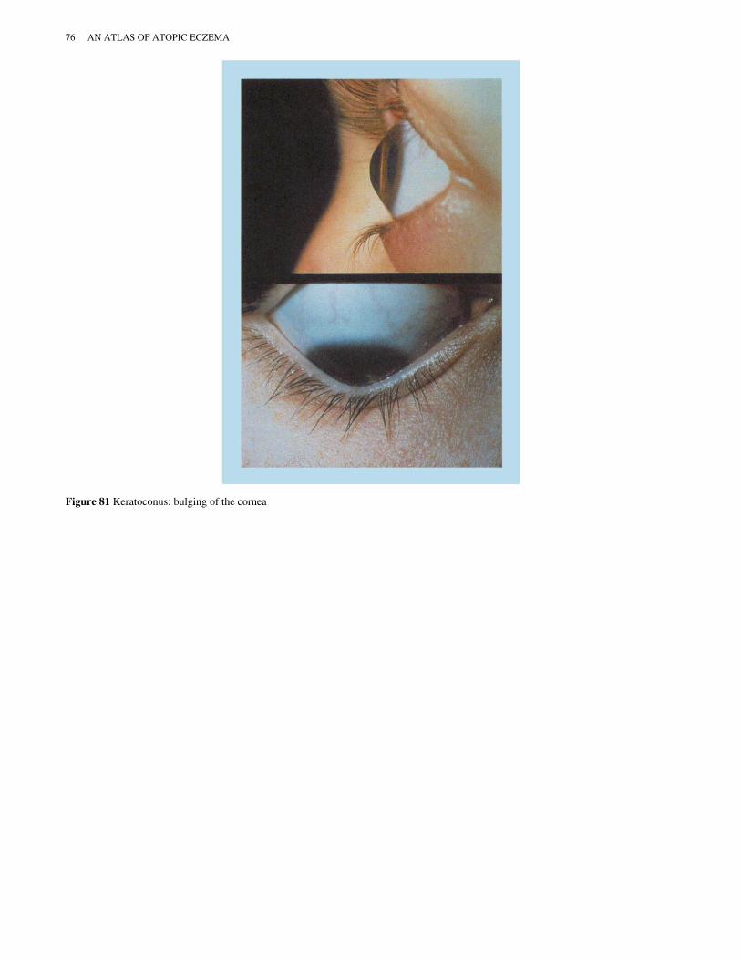
Figure 81 Keratoconus: bulging of the cornea
76 AN ATLAS OF ATOPIC ECZEMA

Figure 82 Dry scaly skin in ichthyosis vulgaris. Sparing of the flexures is a characteristic feature
CLINICAL FEATURES 77

Figure 83 Molluscum contagiosum in an individual with atopic eczema in the axilla
78 AN ATLAS OF ATOPIC ECZEMA

Figure 84 Vesicular eruption of the face in eczema herpeticum
Figure 85 Crusted/vesicular eruption around the eyes in eczema herpeticum
CLINICAL FEATURES 79

Figure 86 Ulcerated lesions in eczema herpeticum
80 AN ATLAS OF ATOPIC ECZEMA

8Differential diagnosis
OTHER ECZEMATOUS CONDITIONS
Eczema is divided into endogenous and exogenous types. In the latter, identifiable external substances (allergens and irritants)induce the eczema at the site of contact of the substance. In endogenous eczema, the disease is essentially due toconstitutional factors but the exact pathogenetic mechanisms have yet to be elucidated. The classification of endogenouseczema is based on the clinical patterns of the disease. Apart from atopic eczema, the other patterns of endogenous eczemasare seborrheic (infantile and adult forms), discoid or nummular, hand and foot (pompholyx), hypostatic and asteatotic. Theseclinical patterns are not always distinct, and often there may be combined features of two or even three patterns. In addition,an individual with one pattern of endogenous eczema is more likely to develop another at a different time or in combination.
Seborrheic eczema of infancy
This begins at a younger age than atopic eczema. Seborrheic eczema usually presents between the 2nd and 8th week of lifeand certainly before 6 months. It may present as cradle cap, with yellowish greasy scales on the scalp, and then spreads to theface and presents as red scaly patches. An alternative presentation is in the napkin area, so-called nappy rash (Figure 87). Theeruption may be confined to these areas or spread to involve the neck, axillae (Figure 88) and flexures of the limbs. It presentsas confluent red areas. It may also occur on the trunk as red scaly papules, with the scaling having a psoriatic appearance.
Seborrheic eczema of infancy has a good prognosis and, in the majority of infants, the disease will resolve after a fewweeks and virtually in all by the age of 1 year. There is no doubt that, in some infants, the pattern of eczema changes from theseborrheic variety into an atopic pattern and, in these instances, the prognosis and course of the eczema are those of an atopicindividual.
Discoid eczema
As the name implies, this type of eczema manifests in circular plaques. It most commonly affects the limbs (Figure 89) andthe age prevalence is that of young adults. Discoid eczema has a relatively good prognosis, clearing after 2–3 years. In somechildren with atopic eczema in the flexures, discoid lesions of eczema also occur on the extensor limbs. In these individuals,the question arises as to whether the individuals have both atopic and discoid eczema or whether the lesions are amanifestation of one disease, i.e. atopic eczema. When atopic eczema first presents in infants, it is not uncommon for thelesions first to affect the extensor limbs.
Asteatotic eczema
Asteatotic means without grease. Asteatotic eczema is usually seen in the elderly as the aging process leads to a thinning ofthe stratum corneum and loss of adhesion between the corneocytes. Keratin is composed of fats and proteins and the loss of fatfrom the stratum corneum has led to the name asteatotic. Clinically, the defect of the stratum presents as dry scaly skin.Whether the biological defect in the aging skin is the same as in atopic individuals is questionable. It is possible that thoseelderly persons who develop dry skin are basically atopics.
Asteatotic eczema is most common on the legs (Figure 90), although it may occur on the trunk and arms. The inflammatorycomponent is often linear and the eczema may look like a crazy pavement. It has been given the name eczema craquelé, as theeczema occurs where the skin appears to have cracked.
As with atopic eczema, the asteatotic pattern is more common in cold weather, which exacerbates the tendency of thekeratin to crack.

Contact eczema
As has been stated above, contact eczema may be due to chemicals that directly damage the stratum corneum (primaryirritants) or due to substances which elicit a true allergic reaction with antigen-specific T-lymphocytes. Primary irritanteczema is most commonly seen on the hands due to overuse of detergents, either from washing-up liquids and householdcleaning fluids or occupational use, e.g. in hair-dressing from frequent shampooing without gloves. This type of eczema maybe distinguished from atopic hand eczema. The irritant eczema most commonly starts in the web spaces at the base of thefingers and then spreads to involve the fingers and dorsum of the hand (Figure 91). Atopic eczema is common on the back ofthe fingers and dorsum of the hand but it tends to be more patchy and does not usually involve web spaces. It should beremembered that, because of the inherent defect of the stratum corneum in atopics, irritant eczema tends to be more commonin these individuals.
Atopic individuals do not have a higher incidence of allergic contact eczema. In adults, atopic eczema may affect the skinaround the eyes and mouth and those individuals often have dry lips. Allergic contact eczema on the face commonly firstpresents around the eyes (Figure 92), because of the very thin skin at these sites, and is most commonly due to cosmetics.Allergic contact eczema on the lips is usually due to lipstick (Figure 93) and lip balms and, clinically, may beindistinguishable from atopic eczema. It is always worthwhile patch-testing those individuals with an atopic background whodevelop eczema again in later life, particularly if the pattern suggests a possible allergic cause, rather than simply labellingthe eczema of these patients as recurrence of their atopic disease. It should be remembered that allergic contact eczema ispotentially curable if the cause is identified, whereas treatment for atopic eczema is purely suppressive.
SYNDROMES WITH ECZEMATOUS ERUPTIONS
Wiskott-Aldrich syndrome
This is a rare X-linked recessive disorder. The gene for the disease has been mapped to the short arm of the X-chromosome. Itis characterized by eczema and thrombocytopenic purpura with associated bleeding and recurrent infection. The eczemausually occurs in the first month of life and affects the scalp, face, flexures and napkin area and is often indistinguishable fromatopic eczema. The diagnosis of Wiskott-Aldrich syndrome becomes apparent when the other features become manifest.
Figure 87 Seborrheic eczema of infancy: well-demar-cated red plaques in the napkin area. Red scaly patches on the trunk and legs are alsopresent
82 AN ATLAS OF ATOPIC ECZEMA

Netherton's syndrome
This is a rare autosomal recessive disorder. It is characterized by a generalized erythroderma soon after birth, much earlierthan atopic eczema develops. The atopic features include flexural accentuation and pruritus. There is often a family history ofatopy and the individuals may have raised IgE levels.
NON-ECZEMA DISORDERS
Hypertrophic lichen planus
Hypertrophic lichen planus on the legs can resemble lichen simplex lesions of atopic individuals (Figure 94). Both maypresent as purplish plaques, varying from 1 to 10 cm. The lichen planus lesion may occur anywhere on the lower leg, butlichen simplex is most common on the anterior or lateral aspect just above the ankle.
Psoriasis
Psoriasis is rare in infants and children, which is the most common age group for atopic eczema. When psoriasis does occur inchildhood, it often affects the genitalia, groins, and axillae. It is distinguished from eczema by the sharp line of demarcationoccurring between the affected and non-affected skin (Figure 95). Psoriasis also tends to be more red, and crusting is notusually a feature.
Guttate psoriasis is usually seen in children and young adults but characteristically affects the trunk, with small red scalypapules, and there is often a history of a streptococcal sore throat 2 weeks before the rash. Guttate psoriasis is not usuallyconfused with atopic eczema. In adults when psoriasis itches and is excoriated, the distinction between psoriatic andeczematous skin lesions may be difficult.
Scabies
The classical lesions of scabies are burrows appearing on the hands and wrists, and papules on the genitalia in males.However, a generalized eruption, which is a reaction to the mite burrowing in the skin, may occur (Figure 96). This
Figure 88 The flexures of the neck and axillae are common sites for seborrheic eczema of infancy
DIFFERENTIAL DIAGNOSIS 83

generalized eruption has non-specific features of red scaly papules but often shows eczematous features. Thus, patientspresenting with eczematous eruptions of recent onset should always be examined for features of scabies.
Acrodermatitis enteropathica
This is a very rare condition, which is thought to be autosomal recessive. It begins 4–6 weeks after weaning, or earlier, if theinfant is not breast-fed. The first presentation is a rash around the mouth and on the back of the hands. It is this distributionthat often suggests atopic eczema. However, the rash develops blisters and becomes fixed and deep erosions may occur. Thereis associated hair loss and diarrhea with failure to thrive. The disorder is due to impaired zinc absorption and responds to zincsupplements.
Figure 89 Discoid eczema on the limbs. There are no confluent lichenified patches in the popliteal fossae
84 AN ATLAS OF ATOPIC ECZEMA

Figure 90 Asteatotic eczema on the legs: superficial cracking of the skin
DIFFERENTIAL DIAGNOSIS 85

Figure 91 Contact eczema on the back of the hands, involving some of the web spaces and sides of fingers
Figure 92 Contact eczema on the eyelids, an allergic reaction to antibiotic eye ointment
86 AN ATLAS OF ATOPIC ECZEMA

Figure 93 Dry scaly lip due to contact eczema caused by reaction to lipstick
DIFFERENTIAL DIAGNOSIS 87

Figure 94 Hypertrophic lichen planus, with similar appearance to lichen simplex (chronic eczema)
88 AN ATLAS OF ATOPIC ECZEMA

Figure 95 Psoriasis is uncommon in children but may be seen. The well-demarcated plaques are unlike eczema
DIFFERENTIAL DIAGNOSIS 89

Figure 96 Scabies: generalized scaly erythematous eruption. Large red papules on the trunk may also be seen. They are thought to be thesite of the burrowing mite
90 AN ATLAS OF ATOPIC ECZEMA

9Management
EMOLLIENTS
One of the features of atopic eczema is dry skin associated with an abnormality of the skin barrier or stratum corneum. Thisdry and abnormal stratum corneum has impaired barrier function and may play a role in the etiology of the eczema byallowing penetration of irritants and microorganisms. Emollients are used to try to create an artificial skin barrier. Essentially,they contain greasy/fatty substances, which form a barrier against penetration of possible irritants and microorganisms. Theywill also decrease transepidermal water loss and therefore increase hydration of the epidermis.
Emollients have no anti-inflammatory action and thus will have no significant effect on inflamed eczematous skin. Theywill, therefore, have little effect on irritation. Many so-called emollients are oil-in-water emulsions. The greater the watercontent, the less greasy they appear. Emollients with high water content have a cooling effect, due to the latent heat ofevaporation of the water. The cooling effect may be anti-pruritic, but the lower temperature of the skin will tend to decreasecorneocyte bonding and, thus, the barrier function of the stratum corneum, which may be an aggravating factor for eczema.Thus, the main function of emollients is that of possible prevention of eczema rather than a treatment of established disease.
BATHING
Bathing has often been said to aggravate eczema. This may be partly due to the soaps and detergents present in shower gelsand bubble baths, which are degreasing agents and which further damage the stratum corneum. In addition, too hot a bathincreases the skin temperature, which in turn will increase the blood supply to the skin, leading to further release ofinflammatory mediators and increased itching. The latter, in turn, will lead to scratching and aggravation of the eczema.However, bathing may reduce the amount of micro-organisms on the surface of the skin that may play a role in the etiology ofeczema.
Emollients containing fatty substances can be added to the bath water to lessen the effect of soaps and detergents on thestratum corneum. A number of substances, which are not soaps or detergents, can be used as soap substitutes. Although theydo not possess the cleaning properties of soaps and detergents, they will cause less damage to the stratum corneum, which isalready abnormal in atopic eczema. Emulsifying ointment BP is a suitable soap substitute.
CLOTHING
Individuals who are atopic are often intolerant to clothing with ‘rough’ surfaces next to their skin. Wool and synthetic fibers haverough filaments that induce irritation and make the individual uncomfortable. Articles of clothing made of pure cotton are themost appropriate.
CLIMATE
Cold climates tend to aggravate and warm ones tend to be beneficial for atopic eczema. This is thought to be due in part to theeffect of temperature on the corneocyte bonding in the stratum corneum. Corneocyte bonding is temperature-dependent; thelower the temperature, the weaker the bonding. Poor bonding of the skin leads to dryness and impaired barrier function, withaggravation of the eczema.
A warmer climate is also associated with more exposure to sunlight. Ultraviolet light has immuno-suppressive andbacteriocidal properties, both of which are beneficial in decreasing the eczematous process. However, a humid climate,leading to increased sweating and overhydration of the strateum corneum, will aggravate eczema.

DIET
Food allergens have been implicated as a cause of atopic eczema. Parents of children with atopic eczema are often convincedthat certain foods are the cause of the disease. However, in the vast majority of patients, elimination diets have not provedhelpful. One of the main problems is that there are no reliable tests to predict which foods are responsible for inducing theeczema. In addition, food antigens may illicit a type 1, IgE-mediated reaction in the skin, which is essentially urticarial andassociated with irritation but is not an eczematous process.
If parents are convinced that a particular food flares the eczema, then it is reasonable to recommend elimination from thediet for this particular food for a month, followed by gradual re-introduction of the food. If there is definite improvement withan ‘elimination’ diet and definite deterioration with re-introduction, then it is reasonable to advise elimination of this particularfood until the child is older and the eczema is improving. If the elimination diets are pursued, it is important that adequateattention should be paid to dietary supplements, if required.
If dietary antigens are playing a role in the pathogenesis of the eczema, it is unlikely to be one specific antigen. The fault islikely to be in the handling of the antigen in the gut and by the immune system. If there is such a fault, it is likely to apply toseveral if not all of the dietary antigens, and it is not possible or advisable to try to eliminate them all. Fortunately, theimmune system appears to ‘mature’ with age and the eczema improves and disappears, in the majority, in time.
BREAST-FEEDING
It has been claimed that infants who are breast-fed are less likely to develop atopic eczema. However, there are several reportsthat infants who are solely breast-fed and have not taken other foods have developed atopic eczema. Therefore, either food isnot important in the cause of the eczema or the infant is sensitized to dietary antigens that are present in the mother’s milk.
Another benefit of breast-feeding is that secretory IgA is present in breast milk. It has been suggested that secretory IgAbinds to dietary antigens and inhibits the absorption of these potential allergens across the immature gut mucosa.
DELAYED INTRODUCTION OF SOLID FOODS
Animal studies have shown that certain antigens, when fed to newborn mice, will invoke humoral and cell-mediatedresponses, whilst the same allergen given to an adult mouse does not. This observation is also based on an immature responseby the gut and immune system to foreign antigens. It has been suggested that the foods most commonly associated with atopiceczema, egg, cow’s milk, fish and peanuts, should not be introduced to the diet until the age of 1 year. However, in a study inwhich fish and citrus fruits were avoided until 1 year, there was no benefit at 3 years follow-up to avoiding allergic reactionsto those substances45.
AVOIDANCE OF FOOD ANTIGENS IN PREGNANCY
It has been suggested that, if the maternal diet in pregnancy was modified so as to reduce the most common food antigensassociated with atopic eczema, the measure may prove protective. This environmental manipulation has been tried inpregnancy with measures to avoid the house dust mite antigen and subsequent development of respiratory symptoms in atopy,and was shown to be helpful46. Dietary manipulation may prove more difficult in pregnancy since dietary deficiency maydevelop.
PROBIOTICS
Probiotics are cultures of potentially beneficial bacteria of the healthy gut flora. It has been suggested that specific microbes inthe commensal gut flora are more important than infections in promoting anti-allergic responses. These microbes have beenassociated with the following: Th1 type immunity, generation of TGF-� , which suppresses Th2 responses, and induction oforal tolerance and IgA production, an essential component of mucosal immune defence.
In a study of mothers who have had at least one first-degree relative (or partner) with atopic disease (eczema, rhinitis, orasthma), the mother was given lactobacillus GG prenatally and their infants were given it postnatally for 6 months. At the ageof 2 years in those receiving lactobacillus GG, the incidence of atopic eczema was 23% vs. 46% in controls47, a highlysignificant result. It has recently been shown that this benefit is maintained at 4 years follow-up48. The use of probiotics, a simpleand safe procedure, certainly looks promising.
92 AN ATLAS OF ATOPIC ECZEMA

AEROALLERGENS
The aeroallergens that have been implicated in atopic eczema are house dust mites, weeds, animal danders, and molds. It hasbeen suggested that these allergens may not only induce eczema by contact with the skin but also through inhalation49.However, it should be recalled that the induction of a positive patch test to house dust mite could only be achieved after Sell-o-tape stripping of the skin, with the removal of the stratum corneum barrier. Thus, it is questionable whether house dust miteantigens can initiate eczema through the skin. It is possible that they may induce eczema through damaged and excoriated skinand thus be an additional antigenic factor in maintaining and exacerbating the eczema after it has been initiated by otherantigens (e.g. food).
Two studies have shown improvement in atopic eczema with measures to decrease house dust mite exposure50, 51. Thelatter study was controlled and showed significant improvement in the severity of the eczema. Measures to decrease housedust mite antigens are not easy. Bedding, including pillows, mattresses and quilts, should be covered with allergen-impermeable covers. The mattress and floors should be cleaned with high-filtration vacuum cleaners. Carpets should bereplaced by wooden or vinyl floors. Soft toys should be washable and washed at least once a week. Bed linen should be ‘hot-washed’ weekly. Carpets and soft furnishings should be sprayed with a benzyl benzoate preparation weekly.
Although these measures will decrease the exposure to house dust mites, they may not eradicate them totally. Decrease inhouse dust mites will not cure atopics, who are likely to have multiple allergies, but they may help, and be worth trying in,infants and children with severe disease.
ANIMALS
Cat, dog and horse dander can cause urticaria (type 1 hypersensitive immune reactions) in atopic subjects. They may alsoinduce asthma and rhinitis. Whether they induce eczema is debatable but, because of the type 1 responses, they will causeirritation and scratching, which themselves may aggravate pre-existing eczema.
Avoidance of the animal is the most appropriate measure. If there is any doubt as to whether removal of the animals will help,the most appropriate procedure is for the child to stay with a relative for 2 weeks and see if the eczema improves and thenrelapses when they return home.
PSYCHOTHERAPEUTIC MEASURES
Children with atopic diseases are often said to be hyperkinetic. They have boundless energy and seem to need little sleep.Adults with atopic eczema have been reported to have a higher incidence of anxiety, depression, hypochondriasis and hysteria.However, whether these psychological disorders are secondary to their chronic skin problem or whether they are primaryfactors as part of the atopic diathesis is not certain.
In a controlled trial of psychotherapeutic measures in the treatment of eczema in adults (endogenous but only one with atopicdisease), it was shown that the eczema improved in the patients who were receiving psychiatric treatment as opposed to thecontrol group. The treatment consisted of psychotherapy, occasional use of anti-depressants, and training in relaxation andhypnosis52.
Patients should be referred for psychiatric assessment if there is clear-cut evidence of a psychiatric problem or if the patientraises the question whether their eczema could be due to or aggravated by such problems. It should always be rememberedthat chronic physical disease itself may lead to psychiatric problems. Clearing the disease may sometimes be the best option.
INVESTIGATIONS
Patients and/or their parents often believe that atopic eczema is due to an allergic response to food and they may well be right.They demand tests to identify the offending food. But the tests available, the ‘prick’ or epicutaneous tests and RAST for IgEantibodies to food, have not been shown to be helpful in the management (see Chapters 5 and 6). Elimination of foods givingpositive tests does not improve the eczema in the majority of individuals. Patch tests in atopic eczema have also been shownnot to be helpful in the management of the disease.
TOPICAL DRUGS
When prescribing topical preparations, it is important to consider the vehicle for the drug. Topical preparations may beointments, creams or lotions. Ointments are composed of fatty substances and tend to be greasy but may remain on the surfaceof the skin for 8–12 h. Thus, there is a reservoir for the drug for this period of time and the drug will diffuse into the skinduring this period. To treat the skin disorder on a continuous basis, the ointment will have to be applied two to three times a
MANAGEMENT 93

day. As a general rule, ointments tend to be the most appropriate vehicle for drugs for dry scaly rashes such as chronic atopiceczema. Another advantage of the ointment base is that it acts as an artificial barrier preventing transepidermal water loss.This hydrates the stratum corneum and enhances the absorption of the drug into the skin.
Creams are oil-in-water emulsions and are approximately 70% water and 30% ‘fats’. Creams tend to remain on the surfaceof the skin for shorter periods than ointments, as the water evaporates when the cream is applied to the skin. The advantage ofthe cream is that, when the water component evaporates, it cools the skin. This tends to decrease the blood supply to the skinand to lessen the release of inflammatory mediators, thus relieving the itch, but this action is short-lived. Because of theevaporation of the water, the vehicle carrying the drug is on the surface of the skin for a shorter period and, therefore, tendsnot to be as effective as an ointment, unless it is applied more frequently. To achieve a similar drug reservoir and absorptioninto the skin as that attained by ointments, creams will have to be applied four times a day.
Lotions will evaporate even more quickly than creams and, therefore, the drug will be on the surface of the skin for arelatively short duration. Lotions may be alcohol- or water-based, depending on the solubility of the drug. In practice, lotionsare mainly used for disorders of the scalp because creams and ointments are too greasy and unpleasant to use, unless it is forshort periods.
As a rule, ointments are the vehicles of choice for delivery of the drug in eczema. The exceptions are acute weepingeruptions and the intertriginous areas. Creams or lotions should be considered, depending on the situation, because theointment bases are non-miscible with sweat and tissue fluid.
Apart from the base or vehicle for the drug, it is important to prescribe an appropriate quantity of the drug. All too often,insufficient amounts are given for adequate use. If the disease is extensive, it is important to estimate the percentage of skinsurface involved. It takes approximately 30 g to cover the total skin surface of an adult. Thus, if 50% of the body surface isinvolved, 15 g will be required for each application. The body surface for children has to be estimated, but it should beremembered that the surface area to body weight ratio is proportionally greater in children than in adults. This is of relevancewhen considering the absorption of the drug into the circulation and possible systemic side-effects.
Corticosteroids
Topical corticosteroids were introduced for dermato-logical conditions in the 1950s and revolutionized the management ofatopic eczema. Corticosteroids have a strong anti-inflammatory action and this is the reason why they are so effective ineczema. The cellular effects are due to the binding of the glucocorticosteroids (GCS) to the cytosolic GCS receptor innucleated cells. Since most cells have these receptors, the effects of the GCS are multiple. Once the steroids have bound to thereceptor, there is conformational change in the GCS receptor complex; which moves to the nucleus. The GCS complex nowbinds to segments of DNA cell glucocorticoid-responsive elements. As a result, certain parts of the genome are affected,leading to increased or decreased transcription, with resulting increased or decreased cytokine production. GCS will also bindto NF-kB, which has been shown to be a regulator for many cytokines and cell adhesion molecules. Binding of GCS to NF-kBwill switch off cytokine production, particularly of those cytokines concerned with T-cell-mediated inflammation.
Between the 1950s and 1970s, many new synthetic glucocorticosteroids were produced and introduced into dermatology. Theaim was to make these steroids more potent and, therefore, better anti-inflammatory agents. However, the more potent theanti-inflammatory effect, the more potent were the effects on collagen and fibroblasts. One of the actions of corticosteroids isto break down collagen and inhibit fibroblasts, which synthesize collagen. The loss of existing collagen and inhibition of newcollagen formation have the clinical effect of making the skin thinner, which is the main side-effect of topical steroids.
When using topical corticosteroids, there are three points to be considered. First, and most important, is the strength of thesteroids, second, the site(s) to be treated and, third, the age of the patient.
Strength of topical corticosteroids
When using topical glucocorticosteroids, it is imperative that the physician knows the strength of the steroid. Corticosteroidactivity is assessed biologically by the vasoconstrictor test and the fibroblast inhibition assay. The range of activity variesenormously and on some assays, if the weakest glucocorticosteroid is graded as 1 unit, then the strongest is 600 times morepotent. It was the failure to appreciate this wide variation in steroid activity that led to side-effects becoming so common inthe late 1960s and 1970s. Since then, the opinion of the general public is against steroids and they often refuse to use them.Topical steroids have been placed into four groups, depending on their activity, and have been designated as weak, moderate,strong, and very strong53. This has enabled physicians to stop the abuse of topical steroids. It is important to remember that, whenprescribing topical steroids, side-effects are proportional to the strength of steroid x duration of use.
94 AN ATLAS OF ATOPIC ECZEMA

Site
The importance of site is highlighted by the fact that the skin is not of uniform thickness. In areas where the skin is very thin,e.g. face, there will be, first, greater absorption of the steroid and, second, a greater risk of side-effects due to the presence ofless collagen in the dermis. Other sites where the skin is relatively thin are the neck and intertriginous areas, and genitalia. Asa general rule, the thinner the skin, the weaker the steroid should be.
Another important aspect of site to consider is the differences at the intertriginous areas. Not only is the skin relatively thinbut, because the skin surfaces are in contact, the sweat does not evaporate so easily as at other sites and this leads to increasedhydration of the stratum corneum, producing greater absorption of the steroid.
Age
It should be stressed that infants have thinner skins than children. The thinner the skin, the more risk of thinning the skinfurther with steroids. In addition, there is a risk of greater absorption of the steroid and possible systemic side-effects. Thus,very potent steroids should be avoided in infants. It should be remembered that thinning of the skin is part of the agingprocess. This is important when using topical steroids in patients over the age of 75, who are likely to have thin skins.
How to use topical corticosteroids in eczema
It is important to use the weakest possible steroid to treat the eczema and for the shortest period of time. However, the aim oftreatment is to try to clear the eczema and induce a remission. The strength of the steroid will depend on the severity of theeczema and the age of the patient. In infants, weak- or moderate-strength topical steroids are usually effective. Weak onesshould be used for the face. They also can be used at other sites as the initial treatment, but the moderate-strength ones may beused for short periods (i.e. up to 2 weeks) if the eczema is severe
In children, if the eczema is severe, the strong topical steroids are acceptable for the trunk and limbs for periods of up to 2weeks and not more often than every 2 months. Similarly, in adults, very strong topical steroids may be used for a 2-weekperiod but not more frequently than every 2 months. This can be referred to as ‘the rule of 2’: twice daily for 2 weeks but notmore frequently than every 2 months. The aim of using potent or very potent topical steroids for short periods is to try toinduce a remission. There is evidence that this does occur.
Figure 97 Striae, caused by long-term use of very strong topical steroids
MANAGEMENT 95

Auto-sensitization is seen in eczematous conditions. This phenomenon is the development of eczema at distant sites fromthe original patch of acute eczema. Appropriate treatment, in which the goal is to clear the eczema, is more effective thantrying to ‘damp down’ the eczema with weak and ineffective topical steroids.
If the eczema clears, then no topical steroids should be used. The patient should be informed that steroids should not beused as a preventative but only as treatment to clear the disease. If the eczema clears in the majority of the areas with strong/very strong topical steroids after a 2-week period, but there are small areas remaining, then it is permissible to continue foranother week or two for these lesions. If and when the eczema recurs, patients should use weak- or moderate-strength topicalsteroids in the first instance. The more potent ones should be reserved for severe disease.
Side-effects
Local If topical steroids are used correctly, side-effects should not occur. Side-effects are due to the use of too strong apreparation for too long and at the wrong site. Side-effects are due to the thinning of skin resulting from collagen atrophy inthe dermis. Clinically, this may present as striae (Figure 97), purpura (Figure 98), spontaneous ‘bruising’ and telangiectasia(Figure 99). If potent topical steroids are applied to the face, they may give rise to so-called peri-oral eczema, which isexaggerated when the patients attempt to discontinue the steroids. The appearances of the eruption are similar to rosacea, withsmall red papules and pustules, but the distribution is different.
Systemic absorption If large quantities of potent topical steroids are applied to extensive areas over long periods of time,enough steroid may be absorbed to cause suppression of the pituitary-adrenal axis and even Cushingoid features. In adults, thesuppression of the pituitary-adrenal axis would require at least 50 g of clobetasol propionate (a very strong topical steroid) tobe used in 1 week. It would probably take some months for Cushingoid features to appear. To produce similar effects withstrong topical steroids would require greater quantities (i.e. 300 g per week). These side-effects are unlikely to be seen withmoderate or weak topical steroids in adults. However, in children, because of their thinner skin and greater ratio of skinsurface to body weight, there is a greater risk of these side-effects.
Avoidance of side-effects The two golden rules for the limitation of side-effects are:
(1) Topical steroids should not be given on repeat prescriptions. Patients must be seen by their doctor if further supplies arerequired;
(2) Strong/very strong steroids should not be used as maintenance therapy.
Figure 98 Purpura seen in thinning of the skin due to long-term use of strong topical steroids
96 AN ATLAS OF ATOPIC ECZEMA

Intralesional corticosteroids
These have a limited role in atopic eczema in adults. The indication for intralesional corticosteroids is the presence ofresistant patches of lichenified eczema. This treatment should only be carried out by a physician experienced in thistechnique.
Topical tacrolimus
Tacrolimus (FK 506) is a potent immunosuppressive agent, which has been used in transplantation to prevent organ rejection.It acts by binding to an intracellular binding protein (FK BP) and forming a complex, which inhibits the enzyme calcineurin.This enzyme is necessary for activation pathways requiring a rise in intracellular calcium. Drugs that act in this way arereferred to as calcineurin inhibitors. This failure to raise intracellular calcium levels results in a block of the signaltransduction required for cytokine gene expression. This leads to inhibition of cytokine production, including IL-2, IL-3, IL-4,IFN-� and TNF-� . Thus, both Th1 and Th2 cytokine production, an integral part of eczema, is blocked.
Tacrolimus is a smaller molecule than cyclosporin and is absorbed into the skin. It has been shown to be effective intreating atopic eczema54 and has recently been given a licence for use in atopic eczema in adults and children over 2 years ofage for short-term use and intermittent long-term use. Compared to topical steroids, tacrolimus appears to be as effective as amoderate-strength topical steroid. Its place in the management of atopic eczema would appear to be for severe eczema on theface resistant to weak topical steroids. It may also have a role in the treatment of intertriginous areas, where potent topicalsteroids are best avoided.
Side-effects
In approximately one-quarter of patients, there is a burning sensation after application of the drug. This tends to wear off withcontinued use.
The more worrying potential side-effects are those associated with immunosuppression. These would include impairedimmunity to viral infection, e.g. herpes simplex, and the possible long-term effects on the development of skin cancers, asseen with systemic immunosuppressive agents. It is too early to have any information on these side-effects.
Figure 99 Telangiectasia on the face due to strong topical steroids
MANAGEMENT 97

Topical pimecrolimus
This is another calcineurin inhibitor with immuno-suppressive properties similar to those of tacrolimus. Recent treatmentshave shown it to be effective in treating atopic eczema when compared to vehicle alone55. However, when compared tobetametha-sone 17-valerate (a strong topical steroid), it was less effective56. The place of pimecrolimus in the management ofatopic eczema would appear to be similar to that of tacrolimus. The side-effects, real and potential, are the same as those oftacrolimus.
ANTISTAPHYLOCOCCAL TREATMENT
As has been mentioned previously, S.aureus is present in over 90% of atopic eczema lesions. However, whether the organismplays a role in the pathogenesis of the diseases or whether it is secondary to the eczema is as yet not decided. After successfultreatment with both topical steroids and tacrolimus, the organisms are no longer present57,58. These observations seem toimply that the presence of the organism is secondary.
However, in some individuals, the eczema lesions are modified and resemble impetigo, and the picture is referred to asimpetiginized eczema. In this situation, there is no doubt that antibiotics, topical and systemic, improve the infected eczema.The appearance reverts to that of eczema. These individuals often have recurrent episodes of impetiginized eczema, implyingthat they are either S.aureus carriers or that they have an inability to deal with these organisms. If there is repeated clinicalinfection, swabs should be taken from the carrier sites and, if S.aureus is present, topical fusidic acid or mupiricin should beapplied for at least 6 weeks and swabs taken again to be certain that the organisms have been eradicated. Antisepticpreparations, such as chlorhexidine hydrochloride and benzalkonium chloride added to bath water or to an emollient for useafter bathing, will decrease the S.aureus content of the skin and may be helpful in preventing recurrent impetiginized eczema.
It is questionable whether the routine use of topical antibiotics combined with topical steroids can be justified. Althoughthere is some evidence that better results are obtained than with topical steroids alone, there is a risk of inducing resistantstrains of S.aureus with this approach. Antiseptics would be more appropriate if routine antibacterial substances could beshown to definitely improve the eczema.
ANTIVIRAL TREATMENT
One of the more severe complications of atopic eczema is eczema herpeticum (Figures 84–86). This is a widespread eruptiondue to herpes simplex, which usually begins on the face but may appear on the trunk and limbs. The initial lesions are blisters,which may be umbilicated. When the blisters rupture, they may form erosions, which invariably become secondarily infectedwith S.aureus and become crusted. The diagnosis may be missed and simply thought to be impetiginized eczema. Antiviraltreatment with systemic aciclovir or famci-clovir should be given as soon as the diagnosis is suspected. It is important not todelay treatment and wait for confirmation of the infection by laboratory tests. Since secondary infection with S. aureus iscommon, it is advisable to give appropriate systemic antibiotics.
A similar picture to eczema herpeticum may be seen with vaccinia virus. Smallpox has been eradicated and vaccination isno longer required. However, because of the threat of bioterrorism with smallpox virus, vaccination may be reintroduced andphysicians should be aware of so-called eczema vaccinatum, following vaccination with vaccinia virus. The treatment is thesame as that for eczema herpeticum.
ANTIFUNGAL TREATMENT
The yeast organism Pityrosporum orbiculare has been claimed to play a role in atopic eczema, particularly of the head andneck. It has been maintained that appropriate antifungal treatment improves the eczema at these sites35. Topical or systemictriazole antifungal preparations are the most appropriate.
ULTRAVIOLET LIGHT
It has been known for many years that natural sunlight is beneficial for atopic eczema. In certain countries, e.g. NorthernEuropean ones, which do not have much natural sunlight, artificial sources of ultraviolet light have been developed and havebeen shown to be helpful for eczema and psoriasis. In eczema, the role of ultraviolet light may be twofold. First, it hasimmunosuppressive properties that interfere with the function of the Langerhans cells (antigen-presenting) and lymphocytes.Second, ultraviolet light has antibacterial properties and will decrease the number of S.aureus organisms on the skin.
In countries where natural sunlight is not available, there are currently three forms of ultra- violet light treatment. The firstis with high-intensity UVB lamps, so-called narrowband UVB, with the lamps emitting UVB at 311 nm. The second is with
98 AN ATLAS OF ATOPIC ECZEMA

high-intensity UVA-1 (340–400 nm). The third is with PUVA. PUVA stands for psoralens+UVA (long-wave ultravioletlight). Psoralens are photo-sensitizers and increase the effect of the ultraviolet light on the skin. The advantage of narrowbandUVB is that no tablets are required, compared to treatment with PUVA. Psoralens may cause gastrointestinal symptoms,particularly nausea, and subjects have to be cautious about going into the sun and artificial sources of UVA for up to 6–8 hafter taking the drug. If the gastrointestinal upset is severe, it is possible to use psoralens topically. The psoralens are added tobath water and patients lie in the bath for 10 min prior to receiving their UVA treatment.
Narrowband UVB and PUVA should be reserved for chronic and persisting eczema that is not responsive to topicalmeasures. It is not really suitable for infants and young children. There is some evidence (as with very potent topical steroids)that ultraviolet light regimes may induce remissions59.
Climate therapy
Treatment of skin disease at the Dead Sea Center has a beneficial effect on both psoriasis and eczema. The Dead Sea Centerhas two unique properties. First, it is 400 m below sea level and is surrounded by mountains. The water vapor from theevaporation of the sea is retained in the area and the water droplets filter out the UVB rays and not the UVA. The UVB rays(middle-wave length) are the rays that induce the sunburn response. The UVA rays, which are not filtered out, do not causesunburn but have a beneficial effect on eczema and psoriasis and the patients can stay on the beach all day.
The second advantageous effect of the Dead Sea is the high concentration of salt and other minerals that increase thehydration of the strateum corneum. However, any cracks or fissures from the eczema will cause stinging when theconcentrated salt water is in contact with the skin.
SYSTEMIC TREATMENT
This is reserved for severe eczema not responsive to topical measures and ultraviolet light. Fortunately, the number of patientswith atopic eczema not controlled by these measures is very few. The drugs used are systemic corticosteroids and immuno-suppressive ones.
Corticosteroids
These are highly effective and will suppress the eczema. However, if the eczema is severe, it tends to relapse when the steroidsare discontinued. Because of the long-term side-effects of systemic steroids, they are not advocated for long-term treatment.Systemic steroids are indicated in short courses for treatment of a flare-up of atopic eczema. Their advantage is that they actquickly and, for short courses, are relatively safe.
Usually prednisolone, 30 mg daily, is sufficient to suppress the eczema and treatment should be tailed off after 2 weeks.
Cyclosporin
This is a potent immunosuppressive drug that was used to stop rejection of transplanted organs. Its primary site of action is onactivated T-lymphocytes, where it will block cytokine production. It is effec-tive in atopic eczema with an initial dose of 3mg/kg/day. Cyclosporin has three main side-effects. First, it is nephrotoxic. In periods of up to 2 years, the nephrotoxicityappears to be reversible but, if continued for longer periods, the risk of permanent impairment of renal function increases withthe duration of use. Second, cyclosporin may induce hypertension. Thus, a patient’s renal function and blood pressure must bemonitored on a regular basis. The third complication is that long-term immunosuppression may lead to the development ofmalignancy particularly of the skin in sun-exposed areas.
Azathioprine
This is an immunosuppressive agent, whose mode of action is the inhibition of DNA synthesis and which thus inhibitsmitosis. Azathioprine reduces both T-and B-cell proliferation. Its main side-effect is bone marrow suppression. Patients whohave low levels of the enzyme thiopurinemethyl transferase are particularly at risk. This enzyme should be measured beforestarting treatment with azathioprine. If the drug is taken long-term, there is a risk of malig-nancies, particularly lymphomasand skin cancers.
Azathioprine has been shown to have beneficial effects in atopic eczema but there have been no controlled trials and it isnot as effective as cyclosporin. The therapeutic effect of immuno-suppressive drugs in eczema is proportional to theirefficiency as immunosuppressive agents.
MANAGEMENT 99

Mycophenolate mofetil
This is another immunosuppressive drug, whose mode of action is via inhibition of purine bio-synthesis. It has been shown tobe effective in atopic eczema60. As with all current immunosuppressive agents, there are serious potential side-effects, whichinclude bone marrow suppression and the risks of malignancy with long-term immunosuppression.
BIOLOGICAL AGENTS
Monoclonal antibodies
In an attempt to circumvent the side-effects of immunosuppressive drugs, a number of monoclonal antibodies have beenproduced directed against the cytokines and cytokine receptors involved in immune responses in chronic inflammatorydiseases, e.g. rheumatoid arthritis, Crohn’s disease, and psoriasis. The monoclonal antibodies that have been used are againstTNF-� , TNF-� receptor, and IL-2 receptor. The results so far are promising. The treatment is not without side-effects and thelong-term hazards are not known, but, if the patients are immunosuppressed, the risk of malignancy exists, as withimmunosuppressive drugs. As yet, there are no reports of these monoclonal antibodies in the treatment of eczema but they arelikely to be effective.
Cytokines
Recombinant IFN-� , which down-regulates Th2 responses, has been reported to be of benefit in atopic eczema61. However,IFN-� is a constituent cytokine in the chronic phase of atopic eczema and, in this instance, recombinant IFN-� may aggravatethe eczema.
IL-10 is an inhibitory cytokine in inflammation mediated by both Th1 and Th2 cytokines. 11–10 has been reported toimprove psoriasis and it is likely to have a similar effect in atopic eczema. Interestingly, in schistosomiasis infection (aparasitic infection) which invokes Th2 responses, the incidence of atopic eczema is very low, and it has been suggested thatthis may be due to the fact that the infestation also invokes IL-10 production62.
Monoclonal antibody to IgE
Recently, it has been shown that a monoclonal antibody to IgE can block IgE binding to receptors on mast cells andbasophils. This observation has led to a study in which peanut-sensitive individuals were given the monoclonal antibody toIgE and then challenged with increasing amounts of peanut antigen63. It was found that the antibody raised the amount ofantigen required to initiate a type 1 hypersensitivity response. In atopic eczema, it is thought that the IgE bound to antigen-presenting cells may be involved in the pathogenesis of the eczema. In theory, it may be possible to develop an antibody tothat part of the IgE molecule that binds to a receptor on the antigen-presenting cell, and thus inhibit the development of eczema.
THE FUTURE
Atopic eczema may well be an antigen-driven disorder but the primary fault is likely to be in the immune response to multipleforeign proteins, e.g. food and aeroallergens. The disease is unlikely to be due to a single allergen. The fact that the majority ofpatients with atopic eczema ‘grow out’ of their disorder implies a different response by the immune system as the individualmatures. Efforts to block or alter this abnormal response to foreign antigens are required. The initial observation on the use ofprobiotics looks promising and appears to carry little risk. Induction of tolerance by other methods long sought by clinicalimmunologists may well be developed as our knowledge on immune mechanisms increases.
100 AN ATLAS OF ATOPIC ECZEMA

References
1. Coco AF, Cooke RA. On the classification of the phenomenon of hypersensitivities. J Immunol 1923; 8:163–822. Bos JD. Atopiform dermatitis. Br J Dermatol 2002; 147:426–93. Hanifin JM, Rajka G. Diagnostic features of atopic dermatitis. Acta Derm Venereol Suppl (Stockh) 1980; 92:44–74. Williams HC, Burney PGJ, Pembroke AC, et al. The UK working party diagnostic criteria for atopic dermatitis. III. Independent
hospital validation. Br J Dermatol 1994; 131:406–165. McNally N, Phillips D. Geographical studies of atopic dermatitis. In Williams HC, ed. Atopic Dermatitis. Cambridge: Cambridge
University Press, 2000:71–846. Williams HC, Robertson CE, Stewart AW, et al. Worldwide variations in the prevalence of symptoms of atopic eczema in the
international study of asthma and allergies in childhood. J Allergy Clin Immunol 1999; 103:125–387. Charman C,Williams HC. Epidemiology. In Bieber T, Leung DYM, eds. Atopic Dermatitis. New York: Marcel Dekker, 2002:21–428. Herd RM. The morbidity and cost of atopic dermatitis. In Williams HC, ed. Atopic Dermatitis. Cambridge: Cambridge University
Press, 2000:85–959. Schultz-Larsen F, Hanifin JM. Secular change in the occurrence of atopic dermatitis. Acta Derm Venereol Suppl (Stockh) 1992; 176:
7–1210. Schultz-Larsen F. Atopic dermatitis, a genetic-epidemiological study in a population-based twin sample. J Am Acad Dermatol 1993;
28:719–2311. Kay J, Gawkrodger DJ, Mortimer MJ, et al. The prevalence of childhood atopic eczema in a general population. J Am Acad
Dermatol 1994; 30:35–912. Williams HC, Strachan DP. The natural history of childhood eczema: observations from the 1958 British Cohort Study. Br J
Dermatol 1998; 120:834–913. Vickers CFH. The natural history of atopic eczema. Acta Derm Venereol Suppl (Stockh) 1980; 92:113–1514. Williams HC, Wuthrich B. The natural history of atopic dermatitis. In Williams HC, ed. Atopic Dermatitis. Cambridge: Cambridge
University Press, 2000:43–5915. Rystedt L. Long-term follow-up in atopic dermatitis. Acta Derm Venereol Suppl (Stockh) 1986; 114:117–2016. Larsen FS. Genetic epidemiology of atopic eczema. In Williams HC, ed. Atopic Dermatitis. Cambridge: Cambridge University Press,
2000:113–2417. Cookson WO, Moffat MF. The genetics of atopic dermatitis. Curr Opin Allergy Clin Immunol 2002; 5:383–718. Cookson WO, Ubhi B, Lawrence R, et al. Genetic linkage of childhood atopic dermatitis to psoriasis susceptibility loci. Nature
Genet 2001; 27:372–919. Cox HE, Moffat MF, Faux JA, et al. Association of atopic dermatitis to the beta subunit of the high affinity immunoglobulins E
receptor. Br J Dermatol 1998; 138:182–720. Schultz-Larsen F. Genetic epidemiology of atopic dermatitis. In Williams HC, ed. Atopic Dermatitis. Cambridge: Cambridge
University Press, 2000:113–2421. Cookson WO. The alliance of genes and environment in asthma and allergy. Nature 1999; 402:B5–11 22. Prausnitz C, Kustner H. Studien die uber die Ueberemptindlichkeit. Zentralbl Bakteriol 1921;86:160–923. Werfel T, Kapp A. T cells in atopic dermatitis. In Bieber T, Leung DYM, eds. Atopic Dermatitis. New York: Marcel Dekker, 2002:
241–6624. Scalabrin DMF, Baubek S, Perzanowski MS, et al. Use of specific IgE in assessing the relevance of fungal and dust mite allergens in
atopic dermatitis. A comparison with asthmatic and non-asthmatic control subjects. J Allergy Clin Immunol 1999; 104:1273–925. Walley AJ, Chavanas S, Moffat MF, et al. Gene polymorphism in Netherton and common atopic disease. Nature Genetics 2001; 29:
175–826. von Bubnoff D, Novak N, Kraft S, et al. The central role of FceRI in allergy. Clin Exp Dermatol 2003; 28:184–727. Zhang Y, Leaves NI, Anderson GG, et al. Positional cloning of a quantitative trait locus on chromosome 13q14 that influences
immunoglobulin E levels and asthma. Nature Genet 2003; 34:181–628. Elliott K, Forrest S. Genetics of atopic dermatitis. In Bieber T, Leung DYM, eds. Atopic Dermatitis. New York: Marcel Dekker 2002:
81–11029. David TJ, Patel L, Ewing CI, Stanton RHJ. Dietary factors in established atopic dermatitis. In Williams HC, ed. Atopic Dermatitis.
Cambridge: Cambridge University Press, 2000:193–20130. Munkvad M, Danielsen L, Hoj L, et al. Antigen-free diet in adult patients with atopic dermatitis. Acta Derm Venereol (Stockh) 1984;
64:524–831. Devlin J, David TJ, Stanton RHJ. Elemental diet for refractory atopic eczema. Arch Dis Childh 1991; 66:93–932. Hattevig, G, Kjellman B, Johansson SGO, et al. Clinical symptoms and IgE responses to common food problems in atopic and
healthy children. Clin Allergy 1984; 14:551–933. Kleinman RE, Walker WA. Antigen processing and uptake from the intestinal tract. Clin Rev Allergy 1984; 2:25–37
34. Sampson HA. Late phase response to food in atopic dermatitis. Hosp Pract 1987; 22:111–2835. Leung DYM, Bieber T. Atopic dermatitis. Lancet 2003; 361:151–6036. Bygum A, Mortz CG, Andersen KE. Atopy patch tests in young adult patients with atopic dermatitis and controls: dose-response
relationship, objective reading, reproducibility and clinical interpretation. Acta Derm Venereol (Stockh) 2003; 83:18–2337. Abeck D, Ruzicka T. Bacteria and atopic eczema: merely association or etiologic factor. In Ruzick T, Bing, Przybilla B, eds.
Handbook of Atopic Eczema. Berlin: Springer-Verlag, 1991:212–2038. Motala C, Potter PC, Weinberg EG, et al. Anti Staphylococcus aureus-specific IgE in atopic dermati-tis. J Allergy Clin Immunol
1986; 78:583–939. Skov L, Olsen JV, Giorno R, et al Application of staphylococcal enterotoxin B on normal and atopic skin induces upregulation of T
cells by a super antigen mediated mechanism. J Allergy Clin Immunol 2000;105:820–640. Ong PY, Ohtake T, Brandt T, et al. Endogenous antimicrobial peptides and skin infections in atopic dermatitis. N Engl J Med 2002;
347:1151–6041. Schafer T, Ring J. The possible role of environmental pollution in the development of atopic dermatitis. In Williams HC, ed. Atopic
Dermatitis. Cambridge: Cambridge University Press, 2000:155–6842. Alm JS, Swartz J, Lilja G, et al. Atopy in children of families with an anthroposophic lifestyle. Lancet 1999; 353:1485–843. Schultz-Larsen F, Diepgen T, Svensson A. Clinical criteria in diagnosing AD. The Lillehammer Criteria 1994. Acta Derm Venereol
Suppl (Stockh) 1996; 76:115–1944. Rajka G. Atopic Dermatitis. Philadelphia: Saunders 1975:4–3545. Saarinen UM, Kajosaari M. Does dietary elimination in infancy prevent or only postpone a food allergy? A study of fish and citrus
allergy in 375 children. Lancet 1980; 1:166–746. Cutovic A, Simpson BM, Simpson A, et al. Effect of environmental manipulation in pregnancy and early life on respiratory symptoms
and atopy during the first year of life: a randomised trial. Lancet 2001; 358:188–9347. Kalliomaki M, Salminen S, Arvilommi H, et al. Probiotics in primary prevention of atopic disease: a random placebo controlled trial.
Lancet 2001; 357:1076–948. Kalliomaki M, Salminen S, Poussa T, et al. Probiotics and prevention of atopic disease: 4-year follow-up of a randomised placebo-
controlled trial. Lancet 2003; 361:1869–7149. Tupker RA, De Monchy JG, Coenraads PJ, et al. Induction of atopic dermatitis by the inhalation of house dust mites. J Allergy Clin
Immunol 1996; 97:1064–7050. Roberts DL. House dust mite avoidance and atopicdermatitis. Br J Dermatol 1994; 110:735–651. Tan BB, Weald DD, Strickland I, et al. Double blind controlled trial of effect of house dust mite allergen avoidance on atopic
dermatitis. Lancet 1996; 347:15–1852. Brown BG, Bettley FR. Psychiatric treatment of eczema: a controlled trial. Br Med J 1971; 2:729–653. Fry L. Dermatology: An Illustrated Guide. London: Update Publications, 1978:16254. Paller A, Eichenfield LF, Leung DY, et al. A 12-week study of tacrolimus ointment for the treatment of atopic dermatitis in pediatric
patients. J Am Acad Dermatol 2001; 44(Suppl):S47–5755. Kapp A, Papp K, Bingham A, et al. Long-term management of atopic dermatitis in infants with topical pimecrolimus, a non-steroidal
anti-inflamma-tory drug. J Allergy Clin Immunol 2002; 110:277–8456. Luger T, Van Leent EJ, Graeber M, et al. SDZ ASM 981: an emerging safe and effective treatment for atopic dermatitis. Br J
Dermatol 2001; 144:788–9457. Stadler JF, Fleury M, Sourisse M, et al. Local steroid therapy and bacterial skin flora in atopic dermatitis. Br J Dermatol 1994, 131:
536–4058. Remitz A, Kyllonen H, Granlund H, et al. Tacrolimus ointment reduces staphylococcal colonisation of atopic dermatitis lesions. J
Allergy Clin Immunol 2001; 107:196–759. Krutmann JT, Morita A. Phototherapy for atopic dermatitis. In Bieber T, Leung DYM, eds. Atopic Dermatitis. New York: Marcel
Dekker, 2002:501–1860. Grundmann-Kollman M, Podda M, Ochsendorf F. Mycophenolate mofetil is effective in the treatment of atopic dermatitis. Arch
Dermatol 2001; 137:870–361. Hanifin JM, Schneider LC, Leung DY, et al. Recombinant interferon gamma therapy for atopic dermatitis. J Am Acad Dermatol
1993; 28:189–9762. Van der Briggelaar AHJ, Van Ree R, Rodriques LC, et al. Decreased atopy in children infected with Schistoma haematobuim: a role
for parasite induced IL-10. Lancet 2000; 356:1723–763. Leung DYM, Sampson HA, Yunginger JW, et al. Effect of anti-IgE therapy in patients with peanut allergy. N Engl J Med 2003; 348:
986–93
102 AN ATLAS OF ATOPIC ECZEMA

Index
aciclovir 69acrodermatitis enteropathica 76acute eczema 19, 37adhesins 29adhesion molecules 21adolescents,
clinical features in 38juvenile plantar dermatosis in 59, 60
adults,clinical features in 38examples of eczema in 50–56
aeroallergens 28, 79age,
features related to 38incidence and 13, 15onset and 35remission and 15topical corticosteroid use and 81
air pollutants 31allergens,
breast-feeding 27food 27peanuts 27role in eczema 12skin response to 22tests 27for food allergies 27
allergic conjunctivitis 11allergic rhinitis 11,12
incidence with eczema 16, 63links with eczema 12mast cells in 23
allergy, genetics of 17animal dander 28, 79ankles 47antecubital fossa 46, 50anterior neck folds 11anthroposophic lifestyle 32anti-depressants 79antibodies, monoclonal 86antigen-presenting cells 23antigens,
avoidance of food antigens during pregnancy 78dietary 33in breast milk 27in food 27
antimicrobial peptide down-regulation in eczema 31antiviral drugs 69arms,
antecubital fossa involvement 46, 50in adult 50in infants 42lichenification on 48
asteatotic eczema 72, 73asthma 11, 12
incidence in eczema 16, 63increased 14
links with eczema 12mast cells in 23
atopen 11atopic diathesis 17atopic keratoconjunctivitis 66atopic syndrome 12atopiform eczema 11autoantigens 28azathioprine 85
bacteria,antistaphylococcal treatment 84infections with 66role in eczema 28see also Staphylococcus;Streptococcus
bathing 77blood findings in eczema 21breast, eczema on 54breast-feeding 33
role in eczema 27role in management 78
Candida albicans 30cathelicidins 30central cubital fossa 48cheilitis 11, 54children,
clinical features in 38examples of eczema in 44–49juvenile plantar dermatosis in 59
chromosome 1q21 18chromosome 3p24–22 18chromosome 3q14 18chromosome 3q21 18, 25chromosome 5q31 18, 25chromosome 5q35 18chromosome 6p 18chromosome 6q12 26chromosome 11q13 18, 25
103

chromosome 12q 18chromosome 13q13 18chromosome 13q14 18, 25chromosome 14q 18chromosome 15q14–15 18chromosome 16q 18chromosome 17qtel 18chromosome 17q11 26chromosome 17q15 18chromosome 17q21 18chromosome 17q25 18chromosome 18q21 18chromosome 20p 18chromosome linkages to eczema 18chronic eczema 19, 20, 37climate 77
cold 33therapy 85
clinical features 35–70clothing 77conjunctivitis
allergic 11, 67recurrent 11
contact eczema 72, 73, 74corticosteroids,
intralesional 83systemic 85topical 80age and use of 81method of use 81side-effects of use 82site and use of 81strength of 81
creams 80crusting 35, 36
in infant 39, 40of knees 43severe 40
on adult neck 53Cushingoid features 83cutaneous lymphocyte antigen 29, 30cyclosporin 85cytokines,
antimicrobial peptides and 30blood findings in eczema 21eosinophil stimulation by 23production in skin 22use in management 86
dander 28, 79Dead Sea Center 85definitions,
atopic 11dermatitis cf eczema 11extrinsic cf intrinsic 12variations in literature 15
Dennie-Morgan infraorbital fold 11, 63, 65dermatitis cf eczema 11detergents 33diagnosis 71–76
clinical criteria of Hanifin and Rajka 11
clinical criteria of Williams 11diet,
avoidance of food antigens during pregnancy 78dietary antigens 33eczema and 27maternal 33probiotic 79role in management 78
differential diagnosis 71–76discoid eczema 72discoid papular eczema 59, 62domicile, role in eczema 13dry skin 11, 12, 26
causes 26skin function and 26
ears, eczema on 54, 58eczema,
bacteria and 28benefits to management 33candidate genes 25cf dermatitis 11clinical criteria 11clinical features 35–70definitions 11differential diagnosis 71–76epidemiology 13etiological factors 32etiology 25, 25food intolerance and 27genetics 17histology 19–21HLA system and 17infantile 12inflammatory cells in 23loci for 18natural history 15pathogenesis 19–24possible genetic abnormalities in 26T-helper-type responses in 22tests for 27triggers for 33
eczema herpeticum 69, 70eczema vaccinatum 69edema 35emollients 77emotional factors 33enterotoxin B 30environmental factors 13environmental pollution 31eosinophils 23epidemiology 13–14erosions 36erythema 11
confluent 42on back 41
erythrodermia 55etiology 25
abnormal skin barrier 26genetics 25summary of factors 32
104 AN ATLAS OF ATOPIC ECZEMA

excoriations,in adult 51in antecubital fossa 46in infants 39, 43on hands of child 44on legs 44, 48
exocytosis 19eyelids,
contact eczema on 74in adult 52
face,confluent eczema on neck and 55in adult 52in infants 39, 40lichenification on 52secondary infection of 45, 49Staphylococcus aureus infection of 45
famciclovir 69family history 11
and incidence, 17feet, juvenile plantar dermatosis 59, 60fibronectin 29fissures 37
on fingers, 61FK 506 83flexural involvement 12food allergens 27food antigen avoidance during pregnancy 78food intolerance 11
test for food allergies 27fungi,
antifungal treatment 84infections with 66role in eczema 30
genetics,candidate genes 25chromosome linkages to eczema 18family studies 17genome screens 18HLA system and eczema 17maternal effect 18of atopy 9possible abnormalities in eczema 26twin studies 17
genome screens 18glucocorticosteroids 80gluteal folds 47
hands,atopic eczema on 59, 60, 61excoriations on 44fissures on 61in infants 43
hay fever, see under allergic rhinitisherpes simplex virus 11
incidence of warts in eczema patients 69high-affinity receptor for IgE 18histology 19–21
hobbies 15house dust mites 28, 33human � -defensins 30human lymphocyte antigen (HLA) system, role in eczema 17hygiene hypothesis 9, 32hyperkeratosis 35
of foot sole 37 ;hyperkinesis 79hyperpigmentation 55, 56hypersensitivity type IV 21hypertrophic lichen planus 75hypnosis 79hypopigmentation 56
ichthyosis vulgaris 11, 66, 68immediate skin test (type I) reactivity 11immune response 22immune system
down-regulation of antimicrobial peptides 31innate 30
immunoglobulin E (IgE),antibodies,blood findings in eczema 21chromosome linkages 18role in eczema 11,12
high-affinity receptor 18gene for 25
levels in diseases 24levels in eczema 24monoclonal antibody to 86receptors 22mast cells and 23-specific antibodies,Staphylococcus aureus infection and 28tests for 27
immunopathogenesis 21incidence 9, 13, 31
age and 13family history and 17increasing 13industrialization and 13
industrialization, 13infantile eczema 12infants,
clinical features in 38examples of eczema in 39–44seborrheic eczema in 71, 72
infections, 11antistaphylococcal treatment 84antifungal treatment 84antiviral treatment 84bacterial 66facial 45, 49fungal 66leg 45viral 69
inflammatory cells 23inheritance 17interferon-� ,
blood findings in eczema 21production in eczema 22
INDEX 105

interleukins,blood findings in eczema 21locus for interleukin-4 receptor 26production in eczema 22
intolerances,food 11wool 11
intralesional corticosteroids 83itching 11, 38
juvenile plantar dermatosis 59, 60
keratoconjunctivitis 67atopic 66
keratoconus 66, 68keratosis pilaris 11, 63, 64knees,
excoriations in adult 51in infants 43popliteal fossa involvement 45, 47
legs,excoriations on 48and crusting on 44
in child 47in infants 41, 42
LEKTI 25lichen simplex 20, 59, 62lichenification 11, 35, 37
facial 52in black child 49in popliteal fossa 50on arms 48pigmentation loss after 57
lifestyle 13anthroposophic 32
linearity, increased 63, 65lips,
contact eczema on 73, 74eczema on 54, 57
loci,for asthma 18for eczema 18,18for psoriasis 18
lotions 80
Malassezia furfur 30management 77–86
aeroallergens 79animal dander 79antifungal treatment 84antiviral treatment 84bathing 77biological agents 86breast-feeding 78climate 77climate therapy 85
clothing 77corticosteroids,intralesional 83
systemic 85topical 80
delayed weaning 78diet 78emollients 77food antigen avoidance in pregnancy 78future developments in 86investigations 80pimecrolimus 83probiotics 79psychotherapy 79Staphylococcus aureus infection 84systemic treatment 85tacrolimus 83topical preparations 80ultraviolet light 84
mast cells 23maternal effect 18micropapular eruptions 63migraine incidence with eczema 66mild eczema 36
in infant 39molds 28molluscum contagiosum 69mycophenolate mofetil 86
natural history 15–16neck,
confluent eczema on face and 55in adult 53reticulate pigmentation on 54, 57
Netherton’s syndrome 25, 75neuropeptides 33nitrogen oxides 32
occupation 15ointments 80orbital darkening 11
Paget’s disease 54pallor 11, 63patch tests 28pathogenesis 19–24peanuts 27peri-auricular eczema 54perifollicular accentuation 11pharmacological abnormalities 32PHF11 25pigmentation,
altered 54hyperpigmentation 55, 56hypopigmentation 56loss after lichenification 57reticulate on neck 54, 57
pimecrolimus 83pinna, eczema on 54, 58pityriasis alba 11, 59, 62Pityrosporum orbiculare 66plantar dermatosis, juvenile 59, 60plaques 37
106 AN ATLAS OF ATOPIC ECZEMA

pollens 28pollution 9, 31, 33popliteal fossa 45, 47
lichenification in 50pregnancy, avoidance of food antigens during 78probiotics 32, 33, 79prognosis 15
indicators of 15prurigo nodularis 59, 64pruritus 11psoriasis 75, 76
cf histology of eczema 21loci for 18
psychotherapy 79purpura and topical corticosteroid use 82
radioallergoabsorbent tests 27RANTES 26relapsing course 11reticulate pigmentation on neck 54, 57rhinitis (allergic) 11
incidence with eczema 16, 63links with eczema 12mast cells in 23
risk of development 15
satellite lesions 48scabies 75, 76scaling 35, 36
in adult 50, 53in infant 39, 39, 42on scalp and back 41
scratching,chronic eczema and 20Staphylococcus aureus infection and 29
seborrheic eczema 71, 72sex 35shiny foot syndrome 59, 60side-effects,
pimecrolimus 84topical corticosteroids 82topical tacrolimus 83
sites,in adolescents 38in adults 38in children 38in infants 38topical corticosteroid use and 81
skin,barrier function 26colonization by Staphylococcus aureus 29pallor 63see also dry skinthinning and topical corticosteroid use 82
skin-prick tests 27smallpox 69SPINKS 25spongiosis 19Staphylococcus aureus 11, 28
appearance of infections 66
facial infection 45infection in infants 38management of infections 66role in eczema 30superantigens and 29T-cell activation by 29treatment of 84
Streptococci 28stress 33striae and topical corticosteroid use 82subacute eczema 19, 36subcapsular cataracts 11sulfur dioxide 32superantigens,
effects of 30pathogenesis and 29
symmetry 54systemic treatment,
azathioprine 85corticosteroids 85cyclosporin 85mycophenolate mofetil 86
T-cells 23activation by Staphylococcus aureus 29clones 27
T-helper type cells 21T-helper-type responses,
fungi and 30hygiene hypothesis 32in eczema 22Staphylococcus aureus and 29
tacrolimus 83telangiectasia and topical corticosteroid use 83tests 80
IgE-specific antibodies 27patch 28radioallergoabsorbent 27skin-prick 27
thighs 47Toll-like receptors 30topical corticosteroids 80–83topical preparations 80treatment, see under managementTrichophyton rubrum 66trunk, in infants 40, 41twin studies in eczema 17type IV hypersensitivity 21
ultraviolet light 84urbanization 9urticaria, incidence with eczema 66
vaccination 32vaccinia virus 69vascular abnormalities 32vascular cell adhesion molecule 21virus,
antiviral treatment 84eczema herpeticum 70
INDEX 107

infections with 69
warts 60, 69weaning 33
delayed 78weather 77
climate therapy 85cold 33
weeping 37white dermographism 63, 65Wiskott-Aldrich syndrome 75wool intolerance 11wrists, symmetrical involvement 46
xeroderma 63xerosis, see under dry skin
108 AN ATLAS OF ATOPIC ECZEMA