amino acids and proteins … for as biology. amino acids proteins are macromolecules consisting of...
TRANSCRIPT
Amino acids and proteins
… for AS Biology
Amino acids
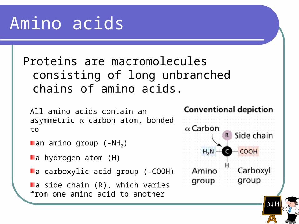
Proteins are macromolecules consisting of long unbranched chains of amino acids.
All amino acids contain an asymmetric carbon atom, bonded to
an amino group (-NH2)
a hydrogen atom (H)
a carboxylic acid group (-COOH)
a side chain (R), which varies from one amino acid to another

Variety of amino acids
The chemical properties of different amino acids are determined by their side chains, and are in turn important in determining the properties of the proteins they make up.
Some amino acids are important as molecules in their own right: for example the formation of urea in the mammalian liver involves a cyclic metabolic pathway involving the amino acids ornithine, citrulline and arginine.
But most amino acids are found in proteins.
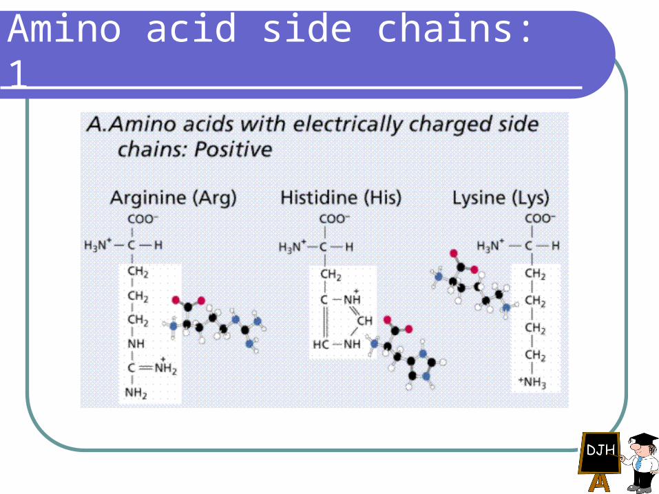
Amino acid side chains: 1
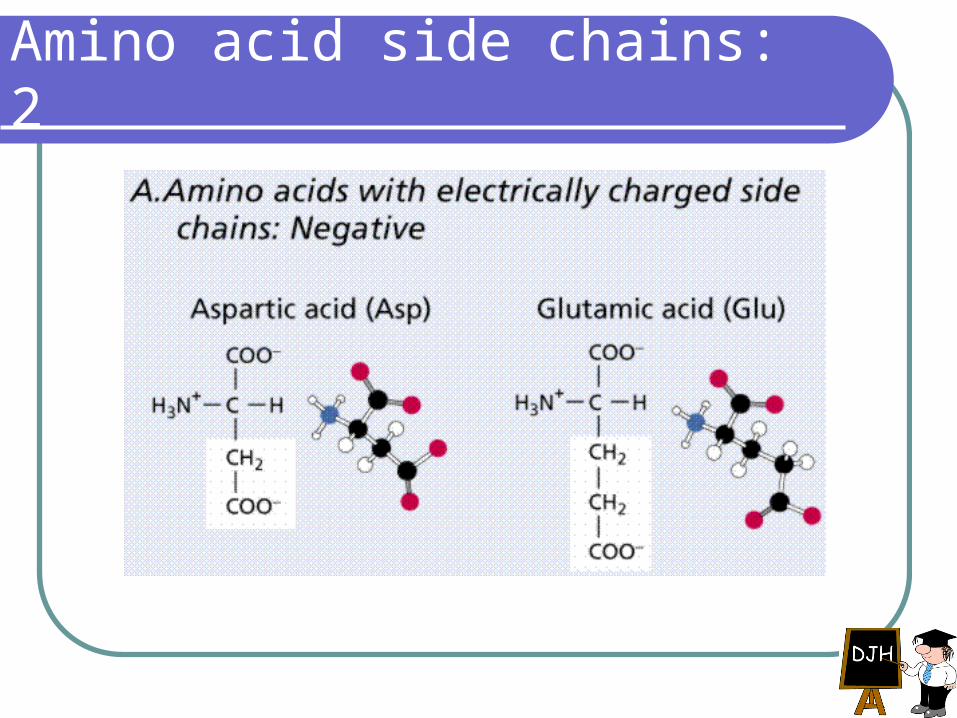
Amino acid side chains: 2
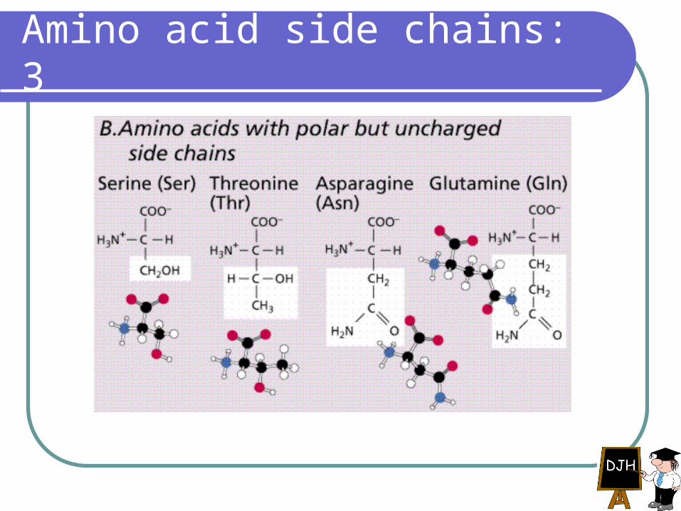
Amino acid side chains: 3
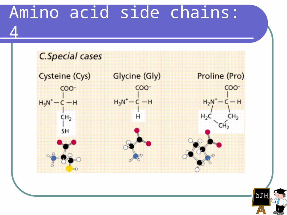
Amino acid side chains: 4
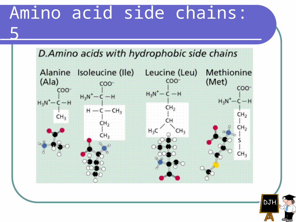
Amino acid side chains: 5
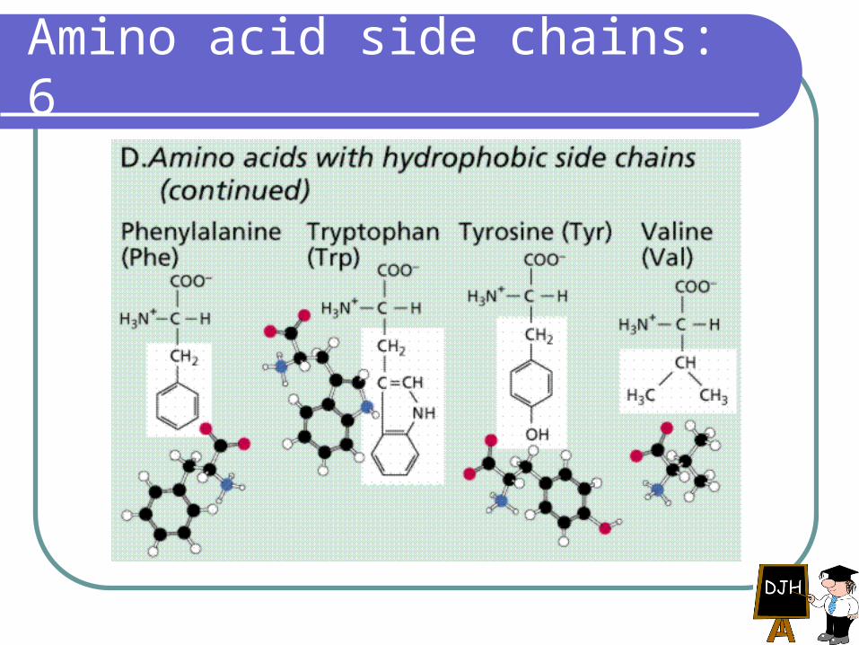
Amino acid side chains: 6
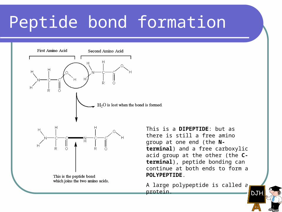
Peptide bond formation
This is a DIPEPTIDE: but as there is still a free amino group at one end (the N-terminal) and a free carboxylic acid group at the other (the C-terminal), peptide bonding can continue at both ends to form a POLYPEPTIDE.
A large polypeptide is called a protein.
Have you understood peptide bonding?
What kind of reaction is involved in forming a peptide bond?
Condensation.Which two atoms are joined by a peptide
bond?C and N.What kind of bond is it?Covalent.
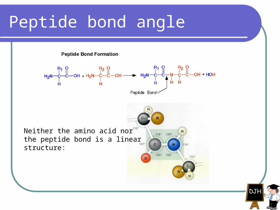
Peptide bond angle
Neither the amino acid nor the peptide bond is a linear structure:
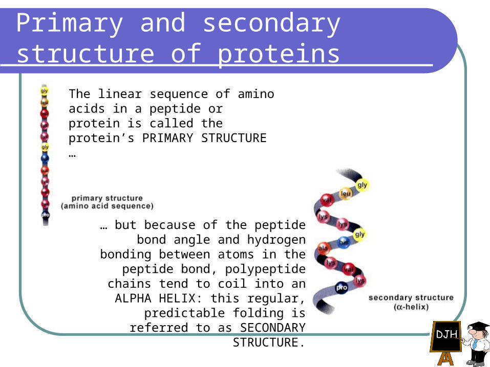
Primary and secondary structure of proteins
The linear sequence of amino acids in a peptide or protein is called the protein’s PRIMARY STRUCTURE …
… but because of the peptide bond angle and hydrogen bonding between
atoms in the peptide bond, polypeptide chains tend to coil into an ALPHA
HELIX: this regular, predictable folding is referred to as SECONDARY
STRUCTURE.
The alpha helix
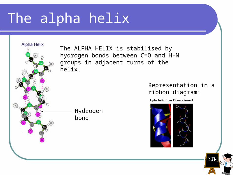
The ALPHA HELIX is stabilised by hydrogen bonds between C=O and H-N groups in adjacent turns of the helix.
Hydrogen bond
Representation in a ribbon diagram:
The beta pleated sheet (1)
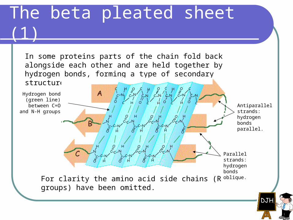
In some proteins parts of the chain fold back alongside each other and are held together by hydrogen bonds, forming a type of secondary structure called a BETA PLEATED SHEET.
Hydrogen bond (green line)
between C=O and N-H groups
For clarity the amino acid side chains (R groups) have been omitted.
Antiparallel strands: hydrogen bonds parallel.
Parallel strands: hydrogen bonds oblique.

The beta pleated sheet (2)
Other ways of representing the beta pleated sheet. In the diagram below, note the R groups (yellow spheres) protruding above and below the plane of the sheet.
Three-dimensional structure of a protein: ribonuclease
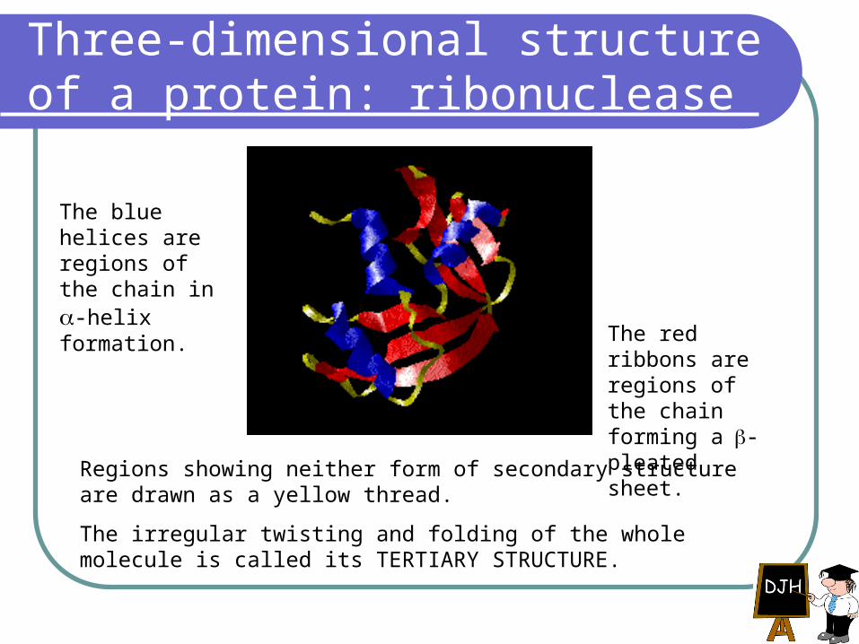
The blue helices are regions of the chain in -helix formation.
The red ribbons are regions of the chain forming a-pleated sheet.
Regions showing neither form of secondary structure are drawn as a yellow thread.
The irregular twisting and folding of the whole molecule is called its TERTIARY STRUCTURE.
Tertiary structure (1)
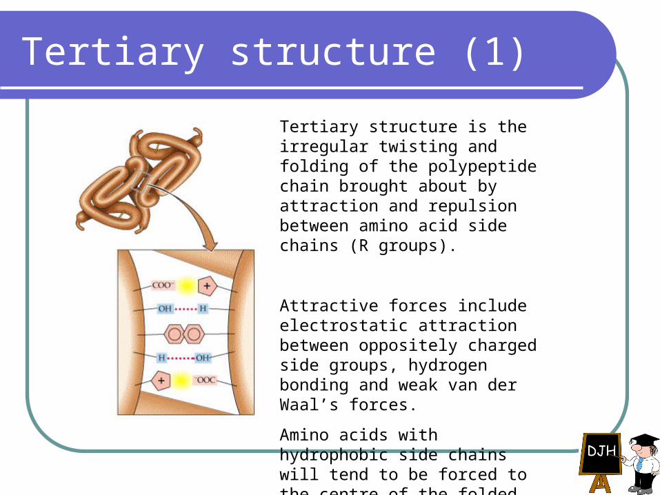
Tertiary structure is the irregular twisting and folding of the polypeptide chain brought about by attraction and repulsion between amino acid side chains (R groups).
Attractive forces include electrostatic attraction between oppositely charged side groups, hydrogen bonding and weak van der Waal’s forces.
Amino acids with hydrophobic side chains will tend to be forced to the centre of the folded molecule, away from water.
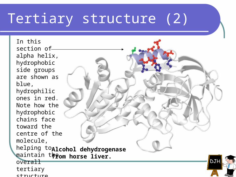
Tertiary structure (2)
In this section of alpha helix, hydrophobic side groups are shown as blue, hydrophilic ones in red. Note how the hydrophobic chains face toward the centre of the molecule, helping to maintain the overall tertiary structure.
Alcohol dehydrogenase from horse liver.
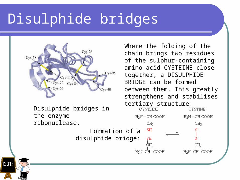
Disulphide bridges
Where the folding of the chain brings two residues of the sulphur-containing amino acid CYSTEINE close together, a DISULPHIDE BRIDGE can be formed between them. This greatly strengthens and stabilises tertiary structure.
Disulphide bridges in the enzyme ribonuclease.
Formation of a disulphide bridge:
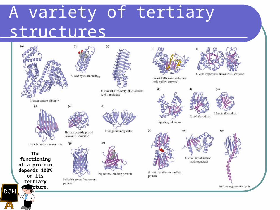
A variety of tertiary structures
The functioning of a protein
depends 100% on its tertiary
structure.
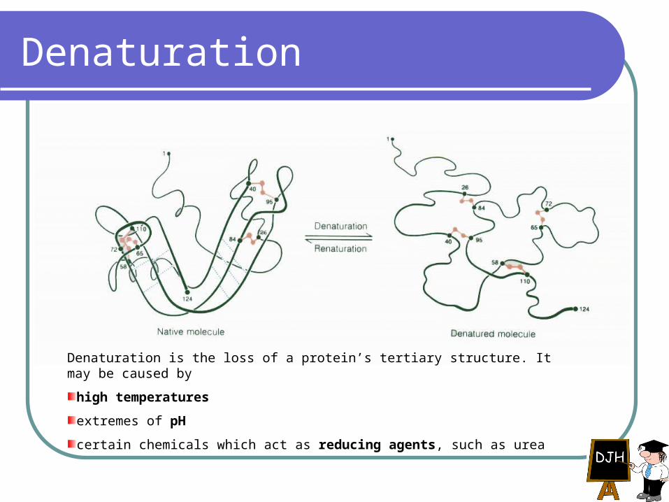
Denaturation
Denaturation is the loss of a protein’s tertiary structure. It may be caused by
high temperatures
extremes of pH
certain chemicals which act as reducing agents, such as urea
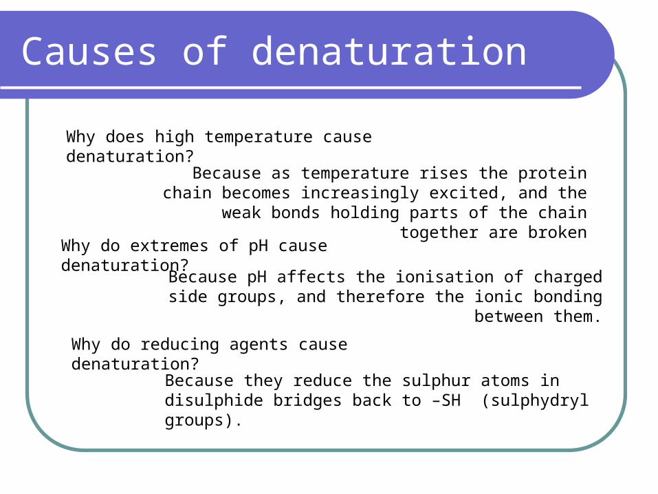
Causes of denaturation
Because as temperature rises the protein chain becomes increasingly excited, and the weak bonds holding parts of
the chain together are broken
Why does high temperature cause denaturation?
Why do extremes of pH cause denaturation?
Because pH affects the ionisation of charged side groups, and therefore the ionic bonding between them.
Why do reducing agents cause denaturation?
Because they reduce the sulphur atoms in disulphide bridges back to –SH (sulphydryl groups).
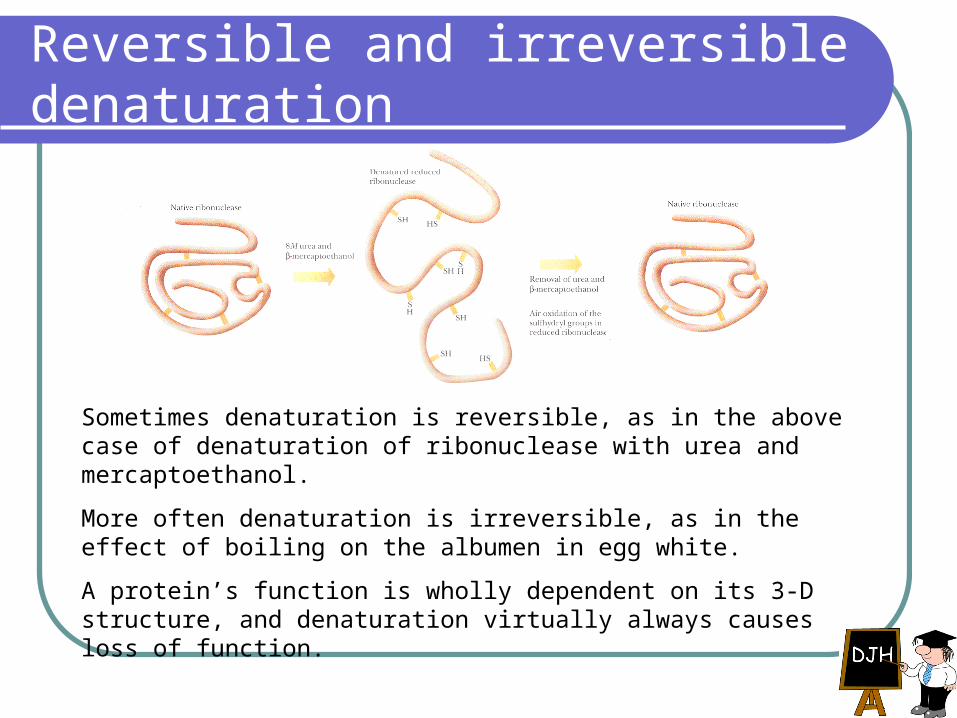
Reversible and irreversible denaturation
Sometimes denaturation is reversible, as in the above case of denaturation of ribonuclease with urea and mercaptoethanol.
More often denaturation is irreversible, as in the effect of boiling on the albumen in egg white.
A protein’s function is wholly dependent on its 3-D structure, and denaturation virtually always causes loss of function.
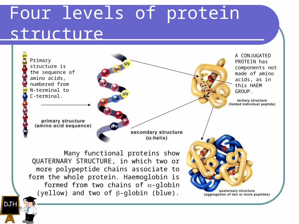
Four levels of protein structure
Primary structure is the sequence of amino acids, numbered from N-terminal to C-terminal.
A CONJUGATED PROTEIN has components not made of amino acids, as in this HAEM GROUP.
Many functional proteins show QUATERNARY STRUCTURE, in which two or more polypeptide
chains associate to form the whole protein. Haemoglobin is formed from two chains of -globin
(yellow) and two of -globin (blue).