amiloride clinical trial in optic neuritis (action) protocol: a
TRANSCRIPT
Amiloride Clinical Trial In OpticNeuritis (ACTION) protocol:a randomised, double blind, placebocontrolled trial
Justin B McKee,1 John Elston,2 Nikos Evangelou,3 Stephen Gerry,4 Lars Fugger,5
Christopher Kennard,1 Yazhuo Kong,1,6 Jacqueline Palace,1 Matthew Craner1
To cite: McKee JB, Elston J,Evangelou N, et al. AmilorideClinical Trial In Optic Neuritis(ACTION) protocol:a randomised, double blind,placebo controlled trial. BMJOpen 2015;5:e009200.doi:10.1136/bmjopen-2015-009200
▸ Prepublication history thispaper is available online. Toview these files please visitthe journal online(http://dx.doi.org/10.1136/bmjopen-2015-009200).
Received 1 July 2015Revised 28 August 2015Accepted 4 September 2015
For numbered affiliations seeend of article.
Correspondence toDr Matthew Craner;[email protected]
ABSTRACTIntroduction: Neurodegeneration is a widely acceptedcontributor to the development of long-term disabilityin multiple sclerosis (MS). While current therapies inMS predominantly target inflammation and reducerelapse rate they have been less effective at preventinglong-term disability. The identification and evaluationof effective neuroprotective therapies within a trialparadigm are key unmet needs. Emerging evidencesupports amiloride, a licenced diuretic, as aneuroprotective agent in MS through acid sensing ionchannel blockade. Optic neuritis (ON) is a commonmanifestation of MS with correlates of inflammationand neurodegeneration measurable within the visualpathways. Amiloride Clinical Trial In Optic Neuritis(ACTION) will utilise a multimodal approach toassess the neuroprotective efficacy of amiloride inacute ON.Methods and analysis: 46 patients will be recruitedwithin 28 days from onset of ON visual symptoms andrandomised on a 1:1 basis to placebo or amiloride10 mg daily. Double-blinded treatment groups will bebalanced for age, sex and visual loss severity by arandom-deterministic minimisation algorithm. Theprimary objective is to demonstrate that amiloride isneuroprotective in ON as assessed by scanning laserpolarimetry of the peripapillary retinal nerve fibre layer(RNFL) thickness at 6 months in the affected eyecompared to the unaffected eye at baseline. RNFL incombination with further retinal measures will also beassessed by optical coherence tomography. Secondaryoutcome measures on brain MRI will include corticalvolume, diffusion-weighted imaging, resting statefunctional MRI, MR spectroscopy and magnetisationtransfer ratio. In addition, high and low contrast visualacuity, visual fields, colour vision andelectrophysiology will be assessed alongside quality oflife measures.Ethics and dissemination: Ethical approval wasgiven by the south central Oxford B research ethicscommittee (REC reference: 13/SC/0022). The findingsfrom ACTION will be disseminated through peer-reviewed publications and at scientific conferences.Trial registration number: EudraCT2012-004980-39, ClinicalTrials.gov Identifier: NCT01802489.
BACKGROUNDOverviewMultiple Sclerosis (MS) is the mostcommon cause of progressive disability inyoung adults in the western world. It is char-acterised by inflammatory demyelinatingplaques with associated axonal and neur-onal damage affecting the central nervoussystem (CNS). The accumulated impact ofneurodegeneration is thought to be themajor pathological substrate for disability inMS.1 One of the major unmet needs in MSis for safe, cost-effective therapeutic inter-ventions that prevent neurodegenerationand thereby the development of disability.
Strengths and limitations of this study
▪ This study examines optic neuritis as a discretecentral nervous system inflammatory event withmeasurable structural and clinical outcomesallowing the time-efficient (6 month) assessmentof neuroprotective therapies.
▪ The repurposing of amiloride, a licenced diureticwith excellent safety record, as a neuroprotectivetherapy is supported by a strong basic and clin-ical science with potential to translate to multiplesclerosis (MS).
▪ A multimodal approach including retinal scansand brain MRI will examine the effect of theintervention from the retina to the brain allowingthe evaluation of sensitive outcome markers andinform structural–functional correlates for futurestudies of optic neuritis.
▪ Neuronal loss can occur early in acute inflamma-tion, therefore recruitment up to 28 days may beoutside the effective ‘therapeutic window’ forneuroprotection.
▪ The primary outcome measure uses theunaffected eye as a powerful comparator, butthis eye may be affected by MS-related changesor previous subclinical optic neuritis, thus con-founding the results.
McKee JB, et al. BMJ Open 2015;5:e009200. doi:10.1136/bmjopen-2015-009200 1
Open Access Protocol
group.bmj.com on April 11, 2018 - Published by http://bmjopen.bmj.com/Downloaded from
The natural history of MS is such that disability accruesslowly in the majority of patients and therefore studiesexamining neuroprotective therapies can be protractedand expensive. Clinical trial paradigms examiningacute inflammatory optic neuritis (ON) may help over-come this barrier by demonstrating a ‘proof-of-concept’neuroprotective benefit for repurposed or noveltherapies.Acute inflammatory ON is a common cause of revers-
ible visual loss in young adults in the UK and westernworld. Patients typically present with painful visual lossthat progresses for a maximum of 2 weeks and thenrecovers over the following 2–6 weeks, with further pro-longed recovery of up to a year.2 ON is a common pre-cursor to the development of MS with approximately50% of patients developing MS within 10 years.3 In add-ition, 70% of patients with MS will show evidence ofoptic nerve involvement over time.4 The presence ofinflammatory demyelination and associated axonal lossin ON parallels the pathophysiology observed in otherwhite matter structures in MS5 enabling translationalresearch in ON to be applicable to the wider patientgroup affected by MS.The distinct anatomy and function of the optic nerve
affords unique ways of studying both structural andfunctional changes in the CNS during the acute andrecovery phases of ON. The retinal nerve fibre layer(RNFL) is a major site of axonal loss after ON.6 Recentadvances in retinal imaging allow accurate in vivo esti-mates of RNFL thickness, with the use of scanning laserpolarimetry (GDx) and optical coherence tomography(OCT). High-resolution spectral domain OCT (SDOCT) with segmentation allows precise measurementof specific retinal layers using computerised algorithmsand quantitation of both peripapillary and macularRNFL as well as the ganglion cell (GCL) and otherretinal layers. Both GDx and SD OCT have recentlybeen employed in neuroprotective trial frameworks7
allowing neurodegeneration to be quantified in vivoboth sensitively and longitudinally.8–10 Moreover, OCTmeasures of RNFL loss correlate with clinical outcomemeasures of visual acuity and disability as measured bythe Expanded Disability Status Scale (EDSS). Surrogatemarkers of neurodegeneration such as MRI measuresof cerebral brain atrophy also correlate with RNFLloss.11 Thus, ON affords the opportunity to assess theimpact of neuroprotective strategies in inflammatoryCNS disease.Currently there are no therapeutic strategies that have
been shown to improve the visual outcome in ON. Oralor intravenous methylprednisolone has been shown tohasten the visual recovery of ON but not to affect thefinal visual outcome.2 Thus, by assessing amiloride, thistrial aims to meet the major unmet need for cost-effective, safe, neuroprotective strategies that reduceboth axonal loss and disability in ON and by extensionto MS.
Translating the basic scienceRecent advances in molecular biology have shed newlight on the mechanisms of axonal loss and on potentialtherapeutic targets in ON and MS.While axonal loss in MS is likely to be multifactorial,
a key end point is the influx of sodium (Na+) andcalcium (Ca2+) ions through ion channels and exchan-gers.12 Recent evidence has implicated the AcidSensing Ion Channel type 1 (ASIC1), capable offluxing both Na+ and Ca2+, as a mediator of neuronalinjury in stroke and more recently in CNSinflammation.13 14
Amiloride, a licenced diuretic with a proven safetyrecord, acting through ASIC1 blockade was found to besignificantly neuroprotective and myeloprotective inanimal models of MS.13 Furthermore, a recent singlearm, open label, longitudinal run-in trial of amiloride inprimary progressive patients with MS showed a signifi-cant reduction in the rate of brain atrophy on theamiloride treatment phase, compared to the run inphase. In tandem with this, diffusion tensor imagingindices of tissue damage were significantly reducedduring the treatment phase in clinically relevant whitematter structures suggesting a neuroprotective benefit inpatients with MS.15
This converging basic and clinical science stronglysupports a role for further testing of amiloride as anovel neuroprotective drug in the treatment of ONand MS.
METHODSObjectives and outcome measuresThe Amiloride Clinical Trial in ON (ACTION) incorpo-rates multimodal techniques covering imaging modal-ities of GDx, OCT and MRI combining with clinical andfunctional outcomes.
Primary objectiveTo evaluate the potential neuroprotective efficacy ofamiloride in ON through the surrogate measure ofretinal nerve fibre layer (RNFL) measurement.
Outcome measureGDx determined difference in peripapillary RNFL thick-ness between affected eye at 6 months and unaffectedfellow eye at baseline.In addition secondary outcome measures of the
primary objective will be made by assessing GDx deriveddifference in the affected eye at 12 months compared tothe baseline unaffected eye, and the same measure-ments of RNFL will be made using OCT. Further OCTsecondary outcome measures will utilise retinal layer seg-mentation to determine macular RNFL thickness andvolume as well as GCL measurements.
2 McKee JB, et al. BMJ Open 2015;5:e009200. doi:10.1136/bmjopen-2015-009200
Open Access
group.bmj.com on April 11, 2018 - Published by http://bmjopen.bmj.com/Downloaded from
Secondary objectivesSecondary objective 1To assess markers of neurodegeneration in ON and theneuroprotective effect of amiloride through MRIoutcomes.
Outcome measuresMRI surrogate markers of white and grey matter injuryand connectivity derived on 3 T MRI at baseline, 6 and12 months, including:▸ Diffusion-weighted imaging (DWI) measures of frac-
tional anisotropy (FA), mean diffusivity (MD), axialand radial diffusivity (RD) of the postchiasmal visualpathways;
▸ High-resolution T1-weighted imaging of the corticaland subcortical grey matter volume;
▸ MR spectroscopy (MRS) measures ofN-acetylaspartate (NAA) in the visual cortex;
▸ Resting state functional MRI (RS-fMRI) patterns ofactivity;
▸ Magnetisation transfer imaging (MTI) derived mag-netisation ratio (MTR) of the white and grey matter.
Secondary objective 2To assess whether amiloride improves functional andvisual outcome following ON.
Outcome measuresHigh and low contrast visual acuity at baseline, 6 and12 monthsHumphrey visual fields (HVF) at baseline, 6 and
12 months.Fansworth Munsell 100-hue colour vision test (FM100)
at baseline and 6 monthsLatency and amplitude of waveforms in pattern visu-
ally evoked potential (PVEP) and pattern electroretino-gram (PERG) at baseline and 6 monthsQuality of life scores on the national eye institutes
25-point visual functioning questionnaire (NEI-VFQ-25)and the 10-point neuro-ophthalmic supplement(10-NOS).
Secondary objective 3To confirm ON as a sensitive and efficient model forneuroprotection in a clinical trials framework.
Outcome measuresA combination of outcome measures of MRI, RNFL,electrophysiology and clinical outcome measures willindividually determine the validity of these measuresand correlations will also support this trial platform forfurther neuroprotective studies.
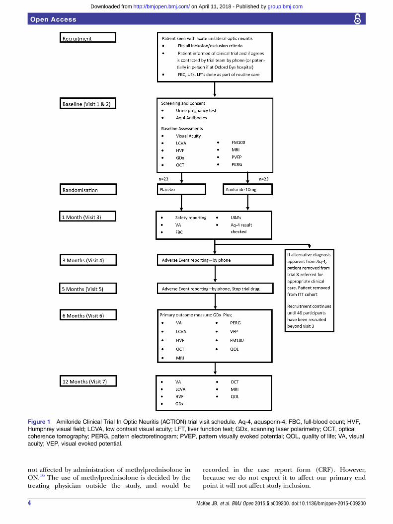
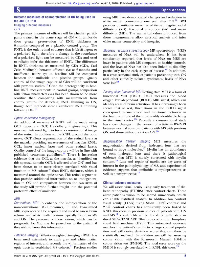
Study designThe ACTION trial is a phase II randomised, doubleblind, investigator led, parallel group placebo controlledtrial to examine the neuroprotective efficacy of
amiloride treatment following ON (figure 1). We willrecruit 46 participants who have either a clinically iso-lated syndrome of ON, or ON in participants with MSpreviously unaffected by ON. Participants will be rando-mised on a 1:1 basis to amiloride or placebo for5 months treatment. The primary outcome measure willbe peripapillary RNFL thickness on GDx at 6 monthscompared to unaffected fellow eye at baseline.Secondary outcome measures will be visual outcomes(visual acuity, LCVA, visual field and colour vision), MRIand GDx/OCT measures at baseline, 6 and 12 months,PVEP and PERG at baseline and 6 months. The12 month measurements will assess if the potentialbenefit from amiloride is sustained and whether or nota rebound neurodegeneration occurs when off activedrug. Placebo treatment identical to amiloride andblinding of both the investigators and participants totreatment is being utilised to minimise bias. We proposeto combine longitudinal measurements of RNFL thick-ness determined by both GDx and OCT to provide evi-dence of the neuroprotective efficacy of amiloride. Thiswill be supplemented by clinical and electrophysiologicaloutcomes and non-conventional MRI measures. In thetreatment arm of the study we will use 10 mg of amilor-ide. This replicates the efficacious and well-tolerateddose used in our preliminary study in primary progres-sing multiple sclerosis participants, and is consistent withthe licenced dose of amiloride.We aim to randomise and initiate treatment within a
maximum of 28 days from onset of visual symptoms butit is expected that a median of 14 days will be achieved.This timeframe has been chosen because longitudinalstudies have shown that RNFL loss is greatest within3 months after onset of ON.10 Therefore, initiation oftherapy as early as possible is likely to positively influ-ence the primary outcome measure; showing thatamiloride has a neuroprotective effect. Treatment will befor a 5-month period from randomisation; this will allowa therapeutic efficacy during the early period of themajority of RNFL loss. This allows for a 4 week ‘washout’period prior to imaging, to ameliorate any potentialinfluence on tissue volume from the diuretic effect ofamiloride. Similarly, imaging outcomes incorporatingvolumetric analyses (MRI, OCT, GDx) will be acquiredprior to randomisation to minimise any potential con-founding effect of the drug. In addition, MRI prior torandomisation will mitigate potential inclusion of com-pressive optic neuropathies.In current clinical practice some patients may be given
a course of oral or intravenous methylprednisolone atthe initial presentation of ON. Methylprednisolone hasbeen shown to hasten the recovery of ON, but notchange the visual outcome.2 Given the systemic sideeffects of high-dose steroids, this is usually restricted to asmall group of patients in whom symptoms are severelyrestricting their activities of daily living. It has beenshown that our primary outcome of RNFL thickness is
McKee JB, et al. BMJ Open 2015;5:e009200. doi:10.1136/bmjopen-2015-009200 3
Open Access
group.bmj.com on April 11, 2018 - Published by http://bmjopen.bmj.com/Downloaded from
not affected by administration of methylprednisolone inON.16 The use of methylprednisolone is decided by thetreating physician outside the study, and would be
recorded in the case report form (CRF). However,because we do not expect it to affect our primary endpoint it will not affect study inclusion.
Figure 1 Amiloride Clinical Trial In Optic Neuritis (ACTION) trial visit schedule. Aq-4, aqusporin-4; FBC, full-blood count; HVF,
Humphrey visual field; LCVA, low contrast visual acuity; LFT, liver function test; GDx, scanning laser polarimetry; OCT, optical
coherence tomography; PERG, pattern electroretinogram; PVEP, pattern visually evoked potential; QOL, quality of life; VA, visual
acuity; VEP, visual evoked potential.
4 McKee JB, et al. BMJ Open 2015;5:e009200. doi:10.1136/bmjopen-2015-009200
Open Access
group.bmj.com on April 11, 2018 - Published by http://bmjopen.bmj.com/Downloaded from
Outcome measures of neuroprotection in ON being used inthe ACTION trialImaging outcome measuresGDXThe primary measure of efficacy will be whether partici-pants treated in the acute stage of ON with amilorideshow greater preservation of RNFL thickness at6 months compared to a placebo control group. TheRNFL is the only retinal structure that is birefringent toa polarised light, therefore a change in the retardanceof a polarised light can be measured by GDx and usedto reliably infer the thickness of RNFL. The differencein RNFL thickness, as measured by GDx (GDx, CarlZeiss Meditech) between affected eye at 6 months andunaffected fellow eye at baseline will be comparedbetween the amiloride and placebo groups. Qualitycontrol of the image capture of GDx will be consistentwith previous studies.17 Given the heterogeneity in base-line RNFL measurements in control groups, comparisonwith fellow unaffected eyes has been shown to be moresensitive than comparing with measurements fromcontrol groups for detecting RNFL thinning in ON,though both methods show a significant RNFL thinningfollowing ON.18
Optical coherence tomographyAn additional measure of RNFL will be made usingOCT (Spectralis OCT, Heidelberg Engineering). Thisuses near infra-red light to form a cross-sectional imageof the retina. In addition to the RNFL around the opticnerve, OCT allows segmentation of the retinal layers atthe macula, providing measurements of macular RNFL,GCL, inner nuclear layer and outer retinal layers.Quality control of the image capture will be in line withpublished consensus guidelines.19 There is increasingevidence that the GCL at the macula, as identified onthis spectral domain OCT, is affected after ON8 and hasbeen shown to be more closely correlated with visualfunction in MS cohorts20 than RNFL thickness, which ismeasured around the optic nerve. This retinal segmenta-tion provides additional information on neurodegenera-tion in ON and comparison between the two arms ofthe study will provide further insight into the potentialprotective effect of amiloride.
MRIConventional MRI To enhance the interpretation of thenon-conventional MRI measures, T1 and T2-weightedMRI sequences will be acquired and used to assess brainvolume and white matter lesions typically found in MSand ON. The presence of these lesions, which can beprognostic for MS, may be passed on to the patient ifthey wish to know this information.
Diffusion imaging Diffusion-weighted imaging (DWI) hasbeen used extensively to study grey and white matterregions of interest, and recently the white matter of theoptic tracts in established MS cohorts.21 Previous studies
using MRI have demonstrated changes and reduction inwhite matter connectivity one year after ON.22 DWIprovides quantitative measures of tissue integrity; radialdiffusivity (RD), fractional anisotropy (FA) and meandiffusivity (MD). The numerical values produced fromthese measurements allow statistical analysis and inferwhite matter connectivity and integrity.
Magnetic resonance spectroscopy MR spectroscopy (MRS)measures of NAA will be undertaken. It has beenconsistently reported that levels of NAA on MRS arelower in patients with MS compared to healthy controls,and the level of NAA has also been linked to disability,particularly in the early stages of disease.23 In addition,in a cross-sectional study of patients presenting with ONand other clinically isolated syndromes, levels of NAAwere low.24
Resting state functional MRI Resting state MRI is a form offunctional MRI (fMRI). FMRI measures the bloodoxygen level-dependant (BOLD) MRI signal, which canidentify areas of brain activation. It has increasingly beenshown that at rest, fluctuations in the BOLD signalcorrespond to anatomical and functional networks inthe brain, with one of the most readily identifiable beingin the visual cortex.25 Recently a cross-sectional studyhas shown changes in the pattern of activity in RS-fMRIbetween normal controls, patients with MS with previousON and those without previous ON.26
Magnetisation transfer imaging MTI measures themagnetisation derived from hydrogen ions that arebound to large molecules.27 Myelin has an abundanceof such hydrogen ions and there is pathologicalevidence that MTI is closely correlated with myelincontent.28 Loss and repair of myelin are key areas ofinterest in the pathophysiology of MS, and experimentalevidence suggests that amiloride is myeloprotective aswell as neuroprotective.13
Clinical outcome measuresWe will assess visual acuity using early treatment of dia-betic retinopathy (ETDRS) letter contrast charts. Theseallow patient’s vision to be scored numerically, whichcan enable statistical analysis. In addition, low contrastvisual acuity (LCVA) using Sloan 1.25% contrast and2.5% contrast charts has consistently been linked toRNFL thickness in previous studies of patients with ONand MS.11 Visual fields will be tested using the standar-dised SITA-STANDARD 30–2 protocol on the Humphreyvisual field machine (HVF). This automated sequencematches the patient’s results to a large control popula-tion and will derive deviation scores that can then bestatistically analysed. In addition we will be assessingcolour vision with the Farnsworth Munsell 100-Huecolour vision test (FM100). The total error score on theFM100 is strongly correlated with RNFL thickness.29
McKee JB, et al. BMJ Open 2015;5:e009200. doi:10.1136/bmjopen-2015-009200 5
Open Access
group.bmj.com on April 11, 2018 - Published by http://bmjopen.bmj.com/Downloaded from
Electrophysiological outcome measuresProlongation of the latency of the P100 deflection of thepattern visually evoked potential (PVEP) are characteris-tic in acute ON. This then improves but remains delayedin absolute terms after visual recovery,30 thus providing auseful diagnostic tool that is used in clinical practice,and as an outcome measure in therapeutic trials.Furthermore the pattern electroretinogram (PERG) aidsthe interpretation of the PVEP by providing an assess-ment of macular function. The amplitude of the PERGis partially derived from the GCL component of theRNFL,31 and reductions in it are readily identifiable inON.32
Quality of LifeWe aim to measure vision-related quality of life scoresusing the standardised national eye institute visual func-tioning questionnaire (NEI-VFQ-25) with a 10-itemneuro-ophthalmic supplement (10-NOS). TheNEI-VFQ-25 provides a standardised quality of life scorefor ophthalmic disease validated across ophthalmic epi-demiological studies.33 Both NEI-VFQ-25 and 10-NOS
have been significantly correlated with segmentedretinal layers on spectral domain OCT in ON.34
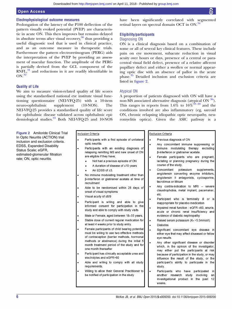
Eligibility/participantsDiagnosing ONON is a clinical diagnosis based on a combination ofsome or all of several key clinical features. These includepain on eye movement, subacute reduction in visualacuity over hours or days, presence of a central or para-central visual field defect, presence of a relative afferentpupillary defect and either a swollen or normal appear-ing optic disc with an absence of pallor in the acutephase.35 Detailed inclusion and exclusion criteria arelisted in figure 2.
Atypical ONA proportion of patients diagnosed with ON will have anon-MS associated alternative diagnosis (atypical ON 36).This ranges in reports from 1.6% to 16%37–39 and theconditions involved are also inflammatory (eg, sarcoidON, chronic relapsing idiopathic optic neuropathy, neu-romyelitis optica). Given the ASIC pathway is a
Figure 2 Amiloride Clinical Trial
In Optic Neuritis (ACTION) trial
inclusion and exclusion criteria.
EDSS, Expanded Disability
Status Scale; eGFR,
estimated-glomerular filtration
rate; ON, optic neuritis.
6 McKee JB, et al. BMJ Open 2015;5:e009200. doi:10.1136/bmjopen-2015-009200
Open Access
group.bmj.com on April 11, 2018 - Published by http://bmjopen.bmj.com/Downloaded from
downstream mechanism of neurodegeneration ingeneral CNS inflammation, we would not anticipate thatamiloride would be detrimental to these groups.The clinical course of an atypical ON is such that the
diagnosis may not be discriminated from typical ON untilafter a poor recovery of vision in the initial weeks afterON. In addition, in the case of neuromyelitis optica, thediagnosis can be confirmed following positiveantiaquaporin-4 antibody testing. However, this result willnot be available until visit 3 (at 4 weeks). Once identified,these participants will be removed from theintention-to-treat (ITT) cohort and referred for appro-priate care. Other differential diagnoses will remain inthe ITT cohort and also be referred for appropriate clin-ical care. However, should these participants requireimmunosuppressive treatment they will be removed fromthe final analysis. We accounted for a 10% rate of atypicalON in our study powering, in addition to removing parti-cipants with neuromyelitis optica from the ITT cohort.We would aim to have a much smaller number of residualatypical ON than this, however, given the need for earlyrecruitment necessary for neuroprotection, we recognisethe potential for atypical ON to enter the cohort.
Ischaemic optic neuropathiesThe age group eligibility criteria for this trial wereselected to minimise the potential inclusion of patientswith ischaemic optic neuropathies (anterior ischaemicoptic neuropathy). While these can be confused withON at initial diagnosis, they generally differ in their clin-ical presentation and natural history. Participants subse-quently diagnosed with ischaemic optic neuropathy willbe removed from the ITT analysis. Moreover, we do notanticipate any harm from amiloride in these groups asanimal models of CNS ischaemia, have implicatedASIC1 as an important factor in neuronal injury withblockade providing neuroprotective benefit.40
Study proceduresStudy procedures will all take place at the John Radcliffehospital and the FMRIB centre at the John Radcliffesite.
Screening and eligibility assessmentParticipants will be identified at the time of their presen-tation with ON, by the clinical care team at the trial site(Oxford) or patient identification centres (PIC) andgiven the participant information leaflet (PIL) at thatpoint. With the patient’s permission, which will be docu-mented in the notes by the treating ophthalmologist orneurologist, they will be telephoned by the trial teamand invited to come to the trial centre in Oxford.Prescreening will take place in the form of discussion
between the trial centre and the recruiting physician inorder to ensure that the patient meets all the medicalinclusion and exclusion criteria. Participants will havehad an eye examination as part of their routine clinicalcare, this will only be repeated by the trial team at
screening if the trial team deem it necessary to confirmthe diagnosis of ON.
Informed consentWritten and verbal versions of the participant informa-tion and informed consent will be presented to theparticipants.Written informed consent will then be obtained by
means of participant dated signature and dated signa-ture of the named trial investigator who discussed thetrial and obtained the informed consent.
RandomisationSubject numbers will be assigned sequentially as eachsubject enters the study. The subjects will be assigned astudy drug through a centralised randomisation softwarehosted by the UKCRC registered Oxford CognitiveHealth and Neuroscience Clinical Trials Unit. The ran-domisation will be performed by study investigatorswhose access to the software does not allow unblinding.If requested on clinical grounds the decision to unblindwill be made by the chief investigator. The randomisa-tion log and unblinding envelopes are held in the JohnRadcliffe Pharmacy with a 24 h on call service. Arandom-deterministic minimisation algorithm will beused to produce treatment groups balanced for import-ant prognostic factors. The first 10% of participants willbe allocated randomly without minimisation to avoidpredictability. Subsequently the minimisation algorithmwill be applied with an allocation ratio that is not fullydeterministic: there will be an 80% bias in favour of allo-cations that minimise the imbalance.The randomisation algorithm will minimise for the fol-
lowing three variables related to prognosis at baseline:▸ Sex (male or female)▸ Number of weeks since onset of symptoms (<2 weeks;
≥2 weeks)▸ Severity of visual acuity (between ≤6/9 and ≥6/18, ie,
mildly affected; <6/18 and worse, ie, severelyaffected).The study drug will be labelled with the study number
and unique pack identification number. The two treat-ments amiloride and placebo will be indistinguishable.
StatisticsSample sizeIt is estimated that the maximum total number of parti-cipants randomised will be 50 (25 per arm). Thirty sixparticipants are required to detect a difference of7.4 μm between groups on the primary outcome (differ-ence in RNFL between affected and unaffected eye).This assumes a SD of 6.6, power of 90% and a 5% sig-nificance level. Allowing for a 10% drop out rate thisincreases to 40. However, as described previously, it isthought that approximately 10% of participants mayhave atypical ON. Therefore, to ensure that there isenough power to detect this difference in the typical
McKee JB, et al. BMJ Open 2015;5:e009200. doi:10.1136/bmjopen-2015-009200 7
Open Access
group.bmj.com on April 11, 2018 - Published by http://bmjopen.bmj.com/Downloaded from
ON subgroup we will need to randomise a total of 46participants.It is likely that patients found to have neuromyelitis
optica (NMO) as described above will be given furthermedications that are potential confounding factors(listed in exclusion criteria) and they will not beincluded in the main ITT analysis. Therefore, random-isation will continue until a total of 46 participants havebeen randomised and reached the 4-week point withoutbeing diagnosed with NMO. It is thought that amaximum of 8% of participants randomised could haveNMO, increasing the possible sample size to 50.
Quality control and quality assuranceThere will be an independent data and safety monitor-ing committee (DMC) made up of an independent con-sultant neurologist, an independent consultantophthalmologist, an independent statistician and thetrial statistician. They will meet and perform interimanalysis according to the DMC charter.The trial investigators and the DMC will have access to
the final trial data set.Regular monitoring will be performed according to
ICH GCP by Oxford Cognitive Health and NeuroscienceClinical Trials Unit. Data will be evaluated for compli-ance with the protocol and accuracy in relation tosource documents.Overall supervision of the trial will be co-ordinated
through a trial steering committee consisting of anexternal consultant neurologist with experience in clin-ical trials in neuroprotection in ON and MS, the PI, thestatistician, the coapplicants, a lay member and researchnurses. The trial steering committee will meet every6 months supplemented with teleconferencing asrequired by its members.
ConfidentialityData will be stored within a firewall and password-protected computer system within a swipe-card securedbuilding. Investigators will ensure that the participants’anonymity is maintained. The participants will be identi-fied only by initials and a participants ID number on theCRF and any electronic database. All documents will bestored securely and only accessible by trial staff andauthorised personnel. The study will comply with theData Protection Act which requires data to be anon-ymised as soon as it is practical to do so.
AnalysisInclusion in analysisThe primary analysis will be carried out on a modifiedintention to treat cohort. This cohort will include allrandomised participants (analysed in the group to whichthey were randomised) with the exception of those diag-nosed with NMO in the first 4 weeks fromrandomisation.A further sensitivity analysis will be carried out includ-
ing all randomised participants with typical ON only.
They will be analysed in the group to which they wererandomised.
Analysis of primary outcomeThe primary outcome will be presented using meansand SDs and the difference between the two groups willbe presented as the difference in means with 95% CI.The primary outcome will be analysed using an analysisof covariance to assess the treatment effect at 6 months,adjusting for baseline value and minimisation factors.We will formally assess the distribution of the data forevidence of departure from normality and, if necessary,transform the data or analyse using a non-parametricequivalent. A 5% level of significance will be used.
Analysis of secondary outcomesContinuous secondary outcomes (OCT, letter score,electrophysiological data, colour vision data, visual fielddata and quality of life data) will be analysed in thesame way as the primary outcome.MRI analysis will be performed using FSL tools
(http://www.fmrib.ox.ac.uk/fsl)41 and will include botha region of interest approach and measures of wholebrain metrics to assess for differences between amilorideand placebo groups.
DISCUSSIONNeuroprotective strategies and efficient means to testthem within a clinical trial setting remain key unmetneeds in ON and MS. ACTION is a phase II clinical trialaiming to provide proof of concept that amiloride isneuroprotective in acute inflammatory ON. The trialencompasses a multimodal approach combining struc-tural and functional outcomes with clinical measuresenabling the refinement of future trial designs.However, the recruitment of acute patients with ON
within a short time window to a clinical trial setting canbe challenging and requires a high degree of cooperationbetween clinical and research staff in order to give poten-tial participants the opportunity to take part. Despite this,within the spectrum of CNS structures affected by MS,one of the major values of using ON to assess neuropro-tection is that the visual system provides unique oppor-tunities to do so in a timely and efficient manner.
Ethics and disseminationThe findings from ACTION will be disseminatedthrough peer-reviewed publications and at scientific con-ferences. Each participant will receive a summary of theoverall results.
Author affiliations1Division of Clinical Neurology, Nuffield Department of Clinical Neurosciences,University of Oxford, John Radcliffe Hospital, Oxford, UK2Oxford Eye Hospital, John Radcliffe Hospital, Oxford, UK3Department of Clinical Neuroscience, University of Nottingham MedicalSchool, Queens Medical Centre, Nottingham, UK4Centre for Statistics in Medicine, University of Oxford, Oxford, UK
8 McKee JB, et al. BMJ Open 2015;5:e009200. doi:10.1136/bmjopen-2015-009200
Open Access
group.bmj.com on April 11, 2018 - Published by http://bmjopen.bmj.com/Downloaded from
5Weatherall Institute of Molecular Medicine, University of Oxford, JohnRadcliffe Hospital, Oxford, UK6Nuffield Department of Clinical Neurosciences, Oxford Centre for FunctionalMRI of the Brain (FMRIB), University of Oxford, John Radcliffe Hospital,Oxford, UK
Acknowledgements The authors would like to thank Abigail Koelewyn for herhelp in formatting of the manuscript and figures, and Dr Charles Cottrial forhis help with the electrophysiological outcomes in the trial.
Contributors JBM and MJC designed the trial protocol, with scientific insightand contributions from CK, JE, JP, NE, LF and YK. JBM prepared the firstdraft of this manuscript and it was reviewed by MC, CK, JE, JP, NE, LF, SGand YK. Revisions were performed by JBM and MJC.
Funding This work is supported by the Multiple Sclerosis Society in the UK,grant reference 952/11.
Competing interests MJC received an unrestricted grant from Biogen fortechnical equipment used within the study. The remaining author’s declare nocompeting interests.
Ethics approval South Central Oxford Rec B. ref: 13/SC/022.
Provenance and peer review Not commissioned; externally peer reviewed.
Open Access This is an Open Access article distributed in accordance withthe Creative Commons Attribution Non Commercial (CC BY-NC 4.0) license,which permits others to distribute, remix, adapt, build upon this work non-commercially, and license their derivative works on different terms, providedthe original work is properly cited and the use is non-commercial. See: http://creativecommons.org/licenses/by-nc/4.0/
REFERENCES1. Trapp BD, Ransohoff R, Rudick R. Axonal pathology in multiple
sclerosis: relationship to neurologic disability. Curr Opin Neurol1999;12:295–302.
2. Pau D, Al Zubidi N, Yalamanchili S, et al. Optic neuritis. Eye (Lond)2011;25:833–42.
3. Beck RW, Gal RL, Bhatti MT, et al. Visual function more than 10years after optic neuritis: experience of the optic neuritis treatmenttrial. Am J Ophthalmol 2004;137:77–83.
4. Sisto D, Trojano M, Vetrugno M, et al. Subclinical visual involvementin multiple sclerosis: a study by MRI, VEPs, frequency-doublingperimetry, standard perimetry, and contrast sensitivity. InvestOphthalmol Vis Sci 2005;46:1264–8.
5. Toussaint D, Perier O, Verstappen A, et al. Clinicopathological studyof the visual pathways, eyes, and cerebral hemispheres in 32 casesof disseminated sclerosis. J Clin Neuroophthalmol 1983;3:211–20.
6. Kerrison JB, Flynn T, Green WR. Retinal pathologic changes inmultiple sclerosis. Retina 1994;14:445–51.
7. Suhs KW, Hein K, Sattler MB, et al. A randomized, double-blind,phase 2 study of erythropoietin in optic neuritis. Ann Neurol2012;72:199–210.
8. Syc SB, Saidha S, Newsome SD, et al. Optical coherencetomography segmentation reveals ganglion cell layer pathology afteroptic neuritis. Brain 2012;135(Pt 2):521–33.
9. Trip SA, Schlottmann PG, Jones SJ, et al. Scanning laserpolarimetry quantification of retinal nerve fiber layer thinningfollowing optic neuritis. J Neuroophthalmol 2010;30:235–42.
10. Henderson AP, Altmann DR, Trip AS, et al. A serial study of retinalchanges following optic neuritis with sample size estimates for acuteneuroprotection trials. Brain 2010;133:2592–602.
11. Petzold A, de Boer JF, Schippling S, et al. Optical coherencetomography in multiple sclerosis: a systematic review andmeta-analysis. Lancet Neurol 2010;9:921–32.
12. Stys PK. General mechanisms of axonal damage and its prevention.J Neurol Sci 2005;233:3–13.
13. Vergo S, Craner MJ, Etzensperger R, et al. Acid-sensing ion channel1 is involved in both axonal injury and demyelination in multiplesclerosis and its animal model. Brain 2011;134(Pt 2):571–84.
14. Friese MA, Craner MJ, Etzensperger R, et al. Acid-sensing ionchannel-1 contributes to axonal degeneration in autoimmuneinflammation of the central nervous system. Nat Med 2007;13:1483–9.
15. Arun T, Tomassini V, Sbardella E, et al. Targeting ASIC1 in primaryprogressive multiple sclerosis: evidence of neuroprotection withamiloride. Brain 2013;136(Pt 1):106–15.
16. Naismith RT, Tutlam NT, Xu J, et al. Optical coherence tomographyis less sensitive than visual evoked potentials in optic neuritis.Neurology 2009;73:46–52.
17. Frohman EM, Dwyer MG, Frohman T, et al. Relationship of opticnerve and brain conventional and non-conventional MRI measuresand retinal nerve fiber layer thickness, as assessed by OCT andGDx: a pilot study. J Neurol Sci 2009;282:96–105.
18. Kupersmith MJ, Mandel G, Anderson S, et al. Baseline, one andthree month changes in the peripapillary retinal nerve fiber layer inacute optic neuritis: relation to baseline vision and MRI. J Neurol Sci2011;308:117–23.
19. Tewarie P, Balk L, Costello F, et al. The OSCAR-IB consensuscriteria for retinal OCT quality assessment. PLoS ONE 2012;7:e34823.
20. Saidha S, Syc SB, Durbin MK, et al. Visual dysfunction in multiplesclerosis correlates better with optical coherence tomographyderived estimates of macular ganglion cell layer thickness thanperipapillary retinal nerve fiber layer thickness. Mult Scler2011;17:1449–63.
21. Dasenbrock HH, Smith SA, Ozturk A, et al. Diffusion tensor imagingof the optic tracts in multiple sclerosis: association with retinalthinning and visual disability. J Neuroimaging 2011;21:e41–9.
22. Ciccarelli O, Toosy AT, Hickman SJ, et al. Optic radiation changesafter optic neuritis detected by tractography-based group mapping.Hum Brain Mapp 2005;25:308–16.
23. De Stefano N, Narayanan S, Francis GS, et al. Evidence of axonaldamage in the early stages of multiple sclerosis and its relevance todisability. Arch Neurol 2001;58:65–70.
24. Wattjes MP, Harzheim M, Lutterbey GG, et al. High field MR imagingand 1H-MR spectroscopy in clinically isolated syndromes suggestiveof multiple sclerosis: correlation between metabolic alterations anddiagnostic MR imaging criteria. J Neurol 2008;255:56–63.
25. Wang K, Jiang T, Yu C, et al. Spontaneous activity associated withprimary visual cortex: a resting-state FMRI study. Cerebral Cortex2008;18:697–704.
26. Gallo A, Esposito F, Sacco R, et al. Visual resting-state network inrelapsing-remitting MS with and without previous optic neuritis.Neurology 2012;79:1458–65.
27. Audoin B, Fernando KT, Swanton JK, et al. Selective magnetizationtransfer ratio decrease in the visual cortex following optic neuritis.Brain 2006;129(Pt 4):1031–9.
28. Schmierer K, Scaravilli F, Altmann DR, et al. Magnetization transferratio and myelin in postmortem multiple sclerosis brain. Ann Neurol2004;56:407–15.
29. Henderson AP, Altmann DR, Trip SA, et al. Early factors associatedwith axonal loss after optic neuritis. Ann Neurol 2011;70:955–63.
30. Brusa A, Jones SJ, Plant GT. Long-term remyelination after opticneuritis: a 2-year visual evoked potential and psychophysical serialstudy. Brain 2001;124:468–79.
31. Holder GE. The incidence of abnormal pattern electroretinography inoptic nerve demyelination. Electroencephalogr Clin Neurophysiol1991;78:18–26.
32. Fraser CL, Holder GE. Electroretinogram findings in unilateral opticneuritis. Doc Ophthalmol 2011;123:173–8.
33. Sakai RE, Feller DJ, Galetta KM, et al. Vision in multiple sclerosis:the story, structure-function correlations, and models forneuroprotection. J Neuroophthalmol 2011;31:362–73.
34. Walter SD, Ishikawa H, Galetta KM, et al. Ganglion cell loss inrelation to visual disability in multiple sclerosis. Ophthalmology2012;119:1250–7.
35. Costello F, Hodge W, Pan YI, et al. Tracking retinal nerve fiber layerloss after optic neuritis: a prospective study using optical coherencetomography. Mult Scler 2008;14:893–905.
36. Hickman SJ, Ko M, Chaudhry F, et al. Optic neuritis: an updatetypical and atypical optic neuritis. Neuroophthalmology2008;32:237–48.
37. Hickman SJ, Dalton CM, Miller DH, et al. Management of acuteoptic neuritis. Lancet 2002;360:1953–62.
38. Wakakura M, Minei-Higa R, Oono S, et al. Baseline features ofidiopathic optic neuritis as determined by a multicenter treatment trialin Japan. Optic Neuritis Treatment Trial Multicenter CooperativeResearch Group (ONMRG). Jpn J Ophthalmol 1999;43:127–32.
39. Storoni M, Pittock SJ, Weinshenker BG, et al. Optic neuritis in anethnically diverse population: higher risk of atypical cases in patientsof African or African-Caribbean heritage. J Neurol Sci2012;312:21–5.
40. Xiong Z, Zhu X, Chu X, et al. Neuroprotection in ischemia: blockingcalcium-permeable acid-sensing ion channels. Cell2004;118:687–98.
41. Jenkinson M, Beckmann CF, Behrens TE, et al. FSL. Neuroimage2012;62:782–90.
McKee JB, et al. BMJ Open 2015;5:e009200. doi:10.1136/bmjopen-2015-009200 9
Open Access
group.bmj.com on April 11, 2018 - Published by http://bmjopen.bmj.com/Downloaded from
blind, placebo controlled trial(ACTION) protocol: a randomised, double Amiloride Clinical Trial In Optic Neuritis
Matthew CranerFugger, Christopher Kennard, Yazhuo Kong, Jacqueline Palace and Justin B McKee, John Elston, Nikos Evangelou, Stephen Gerry, Lars
doi: 10.1136/bmjopen-2015-0092002015 5: BMJ Open
http://bmjopen.bmj.com/content/5/11/e009200Updated information and services can be found at:
These include:
References http://bmjopen.bmj.com/content/5/11/e009200#ref-list-1
This article cites 41 articles, 1 of which you can access for free at:
Open Access
http://creativecommons.org/licenses/by-nc/4.0/non-commercial. See: provided the original work is properly cited and the use isnon-commercially, and license their derivative works on different terms, permits others to distribute, remix, adapt, build upon this workCommons Attribution Non Commercial (CC BY-NC 4.0) license, which This is an Open Access article distributed in accordance with the Creative
serviceEmail alerting
box at the top right corner of the online article. Receive free email alerts when new articles cite this article. Sign up in the
CollectionsTopic Articles on similar topics can be found in the following collections
(107)Ophthalmology (460)Neurology
Notes
http://group.bmj.com/group/rights-licensing/permissionsTo request permissions go to:
http://journals.bmj.com/cgi/reprintformTo order reprints go to:
http://group.bmj.com/subscribe/To subscribe to BMJ go to:
group.bmj.com on April 11, 2018 - Published by http://bmjopen.bmj.com/Downloaded from