alkb recognition of a bulky dna base adduct stabilized by chemical cross-linking
TRANSCRIPT
SCIENCE CHINA Chemistry
© Science China Press and Springer-Verlag Berlin Heidelberg 2010 chem.scichina.com www.springerlink.com
*Corresponding author (email: [email protected])
• ARTICLES • January 2010 Vol.53 No.1: 86–90
doi: 10.1007/s11426-010-0008-0
AlkB recognition of a bulky DNA base adduct stabilized by chemical cross-linking
YI ChengQi1 & HE Chuan1,2*
1Department of Chemistry, University of Chicago, Chicago, Illinois 60637, USA; 2Department of Chemical Biology, College of Chemistry and Molecular Engineering, Peking University, Beijing 100871, China
Received September 19, 2009; accepted October 9, 2009
E. coli AlkB is a direct DNA/RNA repair protein that oxidatively reverses N1 alkylated purines and N3 alkylated pyrimidines to regular bases. Previous crystal structures have revealed N1-methyl adenine (1-meA) recognition by AlkB and a unique base flipping mechanism, but how the AlkB active site can accommodate bulky base adducts is largely unknown. Employing a pre-viously developed chemical cross-linking technique, we crystallized AlkB with a duplex DNA containing a caged thymine base (cagedT). The structure revealed a flexible hairpin lid and a reorganized substrate recognition loop used by AlkB to ac-commodate cagedT. These observations demonstrate, at the molecular level, how bulky DNA adducts may be recognized and processed by AlkB.
DNA/RNA repair, AlkB, caged thymine, disulfide cross-linking, bulky base adduct
1 Introduction
DNA can be damaged by environmental factors, such as UV lights and chemotherapy agents, or by chemicals generated from normal metabolic processes, e.g. reactive oxygen spe-cies. Among these DNA damaging factors, alkylation agents constitute a large class which can generate cytotoxic and/or mutagenic DNA lesions. When E. coli is challenged with methylating agents, four genes, ada, aidB, alkA and alkB, are upregulated during the adaptive response pathway [1]. The Ada protein is a multifunctional methyltransferase and transcription activator as well; AidB, the exact function of which is still unknown, was proposed to prevent DNA alkylation damage by destroying alkylating agents; AlkA is a glycosylase with a broad range of substrates; and AlkB is a demethylase that oxidatively repairs N1-methyl adenine (1-meA) and N3-methyl cytosine in DNA/RNA [2, 3]. Sev-eral AlkB human homologs, ABH1, ABH2, ABH3 and
FTO, have been shown to possess similar demethylation activity [4–9]. ABH2 is established as a house-keeping en-zyme that guards the mammalian genome against 1-meA and FTO has been linked to obesity in humans [8, 10].
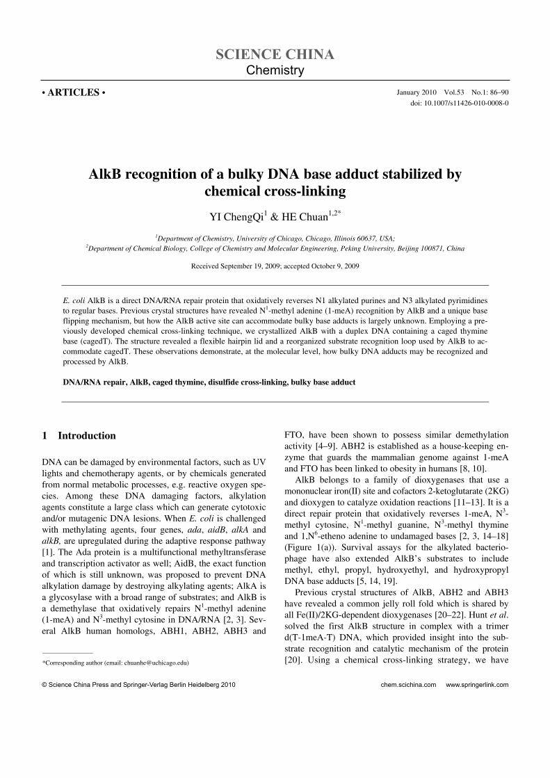
AlkB belongs to a family of dioxygenases that use a mononuclear iron(II) site and cofactors 2-ketoglutarate (2KG) and dioxygen to catalyze oxidation reactions [11–13]. It is a direct repair protein that oxidatively reverses 1-meA, N3- methyl cytosine, N1-methyl guanine, N3-methyl thymine and 1,N6-etheno adenine to undamaged bases [2, 3, 14–18] (Figure 1(a)). Survival assays for the alkylated bacterio-phage have also extended AlkB’s substrates to include methyl, ethyl, propyl, hydroxyethyl, and hydroxypropyl DNA base adducts [5, 14, 19].
Previous crystal structures of AlkB, ABH2 and ABH3 have revealed a common jelly roll fold which is shared by all Fe(II)/2KG-dependent dioxygenases [20–22]. Hunt et al. solved the first AlkB structure in complex with a trimer d(T-1meA-T) DNA, which provided insight into the sub-strate recognition and catalytic mechanism of the protein [20]. Using a chemical cross-linking strategy, we have
YI ChengQi, et al. Sci China Chem January (2010) Vol.53 No.1 87
Figure 1 Base lesions that are repaired by AlkB (a) and a bulky caged base, cagedT (b), used in this study. CagedT will be decaged, within minutes, under irradiation with 365 nm UV light to give a regular thymine base.
solved crystal structures of AlkB with a bound double- stranded DNA (dsDNA) [22]. These structures revealed a unique base-flipping mechanism exploited by AlkB and explained its preference towards single-stranded DNA (ssDNA); we have also obtained ABH2-dsDNA structures which established ABH2 as a duplex DNA-binding protein and provided molecular basis for future design of inhibitors for this human protein [22]. A crystal structure of ABH3 reported by Slupphaug et al. showed the self-oxidation of an active site leucine that might protect against uncoupled generation of dangerous oxygen radicals [21].
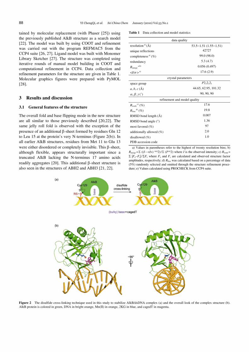
In all of these structures, detailed interactions between 1-meA and protein active site residues have been revealed; however, how AlkB potentially accommodates DNA ad-ducts bigger than a methyl group remains unknown. To answer this question, we crystallized AlkB bound to a 13mer dsDNA containing a 6-nitropiperonyloxymethyl (NPOM) caged thymine (Figure 1(b)), using the previously developed disulfide cross-linking technique [22, 23] (Figure 2(a)). This new structure revealed the high flexibility of a hairpin lid (67 Leu–79 Ser) and a substrate recognition loop (Lys 134–139 Leu), which allows AlkB to accommodate bulky adducts in its active site.
2 Materials and method
2.1 Oligonucleotide synthesis
Oligonucleotides containing cagedT and disulfide-tethered cytosine were prepared by incorporating the “cagedT” phosphoramidite (BERRY&ASSOCIATES, Inc.) and O4- triazolyl-dU-CE phosphoramidite (Glen Research, Inc.) at the desired position during solid-phase synthesis [23]. To deprotect the oligonucleotides, 180 μL 1:1 cystamine:water solution was added to the solid support from a 1 umol-scale DNA synthesis and the mixture was left at room tempera-ture for 16 h before further purification. All synthetic oli-gonucleotides were purified with reverse-phase HPLC. The
lyophilized DNA residue was dissolved in 50 μL buffer (10 mM Tris, pH 7.4).
2.2 Cross-linking, purification and crystallization of the AlkB-dsDNA complexes
A truncated AlkB with deletion of the N-terminal 11 amino acids was cloned into a pET30a vector (Novagen) and overexpressed in E. coli BL21(DE3) [20]. The protein was purified following a previously described procedure [24]. The Ser 129 to Cys mutation was introduced using the QuikChange Site-Directed Mutagenesis Kit (Stratagene). Synthetic oligonucleotides (DNA: 5′-TAGGTAA(cagedT)
AC*CGT-3′ and its complement 5′-AACGGTA_TACCT-3′; C* is a disulfide-tethered cytosine and “_” stands for an abasic site) were synthesized, annealed, and cross-linked to the S129C mutant AlkB as described [23]. The covalently linked AlkB-dsDNA complexes were purified using Mono- Q anion exchange chromatography (GE Healthcare), after which the buffer was exchanged to 100 mM NaCl, 10 mM Tris-HCl (pH 7.4), and the complexes were concentrated to 12–15 mg/mL and filtered with 0.22 μm filters. The com-plexes were crystallized by the hanging drop method at 4°C. 1.0 mM MnCl2 (maganese ion occupies the metal site but doesn’t support catalysis) and 2.0 mM 2KG were added to the complexes, and they were mixed in a 1:1 ratio with well solution containing 100 mM NaCl, 50 mM MgCl2, 100 mM cacodylate (pH 6.5), and 20% w/v PEG 8K. Crystals ap-peared after 2–4 days and were allowed to grow for several weeks. They were transferred into a cryo-protectant solution containing the reservoir solution plus 20% glycerol, and frozen in liquid nitrogen for X-ray diffraction data collec-tion.
2.3 Structure determination, refinement and model building
The phase of the AlkB-DNA complex structure was ob-
88 YI ChengQi, et al. Sci China Chem January (2010) Vol.53 No.1
tained by molecular replacement (with Phaser [25]) using the previously published AlkB structure as a search model [22]. The model was built by using COOT and refinement was carried out with the program REFMAC5 from the CCP4 suite [26, 27]. Ligand model was built with Monomer Library Sketcher [27]. The structure was completed using iterative rounds of manual model building in COOT and computational refinement in CCP4. Data collection and refinement parameters for the structure are given in Table 1. Molecular graphics figures were prepared with PyMOL [28].
3 Results and discussion
3.1 General features of the structure
The overall fold and base flipping mode in the new structure are all similar to those previously described [20,22]. The same jelly roll fold is observed with the exception of the presence of an additional β-sheet formed by residues Gln 12 to Leu 15 at the protein’s very N-terminus (Figure 2(b)). In all earlier AlkB structures, residues from Met 11 to Glu 13 were either disordered or completely invisible. This β-sheet, although flexible, appears structurally important since a truncated AlkB lacking the N-terminus 17 amino acids readily aggregates [20]. This additional β-sheet structure is also seen in the structures of ABH2 and ABH3 [21, 22].
Table 1 Data collection and model statistics
data quality
resolution a) (Å) 53.5–1.51 (1.55–1.51)
unique reflections 42727
completeness a) (%) 99.0 (98.0)
redundancy 5.3 (4.7)
Rmerge a,b) 0.056 (0.497)
<I/σ > a) 17.6 (2.9)
crystal parameters
space group P212121
a, b, c (Å) 44.65, 62.95, 101.32
α, β, γ (˚) 90, 90, 90
refinement and model quality
Rwork c) (%) 17.6
Rfree d) (%) 19.8
RMSD bond length (Å) 0.007
RMSD bond angle (°) 1.36
most favored (%) 97
additionally allowed (%) 2.0
disallowed (%) 1.0
PDB accession code
a) Values in parentheses refer to the highest of twenty resolution bins; b) Rmerge = Σ ((I − <I>) **2) / Σ (I**2) where I is the observed intensity; c) Rwork =
Σ |Fo−Fc| / ΣFc where Fo and Fc are calculated and observed structure factor amplitudes, respectively; d) Rfree was calculated based on a percentage of data (5%) randomly selected and omitted through the structure refinement proce-dure; e) Values calculated using PROCHECK from CCP4 suite.
Figure 2 The disulfide cross-linking technique used in this study to stabilize AlkB/dsDNA complex (a) and the overall look of the complex structure (b). AlkB protein is colored in green, DNA in bright orange, Mn(II) in orange, 2KG in blue, and cagedT in magenta.
YI ChengQi, et al. Sci China Chem January (2010) Vol.53 No.1 89
DNA has two slightly different conformations at A6 and A7 positions in this structure, but in both cases a similar distortion of the cagedT-containing strand is observed: A7 and A9 are squeezed together, with A7 being inverted by residues Thr 51 to Gly53 [22]. CagedT is completely flipped out of the duplex DNA and A8’ intercalates between A6 and A7 (Figure 2(b)).
3.2 Recognition of the cagedT at the active site
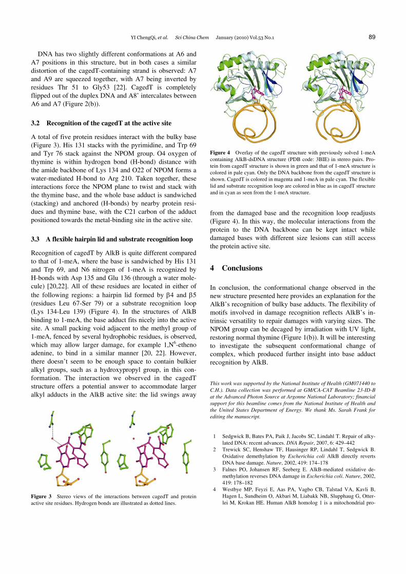
A total of five protein residues interact with the bulky base (Figure 3). His 131 stacks with the pyrimidine, and Trp 69 and Tyr 76 stack against the NPOM group. O4 oxygen of thymine is within hydrogen bond (H-bond) distance with the amide backbone of Lys 134 and O22 of NPOM forms a water-mediated H-bond to Arg 210. Taken together, these interactions force the NPOM plane to twist and stack with the thymine base, and the whole base adduct is sandwiched (stacking) and anchored (H-bonds) by nearby protein resi-dues and thymine base, with the C21 carbon of the adduct positioned towards the metal-binding site in the active site.
3.3 A flexible hairpin lid and substrate recognition loop
Recognition of cagedT by AlkB is quite different compared to that of 1-meA, where the base is sandwiched by His 131 and Trp 69, and N6 nitrogen of 1-meA is recognized by H-bonds with Asp 135 and Glu 136 (through a water mole-cule) [20,22]. All of these residues are located in either of the following regions: a hairpin lid formed by β4 and β5 (residues Leu 67-Ser 79) or a substrate recognition loop (Lys 134-Leu 139) (Figure 4). In the structures of AlkB binding to 1-meA, the base adduct fits nicely into the active site. A small packing void adjacent to the methyl group of 1-meA, fenced by several hydrophobic residues, is observed, which may allow larger damage, for example 1,N6-etheno adenine, to bind in a similar manner [20, 22]. However, there doesn’t seem to be enough space to contain bulkier alkyl groups, such as a hydroxypropyl group, in this con-formation. The interaction we observed in the cagedT structure offers a potential answer to accommodate larger alkyl adducts in the AlkB active site: the lid swings away
Figure 3 Stereo views of the interactions between cagedT and protein active site residues. Hydrogen bonds are illustrated as dotted lines.
Figure 4 Overlay of the cagedT structure with previously solved 1-meA containing AlkB-dsDNA structure (PDB code: 3BIE) in stereo pairs. Pro-tein from cagedT structure is shown in green and that of 1-meA structure is colored in pale cyan. Only the DNA backbone from the cagedT structure is shown. CagedT is colored in magenta and 1-meA in pale cyan. The flexible lid and substrate recognition loop are colored in blue as in cagedT structure and in cyan as seen from the 1-meA structure.
from the damaged base and the recognition loop readjusts (Figure 4). In this way, the molecular interactions from the protein to the DNA backbone can be kept intact while damaged bases with different size lesions can still access the protein active site.
4 Conclusions
In conclusion, the conformational change observed in the new structure presented here provides an explanation for the AlkB’s recognition of bulky base adducts. The flexibility of motifs involved in damage recognition reflects AlkB’s in-trinsic versatility to repair damages with varying sizes. The NPOM group can be decaged by irradiation with UV light, restoring normal thymine (Figure 1(b)). It will be interesting to investigate the subsequent conformational change of complex, which produced further insight into base adduct recognition by AlkB.
This work was supported by the National Institute of Health (GM071440 to C.H.). Data collection was performed at GM/CA-CAT Beamline 23-ID-B at the Advanced Photon Source at Argonne National Laboratory; financial support for this beamline comes from the National Institute of Health and the United States Department of Energy. We thank Ms. Sarah Frank for editing the manuscript.
1 Sedgwick B, Bates PA, Paik J, Jacobs SC, Lindahl T. Repair of alky-lated DNA: recent advances. DNA Repair, 2007, 6: 429–442
2 Trewick SC, Henshaw TF, Hausinger RP, Lindahl T, Sedgwick B. Oxidative demethylation by Escherichia coli AlkB directly reverts DNA base damage. Nature, 2002, 419: 174–178
3 Falnes PO, Johansen RF, Seeberg E. AlkB-mediated oxidative de-methylation reverses DNA damage in Escherichia coli. Nature, 2002, 419: 178–182
4 Westbye MP, Feyzi E, Aas PA, Vagbo CB, Talstad VA, Kavli B, Hagen L, Sundheim O, Akbari M, Liabakk NB, Slupphaug G, Otter-lei M, Krokan HE. Human AlkB homolog 1 is a mitochondrial pro-
90 YI ChengQi, et al. Sci China Chem January (2010) Vol.53 No.1
tein that demethylates 3-methylcytosine in DNA and RNA. J Biol Chem, 2008, 283: 25046–25056
5 Duncan T, Trewick SC, Koivisto P, Bates PA, Lindahl T, Sedgwick B. Reversal of DNA alkylation damage by two human dioxygenases. Proc Natl Acad Sci U S A, 2002, 99: 16660–16665
6 Aas PA, Otterlei M, Falnes PO, Vagbo CB, Skorpen F, Akbari M, Sundheim O, Bjoras M, Slupphaug G, Seeberg E, Krokan HE. Hu-man and bacterial oxidative demethylases repair alkylation damage in both RNA and DNA. Nature, 2003, 421: 859–863
7 Lee DH, Jin SG, Cai S, Chen Y, Pfeifer GP, O'connor TR. Repair of methylation damage in DNA and RNA by mammalian AlkB homo-logues. J Biol Chem, 2005, 280: 39448–39459
8 Gerken T, Girard CA, Tung YC, Webby CJ, Saudek V, Hewitson KS, Yeo GS, Mcdonough MA, Cunliffe S, Mcneill LA, Galvanovskis J, Rorsman P, Robins P, Prieur X, Coll AP, Ma M, Jovanovic Z, Farooqi IS, Sedgwick B, Barroso I, Lindahl T, Ponting CP, Ashcroft FM, O'rahilly S, Schofield CJ. The obesity-associated FTO gene en-codes a 2-oxoglutarate-dependent nucleic acid demethylase. Science, 2007, 318: 1469–1472
9 Jia G, Yang CG, Yang S, Jian X, Yi C, Zhou Z, He C. Oxidative de-methylation of 3-methylthymine and 3-methyluracil in sin-gle-stranded DNA and RNA by mouse and human FTO. FEBS Lett, 2008, 582: 3313–3319
10 Ringvoll J, Nordstrand LM, Vagbo CB, Talstad V, Reite K, Aas PA, Lauritzen KH, Liabakk NB, Bjork A, Doughty RW, Falnes PO, Kro-kan HE, Klungland A. Repair deficient mice reveal mABH2 as the primary oxidative demethylase for repairing 1meA and 3meC lesions in DNA. EMBO J, 2006, 25: 2189–2198
11 Yi C, Yang CG, He C. A non-heme iron-mediated chemical de-methylation in DNA and RNA. Acc Chem Res, 2009
12 Mishina Y, Duguid EM, He C. Direct reversal of DNA alkylation damage. Chem Rev, 2006, 106: 215–232
13 Sedgwick B. Repairing DNA-methylation damage. Nat Rev Mol Cell Biol, 2004, 5: 148–157
14 Delaney JC, Essigmann JM. Mutagenesis, genotoxicity, and repair of 1-methyladenine, 3-alkylcytosines, 1-methylguanine, and 3-methyl- thymine in alkB Escherichia coli. Proc Natl Acad Sci U S A, 2004, 101: 14051–14056
15 Falnes PO. Repair of 3-methylthymine and 1-methylguanine lesions
by bacterial and human AlkB proteins. Nucleic Acids Res, 2004, 32: 6260–6267
16 Koivisto P, Robins P, Lindahl T, Sedgwick B. Demethylation of 3-methylthymine in DNA by bacterial and human DNA dioxygenases. J Biol Chem, 2004, 279: 40470–40474
17 Delaney JC, Smeester L, Wong C, Frick LE, Taghizadeh K, Wishnok JS, Drennan CL, Samson LD, Essigmann JM. AlkB reverses etheno DNA lesions caused by lipid oxidation in vitro and in vivo. Nat Struct Mol Biol, 2005, 12: 855–860
18 Mishina Y, Yang CG, He C. Direct repair of the exocyclic DNA ad-duct 1,N6-ethenoadenine by the DNA repair AlkB proteins. J Am Chem Soc, 2005, 127: 14594–14595
19 Dinglay S, Gold B, Sedgwick B. Repair in Escherichia coli AlkB mutants of abasic sites and 3-methyladenine residues in DNA. Mutat Res, 1998, 407: 109–116
20 Yu B, Edstrom WC, Benach J, Hamuro Y, Weber PC, Gibney BR, Hunt JF. Crystal structures of catalytic complexes of the oxidative DNA/RNA repair enzyme AlkB. Nature, 2006, 439: 879–884
21 Sundheim O, Vagbo CB, Bjoras M, Sousa MM, Talstad V, Aas PA, Drablos F, Krokan HE, Tainer JA, Slupphaug G. Human ABH3 structure and key residues for oxidative demethylation to reverse DNA/RNA damage. EMBO J, 2006, 25: 3389–3397
22 Yang CG, Yi C, Duguid EM, Sullivan CT, Jian X, Rice PA, He C. Crystal structures of DNA/RNA repair enzymes AlkB and ABH2 bound to dsDNA. Nature, 2008, 452: 961–965
23 Mishina Y, He C. Probing the structure and function of the Es-cherichia coli DNA alkylation repair AlkB protein through chemical cross-linking. J Am Chem Soc, 2003, 125: 8730–8731
24 Mishina Y, Chen LX, He C. Preparation and characterization of the native iron(II)-containing DNA repair AlkB protein directly from Escherichia coli. J Am Chem Soc, 2004, 126: 16930–16936
25 Read RJ. Pushing the boundaries of molecular replacement with maximum likelihood. (2001, 57: 1373–1382). Acta Crystallogr Sec D-Bio Crystallogr, 2003, 59: 404–404
26 Emsley P, Cowtan K. Coot: model-building tools for molecular graph-ics. Acta Crystallogr Sec D-Bio Crystallogr, 2004, 60: 2126–2132
27 Bailey S. The Ccp4 suite: programs for protein crystallography. Acta Crystallogr Sec D-Bio Crystallogr, 1994, 50: 760–763
28 Delano W L D S, Palo Alto, Ca. 2002